2018, 4(2): 1-6
Molecular identification of aflatoxigenic
Aspergillus
species in
feedstuff samples
Nooshin Sohrabi
1*, Morteza Taghizadeh
21 Department of Biology, Payame Noor University, Tehran, Iran
2 Department of Research and Development, Razi Vaccine and Serum Research Institute, Agricultural Research Education and Extension
Organization, Karaj, Iran
Article Info A B S T R A C T
Article type:
Original article
Background and Purpose: Aflatoxins are naturally produced by some species of
Aspergillus, such as A. flavus and A. parasiticus. Aflatoxins reportedly have carcinogenic effects on human, poultry, and livestock, and therefore could be linked to severe human illnesses. Aflatoxin biosynthesis pathway involves different clustered genes, including structural, regular, and unassigned genes. The present study was conducted to detect aflR, aflP, and aflD as three important genes contributing to aflatoxin B1 production cycle in Aspergillus species isolated from the feedstuffs of animal husbandry.
Materials and Methods: This study was conducted on 25 isolates of A. flavus, A. parasiticus, A. nomius, and A. nidulans, isolatedfrom animal feedstuff as a test group. The test group was compared with two standard strains (i.e., A. flavus and A. parasiticus) as aflatoxigenic reference organisms and negative controls (i.e., A. fumigatus, A.fusarium, and A.penicillium) in terms of the presence of aflR, aflP, and
aflD genes using polymerase chain reaction (PCR). The determination of the toxigenicity and aflatoxin production of isolated Aspergillus specieswas accomplished using thin-layer chromatography (TLC) and high-performance liquid chromatography (HPLC).
Results:The results obtained by the amplification of the selected genes by PCR method for the detection of aflatoxigenic Asprgillus species were significantly correlated with TLC and HPLC results. Accordingly, all samples, having positive results for aflatoxin B1 production in TLC and HPLC, were able to show the amplification of three target genes. However, 4 cases out of 6 (66%) non-aflatoxigenic isolates were positive for three or two genes.
Conclusion: Based on the findings, the molecular detection of aflatoxin biosynthesis genes (i.e., aflP, aflD, and aflR) could be considered as a quick and reliable method for the detection of aflatoxigenic Aspergillus. Furthermore, this method could be useful in planning and implementing strategies targeted toward improving the safety of human or animal food.
Keywords:Aflatoxigenic, Aspergillus, HPLC, PCR, TLC
Article History:
Received: 02 April 2018 Revised: 18 July 2018 Accepted: 05 August 2018
* Corresponding author:
Nooshin Sohrabi
Department of Biology, Payame Noor University, Tehran, Iran.
Email: nsohrabi75@yahoo.com
How to cite this paper
SohrabiN, TaghizadehM. Molecular identification of aflatoxigenic Aspergillus species in feedstuff samples. Curr Med Mycol. 2018; 4(2): 1-6.DOI:10.18502/cmm.4.2.66
Introduction
he fungal contamination of food or feedstuff is considered as a major problem in humans or animals’ health worldwide. Nearly every food can be contaminated by fungal pathogens [1]. Many of these molds are capable of producing one or more toxins, named mycotoxins. Mycotoxins as the second metabolites of the toxigenic fungi are produced in conditions with specific humidity, temperature, and oxygen pressure [1].
Aflatoxins are the most important mycotoxins
reported in many clinical toxicosis. These
carcinogens could be accompanied with a wide range of diseases, such as cancers [2]. According to the
literature, aflatoxins have teratogenic, mutagenic, and carcinogenic effects; therefore, their involvement in human food chain may be a threat to the public health [2, 3]. Furthermore, these mycotoxins have immunogenic capabilities due to having a low molecular weight. However, they can reduce resistance and interfere with induced immunity by vaccine in poultry birds [4].
safety, the contamination of human and animal food with aflatoxins should be detected with a rapid, sensitive, and highly specific method.
Previous studies have introduced molecular methods, such as polymerase chain reaction (PCR), as sensitive, specific, and powerful assays for the detection of aflatoxin [6-8]. Aflatoxin biosynthesis is a complicated pathway involving at least 18 enzymatic steps to produce these toxins from acetyl coenzyme A. Different investigations have been conducted to study the various genes involving in this process. Accordingly, they have introduced 25 genes, such as aflP (omt), aflD (Nor-1), and aflR,
clustered in a 75-kb region of fungal chromosome III [9-11].
The aflR gene regulates the activity of other structural genes in the aflatoxin biosynthesis pathway. On the other hand, aflD (Nor-1) gene plays an essential role in the early conversion of norsolorinic acid into averantin. The aflP (omt) is involved in the conversion of sterigmatocystin into aflatoxin in the late steps of the aflatoxin pathway [12-14].
With this background in mind, the present study was performed to investigate the presence of aflP,
aflD, and aflR genes in four Aspergillus species,
isolated from animal feed samples, using the PCR method. This study also sought to evaluate the relationship of these genes with the ability of isolated fungi in aflatoxin B1 production.
Materials and Methods
Samplingprocess
This study was conducted on a total of 25 isolates of
A. flavus, A. parasiticus, A. nomius, and A. nidulans,
isolated from animal feedstuff in our previous study [15]. Two standard strains, namely A. flavus PFCC 5004 and A. parasiticus PFCC 5018, were included as aflatoxigenic reference organisms. Additionally, three reference strains of non-aflatoxigenic species, including
A. fumigatus, A.fusarium, and A.penicillium, were used
as negative controls.
All of the strains were stored in distilled water (DW) and kept at room temperature for further studies. To obtain a fresh culture, the isolates were streaked in Czapek Dox agar and potato dextrose agar (Zistroyesh, Iran), allowed to grow at 25°C for 7 days.
Extraction of fungal DNA
The fungal DNA was extracted according to our previously described method [15]. The genomic DNA was isolated from the fungal mycelia, harvested from freshly growing cultures in potato dextrose broth. The mixture was transferred to a mortar and ground vigorously with a pestle to form a fine powder. The lysis buffer [1 M Tris-HCl (pH 7.5), 0.05 M EDTA,
0.9 M NaCl, 0.1 M Na2SO, and 1% sodium
dodecylsulfate] was added to the mixture and subjected to a 30-min heat shock treatment at 65°C for 20 min.
The resultant suspension was centrifuged for 5 min at 2,000 g, and the supernatant was transferred to a fresh, labled microfuge tube. The DNA extraction was performed on the supernatant using chloroform:
isoamyl alcohol (24:1) method. The procedure was
followed by centrifugation at 2,000 g for 15 min. The supernatant was then transferred to a fresh
microcentrifuge tube, and equal volumes of
isopropanol was added. Following centrifugation at 2,000 g for 5 min., the supernatant was removed, and the pellet was resuspended in 100 µl distilled water.
Polymerase chain reaction amplification
For the amplification of clustered pathway genes in aflatoxin biosynthesis, the PCR reaction was performed following the method described by Rahimi et al. with some modifications [16]. All of the primers were designed by OLIGO7 software, according to the reference sequence provided by the gene bank. Table 1 tabulates the specifications of primers and target genes.
All of the three genes were amplified by separate reactions following optimization. For this reason, the PCR mixture was prepared by adding 150 ng of DNA
template, 1.5 mM MgCl2, and 10X PCR buffer
containing 50 mM KCl, 1 mM dNTP, 2.5 U of taq polymerase, and 0.3 pmol of each primer, and then reached to 50 µl with distilled water. Table 2 presents the optimization of the thermal cycle of PCR reaction for each gene. The PCR products were electrophoresed on agarose gel (1%), stained with ethidium bromide, and visualized under ultraviolet light using a gel documentation system (Gel Doc 2000; Bio-Rad, Hercules, CA).
Table 1. Specification and nucleotide sequence of the primers used in the study
Gene name Sequence Gene length Melting temperature
afl D (Nor-1)- F 5′-CTCATCACACGCAGGCATCGG-3′
702 62.8
afl D (No-r1)- R 5′-AGATGCCTGCCACACTGTCT-3′ 63.1
afl P (Omt A)-F 5′-CCCATCTCGATAGCGCCTG-3′
611 61.7
afl P (Omt A)-R 5′-GCCACCCATACCTAGATCAAAGC-3′ 60.1
afl R – F 5′- AGAGCTACTGAACGTCCCAT-3′
1458 60.7
afl R – R 5′-ATCAGGTTGCACGAACTGTCC-3′ 61.1
Table 2. Thermal cycle of polymerase chain reaction for each gene
Gene Initial denaturation Denaturation* Annealing* Extension* Final extension
Temp Time Temp Time Temp Time Temp Time Temp Time
afl D 95°C 3 min 95°C 30 sec 61.4°C 40 sec 72°C 30 sec 72°C 7 min
afl P 95°C 3 min 95°C 30 sec 60°C 45 sec 72°C 45 sec 72°C 7 min
afl R 95°C 2 min 95°C 30 sec 58°C 45 sec 72°C 90 sec 72°C 7 min
Determination of aflatoxin production
a) Fluorescence observation on plate
The ability of all fungal samples for aflatoxin production was detected on coconut agar medium (CAM). To this end, the fungi were cultured on CAM and incubated at 30°C in the dark for 7-10 days. The plates were periodically screened for blue fluorescence under ultraviolet (UV) light (365 nm).
b) Qualitative determination of aflatoxin by
thin-layer chromatography
The assessment of aflatoxin levels in isolates was performed after initial extraction. To this end, the samples were cultured in 100 ml yeast extract-sucrose broth medium (Merck, Germany) and incubated in the dark for 7 days at 30°C. Afterwards, the culture was finely mixed with NaCl and methanol 55% for 30 min and filtered through Whatman filter paper No. 1. Then, 100 ml chloroform was added to culture flask and centrifuged at 1,500 g for 15 min at 4°C. Subsequently, the lower phase was transferred to a new tube and evaporated at 80°C for the removal of chloroform.
For the evaluation of aflatoxin-producing ability of fungal isolates, 20 µl of prepared extract was transferred to silica gel paper by Dispenser Capillary for TLC. After the development of plates by chloroform:acetone (1:9) protocol, the fluorescence intensities of the samples were observed under UV light.
c) Quantitative determination of aflatoxin B1 by
high-performance liquid chromatography
The concentrations of aflatoxin B1 were
determined according to a method described by
Namjoo [17] using high-performance liquid
chromatography (HPLC) with a fluorescent detector (Knaur, Germany). To this end, 20 µl of toxin extract was injected into the HPLC reversed-phase columns C18 column (TSKgel ODS- 210,4.6 mm ID×150 mm, Tosoh Bioscience, Japan) and washed at a flow rate of 1 ml/min by water: methanol: acetonitrile (60:30:15 v/v/v). The fluorescence was detected at the excitation and emission wavelengths of 365 and 440 nm, respectively.
The B1 aflatoxin standards were provided from Sigma (St. Louis, MO). Additionally, the different concentrations of aflatoxin B1 standards (i.e., 5, 10, 100, 500, and 1000 ng/ml) were analyzed by HPLC, according to which a standard curve was prepared. The quantities of aflatoxin B1 in samples were detected by comparing the under the curve area of the chromatogram with the standard curve. The total recovery was obtained as 93.4%.
Results
In this study, four Aspergillus species, including A.
flavus (n=17), A. parasiticus (n=5), A. nomius (n=1),
and A. nidulans (n=2), were isolated from the feedstuff
samples. All of the species were identified by their macroscopic and microscopic features and sequencing of the internal transcribed spacer region. The presence of aflP, aflD, and aflR genes were evaluated by PCR using three sets of primers.
Figure 1a-c depicts the PCR products obtained from each gene fragments. The separate bands of aflD, aflP,
and aflR gene fragments could be seen at 702, 611, and 1458 bp, respectively. A heterogenic pattern was
Figurer 1. Detection of aflR, aflP, and aflD genes in genomic DNA extracted from Aspergillus species by polymerase chain reaction (PCR) using a
separated primer set. The amplified PCR products were analyzed by 1% agarose gel electrophoresis. The specific band corresponding to expected molecular size of aflD (A) (702 bp, Lane 1-5), aflP (B) (611 bp, lane 1 and 3), and aflR (C) (1458 bp, lane 1-3 and 6, 7) were detected. Lane M indicates the 100 bp (A and B) and 1 Kb (C) DNA ladder size marker.
A B
C
A B
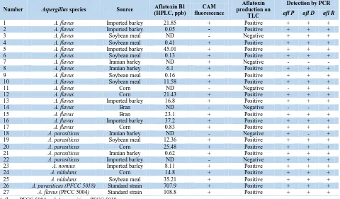
Table 3.Comparision of the results of conventional and molecular assays in terms of aflatoxin production
Number Aspergillus species Source (HPLC, ppb)Aflatoxin B1 fluorescenceCAM
Aflatoxin production on
TLC
Detection by PCR
afl P afl D afl R
1 A. flavus Imported barley 21.85 + Positive + + +
2 A. flavus Imported barley 0.05 - Positive + + +
3 A. flavus Soybean meal ND - Negative + + +
4 A. flavus Soybean meal 0.41 + Positive + + +
5 A. flavus Imported barley 45.01 + Positive + + +
6 A .flavus Soybean meal 0.13 + Positive + + +
7 A. flavus Iranian barley ND + Negative - - -
8 A. flavus Iranian barley 6.1 + Positive + + +
9 A. flavus Soybean meal 0.16 + Positive + + +
10 A. flavus Soybean meal 11.58 + Positive + + +
11 A. flavus Corn ND - Negative - + +
12 A. flavus Corn 21.43 + Positive + + +
13 A. flavus Imported barley 16.8 + Positive + + +
14 A. flavus Bran ND - Negative - - -
15 A. flavus Bran 23.1 + Positive + + +
16 A. flavus Imported barley 37.2 + Positive + + +
17 A. flavus Corn 0.83 + Positive + + +
18 A. parasiticus Iranian barley ND - Negative + - +
19 A. parasiticus Soybean meal 12.36 + Positive + + +
20 A. parasiticus Corn 25.48 + Positive + + +
21 A. parasiticus Iranian barley 0.62 + Positive + + +
22 A. parasiticus Imported barley ND - Negative + + +
23 A. nomius Imported barley 8.11 + Positive + + +
24 A. nidulans Corn 14.8 + Positive + + +
25 A. nidulans Soybean meal 35.21 + Positive + + +
26 A. parasiticus (PFCC 5018) Standard strain 707.9 + Positive + + +
27 A. flavus (PFCC 5004) Standard strain 108.8 + Positive + + +
A. flavus PFCC 5004 and A. parasiticus PFCC 5018
HPLC: high-performance liquid chromatography, CAM: coconut agar medium, TLC: thin-layer chromatography, PCR: polymerase chain reaction
observed in the positive and negative controls. A similar electrophoretic pattern was observed on the PCR products of 20 isolates, which represented the concomitant presence of the three genes. However,
other isolates demonstrated different patterns.
As shown in Table 3, the responsible aflatoxin biosynthesis genes (i.e., aflD, aflP, and aflR), which were greatly detected by PCR method in the positive aflatoxigenic samples, were absent in the negative control species.
The TLC and HPLC methods and the PCR amplification rendered completely similar results regarding the prevalence of target genes and aflatoxin B1 production ability (Table 3). As seen in Table 3, all of fungal isolates that were positive for aflatoxin production based on the TLC and HPLC assays could amplify aflD, aflP, and aflR genes. On the other hand, 4 cases out of 25 samples (16%), which were detected as aflatoxin non-producing Aspergillus, showed a complete set or both of genes. Only 2 (8%) samples, which were negative for aflatoxin production, had negative results in PCR and other conventional methods.
Discussion
Given the technical difficulties of conventional methods in the detection of aflatoxin-producing
Aspergillus, scientists have opted for new and sensitive
methods for the early detection of foodstuff contamination with different aflatoxigenic Aspergillus
species [18-20]. Among the different genes involving in aflatoxin biosynthesis pathway, aflD plays an important role in the early conversion of nosolorinic
acid into averantin. On the other hand, afl P is involved in the conversion of strigmatocystin into aflatoxin in the late steps. Moreover, afl R gene has a key role in the regulation of other genes in aflatoxin biosynthetic pathway [21-23].
In the present study, 92% (n=23) of aflatoxigenic
Aspergillus species gave consistent results regarding
aflatoxin production on CAM as confirmed by TLC and HPLC. However, there was a complete correlation between the results of these tests about the aflatoxin-producing isolates by the presence of three candidate genes and those of other conventional methods. However, 8% (n=2) of the test samples, which were considered as non-producing strains, had negative responses in fluorescence, TLC, and HPLC methods, and showed no bands in PCR reaction for each separated gene. This finding is consistent with those of the previous studies [19, 20].
Previously, we investigated the effect of aflatoxin genes, namely aflP and aflQ, on aflatoxigenic species of A. flavus and A. parasiticus in cattle feed. In the mentioned study, 67.16% and 70.14% of the samples had aflP and aflQ, respectively, and the HPLC findings also confirmed this result [15].
Degola et al. developed a multiplex real time-PCR protocol based on aflD, aflO, and aflQ genes for the discrimination of aflatoxin-producing strains of A.
flavus from the aflatoxin-nonproducing ones. They
reported a good correlation between the target genes expression in nearly all samples [24]. In another study, an optimized multiplex PCR was developed based on
detection of potential aflatoxigenic molds in fermented foods and grains [25].
Houssain et al. reported that the presence of
Aspergillus structural genes (i.e., Nor-1 and Ver-1) and
regulatory gene (i.e., aflR) could be considered as an early indicator of aflatoxin production [26]. In the current study, we found two isolates, which were positive for aflR and negative for aflD or aflP genes. It was observed that all of A. flavus and A. parasiticus
samples could amplify aflR gene. Nonetheless, the presence of this gene is not sufficient for aflatoxin production. These results showed that many other genes and factors account for the aflatoxigenicity of organisms [27]. Schmidt showed that environmental factors have a complex influence on the regulation of aflatoxin biosynthesis genes [28].
Inconsistent with previous reports [19], some non-aflatoxigenic strains of A. flavus and A. parasiticus,
which showed negative results in the conventional methods, could express at least one of the aflP, aflD, or
aflR genes. In line with the previous studies, our results indicated that the lack of aflatoxin production in some aflatoxigenic species could be due to the deletion and simple mutation (replacement of some bases) of aflR,
aflD, and aflP genes or loss of other responsible genes
in aflatoxin production pathway. However, some of the
physiological conditions could affect aflatoxin
biosynthesis [12, 21].
Furthermore, the additional molecular character-rization of these genes or other eventual genes in aflatoxin biosynthesis pathway could be useful for the application of these methods in differentiating between toxinogenic and nontoxinogenic strains of Aspergillus. The results of this study could be helpful for agricultural development and medical or veterinary organization in aflatoxin regulatory controls.
Conclusion
As the findings of the present study indicated, the molecular detection of aflatoxin biosynthesis genes (i.e., aflP, aflD, and aflR) could be considered as a quick and reliable method for the identification of
aflatoxigenic Aspergillus species. Moreover, this
method could be useful in planning and implementing the strategies targeted toward improving the safety of human or animal food.
Acknowledgments
The authors express their gratitude to the Payame Noor University, Tehran, Iran, for financially supporting this study.
Author’s contribution
N. S. and M. T. had equal contribution in data analysis and interpretation. The final manuscript was edited by N. S.
Conflicts of interest
The authors declare no conflicts of interest. The authors are responsible for the content and writing of
the paper.
Financial disclosure
The authors declare no financial interests related to the materials of the study.
References
1. Amaike S, Keller NP. Aspergillus flavus. Annu Rev Phytopathol. 2011; 49:107-33.
2. Gnonlonfin GJ, Hell K, Adjovi Y, Fandohan P, Koudande DO, Mensah GA, et al. A review on aflatoxin contamination and its implications in the developing world: a sub-Saharan African perspective. Crit Rev Food Sci Nutr. 2013; 53(4):349-65.
3. Roze LV, Hong SY, Linz JE. Aflatoxin biosynthesis: current frontiers. Annu Rev Food Sci Technol. 2013; 4:293-311.
4. Gabal MA, Azzam AH. Interaction of aflatoxin in the feed and immunization against selected infectious diseases in poultry. II. Effect on one-day-old layer chicks simultaneously vaccinated against Newcastle disease, infectious bronchitis and infectious bursal disease. Avian Pathol. 1998; 27(3):290-5.
5. Krishnan S, Manavathu EK, Chandrasekar PH.
Aspergillus flavus: an emerging non-fumigatus Aspergillus species of significance. Mycoses. 2009; 52(3):206-22.
6. Hadrich I, Makni F, Neji S, Cheikhrouhou F, Sellami H, Ayadi A. A review molecular typing methods for
Aspergillus flavus isolates. Mycopathologia. 2011; 172(2):83-93.
7. Sadhasivam S, Britzi M, Zakin V, Kostyukovsky M, Trostanetsky A, Quinn E, Sionov E. Rapid Detection and Identification of Mycotoxigenic Fungi and Mycotoxins in Stored Wheat Grain. Toxins (Basel). 2017; 9(10):302. 8. Baranyi N, Kocsubé S, Vágvölgyi C, Varga J. Current
trends in aflatoxin research. Acta Biologica Szegediensis. 2013; 57(2):95-107.
9. Luque MI, Rodríguez A, Andrade MJ, Martín A, Córdoba JJ. Development of a PCR protocol to detect aflatoxigenic molds in food products. J Food Prot. 2012; 75(1):85-94.
10.Jamali M, Karimipour M, Shams-Ghahfarokhi M, Amani A, Razzaghi-Abyaneh M. Expression of aflatoxin genes aflO (omtB) and aflQ (ordA) differentiates levels of aflatoxin production by Aspergillus flavus strains from soils of pistachio orchards. Res Microbiol. 2013; 164(4):293-9.
11.Yabe K, Nakajima H. Enzyme reactions and genes in aflatoxin biosynthesis. Appl Microbiol Biotechnol. 2004; 64(6):745-55.
12.Yu J. Current understanding on aflatoxin biosynthesis and future perspective in reducing aflatoxin contamination. Toxins (Basel). 2012; 4(11):1024-57. 13.Zeng H, Hatabayashi H, Nakagawa H, Cai J, Suzuki R,
Sakuno E, et al. Conversion of 11-hydroxy-O-methylsterigmatocystin to aflatoxin G1 in Aspergillus parasiticus. Appl Microbiol Biotechnol. 2011; 90(2): 635-50.
14.Liu BH, Chu FS. Regulation of aflR and its product, AflR, associated with aflatoxin biosynthesis. Appl Environ Microbiol. 1998; 64(10):3718-23.
15.Rahimi S, Sohrabi N, Ebrahimi MA, Tebyanian M, Taghyzadeh M, Rahimi SA. Studying the effect of aflatoxin genes Aflp and Aflq on Aspergillus flavus and
industrial animal husbandries. Acta Med Aust. 2016; 32:2091.
16.Rahimi S, Sohrabi N, Ebrahimi MA, Tebyanian M, Taghizadeh M, Rahimi S. Application of PCR in the detection of aflatoxinogenic and non-aflatoxinogenic strains of Aspergillus flavus group of cattle feed isolated in Iran. J Mol Biol Res. 2016; 6(1):121-8.
17.Namjoo M, Salamat F, Rajabli N, Hajihoseeini R, Niknejad F, Kohsar F, et al. Quantitative determination of aflatoxin by high performance liquid chromatography in wheat silos in Golestan province, North of Iran. Iran J Public Health. 2016; 45(7):905-10.
18.Bintvihok A, Treebonmuang S, Srisakwattana K, Nuanchun W, Patthanachai K, Usawang S. A rapid and sensitive detection of aflatoxin-producing fungus using an optimized polymerase chain reaction (PCR). Toxicol Res. 2016; 32(1):81-7.
19.Davari E, Mohsenzadeh M, Mohammadi G, Rezaeian-Doloei R. Characterization of aflatoxigenic Aspergillus flavus and A. parasiticus strain isolates from animal feedstuffs in northeastern Iran. Iran J Vet Res. 2015; 16(2):150-5.
20.Scherm B, Palomba M, Serra D, Marcello A, Migheli Q. Detection of transcripts of the aflatoxin genes aflD, aflO, and aflP by reverse transcription-polymerase chain reaction allows differentiation of aflatoxin-producing and non-producing isolates of Aspergillus flavus and
Aspergillus parasiticus. Int J Food Microbiol. 2005; 98(2):201-10.
21.Baquião AC, Rodriges AG, Lopes EL, Tralamazza SM, Zorzete P, Correa B. Expression of genes by aflatoxigenic and nonaflatoxigenic strains of Aspergillus
flavus isolated from Brazil Nuts. Foodborne Pathog Dis. 2016; 13(8):434-40.
22.Abdel-Hadi A, Carter D, Magan N. Temporal monitoring of the nor-1 (aflD) gene of Aspergillus flavus in relation to aflatoxin B₁ production during storage of peanuts under different water activity levels. J Appl Microbiol. 2010; 109(6):1914-22.
23.Liu BH, Chu FS. Regulation of aflR and its product, AflR, associated with aflatoxin biosynthesis. Appl Environ Microbiol. 1998; 64(10):3718-23.
24.Degola F, Berni E, Dall'Asta C, Spotti E, Marchelli R, Ferrero I, et al. A multiplex RT-PCR approach to detect aflatoxigenic strains of Aspergillus flavus. J Appl Microbiol. 2007; 103(2):409-17.
25.Yang ZY, Shim WB, Kim JH, Park SJ, Kang SJ, Nam BS, et al. Detection of aflatoxin-producing molds in Korean fermented foods and grains by multiplex PCR. J Food Prot. 2004; 67(11):2622-6.
26.Hussain A, Afzal A, Irfan M, Abdulla K. Molecular detection of aflatoxin producing strains of Aspergillus flavus from peanut (Arachis Hypogaea). Turkish J Agri Food Sci Technol. 2015; 3(5):335-41.
27.Dehghan P, Zaini F, Rezaei S, Jebali A, Kordbacheh P, Mahmoudi M. Detection of Aflr gene andtoxigenicity of
Aspergillus flavus group isolated from patients with fungal sinusitis. Iran J Public Health. 2008; 37(3):134-41. 28.Schmidt-Heydt M, Abdel-Hadi A, Magan N, Geisen R.