R E S E A R C H
Open Access
Outbreak report: a nosocomial outbreak of
vancomycin resistant
enterococci
in a solid
organ transplant unit
Peter Kreidl
1, Astrid Mayr
1*, Guido Hinterberger
1, Michael Berktold
1, Ludwig Knabl
1, Stefan Fuchs
1,
Wilfried Posch
1, Stephan Eschertzhuber
2, Alois Obwegeser
3, Cornelia Lass-Flörl
1and Dorothea Orth-Höller
1Abstract
Background:Vancomycin resistantenterococci(VRE) are an emerging problem in health care settings. The purpose of the investigation was to assess the extent of the outbreak including environmental contamination and to limit further transmission.
Methods:We used retrospective patient and laboratory data including pulse field gel electrophoresis (PFGE) typing and virulence and resistance gene analysis. For comparison of medians the Mann-Whitney and for comparison of proportions the Fisher exact tests were used.
Results:PFGE typing of VRE strains of an outbreak of 15 VRE cases in a solid transplant unit revealed that nine of the cases belonged to one identical pattern (A), which was only found twice in the environment. Eleven further positive environmental samples showed a different, but identical PFGE pattern E. Only one patient was infected with this environmental strain.
Two of nine (22.2%) PFGE A, but nine of eleven (81.2%) PFGE E samples were positive for gelatinase E (p= 0.01), which is described as enhancing biofilm production, suggesting a survival benefit for this strain on inanimate surfaces. Conclusion:Routine disinfection was not able to stop the cluster, but after repeated enforcement of the infection prevention and control (IPC) bundle such as training, strict adherence to hand hygiene and surface disinfection no further cases were observed. We conclude that certain VRE strains predominate in the environment whereas others predominate in humans. Enforcement of the IPC bundle is essential for controlling VRE outbreaks and reducing further transmission.
Keywords:Solid organ transplant unit, Vancomycin resistantenterococci, Outbreak, Survival benefit, Infection control
Background
Vancomycin resistant enterococci (VRE) are important causes of morbidity and mortality especially in health care settings where they easily disseminate [1]. Effective treatment is limited and thus VRE remain a major chal-lenge for infection control.
Vancomycin resistance of invasive Enterococcus
fae-cium isolates ranges between 0 and 46.3% in countries of the European Union and European Economic Area
(EU/EEA). The proportion of VRE in Austria (4.3% in 2016) was below the EU population weighted mean of 11.8% (95% confidence interval (CI) 11–13%) [2]. Mono-clonal and polyMono-clonal outbreaks of VRE were described worldwide; the largest reported outbreaks comprised up to 72 cases [3].
The most important transmission-routes of VRE are still not fully understood, but cross contamination via the hands of health care workers’(HCW) is believed to play a major role. HCW act as a vector between colo-nized or infected patients, inanimate surfaces and previ-ously unaffected patients [4]. The reservoir for VRE is believed to be the human intestine [5].
* Correspondence:astrid.mayr@i-med.ac.at
1Department of Hygiene, Microbiology and Social Medicine, Division of
Hygiene and Medical Microbiology, Medical University of Innsbruck, Innsbruck, Schoepfstr. 41, 6020 Innsbruck, Austria
Full list of author information is available at the end of the article
Known risk factors for acquisition of VRE include pre-vious use of antibiotics, prolonged hospital stay, under-lying diseases, admission to high risk departments such as oncology, hematology, transplant or intensive care units (ICU), the nurse to patient ratio, and occupancy to a room where previously a patient harboring VRE was admitted [6]. The transmission in high risk wards via contaminated inanimate surfaces is believed to be im-portant but it remains a challenge to quantify the attrib-utable risk [6] of environmental contamination.
We describe an outbreak which was identified on 7th January 2017 after detection of five cases within 2 months following the Orion statement [7].
The objective of this investigation was to assess the extent of the outbreak including the environmental contamination, to limit further transmission and to evaluate the effectiveness of implemented control mea-sures. Further important objectives were to foster infec-tion preveninfec-tion and control (IPC) educainfec-tion by the infection control team (ICT) of the University Hospital Innsbruck (LKI).
Methods Study design
We used retrospective data analysis including PFGE typ-ing, analysis of virulence and resistance genes and pa-tient relevant data from the hospital records.
Participants
Participants were patients admitted to the solid organ trans-plant unit between 15th November 2016 and 30th June 2017 with laboratory confirmation of VRE. Colonization was defined as laboratory confirmation of VRE in a sample of a patient from a non-sterile site without signs of infection such as fever > = 38.5 °C. Infection was defined as laboratory confirmation of VRE in a sample of a patient from a nor-mally sterile site or signs of infection such as fever > = 38.5°.
Case definition
We defined a VRE case as a patient admitted to the transplant unit during the observation period with la-boratory confirmation of VRE in at least one sample ir-respective of the location or type of sampling. We defined a cluster case as a VRE case with the outbreak PFGE pattern A identified between December 2017 and June 2018 and admitted to the transplant unit.
Setting
The period of the outbreak lasted from 15 November 2016 until 30 June 2017.
The LKI is a 1600-bed tertiary-care hospital with sev-eral departments including a solid organ transplant unit.
The setting of the outbreak was the ward of the trans-plant unit consisting of 15 beds in seven rooms (one to
four beds per room) and an adjacent intensive care unit (ICU) consisting of eight beds situated in three separate rooms. Patients were transferred between the standard care unit and the ICU depending on their medical con-dition. The ICT consisted of one senior hospital hygiene specialist and a local hygiene team.
Routine IPC protocol
The internal IPC protocol included the following recom-mendations: 1) twice daily unsupervised routine disinfec-tion of inanimate surfaces of the patient close environment with an aldehyde-free-broad-spectrum disinfectant contain-ing quaternary ammonium compounds (QACs, 2% TPH protect, Schülke & Mayr GmbH, Vienna, Austria) including once daily patient charts, 2) terminal cleaning and disinfec-tion of patient rooms with the above mendisinfec-tioned disinfect-ant and 3) hand hygiene (HH) based on the WHO approach“My Five Moments for Hand Hygiene”[8] using alcohol-based detergents (Sterillium®, BODE Chemie GmbH, Hamburg, Germany). Surveillance of hand hygiene compliance is conducted through bi-annual audits based on the protocol from the Robert Koch Institute [9], measur-ing adherence to HH. The outcomes of the audits were cat-egorized as sufficient or insufficient per unit but lacked a coding system to quantify the adherence to HH on an indi-vidual basis. Annual trainings of the IPC protocol through verbal presentations and ward round sessions were rou-tinely conducted. Data on baseline IPC knowledge of HCW were not routinely collected. IPC guidelines did not require pre-admission VRE screening of patients, unless 1) patients had a positive history of exposure in a high endemic coun-try, 2) were transferred from a long-term-care-facility or 3) were previously known having been infected or colonized with VRE.
According to the IPC protocol, each newly identified patient either colonized or infected by VRE irrespective of the patient being part of an outbreak, triggered a rou-tine infection control response consisting of following measures: isolation precautions, defined as placement in a single room, if possible, and, contact precautions de-pending on the estimated potential for transmission in-cluding personal protective equipment.
Intervention during the outbreak
basis upon the urgency of the situation. Thus informed consent was not obtained.
Frequency of ward round training was increased. The choice of HH antisepsis and environmental disinfection during the outbreak remained the same and compliance with HH was observed by the ICT. No additional decon-tamination procedures such as fogging or steaming were implemented.
Culturing and typing Patient samples
We collected routine clinical samples and rectal screen-ing swabs from patients accordscreen-ing to the IPC protocol (no informed consent was requested for rectal sam-pling), cultivated them on blood agar (Becton Dickinson, Heidelberg, Germany) and selective ChromID VRE Agar (Biomerieux, Marcy-l’Étoile, France) and incubated them for 48 h at 37 °C under aerobic conditions. Suspect col-onies were identified by matrix-assisted laser desorption ionization time-of-flight mass spectrometry (MALDI--TOF, Bruker, Bremen, Germany). Antibiotic susceptibility testing was performed according to the European Com-mittee on Antimicrobial Susceptibility Testing (EUCAST version 6.0, 2016) protocol [10].Enterococciwere classified as VRE if the minimal inhibitory concentration of vanco-mycin was above 4 mg/L identified by E-Test (Biomerieux, Marcy-l’Étoile, France) [11]. Additional patient data such as diagnosis, duration of hospital stay, outcome, type and time of transplantation were obtained from the medical records of the LKI.
Environmental samples
Unheralded sampling of environment was conducted
ac-cording to Galvins protocol (Galvin) using
Columbia-III-Sheep blood agar 5% (Oxoid Limited, Basingstoke, UK) or Tryptic soy agar with neutralizers (VWR International, Radnor, USA) contact plates with a press on time of 10 seconds without any lateral move-ment. Surfaces were selected at different locations close and distant from the respective patients [12] during three occasions: immediately after the alert of the cluster on 18 January, and twice after enforcement of control measures (30th January and 2nd February 2017, respect-ively). Data recorded were date, type and location of sampling, laboratory findings and PFGE results. Direct unheralded observation of hygienic measures was con-ducted to identify potential gaps of adherence to infec-tion control measures. Patient close samples were defined as samples obtained from the vicinity of patients such as the bedrail, bedframe, beside tables, clamps for urine-bags, drug perfusors, stethoscope and bedside monitors [13].
Microbial monitoring of HCWs hands was performed by imprinting all fingertips including the thumb onto
Columbia agar containing 5% sheep blood (Becton, Dickenson and Company, Franklin Lakes, USA) for ap-proximately 5 seconds [14, 15]. Confirmation of VRE was conducted as described above.
PFGE
Molecular Typing was performed using pulse field gel electrophoresis (PFGE) according to the protocols pub-lished by the Centers for Disease Control and Prevention [16]. Extracted bacterial DNA was restricted using Smal enzyme. Strain typing was performed by manual PFGE restriction pattern analysis [17] and by computational analysis of band differences using GelJ Software Version 2 [18]. Dendrogram analysis of PFGE patterns was per-formed using Dice algorithm and Single Linkage cluster-ing. Genotypically related strains with similarity > 90% were considered belonging to the same pattern.
Genetic analysis
Vancomycin resistance genotypes were determined ac-cording to the protocol from Jayaratne et al. [19]. The enterococcal surface antigen was identified according to the protocol by Toledo-Arana [20], the cytolysin activa-tor according to Vankerckhoven [21] and the gelatinase E according to Hancock’s protocol [22].
Infection related outcomes
Infection related outcomes, including colonization, in-fection and patient survival were assessed.
The specimen processing and turn-around time were performed according to local laboratory protocol from 2016.
Sample size
The sample size was limited to the number of identified patients (n= 15).
Statistical methods
Variables of interest were stored in excel database in-cluding age, gender, number of isolates obtained includ-ing positives, date of available laboratory result, source and material of samples, length of hospital stay, previous antibiotic treatment, previous confirmation of VRE in any sample, diagnosis, other identified pathogens, and patient outcome.
Results
Event description
On 7th January 2017 the outbreak response team of the LKI informed the transplant unit about a potential out-break of VRE.
Analysis of historical data revealed that between May 2012 and October 2016, a total of 53 VRE cases were identified at this unit. The median incidence was one (0–3) case per month and the annual median incidence was 11 cases (6–16 cases) per year for the period 2013– 2016. In comparison to previous years, the observed number of five VRE cases in a period of less than 2 months clearly exceeded the expected value suggesting an outbreak.
VRE cases
After a period of 4 months without any detection of VRE from the transplant unit, the potential index case was confirmed on 15th November 2016. He was a 58 year old male suffering from IgA Nephritis who re-ceived a kidney transplant on 12th November 2016, 3 days prior to the diagnosis of VRE in a single sample of a retroperitoneal surgical-site-drainage. The patient was classified as being infected.
Between 15th November 2016 and 30th June 2017, VRE was confirmed in at least one sample of 14
additional patients (Fig.1). Of these, ten patients (66.7%) were males with a median age of 56.5 years (50– 71 years). The five female patients showed a median age of 70 years (60–79 years) (p= 0.003) (Table1).
VRE was first isolated twice from blood cultures, five times from urine samples, four times from wound drainage and once each from a tissue, stool, and rectal swab and from a broncho-alveolar lavage sample. Three of the patients were classified as being colonized, the remaining twelve as being infected with VRE. The median length of hospital stay was 24 days (10–112 days). All but one patient underwent trans-plantation and received immunosuppression. Twelve pa-tients survived (80%) and three died (Table1). In all patients
Enterococcus faeciumwas identified, one patient harbored a tigecycline resistant, two a linezolid resistant and fourteen teicoplanin resistant strains.
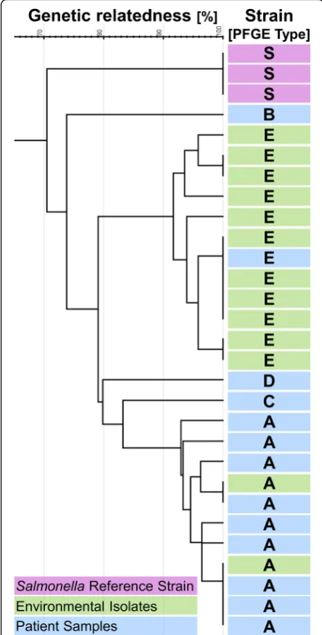
PFGE patterns of cases
PFGE patterns were available for 13 of 15 patients (86.7%). Among those, five different PFGE patterns were identified and subsequently classified as patterns A to E. Nine of the thirteen cases (69.2%) were classified as PFGE pattern A (further called cluster strain, Table1, Fig.2).
All 10 of the 15 patient isolates (66.7%) available for vancomycin resistance genotyping (vanR) revealedvanA,
irrespective of their PFGE pattern. The only patient iso-late with pattern E, was not viable.
Cluster cases
In total, 194 patient samples were obtained from cluster cases, in 26 of them (13.4%) VRE was confirmed. The median number of samples investigated per cluster case was 20 (12–34) and the median number of positive sam-ples was one (1–12). Five patients only had one sample positive for VRE. The median duration of confirmation between the first and the last positive sample was 4 days (1–33 days). The median interval between admission and first isolation of VRE was 13 days (same day-22 days) (n= 14). One additional patient already tested positive in another unit during the outbreak period, but the iso-late was different from the cluster strain (PFGE C). From all other cases no information of previous VRE carriage was available as no routine VRE screening was conducted prior to this outbreak. VRE was first identified in following materials from the nine cluster cases: Blood (n= 1), (wound) drains (n= 4), rectal swab (n= 1), and urine (n= 3).
VRE screening
Pre-admission rectal screening was implemented after the outbreak alert. Among 169 patients screened until the end of 2017, only one was found to be VRE positive (0.6%).
Environmental and hand samples
A total of 139 hand (n= 26; 18.7%) and environmental (n= 113, 81.3%) samples were obtained during three un-heralded on-site visits; nearly half of them (n= 68, 48.9%) were obtained prior to the first enforcement of control measures. More samples taken prior to en-hanced disinfection (n= 10; 14.7%) were positive for VRE compared to post disinfection sampling (n= 4; 5.6%) (p= 0.07). Among the thirteen VRE positive envir-onmental samples the cluster strain (PFGE A) was iden-tified twice: once from a clamp of a urine-bag prior and
once on a patients chart after the first enforcement of control measures. All other eleven VRE positive environ-mental samples revealed one identical PFGE pattern E which was distinct from the patient cluster strain (Fig.2). Pattern E was confirmed only in one patient, who was ad-mitted 2 days prior to the first environmental sampling. Eight of these positive non-cluster strains (PFGE E) were identified during the first on-site visit. Six of them were determined in the patient close environment and two out-side patient rooms. During the third visit the environmen-tal PFGE E strain was identified again three times. Once from the patient close environment (urine clamp bag) of the patient infected with PFGE E; and twice from a bed lo-cated in the corridor (Fig. 3). All tested environmental strains werevanA positive.
One VRE positive hand sample of a HCW was not available for PFGE typing.
Virulence genes
Twenty three samples were available for virulence gene testing (13 environmental and 10 patient samples).
Two of nine (22.2%) PFGE A (six patient samples), but nine of 11 (81.2%) PFGE E (all environmental samples) were positive for gelatinase E (p= 0.0123). The two gela-tinase E positive PFGE A samples were one human sam-ple from a patient already discharged at the time of the environmental sampling and one environmental sample from a urinary bag clamp obtained during the first on-site visit which could not be linked to a specific patient.
All investigated samples were positive for enterococcal surface protein and negative for cytolysin activator.
Control measures
The first of the three unheralded on-site visit including environmental and hand sampling was undertaken on 18th January 2017. Lack of adherence to hand hygiene was observed and therefore enforcement of control mea-sures was stressed by the infection control hygiene team. At the time of this first visit, five patients were admitted
Table 1Patient characteristics
Categories N/total or median (mean; range) % Demographic characteristics Males 5/15 67 Age 59 yrs. (61.3 yrs.; 50–79 yrs) – Clinical findings Infected (versus colonized) 12/15 80
Deceased 3/15 20
Laboratory sampling Number of samples investigated per patient 24 (31; 8–105) – Number of positive samples per patient 2 (6.1; 1–52) – Laboratory findings PFGE pattern A (cluster strain) 9/13 69
(two with PFGE A, one each with PFGE B and E and one without a viable isolate) and six already discharged (five of whom with PFGE A and one with PFGE C).
The second sampling on 30th January, performed after extended cleaning and disinfection procedures towards patients’distant zones did not detect any environmental contamination. Rectal screening targeting all staff was conducted the same day. The participation rate was 79% (94 of 119), all samples remained negative. Non-participation was mainly due to absence; No consent was obtained as
participation was voluntary. In the current IPC protocol HCW hand sampling but no rectal swabbing is included.
During the last on-site visit on 2nd February, VRE was detected in four of 47 (8.51%) environmental samples. Three of those revealed PFGE E, all from patient-close sites of the patient infected with PFGE E, the cluster strain (PFGE A) was isolated from a patient chart. At the time of the third visit, five patients (two with PFGE A) were still admitted to the ward; four further cases, two with the cluster PFGE A were identified later. En-forcement of IPC measures was rigorously stressed again and four ward round sessions were conducted. Insuffi-cient adherence to HH according to the IPC protocol was documented during the first visit. During the second and third visit the adherence to HH was evaluated to be sufficient. The terminal cleaning was audited by the ICT and adherence was defined as sufficient already at the first visit. The effect of teaching was not measured.
Discussion
We describe an outbreak of fifteen VRE cases in a solid organ transplant unit in late 2016 and early 2017, of which nine patients revealed an identical PFGE pattern A, further called the cluster strain.
During the first visit six VRE positive environmental samples were identified in the patient close environment which suggests lack of effective decontamination despite the audited terminal disinfection which was considered sufficient. Additionally, lack of adherence to strict hand hygiene was observed and compliance to HH was classi-fied as insufficient. The two positive samples identiclassi-fied from a laundry rack suggest cross-contamination via HCW hands, as all patients were immobile. The fact, that the majority of environmental strains were identical - although distinct from the outbreak strain - may sug-gest that also prior to the first sampling environmental contamination via HCW hands may have occurred. HH adherence during audits improved from insufficient dur-ing the first visit to sufficient durdur-ing the second and third visit.
Enforcement of ICP measures resulted in a decrease of the proportion of VRE contaminated environmental sam-ples, although it was not significant (Fig.3). Nevertheless, reconfirmation of the non-cluster strain during the third site visit may be due to recontamination and insufficient compliance to hand hygiene. In addition, the confirmation of the cluster strain on a patient chart after enforcement of control measures suggests the cross-contamination via HCW hands as well as ineffective decontamination of the patient chart. Decontamination of patient charts is de-scribed as effective measure to decrease horizontal trans-fer of organisms and to prevent transmission of health care associated infections [23,24].
We speculate that the lack of strict adherence to ad-equate hand hygiene might have led to contamination of the environment. This is supported by the facts that firstly, contamination was observed in patient distant areas such as laundry racks and secondly, that contaminated patient charts are described being a result of lack of adherence to recommended HH [25]. This may have played an import-ant role in triggering the outbreak. Tight working spaces in the ICU unit favor the patient-to-patient contact [26] and may have resulted in a higher risk for transmission of VRE.
Hayden concludes that the role of environmental con-tamination in nosocomial cross-transmission of VRE is still unresolved but enforcement of environmental de-contamination was both associated with reduction of surface contamination and contamination of HCWs’ hands despite only moderate adherence to proper hand hygiene [27]. Proper environmental decontamination may reduce the risk of VRE outbreaks in hospital set-tings as suggested in previous studies. Dancer et al. [28] suggest to focus more on patient close hand-touch sites rather than on general surfaces and bathrooms.
The observed higher median age of male VRE patients rather reflects the distribution of admitted patients than being associated with VRE colonization or infection [29]. The pre-admission rectal screening implemented since the identification of the outbreak revealed less than one positive per hundred investigated patients. VRE screen-ing in a tertiary hospital settscreen-ing is suggested to decrease the incidence in routine patient care and even more im-portant in an outbreak situation [30–32]. Therefore we speculate that most of the cluster cases acquired the VRE during the hospital stay.
The cluster strain PFGE A was identified only twice in the environment; all other environmental strains re-vealed an identical PFGE E pattern. This PFGE E pattern was found only in one patient. The fact, that significantly more PFGE E compared to PFGE A strains were produ-cing gelatinase E, which is described as enhanprodu-cing bio-film production, suggests a survival benefit on inanimate surfaces [33]. Accumulation of mobile genetic elements including plasmids, pathogenicity islands, resistance transposons other fitness islands, phages and surface types [34, 35] may also have contributed to better sur-vival in the environment.
The limitations of our study are that we used retro-spective data analysis which made selection of appropri-ate controls difficult. Also logistical reasons hindered us to conduct a case control study. Therefore we were nei-ther able to identify risk factors for acquisition of VRE nor to identify the source of infection. Furthermore, nei-ther retrospective data where patients were located within the ward at the time of admission nor informa-tion on staffing was available. A higher patient-staff ratio might have influenced the transmission risk. The only PFGE E strain from the patient could not be recovered from the archived skim-milk stock and was hence also not available for genotyping.
Conclusions
We conclude that enforcement of strict adherence to the existing IPC protocol and assessment of adherence are es-sential for controlling VRE outbreaks and reducing further transmission. We cannot exclude the role of other compo-nents in the multi-modal IPC plan instituted which
ultimately may have played a role in the control of the out-break. Unheralded visits are useful tools to further improve adherence to IPC protocols. Frequently touched surfaces such as patient charts are difficult to decontaminate and therefore require particular attention. We conclude that this outbreak has resulted in better control of future VRE out-breaks. HCWs reported a better understanding for the need of rigorous adherence to control measures. We expect fur-ther sporadic cases in future.
Abbreviations
CI:Confidence interval; EU/EEA: European Union and European Economic area; HCW: Health care worker; HH: Hand hygiene; ICT: Infection control team; ICU: Intensive care unit; IPC: Infection prevention and control; LKI: University Hospital Innsbruck (Landeskrankenhaus Innsbruck); PFGE: Pulse filed gel electrophoresis; VRE: Vancomycin resistantenterococci
Acknowledgements
The authors want to thank the staff of the unit and laboratory and the ICT for the good and transparent collaboration.
Availability of data and materials
The datasets used and/or analyzed during the current study are available from the corresponding author on reasonable request.
Authors’contributions
SE provided patient data regarding disease and transplant and reviewed the draft manuscript, MB and LK analyzed the patient samples and the antimicrobial susceptibility testing, SF and WP conducted the molecular examinations. AM and GH conducted and analyzed the environmental sampling, AO, CLF and DO reviewed the manuscript, PK analyzed and interpreted the data and drafted the manuscript. All authors read and approved the final manuscript.
Ethics approval and consent to participate
The Ethics Committee of the Medical University of Innsbruck confirms that for retrospective observational studies no ethics committee approval is required by Austrian law.
Consent for publication Not applicable
Competing interests
The authors declare that they have no competing interests.
Publisher’s Note
Springer Nature remains neutral with regard to jurisdictional claims in published maps and institutional affiliations.
Author details
1Department of Hygiene, Microbiology and Social Medicine, Division of
Hygiene and Medical Microbiology, Medical University of Innsbruck, Innsbruck, Schoepfstr. 41, 6020 Innsbruck, Austria.2Department of Anesthesia
and Critical Care, Centre of Operative Medicine, Medical University of Innsbruck, Anichstr. 35, 6020 Innsbruck, Austria.3Department of
Neurosurgery, University Hospital of Innsbruck, Anichstr. 35, 6020 Innsbruck, Austria.
Received: 19 April 2018 Accepted: 4 July 2018
References
1. Gastmeier P, Schroder C, Behnke M, Meyer E, Geffers C. Dramatic increase in vancomycin-resistant enterococci in Germany. J Antimicrob Chemother. 2014;69(6):1660–4.
2. ECDC. Surveillance of antimicrobial resistance in Europe, 2016 2017 [cited 2018 13 Mar]. Available from:https://ecdc.europa.eu/en/publications-data/ antimicrobial-resistance-surveillance-europe-2016.
3. Ulrich N, Gastmeier P. Where is the difference between an epidemic and a high endemic level with respect to nosocomial infection control measures? An analysis based on the example of vancomycin-resistant enterococcus faecium in hematology and oncology departments. GMS Hyg Infect Control. 2017;12:Doc14.
4. Hayden MK, Blom DW, Lyle EA, Moore CG, Weinstein RA. Risk of hand or glove contamination after contact with patients colonized with vancomycin-resistant enterococcus or the colonized patients’ environment. Infect Control Hosp Epidemiol. 2008;29(2):149–54. 5. Brodrick HJ, Raven KE, Harrison EM, Blane B, Reuter S, Torok ME, et al.
Whole-genome sequencing reveals transmission of vancomycin-resistant enterococcus faecium in a healthcare network. Genome Med. 2016;8(1):4. 6. Huang SS, Datta R, Platt R. Risk of acquiring antibiotic-resistant bacteria from
prior room occupants. Arch Intern Med. 2006;166(18):1945–51.
7. Stone SP, Cooper BS, Kibbler CC, Cookson BD, Roberts JA, Medley GF, et al. The ORION statement: guidelines for transparent reporting of outbreak reports and intervention studies of nosocomial infection. J Antimicrob Chemother. 2007;59(5):833–40.
8. Organization WH. WHO Guidelines on Hand Hygiene in Health Care: First Global Patient Safety Challenge Clean Care is Safer Care 2009 [cited 2018 21 June]. Available from:http://apps.who.int/iris/bitstream/handle/10665/ 44102/9789241597906_eng.pdf;jsessionid=
0353CC69A96F2B5A26F019487149B213?sequence=1.
9. Robert Koch Institute NRfSvnI. Hand-KISS 2017 [cited 2018 21 June]. Available from:http://www.nrz-hygiene.de/surveillance/kiss/hand-kiss/. 10. Testing TECoAS. Breakpoint tables for interpretation of MICs and zone
diameters. Version 6.0, 2016.
11. Biomerieux. Gebrauchsfertige Teststreifen zur direkten Bestimmung der minimalen Hemmkonzentration [cited 2018 21 June]. Available from:http:// www.biomerieux.de/klinische-diagnostik/etestr.
12. Galvin S, Dolan A, Cahill O, Daniels S, Humphreys H. Microbial monitoring of the hospital environment: why and how? J Hosp Infect. 2012;82(3):143–51. 13. Wille I, Mayr A, Kreidl P, Bruhwasser C, Hinterberger G, Fritz A, et al.
Cross-sectional point prevalence survey to study the environmental
contamination of nosocomial pathogens in intensive care units under real-life conditions. J Hosp Infect. 2018;98(1):90–5.
14. Bruhwasser C, Hinterberger G, Mutschlechner W, Kaltseis J, Lass-Florl C, Mayr A. A point prevalence survey on hand hygiene, with a special focus on Candida species. Am J Infect Control. 2016;44(1):71–3.
15. Creamer E, Dorrian S, Dolan A, Sherlock O, Fitzgerald-Hughes D, Thomas T, et al. When are the hands of healthcare workers positive for methicillin-resistant Staphylococcus aureus? J Hosp Infect. 2010;75(2):107–11. 16. CDC. Unified PulsedField Gel Electrophoresis (PFGE) Protocol for Gram
Positive Bacteria 2012 [Available from:https://www.cdc.gov/hai/pdfs/ labsettings/Unified_PFGE_Protocol.pdf.
17. Tenover FC, Arbeit RD, Goering RV, Mickelsen PA, Murray BE, Persing DH, et al. Interpreting chromosomal DNA restriction patterns produced by pulsed-field gel electrophoresis: criteria for bacterial strain typing. J Clin Microbiol. 1995;33(9):2233–9.
18. Heras J, Dominguez C, Mata E, Pascual V, Lozano C, Torres C, et al. GelJ--a tool for analyzing DNA fingerprint gel images. BMC Bioinformatics. 2015;16:270. 19. Jayaratne P, Rutherford C. Detection of clinically relevant genotypes of
vancomycin-resistant enterococci in nosocomial surveillance specimens by PCR. J Clin Microbiol. 1999;37(6):2090–2.
20. Toledo-Arana A, Valle J, Solano C, Arrizubieta MJ, Cucarella C, Lamata M, et al. The enterococcal surface protein, Esp, is involved in enterococcus faecalis biofilm formation. Appl Environ Microbiol. 2001;67(10):4538–45.
21. Vankerckhoven V, Van Autgaerden T, Vael C, Lammens C, Chapelle S, Rossi R, et al. Development of a multiplex PCR for the detection of asa1, gelE, cylA, esp, and hyl genes in enterococci and survey for virulence determinants among European hospital isolates of enterococcus faecium. J Clin Microbiol. 2004;42(10):4473–9.
22. Hancock LE, Perego M. The enterococcus faecalis fsr two-component system controls biofilm development through production of gelatinase. J Bacteriol. 2004;186(17):5629–39.
23. Zimbudzi E, Stuart RL, Korman TM, Kotsanas D. Contamination of renal patients’hospital chart covers with vancomycin-- resistant enterococci: handle with care. Australas Med J. 2011;4(10):538–41.
25. Russotto V, Cortegiani A, Raineri SM, Giarratano A. Bacterial contamination of inanimate surfaces and equipment in the intensive care unit. J Intensive Care. 2015;3:54.
26. Ulrich N, Vonberg RP, Gastmeier P. Outbreaks caused by vancomycin-resistant enterococcus faecium in hematology and oncology departments: a systematic review. Heliyon. 2017;3(12):e00473.
27. Hayden MK, Bonten MJ, Blom DW, Lyle EA, van de Vijver DA, Weinstein RA. Reduction in acquisition of vancomycin-resistant enterococcus after enforcement of routine environmental cleaning measures. Clin Infect Dis. 2006;42(11):1552–60. 28. Dancer SJ. The role of environmental cleaning in the control of
hospital-acquired infection. J Hosp Infect. 2009;73(4):378–85.
29. Schold JD, Buccini LD, Goldfarb DA, Flechner SM, Hsich E, Mason D, et al. Patient participation in research among solid organ transplant recipients in the United States. Transplantation. 2011;91(12):1424–35.
30. Escaut L, Bouam S, Frank-Soltysiak M, Rudant E, Saliba F, Kassis N, et al. Eradication of an outbreak of vancomycin-resistant enterococcus (VRE): the cost of a failure in the systematic screening. Antimicrob Resist Infect Control. 2013;2(1):18.
31. Humphreys H. Controlling the spread of vancomycin-resistant enterococci. Is active screening worthwhile? J Hosp Infect. 2014;88(4):191–8. 32. Popiel KY, Miller MA. Evaluation of vancomycin-resistant enterococci
(VRE)-associated morbidity following relaxation of VRE screening and isolation precautions in a tertiary care hospital. Infect Control Hosp Epidemiol. 2014; 35(7):818–25.
33. Banerjee T, Anupurba S. Prevalence of virulence factors and drug resistance in clinical isolates of enterococci: a study from North India. J Pathog 2015; 2015:692612.
34. Gaca AO, Gilmore MS. Killing of VRE enterococcus faecalis by commensal strains: evidence for evolution and accumulation of mobile elements in the absence of competition. Gut Microbes. 2016;7(1):90–6.