R E V I E W
Open Access
Synthetic Biology Goes Cell-Free
Aidan Tinafar, Katariina Jaenes and Keith Pardee
*Abstract
Cell-free systems (CFS) have recently evolved into key
platforms for synthetic biology applications. Many
synthetic biology tools have traditionally relied on
cell-based systems, and while their adoption has
shown great progress, the constraints inherent to the
use of cellular hosts have limited their reach and
scope. Cell-free systems, which can be thought of as
programmable liquids, have removed many of these
complexities and have brought about exciting
opportunities for rational design and manipulation of
biological systems. Here we review how these simple
and accessible enzymatic systems are poised to
accelerate the rate of advancement in synthetic
biology and, more broadly, biotechnology.
Moving towards a new bioengineering platform
Since its emergence, the field of synthetic biology has
given rise to the development of many technologies that
are implemented using the whole cell [
1
]. These have
in-cluded biosensors capable of detecting broad ranges of
analytes [
2–5
], systems that can count [
6
] or perform
complex logic [
7–10
], engines for the bioproduction of
valuable commodities [
11–14
], gene-circuit-driven
chas-sis for regenerative medicine [
15
,
16
], and engineered
CAR-T cells [
17
]. Such technologies are on track to
transform many aspects of modern life, yet their
require-ment for a cellular host has limited their reach and
scope. For example, concerns over biosafety have
re-stricted the use of engineered cells, and the systems they
host, largely to laboratory settings. The self-replicability
of cell-based systems carries the risk of
“
escape
”
or
con-tamination that could impact human health, food
secur-ity, and the environment. While the development of
safeguards to prevent these types of events is an active
area of research [
18
,
19
], failure-free implementation of
such systems is not a trivial task.
Another substantial limitation of cell-based synthetic
biology is the requirement for laborious genetic
encod-ing of its design features into a livencod-ing cell, which can
limit its functionality and significantly slow down
de-sign
–
build
–
test cycles. In cell-based systems, genetic
in-structions often need to be assembled into a vector,
imported into the cell, and maintained by using a
select-able marker or by genomic integration. Only then can
the instructions be evaluated. Furthermore, designs must
be iteratively tested to minimize cross-talk with
en-dogenous molecular programs while balancing between
the metabolic burden on the cellular host and the
de-sired outcome.
Cell-free systems offer a means to circumvent many of
these limitations. They were originally conceived as tools to
facilitate in vitro protein synthesis and consist of molecular
machinery extracted from cells. They typically contain
en-zymes necessary for transcription and translation, and
ac-cordingly are able to perform the fundamental processes of
the central dogma (DNA
➔
RNA
➔
protein) independent of
a cell. These systems can be derived from eukaryotes (e.g.,
vertebrates, plants, insects, fungi) [
20–27
] or prokaryotes
(e.g.,
Escherichia coli
,
Vibrio natriegens
,
Bacillus subtilis
)
[
28–43
] and may be prepared as either purified
compo-nents [
36
,
44
] or semi-processed cellular extracts [
38
]. CFS
can be made sterile via simple filtration, which provides for
a biosafe format for use outside of the lab.
The open nature of CFS means that there is no
phys-ical barrier (e.g., a cell wall) to programming and
modifi-cation. CFS can be augmented with proteins or small
molecules that improve the performance of synthetic
gene networks [
45
,
46
] or the productivity of reactions
[
39
,
47
]. More importantly, genetically encoded
instruc-tions can be added directly to CFS at desired
concentra-tions and stoichiometries using linear or circular
formats. This means that conceptual designs can go
from computational instructions to chemical synthesis
and amplification (e.g., through PCR) to CFS without
the need for selective markers or cell-based cloning
steps. Such simplicity allows for rapid prototyping of
molecular tools.
© The Author(s). 2019Open AccessThis article is distributed under the terms of the Creative Commons Attribution 4.0 International License (http://creativecommons.org/licenses/by/4.0/), which permits unrestricted use, distribution, and reproduction in any medium, provided you give appropriate credit to the original author(s) and the source, provide a link to the Creative Commons license, and indicate if changes were made. The Creative Commons Public Domain Dedication waiver (http://creativecommons.org/publicdomain/zero/1.0/) applies to the data made available in this article, unless otherwise stated.
* Correspondence:keith.pardee@utoronto.ca
Importantly, CFS can be freeze-dried, enabling room
temperature storage and distribution [
46
,
48
].
Freeze-dried cell-free (FD-CF) systems can then be activated at
the time of need simply by adding water [
46
]. This
fea-ture has been used to deploy biosafe, genetically encoded
tools outside of the laboratory as diagnostics and as
plat-forms for biomanufacturing [
49
,
50
], as well as their
de-ployment in altogether new contexts, such as global
health and education.
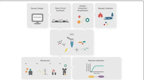
Below we will discuss how CFS are enabling new
tech-nologies and accelerating the coming revolution in
bio-engineering, highlighting some of the most active areas
of research in the cell-free community (Fig.
1
).
Development of sensors
Molecular recognition underlies almost every biological
process, including the nucleic acid base pairing that
im-parts specific syntax to the central dogma. Scientists and
engineers have long worked to usher these processes
into cell-free in vitro environments to understand and
exploit their underlying molecular mechanisms for
pur-poses such as diagnostics and detection of molecules.
One of the fruits from such efforts is the polymerase
chain reaction (PCR), which is now an indispensable tool
utilized in most molecular biology laboratories,
includ-ing those for clinical diagnostics. There is currently a
growing need for de-centralized, portable diagnostics
that can be rapidly deployed in the field, for instance
during infectious disease outbreaks or for agricultural
purposes. However, sensing technologies such as PCR
and others have largely remained confined to
laborator-ies in large urban centers due to their requirement for
specialized equipment and personnel.
The biosafe and stable nature of FD-CF systems offers
an alternative molecular venue to address the unmet
need for distributed and low-cost sensing. Here, the
transcription and translation properties of CFS can be
used to host gene circuit-based sensors that can detect
small molecules and nucleic acids with exquisite
sensi-tivity and specificity. Many of the biosensors and circuits
that have been developed for cell-based applications can
be operated in the cell-free environment. These include,
among others, many classic switches (e.g., TetO- and
LacI-based systems), logic gates, negative feedback loops,
transcriptional cascades [
37
,
41
,
53–56
] and ring
oscilla-tors [
57
]. This cross-compatibility between CFS and
cell-based systems has also been exploited for rapid
proto-typing of regulatory elements that can be brought back
to the cell-based environment.
FD-CF systems do not
require a
temperature-controlled environment and cold-chain logistics intrinsic
to many other diagnostic approaches, as they remain
Fig. 1Cell-free protein expression systems and their applications. Capitalizing on their open nature, CFS can be rationally assembled to include cell lysates, purified proteins, energy sources (e.g., ATP), amino acids, other substrates (such as modified tRNAs and membrane mimics) and RNA or DNA (circular or linear). CFS can be applied in portable diagnostic devices [46,50] and also hold great potential for biomolecular
active for at least a year without refrigeration, enabling
room temperature storage and distribution [
46
]. This,
however, does not circumvent the challenges arising
from handling these molecular tools in liquid phase
—
for
instance upon their resuspension outside of the
labora-tory environment. Inspired by systems like pH paper
and lateral-flow diagnostics, we embedded FD-CF
reac-tions into porous materials (e.g., paper), demonstrating
that low-volume reactions (1
–
2
μ
L) could readily be
achieved within this medium. Such paper-based cell-free
systems enabled the deployment of poised synthetic gene
networks outside of the laboratory in a contained and
biosafe format for the first time [
46
].
With this new ruggedized paper-based format, simple
sensing such as anhydrotetracycline (ATc)-inducible
ex-pression of GFP and mCherry was established [
46
].
However, to demonstrate the real-world potential for
this system, a sensing platform that could be rationally
designed to detect a wide range of practical analytes was
needed. This was realized with the introduction of
toe-hold switches [
58
], a new class of riboregulators, into
FD-CF reactions. The use of toehold switches, which
can be designed to recognize virtually any sequence of
interest, was first demonstrated in paper-based FD-CF
reactions for the detection of genes responsible for
anti-biotic resistance and strain-specific detection of the
Ebola virus [
46
]. While the demonstration of this sensing
capacity in a portable format was exciting, the system
lacked the sensitivity necessary to detect RNA levels
generally present in patient samples.
This sensitivity challenge was addressed by placing
an isothermal amplification step (e.g., NASBA) in the
workflow upstream of the cell-free reaction. This
im-proved the threshold of detection by orders of
magni-tude (10
6). Since isothermal amplification is a
primer-directed
process,
combination
with
toehold-based
sensing results in two sequence-specific checkpoints.
An opportunity to test out the improved system
pre-sented itself in early 2016 when the outbreak of the
mosquito-borne Zika virus was reported in Brazil.
With the improved embodiment, FD-CF toehold
sen-sors could detect all global strains of the Zika virus
at clinically relevant concentrations (down to 2.8
fem-tomolar) from viremic plasma [
50
]. Moreover,
pow-ered by the first CRISPR-based system in an in vitro
diagnostic system, viral genotypes could be
distin-guished with single base pair resolution (e.g.,
Ameri-can vs AfriAmeri-can Zika strains). Most recently the Collins
group extended these concepts in a
tour de force
ef-fort that demonstrated quantitative detection of ten
gut bacterial species from patient samples [
59
]. This
work demonstrated detection at clinically relevant
concentrations with sensing performance that mapped
well with parallel measurements done with RT-qPCR.
It also showcased the ability to detect a toxin-related
sequence for the diagnosis of
Clostridium difficile
infections.
Following the initial work outlining the potential for
the FD-CF format, a body of work ensued demonstrating
many biosensing applications and improvements on
FD-CF preparations. In one of the earliest examples, Duyen
et al. developed a sensor for the detection of antibiotic
contamination based on protein synthesis inhibition
caused by some antibiotics [
60
]. The Freemont group
applied their expertise in CFS to develop sensors for the
detection of
Pseudomonas aeruginosa
in cystic fibrosis
patient samples [
61
], demonstrating that the
quorum-sensing molecule from
P. aeruginosa
(3-oxo-C12-HSL)
could be detected down to low nanomolar
concentra-tions. Another novel approach used CFS to express
engi-neered protein fusions containing nuclear receptor
ligand binding domains for the detection of
endocrine-disrupting compounds [
62
,
63
]. This work showcased
sensitivity in the nanomolar range, and, interestingly,
demonstrated that CFS could operate in the presence of
contaminants in environmental and clinical samples. In
another example, detection of mercury contamination
using the mercury(II)-responsive transcriptional
repres-sor MerR was accomplished [
45
] (Fig.
2
).
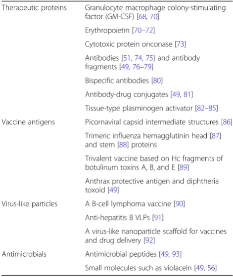
Manufacturing of therapeutics
Another active area in CFS research is the
biomanufac-turing of therapeutics and other protein-based reagents.
Natural biological systems have evolved a remarkable
capacity to synthesize a variety of molecules ranging
from metabolites to biopolymers. Cell-free protein
ex-pression systems allow the incorporation of such
reac-tions into a highly controlled process that allows
production of molecules as needed and in the field. Our
primary focus here will be on a subset of biopolymers,
namely therapeutic proteins. The ongoing work in this
field rests on decades of research that have led to the
productive and practical systems currently available [
28
,
29
,
36–38
,
40
]. Recent advances in high-throughput
preparation techniques [
40
,
45
] and in the development
of systems that can use more economical energy sources
[
64
,
65
] have made CFS highly accessible. Meanwhile,
significant strides are being made towards resolving
vari-ous protein folding issues and shortcomings in
post-translational modifications [
66
] associated with
trad-itional CFS. Recent advances have showcased the
poten-tial for scaling up cell-free reactions, with some having
demonstrated reaction volumes reaching 100 liters [
67
,
68
] to 1000 liters [
69
]. Cell-free expression has been
used as a platform for the production of a wide range of
potential therapeutics, some of which have been
summa-rized in Table
1
. A number of these products have been
Two primary modes of CFS have been pursued. The
first, used by commercial efforts such as Sutro [
94
],
fo-cuses on large, centralized production. This approach
le-verages the advantages of synthesis outside of the cell
for biomanufacturing. For these applications, CFS not
only allow for rapid production, but also significantly
speed up the drug development process [
95
].
Remark-ably, Sutro has reportedly increased their cell-free
pro-duction to an incredible 1000 liters [
69
], showcasing the
scalability of centralized cell-free production. The
sec-ond mode uses FD-CF systems to de-centralize
bioma-nufacturing capacity for small-batch production of
therapeutics, with applications in global health and
emergency response [
49
,
73
,
96
,
97
]. Using this mode of
production, we have recently demonstrated the
proof-of-concept capacity to manufacture over 50 therapeutics
and lab reagents, including proteins (e.g., vaccines,
anti-bodies, and antimicrobial peptides) and small molecules
[
49
], with applications outside of the laboratory setting.
Cell-free biomanufacturing is particularly well-suited
for vaccine production due to its potential for rapid
scale-up in response to public health emergencies.
Suc-cessful cell-free expression of a number of recombinant
vaccines (e.g., botulinum, diphtheria, anthrax) has been
demonstrated [
49
,
86–90
,
98
], with some having been
validated in animal models, such as mice [
49
,
90
].
Con-sidering the low dose requirements (microgram range)
for many of these therapeutics, commercialization of
CFS-derived vaccines will likely see rapid growth in the
coming years. Production of antibodies has also been an
area of focus for the cell-free community [
20
,
49
,
51
,
74–80
,
99
,
100
]. Due to their compact size and relatively
high expression levels in CFS, single-domain antibodies
have garnered particular attention and seem strategically
well-placed to serve the emerging needs in personalized
medicine, i.e., for therapeutics and diagnostics.
Antibiotic resistance has been recognized as a major
threat to global health, resulting in approximately two
million illnesses and 23,000 deaths in the US alone every
year [
101
]. Accordingly, cell-free production of
anti-microbial compounds, including antianti-microbial peptides
and small molecule drugs, has become the focus of some
groups [
49
,
93
]. A number of labs have also
demon-strated the power of CFS to express phages [
56
,
102
–
104
]. The upward trend in the reported antibiotic
resist-ance cases has led to a resurgence in viewing phage
Fig. 2Overview of the use of biosensors in CFS. The general workflow usually involves in silico design of gene circuits encoding biosensors and reporter proteins, followed by chemical synthesis of such circuits. Meanwhile, patient or environmental samples are collected, target analytes are extracted, and, in some cases, amplified. The gene circuits and target analytes are then added to CFS. Examples of biosensors in CFS have includeda) mercury (II) detection using the MerR repressor[45],b) viral and bacterial nucleic acid sensing using toehold switch-based sensors [46,
therapy as a potentially viable alternative to current
anti-biotic regimens [
101
,
105
]. The use of phages has also
been evaluated as an effective treatment strategy for a
number of plant diseases, with some phages now being
commercially available for mass consumption [
106
].
CFS-based production of these non-traditional
antimi-crobials could play a significant role in battling the
anti-biotic resistance crisis and could also help improve food
security around the globe.
Below, we will highlight some of the areas in which
CFS have shown great potential for enhancing current
methods of therapeutics development and
manufactur-ing. These advances are rapidly transforming CFS into
an integral part of the manufacturing ecosystem.
Membrane proteins
While approximately 70% of all drugs act on membrane
proteins[
107
], working with these proteins is notoriously
difficult due to their enrichment in hydrophobic surfaces.
Cell-based expression of membrane proteins is often
fraught with challenges, such as toxicity caused by their
membrane incorporation or their incompatibility with the
host
’
s physiology [
108
]. Recently, cell-free approaches have
been used to tackle this challenging category of proteins,
the coding sequences of which comprise 20
–
30% of all
known genes [
107
]. When compared to current cell-based
methods, CFS can be a powerful tool in the production of
soluble active membrane proteins [
109
]. The ability to
integrate steps that can tackle the challenging aspects of
membrane protein synthesis is particularly valuable. For
in-stance, previous efforts in cell-based systems have
demon-strated that membrane mimics can be successfully used to
synthesize and stabilize a wide range of membrane proteins
such as G-protein-coupled receptors [
110
,
111
], the
epider-mal growth factor receptor [
71
], hepatitis C virus
mem-brane proteins [
112
], and an ATP synthase [
109
,
113
].
These mimics include surfactants, liposomes, and
nano-discs [
114–116
] and can be added directly to CFS
co-translationally or post-co-translationally. There is also evidence
suggesting that functioning single-span membrane proteins
can be synthesized simply in the presence of an oil
–
water
interface (e.g., through the use of emulsions) [
117
].
Macromolecular production
Molecular research has highlighted the importance of
protein
–
protein interactions and the resulting complexes
that these interactions can generate. Whether it is for
the biophysical study of these complexes or as vehicles
for new therapeutic delivery (e.g., virus-like scaffolds for
vaccines), there is a growing need for developing robust
tools aimed at synthesis of such complexes. As in the
case of membrane proteins, CFS have also demonstrated
higher yields, compared to in vivo strategies, in the
pro-duction of macromolecular assemblies such as virus-like
particles (VLPs) [
109
]. Groundbreaking work by the
Swartz group, demonstrating the cell-free expression of
hepatitis B core antigen VLP (2 subunits) [
91
] in an
E.
coli
-based cell-free system, opened the door to other
re-searchers expressing a variety of macromolecular
assem-blies including the
E. coli
RNA polymerase (5 subunits)
[
118
] and an ATP synthase (25 subunits) [
113
]. Earlier
work with reticulocyte lysate had also demonstrated
cell-free expression of the human T-cell receptor (7
sub-units) [
119
]. Remarkably, a number of bacteriophages
have now also been successfully expressed in CFS,
in-cluding the T4 phage, which structurally contains 1500
proteins from 50 genes [
56
,
102–104
] (Fig.
3
).
Non-identical subunits of a protein complex are often
referred to as hetero subunits. In some instances, such
hetero subunits require co-translation to yield active
complexes [
120
]. Thus, the ability of CFS to
concur-rently translate multiple mRNAs facilitates the
produc-tion of active complexes composed of a number of
different subunits [
121
]. Some CFS such as
E. coli
-based
preparations are generally not capable of producing
pro-teins that contain disulfide bonds, which are critical to
numerous pharmaceutically relevant proteins (e.g.,
anti-bodies and many cytokines) [
121
]. However, recent
ef-forts have augmented these systems to enable the
production of complex proteins requiring multiple
disul-fide bonds [
85
,
99
,
122
], expanding the range of
therap-ies that can be made in CFS.
Table 1
Examples of potential therapeutics expressed in CFS to
date
Therapeutic proteins Granulocyte macrophage colony-stimulating factor (GM-CSF) [68,70]
Erythropoietin [70–72]
Cytotoxic protein onconase [73]
Antibodies [51,74,75] and antibody fragments [49,76–79]
Bispecific antibodies [80]
Antibody-drug conjugates [49,81]
Tissue-type plasminogen activator [82–85]
Vaccine antigens Picornaviral capsid intermediate structures [86]
Trimeric influenza hemagglutinin head [87] and stem [88] proteins
Trivalent vaccine based on Hc fragments of botulinum toxins A, B, and E [89]
Anthrax protective antigen and diphtheria toxoid [49]
Virus-like particles A B-cell lymphoma vaccine [90]
Anti-hepatitis B VLPs [91]
A virus-like nanoparticle scaffold for vaccines and drug delivery [92]
Antimicrobials Antimicrobial peptides [49,93]
Modification of proteins and codon tables
Effectiveness of many protein-based therapeutics hinges
upon precise control over natural or non-natural
modifica-tion of their peptide sequences. One of the most
compel-ling uses of such modifications is in the development of
antibody
−
drug conjugates (ADCs), which are quickly
gaining favor as a new class of therapeutics against cancer.
Classic conjugation techniques result in a heterogeneous
mixture of labeled antibodies due to their reliance on
arbi-trary conjugation to multiple amino acid side chains.
Recent studies, however, suggest that pharmacologic
prop-erties of ADCs could be improved through site-specific
conjugation. Non-natural amino acids provide an efficient
avenue for such site-specific conjugation [
123
]. To date,
co-translational incorporation of over 100 different
non-natural amino acids has been demonstrated in vivo [
124
],
allowing for a wide range of modifications [
125–129
]. Many
of these modifications have been demonstrated in the
cell-free context for a variety of applications, including
orientation-controlled immobilization [
92
,
98
] and
site-specific functionalization (e.g., phosphorylation [
130
],
PEGylation [
131
], or drug conjugation [
81
]) [
132–134
].
CFS platforms circumvent some of the cell-based
tox-icity and permeability limitations and offer greater control
and versatility in making protein modifications [
109
,
135
].
Incorporation of non-natural amino acids in cell-based
approaches has typically relied on repurposing stop
co-dons to minimize the negative impacts of recoding on
cell-viability [
109
]. In a cell-free system, however, the
en-tire codon table can in theory be reprogrammed, allowing
not only for the incorporation of non-natural amino acids,
but also for the creation of entirely novel codon tables.
Taken to its extreme, the latter could help with the
protection of intellectual property. DNA sequences
could be obfuscated such that they are rendered
non-functional outside of their specialized cell-free context.
This obfuscated code would make proprietary designs
difficult to copy. Codon obfuscation could also pose
ser-ious challenges for the detection of DNA sequences that
may be employed by malevolent entities. For example,
DNA synthesis companies would have a much more
dif-ficult time screening against DNA sequences that could
be used for nefarious activities (e.g., bioterrorism).
Re-cent work has shown that the size of the codon table
can also be expanded by augmenting the four-letter
gen-etic alphabet with unnatural base pairs [
136
,
137
]. Thus,
proteins made in CFS could
—
at least in theory
—
hold an
unlimited number of non-natural amino acids.
CFS can also be employed for making naturally
occur-ring modifications to proteins. An example of these is
the grafting of sugars (i.e., glycans) referred to as
glyco-sylation. Successful production of many therapeutics is
often contingent upon highly efficient glycosylation, as
lack of proper glycosylation can reduce the efficacy and
circulation half-life of many therapeutic proteins [
138
].
Some CFS (e.g., insect, Chinese hamster ovary, and
hu-man K562 extract-based systems) are inherently capable
of glycosylation. However, their repertoire of glycan
structures tends to be limited to those naturally
synthe-sized by their lysates
’
source cell type. Additionally,
gly-cosylation in these systems often requires recapitulation
of the source cell
’
s protein trafficking mechanisms [
109
].
Thus, creation of synthetic glycosylation pathways in
CFS has become an area of focus in recent years [
135
,
139
]. Success in this domain will likely serve as a key
catalyst in bringing cell-free-produced vaccines and
other therapeutics to the masses. Figure
4
outlines some
of the possible protein modifications in CFS.
Directed evolution
Directed evolution is a powerful tool for aptamer and
pro-tein engineering that uses iterative rounds of mutagenesis
and selection to modify or tune specific bimolecular
prop-erties (e.g., an enzyme
’
s substrate activity). Utility of
apta-mers or proteins, in a given context, with respect to their
corresponding nucleotide sequences is often described as
a fitness landscape. Directed evolution provides a
mas-sively parallel method for searching through a fitness
land-scape to find optimal variants and their corresponding
genotypes [
144
]. This generally requires one-to-one
map-ping of phenotype to genotype. Although cells have a
built-in capacity for such mapping due to their
compart-mentalized nature, using cells to conduct directed
evolu-tion can impose limits on the size of candidate libraries
screened, and restricts the type of solvents, buffers, and
temperatures that can be sampled [
145
]. As a result,
cell-free directed evolution platforms have gained favor [
145
],
starting with the first truly cell-free systems published in
the late 90s [
146
,
147
]. More recently, connecting
pheno-type to genopheno-type has been accomplished through artificial
compartmentalization (e.g., using emulsion, microbeads,
and liposomes) [
145
,
148–151
]. Applications have
in-cluded the design and optimization of Fab antibody
frag-ments [
77
,
152
], membrane proteins [
151
], and, as we will
discuss below, enzyme discovery [
52
].
Platform for discovery
Engineered transcription and translation systems can
also greatly catalyze research in the laboratory. As
previ-ously mentioned, the absence of a cell wall means that
candidate genes can be readily screened for function. It
also means that substrates, including those difficult to
use in the cellular context, can easily be brought into
contact with enzyme libraries to screen for novel
reac-tions. Below we look at some of the recent work using
CFS as a platform for discovery.
Biosynthetic pathways
From the early days of synthetic biology, it was clear that
there was great potential for synergy with the field of
chemical synthesis. Metabolic pathways responsible for
the synthesis of valuable compounds (e.g., drugs, scents,
and flavors) were thus moved out of organisms that did
not easily lend themselves to production and into
heterol-ogous hosts, such as yeast. This microorganism-based
ap-proach has been incredibly successful and has led to the
assembly of genes from disparate sources to create
engi-neered pathways. Enzyme-based catalysis has the
advan-tage of allowing for stereo-selectivity in aqueous,
low-energy reactions (e.g., green chemistry) [
153
]. By
lever-aging naturally occurring pathways, it has been possible to
generate tremendous chemical diversity, as seen in
isopre-noids, from simple precursors [
154
]. An exemplar of this
approach is the synthesis of amorpha-4,11-diene and
arte-misinic acid, which are precursors to the anti-malarial
compound artemisinin [
154–157
]. This process has been
repeated for other pharmaceutical pathways, enabling the
production of opioids [
158
,
159
] and taxol [
160
], as well
as for the generation of molecules for the energy industry
and the agriculture sector [
13
,
161
].
While microorganisms are currently a mainstay for
biomanufacturing of commodities, their use for these
purposes is nontrivial. For example, assembly,
fine-tuning, and host strain integration of the
industrial-ized pathway for the bioproduction of artemisinic acid
is estimated to have taken over 150 person-years
[
162
]. Another challenge to microbial bioproduction
is that a significant portion of inputs are lost to
gen-eral cellular metabolism and growth, reducing
effi-ciency of the overall process [
67
,
134
]. Cell-free
synthetic biology alleviates some of these challenges.
For instance, enzyme discovery
—
the identification of
enzymes that can be used for biosynthetic purposes
—
via CFS has proven to be effective. Enzymes and their
homologs can be rapidly screened for performance
without the cumbersome steps required for cell-based
screening (e.g., plasmid assembly and transformation).
This approach can be extended to simple prototyping
of pathways or the automated multiplexed shuffling of
complex pathway components. Unlike with cell-based
prototyping, the cell-free environment allows for the
use of enzymes encoded as linear constructs (DNA or
RNA). Substrate preference can also be evaluated
without the need for enzyme purification.
In many cases, enzymes and pathways discovered in
CFS will be brought back into cells for scale-up [
163
].
However, there is a growing case for using CFS
dir-ectly as the production medium. Commercial ventures
(e.g., Sutro, Greenlight) have already demonstrated
that CFS can provide economic advantages for the
production of protein and RNA products [
69
]. Thus,
it would be reasonable to think that a similar
ap-proach could provide a viable source of high-value
small molecules. Such systems have the advantage of
enabling bioproduction without metabolic
inefficien-cies, toxicity limitations, complex extraction steps, or
the need for integration into a host strain [
67
,
134
,
164
]. Combined with the capacity for efficient
proto-typing, these systems are generating significant
enthu-siasm. The field is now beginning to focus on more
complex pathways (more than eight enzymes) and
lar-ger reaction volumes (> 100 L) [
67
].
Single enzyme reactions are highly simplified cell-free
systems that have been used for decades at scale for
washing (e.g., dish and laundry detergents) and for
pro-cessing food, wood pulp, and fuel [
165
]. Once fully
oper-ationalized, more complex cell-free enzymatic pathways
could revolutionize the chemical industry and enable
greater accessibility to bioproduction. Earlier attempts at
engineering such pathways outside of a cell were
gener-ally made by using purified components. These pathways
have included those designed for the production of
amorpha-4,11-diene [
166
], isoprene [
167
], fatty acids
[
168
], and nucleotides [
169
]. Recent work has showcased
the use of 27 purified enzymes that can work together to
convert glucose into terpenes such as limonene, pinene,
and sabinene [
170
]. Here, production can operate
con-tinuously for 5 days with a single addition of glucose,
with glucose conversion of greater than 95%, to generate
high product concentrations (> 15 g/L) that are well
above levels toxic to microbes. While exciting,
expres-sion and purification of each individual component for
such an approach is quite laborious.
Transitioning these metabolic pathways into CFS, where
expression of enzyme-encoding sequences could lead to the
self-assembly of pathways, would be incredibly enabling. To
date, a number of reports have validated this approach.
Three- and six-enzyme pathways have recently been
gener-ated de novo from DNA inputs in CFS to produce
N-acetylglucosamine and a peptidoglycan precursor,
respect-ively [
171
,
172
]. A five-enzyme pathway that transforms
tryp-tophan into a bioactive pigment called violacein has also
been demonstrated [
49
,
56
]. Additionally, a combinatorial
strategy has recently been used to build a 17-step enzyme
pathway for n-butanol [
173
]. It is intriguing to envision how
this approach could influence the synthesis of high-value
commodities (e.g., small-molecule drugs, cosmetic
ingredi-ents, food additives, and scented compounds), and move
production towards more sustainable enzyme-catalyzed
processes.
The cell-free assembly of engineered metabolic
path-ways has led to parallel approaches in the areas of
en-ergy production, biomaterials, and even the development
of artificial cells. Below we introduce some of the related
efforts in these fields.
Energy storage and generation
Cell-free enzymatic pathways have recently been used to
create biobatteries with small environmental footprints
and energy-storage densities superior to that of current
lithium-ion devices [
174
]. Moreover, previous studies
have demonstrated ATP generation on electrode surfaces
[
175
,
176
]. Since both the assembly of ATP synthase
[
113
] and the synthesis of membrane proteins into
teth-ered lipid bilayers [
177
] have been shown in CFS, one
potential application of CFS could be rapid prototyping
and construction of novel energy-generating biodevices
that would be capable of producing electricity from
low-value commodities (i.e., biomass or waste) [
109
]. One
could readily imagine CFS simply powered by light [
178
]
or electricity, which could help lower the cost of
manu-facturing industrially relevant biomolecules as discussed
above.
Biomaterials
As noted earlier, CFS have not only been used to screen
the natural diversity of enzymes, but also to sculpt
en-zymatic activity. In an example of this, Bawazer et al
.
used CFS to synthesize solid-state materials [
52
]. A
cell-free system was used to exert evolutionary selection on
biomineralizing enzymes called silicateins that are
cap-able of synthesizing silicon dioxide or titanium dioxide.
DNA fragments coding for two isoforms of silicatein
were digested and reassembled by DNA shuffling to
cre-ate a library of chimeric enzymes. Through a clever
scheme of selection, variants were then chosen for their
ability to deposit silica or titanium dioxide onto
microbeads in an oil-water emulsion. The success of this
methodology through the use of CFS raises the exciting
prospect of using green chemistry for the deposition of
semi-conductor materials. This type of green deposition
could also be modified such that it is guided by a
CFS-compatible photolithography technique similar to that
demonstrated by the Bar-Ziv group [
55
,
179
,
180
].
Artificial cells
platform for engineering artificial cells [
37
,
151
,
183–
185
]. Artificial cells have many important applications;
they can be used to link phenotype to genotype in vitro
for directed evolution applications, and to spatially
sep-arate synthesis of different proteins [
185
]. There is also
evidence indicating that confinement, a feature common
to many types of artificial cells, can be used to boost
protein expression yields of CFS [
186
]. Furthermore,
artificial cells may allow for prolonged expression
with-out relying on traditional dialysis methods that are often
used to provide a continuous supply of reaction
precur-sors. For example, early work by the Noireaux group
showed that membrane-based artificial cells can be
aug-mented with
α
-hemolysin pore proteins from
Staphylo-coccus aureus
in order to achieve selective permeability
for nutrients [
182
,
187
].
Artificial cells may also be constructed in the form of
solid-state two-dimensional compartments. Silicon has
been used to fabricate two-dimensional artificial cells
capable of carrying out many of the features possible in
cell-based systems. These features include simple
metab-olism, operation of gene circuits (e.g., oscillators), and
even communication between compartments. Control
over fabrication geometry allows for precise evaluation
of the effects of diffusion gradients and can help tune
protein turnover [
55
,
179
].
Looking forward, perhaps one of the most exciting
and promising applications of artificial cells is the ability
to express membrane proteins efficiently. This could
allow for cell-free engineering of signaling pathways
[
188
], such as those involving G-protein-coupled
recep-tors (GPCRs) [
189
,
190
]. Approximately 34% of all
FDA-approved drugs act on GPCR targets [
191
]. As such,
artificial cells could become an invaluable tool in the
drug discovery process. Artificial cells also have the
po-tential to be used for in vivo therapeutics. For example,
they could be designed to perform sensing, logic, or
therapeutic functions. Artificial cells may be designed to
accumulate at a tumor site through the enhanced
per-meability and retention (EPR) effect [
192
] or by using
targeting molecules on their surface. They can also be
constructed to protect therapeutic enzymes while being
permeable to specific substrates and products, thus
in-creasing active circulation time and expanding their
therapeutic potential [
193
,
194
].
Education
Given their potential for biosafety and portability,
cell-free systems offer a great platform for teaching
key concepts in synthetic biology. The Cold Spring
Harbor Laboratory course in synthetic biology, for
ex-ample, includes modules that utilize cell-free systems
[
195
]. In recent work led by Jim Collins and Michael
Jewett, the ability of CFS to support on-demand and
on-site sensing and manufacturing was further
ex-tended to bring synthetic biology capabilities to the
classroom [
196
,
197
]. Here FD-CF components were
used to create kits that enable students to experience
rational design of reactions, such as creating their
own unique colors by mixing DNA coding for
differ-ent fluorescdiffer-ent proteins. Other applications included
the on-demand creation of fluorescent hydrogels,
scents, and even sensors that could distinguish
be-tween DNA from banana, kiwi, and strawberry.
Reflecting an important trend in the field of synthetic
biology, this work included the testing of tools under
field conditions with the help of high school students.
This work sets the important groundwork for
inspir-ing curiosity and passion in students who will drive
the next generation of synthetic biology.
The future of biotechnology with cell-free systems
The merger of cell-free systems with the vast array of
genetically programmable tools is transforming the
syn-thetic biology landscape, creating powerful in vitro
plat-forms. These platforms have already begun to bring
about de-centralization of health care through portable
diagnostics and drug manufacturing. They also have
great potential for the efficient, centralized production
of high-value commodities. Cell-free synthetic biology
approaches will take biology and biotechnology to new
horizons and will surely produce many creative and
un-expected outcomes. We expect the field to continue to
expand and to merge with other engineered systems.
One could envision programmed interactions with
mate-rials on the nano-scale and interplay with a variety of
engineered enzymes. We are excited to see how CFS will
bring synthetic biology closer to electronics,
computa-tion, and machine learning.
Acknowledgments
Not applicable.
Authors’contributions
AT and KP co-authored and edited the manuscript. KJ edited the manuscript and created the figures as well as their legends. All authors read and ap-proved the final manuscript.
Funding
This work was supported by the CIHR Foundation Grant Program (201610FDN-375469), CIHR/IDRC team grant (149783), and the Canada Research Chair Program (CIHR, 950-231075) to K.P.
Availability of data and materials
Not applicable.
Competing interests
References
1. Clancy K, Voigt CA. Programming cells: towards an automated‘Genetic Compiler’. Curr Opin Biotechnol. 2010;21(4):572–81.https://doi.org/10.1016/j. copbio.2010.07.005.
2. van der Meer JR, Belkin S. Where microbiology meets microengineering: design and applications of reporter bacteria. Nat Rev Microbiol. 2010;8(7): 511–22.https://doi.org/10.1038/nrmicro2392.
3. Mao N, Cubillos-Ruiz A, Cameron DE, Collins JJ. Probiotic strains detect and suppress cholera in mice. Sci Transl Med. 2018;10(445):eaao2586.https://doi. org/10.1126/scitranslmed.aao2586.
4. Siciliano V, DiAndreth B, Monel B, Beal J, Huh J, Clayton KL, et al. Engineering modular intracellular protein sensor-actuator devices. Nat Commun. 2018;9(1):1881.https://doi.org/10.1038/s41467-018-03984-5. 5. Kotula JW, Kerns SJ, Shaket LA, Siraj L, Collins JJ, Way JC, et al.
Programmable bacteria detect and record an environmental signal in the mammalian gut. Proc Natl Acad Sci U S A. 2014;111(13):4838–43.https://doi. org/10.1073/pnas.1321321111.
6. Friedland AE, Lu TK, Wang X, Shi D, Church G, Collins JJ. Synthetic gene networks that count. Science. 2009;324(5931):1199–202.https://doi.org/1 0.1126/science.1172005.
7. Green AA, Kim J, Ma D, Silver PA, Collins JJ, Yin P. Complex cellular logic computation using ribocomputing devices. Nature. 2017;548(7665):117–21.
https://doi.org/10.1038/nature23271.
8. Kitada T, DiAndreth B, Teague B, Weiss R. Programming gene and engineered-cell therapies with synthetic biology. Science. 2018;359(6376): eaad1067.https://doi.org/10.1126/science.aad1067.
9. Simpson ML, Sayler GS, Fleming JT, Applegate B. Whole-cell biocomputing. Trends Biotechnol. 2001;19(8):317–23.
10. Yehl K, Lu T. Scaling computation and memory in living cells. Curr Opin Biomed Eng. 2017;4:143–51.https://doi.org/10.1016/j.cobme.2017.10.003. 11. Anderson LA, Islam MA, Prather KLJ. Synthetic biology strategies for
improving microbial synthesis of“green”biopolymers. J Biol Chem. 2018; 293(14):5053–61.https://doi.org/10.1074/jbc.TM117.000368.
12. Fossati E, Ekins A, Narcross L, Zhu Y, Falgueyret J-P, Beaudoin GAW, et al. Reconstitution of a 10-gene pathway for synthesis of the plant alkaloid dihydrosanguinarine in Saccharomyces cerevisiae. Nat Commun. 2014;5: 3283.https://doi.org/10.1038/ncomms4283.
13. Smanski MJ, Zhou H, Claesen J, Shen B, Fischbach MA, Voigt CA. Synthetic biology to access and expand nature’s chemical diversity. Nat Rev Microbiol. 2016;14(3):135–49.https://doi.org/10.1038/nrmicro.2015.24.
14. Nielsen J, Keasling JD. Engineering cellular metabolism. Cell. 2016;164(6): 1185–97.https://doi.org/10.1016/j.cell.2016.02.004.
15. Wagner TE, Becraft JR, Bodner K, Teague B, Zhang X, Woo A, et al. Small-molecule-based regulation of RNA-delivered circuits in mammalian cells. Nat Chem Biol. 2018;14(11):1043–50. https://doi.org/10.1038/s41589-018-0146-9.
16. Scheller L, Strittmatter T, Fuchs D, Bojar D, Fussenegger M. Generalized extracellular molecule sensor platform for programming cellular behavior. Nat Chem Biol. 2018;14(7):723–9.https://doi.org/10.1038/s41589-018-0046-z. 17. Cho JH, Collins JJ, Wong WW. Universal chimeric antigen receptors for
multiplexed and logical control of T cell responses. Cell. 2018;173(6):1426– 1438.e11.https://doi.org/10.1016/j.cell.2018.03.038.
18. Lee JW, Chan CTY, Slomovic S, Collins JJ. Next-generation biocontainment systems for engineered organisms. Nat Chem Biol. 2018;14(6):530–7.https:// doi.org/10.1038/s41589-018-0056-x.
19. Jia B, Qi H, Li B-Z, Pan S, Liu D, Liu H, et al. Orthogonal ribosome biofirewall. ACS Synth Biol. 2017;6(11):2108–17.https://doi.org/10.1021/acssynbio. 7b00148.
20. Martin RW, Majewska NI, Chen CX, Albanetti TE, Jimenez RBC, Schmelzer AE, et al. Development of a CHO-based cell-free platform for synthesis of active monoclonal antibodies. ACS Synth Biol. 2017;6(7):1370–9.https://doi.org/1 0.1021/acssynbio.7b00001.
21. Mikami S, Masutani M, Sonenberg N, Yokoyama S, Imataka H. An efficient mammalian cell-free translation system supplemented with translation factors. Protein Expr Purif. 2006;46(2):348–57.https://doi.org/10.1016/j.pep.2 005.09.021.
22. Tran K, Gurramkonda C, Cooper MA, Pilli M, Taris JE, Selock N, et al. Cell-free production of a therapeutic protein: Expression, purification, and
characterization of recombinant streptokinase using a CHO lysate. Biotechnol Bioeng. 2018;115(1):92–102.https://doi.org/10.1002/bit.26439. 23. Burgenson D, Gurramkonda C, Pilli M, Ge X, Andar A, Kostov Y, et al. Rapid
recombinant protein expression in cell-free extracts from human blood. Sci Rep. 2018;8(1):9569.https://doi.org/10.1038/s41598-018-27846-8. 24. Ezure T, Suzuki T, Higashide S, Shintani E, Endo K, Kobayashi S-i, et al.
Cell-free protein synthesis system prepared from insect cells by Cell-freeze-thawing. Biotechnol Prog. 2006;22(6):1570–7.https://doi.org/10.1021/bp060110v. 25. Buntru M, Vogel S, Stoff K, Spiegel H, Schillberg S. A versatile coupled
cell-free transcription-translation system based on tobacco BY-2 cell lysates. Biotechnol Bioeng. 2015;112(5):867–78.https://doi.org/10.1002/bit.25502. 26. Harbers M. Wheat germ systems for cell-free protein expression. FEBS Lett.
2014;588(17):2762–73.https://doi.org/10.1016/j.febslet.2014.05.061. 27. Hodgman CE, Jewett MC. Optimized extract preparation methods and
reaction conditions for improved yeast cell-free protein synthesis. Biotechnol Bioeng. 2013;110(10):2643–54.https://doi.org/10.1002/bit.24942. 28. Yang WC, Patel KG, Wong HE, Swartz JR. Simplifying and streamlining
Escherichia coli-based cell-free protein synthesis. Biotechnol Prog. 2012; 28(2):413–20.https://doi.org/10.1002/btpr.1509.
29. Kigawa T, Yabuki T, Matsuda N, Matsuda T, Nakajima R, Tanaka A, et al. Preparation of Escherichia coli cell extract for highly productive cell-free protein expression. J Struct Funct Genomics. 2004;5(1–2):63–8.https://doi. org/10.1023/B:JSFG.0000029204.57846.7d.
30. Moore SJ, Lai H-E, Needham H, Polizzi KM, Freemont PS.Streptomyces
venezuelaeTX-TL - a next generation cell-free synthetic biology tool. Biotechnol J. 2017;12(4):1600678.https://doi.org/10.1002/biot.201600678. 31. Moore SJ, MacDonald JT, Wienecke S, Ishwarbhai A, Tsipa A, Aw R, et al.
Rapid acquisition and model-based analysis of cell-free transcription-translation reactions from nonmodel bacteria. Proc Natl Acad Sci U S A. 2018.https://doi.org/10.1073/pnas.1715806115.
32. Kelwick R, Webb AJ, MacDonald JT, Freemont PS. Development of a Bacillus subtilis cell-free transcription-translation system for prototyping regulatory elements. Metab Eng. 2016;38:370–81.https://doi.org/10.1016/J.YMBEN.2016. 09.008.
33. Li J, Wang H, Jewett MC. Expanding the palette of Streptomyces -based cell-free protein synthesis systems with enhanced yields. Biochem Eng J. 2018;130:29–33.https://doi.org/10.1016/j.bej.2017.11.013.
34. Li J, Wang H, Kwon Y-C, Jewett MC. Establishing a high yielding
streptomyces-based cell-free protein synthesis system. Biotechnol Bioeng. 2017;114(6):1343–53.https://doi.org/10.1002/bit.26253.
35. Failmezger J, Scholz S, Blombach B, Siemann-Herzberg M. Cell-free protein synthesis from fast-growing Vibrio natriegens. Front Microbiol. 2018;9:1146.
https://doi.org/10.3389/fmicb.2018.01146.
36. Shimizu Y, Inoue A, Tomari Y, Suzuki T, Yokogawa T, Nishikawa K, et al. Cell-free translation reconstituted with purified components. Nat Biotechnol. 2001;19(8):751–5.https://doi.org/10.1038/90802.
37. Shin J, Noireaux V. An E.coli cell-free expression toolbox: Application to synthetic gene circuits and artificial cells. ACS Synth Biol. 2012;1(1):29–41.
https://doi.org/10.1021/sb200016s.
38. Jewett MC, Calhoun KA, Voloshin A, Wuu JJ, Swartz JR. An integrated cell-free metabolic platform for protein production and synthetic biology. Mol Syst Biol. 2008;4:220.https://doi.org/10.1038/msb.2008.57.
39. Li J, Gu L, Aach J, Church GM. Improved cell-free RNA and protein synthesis system. PLoS One. 2014;9(9):e106232.https://doi.org/10.1371/journal.pone.01 06232.
40. Kwon Y-C, Jewett MC. High-throughput preparation methods of crude extract for robust cell-free protein synthesis. Sci Rep. 2015;5:8663.https:// doi.org/10.1038/srep08663.
41. Sun ZZ, Hayes CA, Shin J, Caschera F, Murray RM, Noireaux V. Protocols for implementing an Escherichia coli based TX-TL cell-free expression system for synthetic biology. J Vis Exp. 2013;79:e50762.https://doi.org/10.3791/5 0762.
42. Caschera F, Noireaux V. Synthesis of 2.3 mg/ml of protein with an all Escherichia coli cell-free transcription–translation system. Biochimie. 2014;99: 162–8.https://doi.org/10.1016/j.biochi.2013.11.025.
43. Wiegand DJ, Lee HH, Ostrov N, Church GM. Establishing a cell-free Vibrio natriegens expression system. bioRxiv. 2018:331645.https://doi.org/10.11 01/331645.
44. Tuckey C, Asahara H, Zhou Y, Chong S. Protein synthesis using a reconstituted cell-free system. Curr Protoc Mol Biol. 2014;108:16.31.1–22.
45. Didovyk A, Tonooka T, Tsimring L, Hasty J. Rapid and scalable preparation of bacterial lysates for cell-free gene expression. ACS Synth Biol. 2017;6(12): 2198–208.https://doi.org/10.1021/acssynbio.7b00253.
46. Pardee K, Green AA, Ferrante T, Cameron DE, DaleyKeyser A, Yin P, et al. Paper-based synthetic gene networks. Cell. 2014;159(4):940–54.https://doi. org/10.1016/j.cell.2014.10.004.
47. Chan P, Thomas CJ, Sprang SR, Tall GG. Molecular chaperoning function of Ric-8 is to fold nascent heterotrimeric G proteinαsubunits. Proc Natl Acad Sci U S A. 2013;110(10):3794–9.https://doi.org/10.1073/pnas.1220943110. 48. Smith MT, Berkheimer SD, Werner CJ, Bundy BC. Lyophilized Escherichia
coli-based cell-free systems for robust, high-density, long-term storage. Biotechniques. 2014;56(4):186–93.https://doi.org/10.2144/000114158. 49. Pardee K, Slomovic S, Nguyen PQ, Lee JW, Donghia N, Burrill D, et al.
Portable, on-demand biomolecular manufacturing. Cell. 2016;167(1):248– 254.e12.https://doi.org/10.1016/j.cell.2016.09.013.
50. Pardee K, Green AA, Takahashi MK, Connor DHO, Gehrke L, Collins JJ, et al. Rapid, low-cost detection of Zika virus using programmable biomolecular components. Cell. 2016;165(5):1255–66.https://doi.org/10.1016/j.cell.2016.04.059.
51. Yin G, Garces ED, Yang J, Zhang J, Tran C, Steiner AR, et al. Aglycosylated antibodies and antibody fragments produced in a scalable in vitro transcription-translation system. MAbs. 2012;4(2):217–25.https://doi.org/1 0.4161/mabs.4.2.19202.
52. Bawazer LA, Izumi M, Kolodin D, Neilson JR, Schwenzer B, Morse DE. Evolutionary selection of enzymatically synthesized semiconductors from biomimetic mineralization vesicles. Proc Natl Acad Sci U S A. 2012;109(26): E1705–14.https://doi.org/10.1073/pnas.1116958109.
53. Sun ZZ, Yeung E, Hayes CA, Noireaux V, Murray RM. Linear DNA for rapid prototyping of synthetic biological circuits in an Escherichia coli based TX-TL cell-free system. ACS Synth Biol. 2014;3(6):387–97.https://doi.org/10.1021/sb400131a. 54. Takahashi MK, Chappell J, Hayes CA, Sun ZZ, Kim J, Singhal V, et al. Rapidly
characterizing the fast dynamics of RNA genetic circuitry with cell-free transcription-translation (TX-TL) systems. ACS Synth Biol. 2015;4(5):503–15.
https://doi.org/10.1021/sb400206c.
55. Karzbrun E, Tayar AM, Noireaux V, Bar-Ziv RH. Programmable on-chip DNA compartments as artificial cells. Science. 2014;345(6198):829–32.https://doi. org/10.1126/science.1255550.
56. Garamella J, Marshall R, Rustad M, Noireaux V. The all E.coli TX-TL Toolbox 2. 0: A Platform for Cell-Free Synthetic Biology. ACS Synth Biol. 2016;5(4):344– 55.https://doi.org/10.1021/acssynbio.5b00296.
57. Niederholtmeyer H, Sun ZZ, Hori Y, Yeung E, Verpoorte A, Murray RM, et al. Rapid cell-free forward engineering of novel genetic ring oscillators. Elife. 2015;4:e09771.https://doi.org/10.7554/eLife.09771.
58. Green AA, Silver PA, Collins JJ, Yin P. Toehold switches: de-novo-designed regulators of gene expression. Cell. 2014;159(4):925–39.https://doi.org/10.1 016/j.cell.2014.10.002.
59. Takahashi MK, Tan X, Dy AJ, Braff D, Akana RT, Furuta Y, et al. A low-cost paper-based synthetic biology platform for analyzing gut microbiota and host biomarkers. Nat Commun. 2018;9(1):3347.https://doi.org/10.1038/s4146 7-018-05864-4.
60. Duyen TTM, Matsuura H, Ujiie K, Muraoka M, Harada K, Hirata K. Paper-based colorimetric biosensor for antibiotics inhibiting bacterial protein synthesis. J Biosci Bioeng. 2016;123(1):96–100.https://doi.org/10.1016/j.jbiosc.2016.07.015. 61. Wen KY, Cameron L, Chappell J, Jensen K, Bell DJ, Kelwick R, et al. A cell-free
biosensor for detecting quorum sensing molecules inP. aeruginosa-infected respiratory samples. ACS Synth Biol. 2017;6(12):2293–301.https://doi.org/1 0.1021/acssynbio.7b00219.
62. Salehi ASM, Shakalli Tang MJ, Smith MT, Hunt JM, Law RA, Wood DW, et al. Cell-free protein synthesis approach to biosensing hTRβ-specific endocrine disruptors. Anal Chem. 2017;89(6):3395–401.https://doi.org/10.1021/acs. analchem.6b04034.
63. Salehi ASM, Yang SO, Earl CC, Shakalli Tang MJ, Porter Hunt J, Smith MT, et al. Biosensing estrogenic endocrine disruptors in human blood and urine: A RAPID cell-free protein synthesis approach. Toxicol Appl Pharmacol. 2018; 345:19–25.https://doi.org/10.1016/j.taap.2018.02.016.
64. Calhoun KA, Swartz JR. Energizing cell-free protein synthesis with glucose metabolism. Biotechnol Bioeng. 2005;90(5):606–13.https://doi.org/10.1002/ bit.20449.
65. Kim T-W, Kim H-C, Oh I-S, Kim D-M. A highly efficient and economical cell-free protein synthesis system using the S12 extract of Escherichia coli. Biotechnol Bioprocess Eng. 2008;13(4):464–9. https://doi.org/10.1007/s12257-008-0139-8.
66. Carlson ED, Gan R, Hodgman CE, Jewett MC. Cell-free protein synthesis: applications come of age. Biotechnol Adv. 2012;30(5):1185–94.https://doi. org/10.1016/j.biotechadv.2011.09.016.
67. Dudley QM, Karim AS, Jewett MC. Cell-free metabolic engineering: biomanufacturing beyond the cell. Biotechnol J. 2015;10(1):69–82.https:// doi.org/10.1002/biot.201400330.
68. Zawada JF, Yin G, Steiner AR, Yang J, Naresh A, Roy SM, et al. Microscale to manufacturing scale-up of cell-free cytokine production--a new approach for shortening protein production development timelines. Biotechnol Bioeng. 2011;108(7):1570–8.https://doi.org/10.1002/bit.23103. 69. Breaking free from cells; Synthetic biology. Econ. 2017 (May 6).
70. Sullivan CJ, Pendleton ED, Sasmor HH, Hicks WL, Farnum JB, Muto M, et al. A cell-free expression and purification process for rapid production of protein biologics. Biotechnol J. 2016;11(2):238–48.https://doi.org/10.1002/ biot.201500214.
71. Stech M, Brödel AK, Quast RB, Sachse R, Kubick S. Cell-free systems: Functional modules for synthetic and chemical biology. Adv Biochem Eng Biotechnol. 2013;137:67–102.https://doi.org/10.1007/10_2013_185. 72. Brödel AK, Wüstenhagen DA, Kubick S. Cell-free protein synthesis systems
derived from cultured mammalian cells. Methods Mol Biol. 2015;1261:129– 40.https://doi.org/10.1007/978-1-4939-2230-7_7.
73. Salehi ASM, Smith MT, Bennett AM, Williams JB, Pitt WG, Bundy BC. Cell-free protein synthesis of a cytotoxic cancer therapeutic: Onconase production and a just-add-water cell-free system. Biotechnol J. 2016;11(2):274–81.
https://doi.org/10.1002/biot.201500237.
74. Groff D, Armstrong S, Rivers PJ, Zhang J, Yang J, Green E, et al. Engineering toward a bacterial“endoplasmic reticulum”for the rapid expression of immunoglobulin proteins. MAbs. 2014;6(3):671–8.https://doi.org/10.4161/ mabs.28172.
75. Cai Q, Hanson JA, Steiner AR, Tran C, Masikat MR, Chen R, et al. A simplified and robust protocol for immunoglobulin expression in Escherichia coli cell-free protein synthesis systems. Biotechnol Prog. 2015;31(3):823–31.https:// doi.org/10.1002/btpr.2082.
76. Kanter G, Yang J, Voloshin A, Levy S, Swartz JR, Levy R. Cell-free production of scFv fusion proteins: an efficient approach for personalized lymphoma vaccines. Blood. 2007;109(8):3393–9. https://doi.org/10.1182/blood-2006-07-030593.
77. Stafford RL, Matsumoto ML, Yin G, Cai Q, Fung JJ, Stephenson H, et al. In vitro Fab display: a cell-free system for IgG discovery. Protein Eng Des Sel. 2014;27(4):97–109.https://doi.org/10.1093/protein/gzu002.
78. Kawasaki T, Gouda MD, Sawasaki T, Takai K, Endo Y. Efficient synthesis of a disulfide-containing protein through a batch cell-free system from wheat germ. Eur J Biochem. 2003;270(23):4780–6.
79. Stech M, Merk H, Schenk JA, Stöcklein WFM, Wüstenhagen DA, Micheel B, et al. Production of functional antibody fragments in a vesicle-based eukaryotic cell-free translation system. J Biotechnol. 2013;164(2):220–31.
https://doi.org/10.1016/j.jbiotec.2012.08.020.
80. Xu Y, Lee J, Tran C, Heibeck TH, Wang WD, Yang J, et al. Production of bispecific antibodies in“knobs-into-holes”using a cell-free expression system. MAbs. 2015;7(1):231.https://doi.org/10.4161/19420862.2015.989013. 81. Zimmerman ES, Heibeck TH, Gill A, Li X, Murray CJ, Madlansacay MR, et al.
Production of site-specific antibody–drug conjugates using optimized non-natural amino acids in a cell-free expression system. Bioconjug Chem. 2014; 25(2):351–61.https://doi.org/10.1021/bc400490z.
82. Stech M, Quast RB, Sachse R, Schulze C, Wüstenhagen DA, Kubick S. A continuous-exchange cell-free protein synthesis system based on extracts from cultured insect cells. PLoS One. 2014;9(5):e96635.https://doi.org/10.13 71/journal.pone.0096635.
83. Bulleid NJ, Bassel-Duby RS, Freedman RB, Sambrook JF, Gething MJ. Cell-free synthesis of enzymically active tissue-type plasminogen activator. Protein folding determines the extent of N-linked glycosylation. Biochem J. 1992; 286(Pt 1):275–80.https://doi.org/10.1042/BJ2860275.
84. Oh I-S, Kim D-M, Kim T-W, Park C-G, Choi C-Y. Providing an oxidizing environment for the cell-free expression of disulfide-containing proteins by exhausting the reducing activity of Escherichia coli S30 extract. Biotechnol Prog. 2006;22(4):1225–8.https://doi.org/10.1021/bp060051l.
85. Yin G, Swartz JR. Enhancing multiple disulfide bonded protein folding in a cell-free system. Biotechnol Bioeng. 2004;86(2):188–95.https://doi.org/10.1 002/bit.10827.


![Fig. 4 Protein modifications in CFS. Possible protein modificationsinclude but are not limited to glycosylation, disulfide-bondformation, acetylation [140], phosphorylation [141], and PEGylation[131] (which may be accomplished through the use of non-natura](https://thumb-us.123doks.com/thumbv2/123dok_us/8905107.1833571/7.595.56.290.87.287/modifications-modificationsinclude-glycosylation-bondformation-acetylation-phosphorylation-pegylation-accomplished.webp)
