R E S E A R C H
Open Access
Fetal syringomyelia
Anne Guo
1, David Chitayat
2, Susan Blaser
3, Sarah Keating
1and Patrick Shannon
1*Abstract
We explored the prevalence of syringomyelia in a series of 113 cases of fetal dysraphism and hindbrain crowding, of gestational age ranging from 17.5 to 34 weeks with the vast majority less than 26 weeks gestational age. We found syringomyelia in 13 cases of Chiari II malformations, 5 cases of Omphalocele/Exostrophy/Imperforate anus/Spinal abnormality (OEIS), 2 cases of Meckel Gruber syndrome and in a single pair of pyopagus conjoined twins. Secondary injury was not uncommon, with vernicomyelia in Chiari malformations, infarct like histology, or old hemorrhage in 8 cases of syringomyelia. Vernicomyelia did not occur in the absence of syrinx formation. The syringes extended from the sites of dysraphism, in ascending or descending patterns. The syringes were usually in a major proportion anatomically distinct from a dilated or denuded central canal and tended to be dorsal and paramedian or median. We suggest that fetal syringomyelia in Chiari II malformation and other dysraphic states is often established prior to midgestation, has contributions from the primary malformation as well as from secondary in utero injury and is anatomically and pathophysiologically distinct from post natal syringomyelia secondary to hindbrain crowding.
Keywords:Syringomyelia, Fetal, Chiari II malformation, OEIS, Dysraphism, Spinal cord
Introduction
The association between syringomyelia and tive conditions, and in particular the Chiari malforma-tions has been examined in some depth, with a wealth of clinical, radiological, pathological, surgical and experi-mental studies see, for review [1-6]. Current ideas about syrinx formation centre on alterations in cerebrospinal fluid flow near the craniocervical junction and alter-ations in vascular and interstitial fluid dynamics, which by a variety of proposed mechanisms are thought to re-sult in an accumulation of fluid within the cord, and subsequent intramedullary cavitation or dilation of the central canal [1,3,7]. In these conceptions, with a few ex-ceptions, [6], syringomyelia is often thought to be a chronic, progressive, and late consequence of the under-lying malformation or acquired disease [1,5,8,9]. Syringo-myelia in the context of terminal cord myelocystocele, on the other hand, is proposed to be a straightforward bal-looning of the terminal ventricle in communication with a patent central canal [10-12].
Despite the wealth of early pathological descriptions in the post-natal and paediatric literature e.g. [4,5,13-15],
there is only a scant literature on the pathology or mechanisms of syrinx formation in the human fetus. This is perhaps surprising, as large post-mortem series on the subject mention syrinx formation as not infre-quent in fetal, neonatal and paediatric Chiari II malfor-mations [14,15] and there is an ultrasound report of detection of syringomyelia in a pre-term fetus [16], al-though a large radiological series demonstrates detection by MRI be very unusual and late in gestation [17].
In general, a syrinx can be characterised pathologically as an abnormal, fluid filled within the substance of the spinal cord that may or may not be lined by ependyma, and distinguished from the normal central canal by its location, surrounding reactive glia and disruption of ad-jacent structures [2]. Syringes have been classified into two major morphologies: the communicating, or hydro-myelic form, and the paracentral, or syringohydro-myelic or non-communicating form [3-5,18]. The hydromyelic form is generally thought of as a distention of a patent central canal in communication with the IVth ventricle, and is often associated with the Chiari II malformation. Such syringes are generally lined at least in part by epen-dyma, but may be irregularly denuded. The syringomy-elic form is more common in Chiari I malformations, as well as in other lesions obstructing cerebrospinal fluid flow at the craniocervical junction without dysraphism, * Correspondence:[email protected]
1
Department of Pathology and Laboratory Medicine, 6th Floor, Mount Sinai Hospital, 600 University Avenue, Toronto, Ontario M5G 1X5, Canada Full list of author information is available at the end of the article
and is generally represented by an irregular transverse cleft, sometime intersecting the central canal but not representing a mere distension of the canal. In both forms, the abnormal fluid dynamics near the cranio-cervical junction are thought to lead to an increase in trans-pial pressure gradients and an increase in extracel-lular fluid [1,3,7].
In our practice of fetal neuropathology, we have occa-sionally encountered forms of spinal cord cavitation ac-companying dysraphism which do not neatly fit into these categories and distinction between hydromyelic and syringomyelic pathologies may be difficult or arbi-trary. Given the importance of the association between malformation and syringomyelia, it would seem obvious that ideas about syrinx formation in developmental ab-normalities might be clarified or advanced by studying syringomyelia as early as possible in its genesis, and therefore human fetal material might be of some value. We therefore systematically reviewed our experience with fetal syringomyelia over ten years in an autopsy ser-vice specialising in fetal anomalies, high risk pregnancy and genetic disease in order to clarify the anatomy and clinical correlates of fetal syringomyelia.
Materials and methods
We reviewed our experience over 10 consecutive years, using the autopsy records for all cases of syringomyelia and hydromyelia. The histological diagnosis of syringo-myelia is not wholly straightforward, as mild dilations of the central canal can occasionally be seen in the absence of any other central nervous system pathology. Indeed, for some radiologists the preferred term for extensive, centrally placed fluid collections in the setting of Chiari II malformations is hydrosyringomyelia [2]. However, many definitions of syringomyelia denote that the abnor-mal fluid filled space is accompanied by a distortion or injury of adjacent structures [2,18]. Accordingly, we de-fine longitudinal cavitations as syringes if they are within the substance of the spinal cord, with an adjacent reactive glial component, if they are distorting the normal anatomy of the cord and are at least partially outside of the normal central, symmetrical location of the central canal.
When syringomyelia was noted in the diagnostic re-port, the histology and case records were retrieved. If there was any associated central nervous system malfor-mative sequence or syndrome, the archives were also searched and all other cases with that associated condi-tion were retrieved and the histology reviewed. This resulted in the examination of all Chiari type II malformations, Meckel Gruber syndrome, myeloceles, meningoceles, myelo-meningoceles and the omphalocele/exostrophy/imperforate anus/spinal abnormality (OEIS) complex. Given the known associations of syringomyelia, all cases of spinal or cerebral dysraphism or congenital hindbrain crowding such as
iniencephaly, encephalocele, Dandy Walker malforma-tion, posterior fossa cyst, dural sinus malformamalforma-tion, and congenital cerebellar tumors were also retrieved and examined histologically. At our institution our standard procedure is to extract the entire spinal cord in all autopsy cases where consent is provided. In cases less than 24 weeks gestational age, the spinal column is removed with the cord in situ. Where there is known or suspected spinal pathology, the entire cord is sub-mitted at 3 mm intervals. Otherwise, at least 5 sections of spinal cord are examined. Sections are fixed embed-ded and stained according to routine protocols, and where appropriate, immunohistochemistry for epithe-lial membrane antigen, gepithe-lial fibrillary acid protein (GFAP), alpha beta crystallin, neurofilament light chain, myelin basic protein, nestin, vimentin, CD168 and CD63 are employed. We excluded cases where autolysis or mechan-ical artefact made interpretation ambiguous or difficult, or if sampling of spinal cord and column did not follow standard procedure.
Results
The study period encompassed 1,965 neuropathological examinations, amoung which we encountered 112 cases of dysraphism. From these we retrieved 51 cases of Chiari 2 malformation, of which 5 were not included due to inadequate sampling (one case) or excess auto-lytic and mechanical artefact (4 cases). All the remainder demonstrated open neural tube defects in the form of lumbosacral or thoracolumbar myelocele in addition to the usual posterior fossa abnormalities. One displayed in addition an occipital encephalocele. The gestational age ranged from 17.5-24 weeks, and all specimens were the result of pregnancy termination following the detection of fetal anomalies by ultrasound. Forty two of the remaining 46 demonstrated myeloceles with myeloschisis with two demonstrating occipital encephalocele, one lum-bosacral myelomeningocele and one thoracolumbar mye-locystocele. All demonstrated mild enlargement of the central canal outside of the area of spina bifida. Among these cases we identified 12 syringes, all of them in the context of myelocele and myeloschisis. Syringes followed three major morphological distributions as follows.
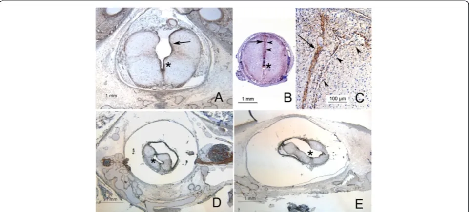
In 4 cases of Chiari II malformation, the cavity which we will term a Type 1 syrinx was a cavity dorsal to the central canal, with its apex at central canal and its base at the dorsal pial surface of the cord (Figure 1), extend-ing rostrally 2–3 levels from the site of spina bifida and in communication with the central canal. In one such case there was a very large myelocystocele, with the dor-sal elements of the cord severely attenuated and extend-ing as a subcutaneous cyst in continuity with underlyextend-ing spina bifida, extending from the rostral sacrum to the lower cervical spine. The dorsal elements of this cyst
Guoet al. Acta Neuropathologica Communications2014,2:91 Page 2 of 9
were composed of an ependymal lined membrane adher-ent to arachnoid applied directly to the subcutaneous connective tissue, and the ventral aspect of the was the splayed open, flattened spinal cord.
In 8 cases, there was a syrinx morphology we refer to as a type 2 syrinx in which the syrinx varied in shape from wedge shaped to slit like, was in continuity with the central canal only for one or two levels adjacent to the myelocele, and with one exception extended rostrally from the myelocele, and generally had a paramedian to midline course, either within or between the dorsal col-umns (Figure 1). In four of the 8 cases of type 2 syrinx, the syrinx ran the length of the cord and also connected to the central canal at the cervicomedullary junction. In three more it was thoracolumbar arising from a lumbo-sacral myelocele, and in the last it extended from a thoracolumbar myelocele rostrally into the cervical cord. In these latter 4, the syrinx did not therefore connect again with the rostral central canal or the IVth ventricle.
A third form of syrinx (type 3) was present in a single case and was a large irregular cavity extending nearly the length of the cord into the lower cervical levels, dis-secting irregularly and laterally into the cord substance, accompanied by severe atrophy of the neural structures and continuous at multiple points along its length with a dilated and distorted central canal (Figure 1). None of the cases demonstrated a simple multisegment cystic
dilation of the central canal. In all cases, the dorsal aspect of the syrinx cavity is represented by a thin membrane containing glial and neural elements with overlying arachnoid.
The morphology of the syrinx lining was variable: par-ticularly the dorsal aspect was often discontinuously lined by ependymal glia with round nuclei and a cu-boidal profile, as opposed to the distinctly columnar ependyma of the central canal. Elsewhere, the cavities were lined by astrocytic glia or their processes, express-ing GFAP, Vimentin, Nestin and more inconstantly alpha beta crystallin (Figure 2).
Among the cases of syringomyelia in Chiari II malfor-mation, 7 of the 13 displayed vernicomyelia, i.e. the pres-ence of amnionic squames in the syrinx cavity or in the spinal subarachnoid space with an associated histiocytic reaction (Figure 2). One was a type 1, five were type 2, and one was a type 3 syrinx. No case of Chiari malfor-mation demonstrated vernicomyelia without syringo-myelia. In the single case of a type 3 syrinx, vernix and reaction was present in the IVthand lateral ventricles as well. In one case of type 2 syrinx the cord showed what we interpret as infarct like pathology and scarring, with an aggregate of foamy macrophages in one area and with patches of gliosis with microglial aggregation in others.
encephalocele without Chiari malformation or spinal dys-raphism (Median age 22 weeks, range = 18 to 26 weeks), two had syringomyelia, and both were in the context of Meckel Gruber syndrome. Of these two cases, both in-volved the cervical cord: one was isolated to the cervical cord, and corresponded to a type 1 syrinx, and one was a type 2 syrinx, extending the length of the cord (Figure 3) with both paramedian and dorsolateral cavitations, none of which were ependymal lined. At the margin of this syr-inx were scattered pigmented histiocytes which stained positively for iron (Figure 3).
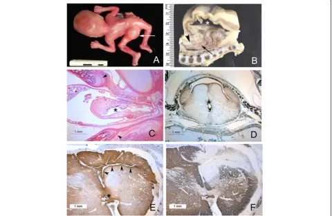
We retrieved eight cases of the omphalocele exostro-phy imperforate anus sacral anomaly syndrome (median age = 21 weeks, range = 18 to 34 weeks). None displayed a concomitant Chiari malformation. All had sacral dys-raphism with low lying conus and a dilated sacral central canal. Five of the eight displayed a large dorsal myelocys-tocele with ascending syringomyelia (Figure 4). The mye-locystocele is composed of a dorsally expanding cyst wherein the dorsal aspect of the cyst is composed of
ependyma resting on arachnoid adherent to subcutaneous fibrous tissue, and the ventral aspect is an opened, flat-tened cord. In two cases the syrinx extended rostrally to the mid-cervical spinal cord. The ascending syringomyelia is in no case ependymal lined, though caudally it is in con-tinuity with the myelocystocele. In one case, where the lumbosacral spinal cord herniated dorsally though the ab-sent neural arch, there was near complete local cavitation of the spinal cord, with reactive gliosis in the remaining cord, and a pattern of syringomyelia similar to that seen in one of the cases of Meckel Gruber, with paramedian and dorsolateral cavitation. The other cases of hindbrain crowding or neural tube defect included 23 cases of anencephaly and 12 of the Dandy Walker malforma-tion, five dural sinus malformations and two congenital cerebellar tumors, and a single case of lumbosacral spina bifida occulta with a very low lying conus without a Chiari malformation, none of which had syringomyelia.
One instance of sacral dysraphism was distinct: this case comprised the abnormal spinal cords conjoined Figure 2Spinal cord pathology associated with syringes in Chiari II malformations. AThe anatomical boundary between syrinx and central canal may be indistinct, with syringes partially lined by ependyma (arrowheads) alternating with areas of glial hyperplasia (arrows,
Immunohistochemistry for nestin).BSecondary changes in a spinal cord with syrinx: macrophages forming dense aggregates (arrow) and accumulating within the neuropil (arrowhead, immunohistochemistry for CD168).CH + E of same case asB, demonstrating aggregate of foamy macrophages.DRegion of gliosis at area indicated by arrowhead inB(immunohistochemistry for alpha beta crystallin).EVernicomyelia with foreign body macrophages (arrow).FImmunohistochemical reactivity of vernix to low molecular weight keratin (Cam 5.2).
Guoet al. Acta Neuropathologica Communications2014,2:91 Page 4 of 9
twins (Figure 4) fused dorsally, with their lumbosacral spinal cords sharing a common neural canal. The lum-bosacral segments of both cords shared a sheath of fibrolipomatous tissue, but were otherwise separate along their lengths. Both cords had dilated central canals which in the thoracic and cervical sections assumed a dorsal, slit like profile extending between the dorsal col-umns. In those sections the dorsal aspect was only de-void of ependyma in small patches but remained in continuity ventrally with the expanded central canal.
Discussion
This series demonstrates that syringomyelia is not un-common in fetal autopsy material, if sought carefully, and can be occasionally expected as part of the suite of several malformative conditions by midgestation. The presence of a spinal cord syrinx in Chiari II malformations in neonatal and fetal specimens has been documented
[4,5,13-15] but it is usually not clear how or whether the authors distinguish between syringomyelia and a persist-ently patent central canal, as the histomorphological distinction between the two is not explicitly defined. Moreover, the morphology and extent of the syringes reported is generally poorly documented. In our series, the fetal syringes are continuous with the site of the dysraphism, are in continuity with the central canal for at least part of their length and can therefore be classi-fied as communicating syringes as one might expect.
In a minority subset of Chiari II malformations, (i.e. the Type 1 syringes, and in the cavities within the pyopagus twins (Figures 1 and 4) the impression is that the cavities represent a local hypoplasia of dorsal elements and a con-comitant expansion of the central canal. The Type 1 syrin-ges would thus represent the rostral end of the area of incomplete dorsal fusion of the cord. However, in the re-mainder of the syringes, these cavities are not simply Figure 3Spinal cord and syrinx pathology in OEIS. AOEIS in a midgestation fetus with large dorsal myelocystocele (arrowhead).BLongitudinal section of caudal spine and myelocystocele in a near term infant with OEIS. The dorsal sac (asterix) is lined by ependyma and continuous with the central canal of the spinal cord (arrow) which herniates dorsally. The dorsal elements of the neural canal are absent in the midline below the upper lumbar spine (arrowhead) and the conus is low lying.CSame specimen as(A), whole mounted axial section of spinal column at the point where the cord herniates through dorsally between the separated spinal laminae (arrowheads). The central canal (asterix) is dilated and irregular with a
expanded central canals, as their morphology is radically different from the normal central canal, they may have prominent non-central components, and there is an obvi-ous disturbance of the normal neural structures. Such sy-ringes were not continuous with the central canal along their entire extent, all were associated with surrounding gliosis, and some were associated with definite evidence of secondary antemortem destructive lesions, such as inflam-mation (i.e. vernicomyelia) or infarct, apparently acquired in utero. These syringes sometimes had multiple areas that were separated from the central canal by white matter
tracts but were still partially lined by ependyma. The con-sistently dorsal median and paramedian aspect of these sy-ringes, their thin dorsal walls and their partial ependymal linings suggest that they are remnants of the central lumen of a neural tube with dorsally hypoplastic elements, as one might expect in a dysraphic spinal cord. However, the presence of axonal tracts separating these cavities from the central canal suggests that the establishment of axonal tracts in the wall of the neural tube may contribute to the establishment of eccentric cavities that represent a partially filled in, abnormal neural tube canal.
Figure 4Syringes in Meckel Gruber and pyopagus twins. ACross section of cervical spinal cord, Meckel Gruber with an occipital encephalocele.Asmall dorsal median cavity (arrow) is continuous ventrally with the dorsal glial raphe, but does not appear to connect to the mildly dilated and dorsally elongated central canal in this plane.BSame case, thoracolumbar cord: large dorsal syrinx with irregular lateral extensions (AandB: Immunohistochemistry for Vimentin).CWall of syrinx in(B), with scattered histiocytes reacting positively for Iron (arrows, Perl’s iron stain).DSacral spinal cords of pyopagus twins stained for alpha beta crystallin: one cord is a hemicord with a collapsed and dilated central canal (asterix). Both cords share a thin fibrolipomatous sheath within a single dural sac. More rostrally, the lumbar spinal cords of each twin separated and had distinct dural sacs within a common neural canal.ECross section of thoracolumbar spinal of the cord oriented as the superior cord inDdemonstrating a dorsally elongated central canal lined by ependyma and extending between the posterior columns.FThe abnormality in(E)is duplicated in the thoracolumbar cord of the other twin.
Guoet al. Acta Neuropathologica Communications2014,2:91 Page 6 of 9
In this series, syringomyelia is present in a number of dysraphic conditions. Moreover, the syringomyelia in each case was predominantly associated with the region of the herniation of the neuraxis through dorsal bony structures: in the Chiari II malformation and OEIS, con-genital syringes ascended from the site of spina bifida, while in both cases of Meckel Gruber, the syringes were in part cervical, and adjacent to the occipital encephalo-cele. This geographical continuity with dysraphism, to-gether with the evidence of infarction and old haemorrhage in some of these cords, suggests that herniation of the spinal cord may lead to local tissue injury and predispose to cavitation. One published series of fetal myeloceles sug-gests that injury secondary to delivery is the dominant spinal cord lesion in the presence of an open neural tube defect [19]. Indeed, agonal haemorrhage without tissue gliosis or reaction is very common in all early fetal neuro-pathological specimens. However, such acute haemorrhage will not produce the gliosis surrounding these cavities that we illustrate here, and the cavities illustrated do not contain blood, and we conclude that these lesions are not second-ary to parturition, but are a consequence of the malforma-tive sequence as well as early secondary events. Although the fetal cord is a fragile pathological specimen, and can be distorted by autolysis and artefact, our examination tech-nique of whole cord spinal column removal and decalcifica-tion with the spinal cordin situ minimises distortion, and because we required the presence of a reactive glial lining in order to identify a syrinx, we discount the possibility that the cavities we observe are artefactual.
The prevalence of vernicomyelia among the cases of Chiari malformations with myelocele deserves comment. Vernicomyelia can be a spectacular and striking feature in fetal specimens [20-23]. Possibly the high prevalence in this series reflects the early gestational age: the avail-able autopsy series exploring the subject demonstrates an increased prevalence of vernicomyelia among fetal and neonatal post mortems as compared to young chil-dren [20]. The reason for this age difference is unclear. Perhaps the sampling of the complete neuraxis is ren-dered easier by the sheer size of the younger fetus, or the offending vernix is cleared during post natal devel-opment, or that the most severe pathologies are over-represented in pregnancy termination and neonatal deaths. It may be that vernicomyelia is only possible in the presence of a significant syrinx that allows reflux of the vernix into the open defect. The high prevalence of vernicomyelia in the presence of syringomyelia, and the absence of vernicomyelia in the absence of syringomyelia suggest that either the secondary inflammatory path-ology of vernicomyelia plays a role in generation of spinal cord pathology, or that vernicomyelia is a marker for severe pathology. Either way, by mid gestation verni-comyelia is prevalent and associated with severe injury
in this series. This suggests that injury to the cord by vernicomyelia is unlikely to be wholly prevented by mid-gestation fetal surgical repair, though indeed such sur-gery might prevent further injury.
Among the central nervous system malformations that accompany syringomyelia are chronic hindbrain crowd-ing conditions, such as the Chiari and Dandy Walker malformations [5]. In these conditions, a wealth of radiological, surgical, clinical and experimental work points to the conclusion that syrinx formation is due to altered cerebrospinal fluid dynamics near the cra-niocervical junction, and suggests that syringomyelia is a late, slowly progressive phenomenon [1,5,8,9]. Typ-ical hydromyelic syringomyelia, as documented pri-marily in infants and children, may develop by term gestation as a consequence of chronically altered cere-brospinal fluid dynamics, leading to a progressive ex-pansion of the central canal in utero. However, such an explanation does not completely account for the morph-ology we see in this series. The syringes present in this series are established by midgestation and do not consist of a dilation of the central canal: many appear to have a prominent developmental component with a failure of complete neural tube closure, hypoplasia of dorsal struc-tures, and the irregular establishment of axonal tracts in the walls of the syrinx (Figures 2 and 3) separating the lumen of the syrinx from the central canal. Moreover, the presence of inflammatory pathology and features suggesting ischemia or haemorrhage suggests that the abnormal anatomy renders these cords vulnerable to secondary insults prior to labor and delivery. Syringo-myelia in dysraphic states may therefore have a substantial developmental component and is at least morphologically distinct from that presenting in later life.
Our results stand in contrast to a recent MRI study [17] that demonstrated syringomyelia in only 1.1% of fetal open neural tube defects, and then only late in ges-tation, and so suggested that the term congenital syr-ingomyelia may be obsolete. However, the Chiari II malformations in our series are exclusively in mid gesta-tion fetuses, and roughly one-fifth demonstrate syringo-myelia. It should be noted that the majority of syringes in the open neural tube defects in our series are slit like, less than 1 mm in diameter, and in spinal cords generally less than 4 mm in diameter (e.g. see Figure 1B and C). We suggest that part of the discrepancy between our re-sults and the radiological series is simply the higher resolution of histology compared to MRI. It may be that the usual hydromyelic form of syringomyelia docu-mented primarily in infants and children results from a progressive expansion of the central canal, and is not generally seen until term or early post natal life, and fetal syringes may expand in post natal life. Also, this series is drawn largely from pregnancy terminations and the overwhelming predominance of myelocele with mye-loschisis suggests that intrinsic developmental abnormal-ities of the spinal cord are probably more severe in this series than in the population of Chiari II malformations in general.
The morphology of the spinal cord abnormality in OEIS is sparsely described in the pathology literature [11,12,24]. To our knowledge, ours is the only study to recognize and demonstrate an anatomical distinction be-tween the caudal hydromyelia of OEIS which is com-mon, and the dramatic ascending syringomyelia distinct from the central canal illustrated here. However, in none of these cases was there an obstruction to cerebrospinal fluid circulation either in the spinal canal or at the cra-niocervical junction except possibly caudally, at the point of dorsal herniation of the cord, nor was there a reason to suspect reduced craniospinal compliance [7]. However, the syringes ascend in the cord, and do not communicate with the cervical central canal or reach the level of the medulla. Common to all five cases of syr-ingomyelia in this series is the presence of a dramatic dorsally dilated myelocystocele with hydromyelia, and an ascending syrinx in continuity with the cystocele caud-ally but rostrcaud-ally diverging from the central canal. The pattern of ascending syrinx from a thin walled terminal bulb suggests as a speculative possibility that in terminal myelocystoceles a syrinx might be formed by fluid forced into the substance of the spinal cord by compression of the bulging dorsal sac, somewhat in the manner of fluid being forced from a turkey baster or bulb pipette.
In summary, syringomyelia in fetal specimens is an un-usual, but not remarkably infrequent accompaniment to a variety of dysraphic states. The pathogenesis of syringo-myelia in Chiari II malformations and other congenital
conditions deserves some re-appraisal, as the morphology is not consistent with the effects of simple alterations of fluid dynamics, nor is syringomyelia a late complication here. Our series suggests that syringomyelia can be ac-quired in the first half of gestation, and represents a com-bination of intrinsically abnormal development, secondary pathology and possibly superimposed and contributing abnormal fluid dynamics. The contribution of in utero in-sults to the development of clinical syringomyelia in the post natal state is not a topic well addressed and will re-quire careful evaluation if fetal surgical therapy for dys-raphism is to become common.
Competing interests
The authors declare that they have no competing interest.
Acknowledgements
The authors would like to acknowledge the skilled dissections of A Wolfe, M Thompson, B Chow, N Saito and C Elliot, and the histological preparations of M Cooke and C Goodwin.
Author details
1
Department of Pathology and Laboratory Medicine, 6th Floor, Mount Sinai Hospital, 600 University Avenue, Toronto, Ontario M5G 1X5, Canada.
2
Department of Clinical Genetics, Mount Sinai Hospital, Toronto, Canada.
3Department of Diagnostic Imaging, Hospital for Sick Children, 555 University
Avenue, Toronto, Ontario M5G 1X8, Canada.
Received: 19 June 2014 Accepted: 23 July 2014
References
1. Driver CJ, Volk HA, Rusbridge C, Van Ham LM (2013) An update on the pathogenesis of syringomyelia secondary to Chiari-like malformations in dogs. Vet J 198(3):551–9
2. Klekamp J, Samii M (2002) Syringomyelia: Diagnosis and Treatment. Springer-Verlag Berlin, Heidelberg
3. Levine DN (2004) The pathogenesis of syringomyelia associated with lesions at the foramen magnum: a critical review of existing theories and proposal of a new hypothesis. J Neurol Sci 220(1–2):3–21
4. Milhorat TH, Capocelli AL, Anzil AP, Kotzen RM, Milhorat RH (1995) Pathological basis of spinal cord cavitation in syringomyelia: analysis of 105 autopsy cases. J Neurosurg 82:802–812
5. Milhorat TH1, Miller JI, Johnson WD, Adler DE, Heger IM (1993) Anatomical basis of syringomyelia occurring with hindbrain lesions. Neurosurgery 32(5):748–54
6. Vandertop WP (2014) Syringomyelia. Neuropediatrics 45(1):3–9 7. Bilston LE, Stoodley MA, Fletcher DF (2010) The influence of the relative
timing of arterial and subarachnoid space pulse waves on spinal perivascular cerebrospinal fluid flow as a possible factor in syrinx development. J Neurosurg 112:808–813
8. McLone DG1, Knepper PA (1989) The cause of Chiari II malformation: a unified theory. Pediatr Neurosci 15(1):1–12
9. Williams H (2008) A unifying hypothesis for hydrocephalus, Chiari malformation, syringomyelia, anencephaly and spina bifida. Cerebrospinal Fluid Res 5:7
10. Carey JC, Greenbaum B, Hall BD (1978) The OEIS complex (omphalocele, exstrophy, imperforate anus, spinal defects). Birth Defects Orig Artic Ser 14(6B):253–63
11. Muthukumar N (2013) Terminal myelocystocele with holocord syringomyelia: short report. J Pediatr Neurosci 8(2):171–2
12. Tandon V1, Garg K, Mahapatra AK (2012) Terminal myelocystocele: a series of 30 cases and review of the literature. Pediatr Neurosurg 48(4):229–35 13. Emery JL, Lendon RG (1972) Clinical implications of cord lesions in
neurospinal dysraphism. Dev Med Child Neurol Suppl 27:45–51
14. Emery JL, Lendon RG (1973) The local cord lesion in neurospinal dysraphism (meningomyelocele). J Pathol 110(1):83–96
Guoet al. Acta Neuropathologica Communications2014,2:91 Page 8 of 9
15. Gilbert JN, Jones KL, Rorke LB, Chernoff GF, James HE (1986) Central nervous system anomalies associated with meningomyelocele, hydrocephalus, and the Arnold-Chiari malformation: reappraisal of theories regarding the pathogenesis of posterior neural tube closure defects. Neurosurgery 18(5):559–64
16. Iruretagoyena JI, Trampe B, Shah D (2010) Prenatal diagnosis of Chiari malformation with syringomyelia in the second trimester. J Matern Fetal Neonatal Med 23(2):184–6
17. Bixenmann BJ, Kline-Fath BM, Bierbrauer KS, Bansal D (2014) Prenatal and postnatal evaluation for syringomyelia in patients with spinal dysraphism. J Neurosurg Pediatr 4:1–6
18. Hinokuma K1, Ohama E, Oyanagi K, Kakita A, Kawai K, Ikuta F (1992) Syringomyelia. A neuropathological study of 18 autopsy cases. Acta Pathol Jpn 42(1):25–34
19. Hutchins GM1, Meuli M, Meuli-Simmen C, Jordan MA, Heffez DS, Blakemore KJ (1996) Acquired spinal cord injury in human fetuses with myelomeningocele. Pediatr Pathol Lab Med 16(5):701–12
20. Jacobs EB, Landing BH, Thomas W (1961) Vernicomyelia. Its bearing on theories of genesis of the Arnold-Chiari complex. Am J Pathol 39:345–53 21. Lam S1, Grandhi R, Greene S (2013) Meconium staining of the brainstem
with open myelomeningocele. J Neurosurg Pediatr 11(2):150–3 22. Midha R, Becker LE (1991) Vernix caseo granulomatous meningitis
(vernicomyelia). Can J Neurol Sci 18(1):63–5
23. Stritzke AI1, Dunham CP, Smyth JA, Steinbok P (2011) Congenital stridor in the context of Chiari malformation type II: the etiological role of vernix caseosa granulomatous meningitis. J Neurosurg Pediatr 8(4):372–6 24. McLone DG, Naidich TP (1985) Terminal myelocystocele. Neurosurgery
16(1):36–43
doi:10.1186/s40478-014-0091-0
Cite this article as:Guoet al.:Fetal syringomyelia.Acta Neuropathologica
Communications20142:91.
Submit your next manuscript to BioMed Central and take full advantage of:
• Convenient online submission
• Thorough peer review
• No space constraints or color figure charges
• Immediate publication on acceptance
• Inclusion in PubMed, CAS, Scopus and Google Scholar
• Research which is freely available for redistribution