R E S E A R C H
Open Access
The left frontal cortex supports reserve
in aging by enhancing functional
network efficiency
Nicolai Franzmeier
1, Julia Hartmann
1, Alexander N. W. Taylor
2, Miguel Á. Araque-Caballero
1, Lee Simon-Vermot
1,
Lana Kambeitz-Ilankovic
3, Katharina Bürger
1,4, Cihan Catak
1, Daniel Janowitz
1, Claudia Müller
1, Birgit Ertl-Wagner
5,
Robert Stahl
5, Martin Dichgans
1,4,6, Marco Duering
1and Michael Ewers
1*Abstract
Background:Recent evidence derived from functional magnetic resonance imaging (fMRI) studies suggests that functional hubs (i.e., highly connected brain regions) are important for mental health. We found recently that global connectivity of a hub in the left frontal cortex (LFC connectivity) is associated with relatively preserved memory abilities and higher levels of protective factors (education, IQ) in normal aging and Alzheimer’s disease. These results suggest that LFC connectivity supports reserve capacity, alleviating memory decline. An open question, however, is why LFC connectivity is beneficial and supports memory function in the face of neurodegeneration. We hypothesized that higher LFC connectivity is associated with enhanced efficiency in connected major networks involved in episodic memory. We further hypothesized that higher LFC-related network efficiency predicts higher memory abilities. Methods:We assessed fMRI during a face-name association learning task performed by 26 healthy, cognitively normal elderly participants. Using beta-series correlation analysis, we computed task-related LFC connectivity to key memory networks, including the default mode network (DMN) and dorsal attention network (DAN). Network efficiency within the DMN and DAN was estimated by the graph theoretical small-worldness statistic. We applied linear regression analyses to test the association between LFC connectivity with the DMN/DAN and small-worldness of these networks. Mediation analysis was applied to test LFC connectivity to the DMN and DAN as a mediator of the association
between education and higher DMN and DAN small-worldness. Last, we tested network small-worldness as a predictor of memory performance.
Results:We found that higher LFC connectivity to the DMN and DAN during successful memory encoding and recognition was associated with higher small-worldness of those networks. Higher task-related LFC connectivity mediated the association between education and higher worldness in the DMN and DAN. Further, higher small-worldness of these networks predicted better performance in the memory task.
Conclusions:The present results suggest that higher education-related LFC connectivity to key memory networks during a memory task is associated with higher network efficiency and thus enhanced reserve of memory abilities in aging.
Keywords:Cognitive reserve, Aging, Memory task fMRI, Small-worldness, Frontoparietal control network
* Correspondence:[email protected]
1Institute for Stroke and Dementia Research, Klinikum der Universität München, Ludwig-Maximilians-Universität (LMU), Feodor-Lynen Straße 17, 81377 Munich, Germany
Full list of author information is available at the end of the article
Background
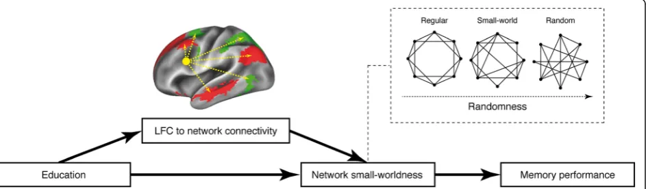
The concept of reserve describes the ability to maintain cognition relatively well during the course of neurode-generation [1]. Protective factors that are associated with higher reserve and a reduced risk of Alzheimer’s disease (AD) at older age include early-life experiences of cogni-tively challenging activities [2, 3], such as higher IQ or greater education [4, 5]. Specifically, higher formal edu-cation has been associated with slower age-related cog-nitive decline [6], reduced risk of AD dementia [4], and relatively stable cognition in the presence of accumulat-ing AD pathology [7–11]. Thus, in normal and patho-logical aging, subjects show variable levels of reserve that may be influenced by life factors such as education. The understanding of those brain mechanisms that underlie reserve is pivotal to developing interventional approaches to directly stimulate and enhance reserve in aging for the prevention of the development of cognitive decline and dementia. In a series of functional magnetic resonance imaging (fMRI) studies, we have recently identified a functional hub region in the left frontal cor-tex (LFC; Brodmann areas 6/44) as a putative neural substrate of reserve. Specifically, we found that higher global functional connectivity of the LFC hub during both resting state and memory task fMRI was associated with (1) greater education and (2) higher memory per-formance relative to the level of age-related hippocampal atrophy, AD-related parietal glucose hypometabolism, or tau pathology [10, 12–15]. This result pattern suggests that LFC connectivity is associated with protective fac-tors (i.e., education) and supports memory-related re-serve in aging and AD. An open question, however, is why LFC connectivity is beneficial and supports memory function in the face of neurodegeneration.
Our lead hypothesis was that higher connectivity of the LFC to major brain networks involved in memory is asso-ciated with enhanced efficiency of these networks. The hy-pothesis was motivated by several previous findings. From a network perspective, the LFC is a key hub of the fronto-parietal control network, which is involved in a broad var-iety of cognitive abilities and hence is also labeled the
“task-positive” network [16]. The frontoparietal control network has been shown to flexibly couple with other net-works in a task-specific way, whereby the degree of con-nectivity to other networks is predictive of higher cognitive performance [17, 18]. Particularly, global con-nectivity of the LFC hub was shown to be associated with higher cognitive control and general cognitive function as measured by fluid IQ in young subjects [19]. Thus, control regions such as the LFC are critical for the regulation of other networks and may enhance their information-processing capacity (i.e., efficiency), which is associated with higher cognitive performance [19, 20]. With regard to reserve in aging and AD, it is thus possible that the
LFC supports reserve by promoting efficient processing capabilities in key memory networks, thereby helping to maintain memory ability relatively well [5].
The efficiency of functional brain networks can be assessed by graph theoretical analysis of the fMRI blood oxygen level dependent signal. In graph theoretical terms, an efficient network is usually considered to allow for fast information transfer (i.e., short pathways to get from a particular node to any other node in the net-work) [21]. A caveat of this approach to quantifying effi-ciency is that random networks show on average a short path length (i.e., high efficiency). However, random works lack topological features of highly organized net-works such as local clustering of connections. Thus, from a functional point of view, a more plausible approach to capturing network efficiency constitutes the measure of “small-worldness,” which takes into account both the shortest path between any two given nodes and the degree of local clustering of connections. High small-worldness can be understood as fast information transfer via short path length in highly structured non-random networks [21, 22]. Previous resting-state fMRI studies have shown that the brain is organized in small-world networks, where hubs such as the LFC are especially important for maintaining small-worldness because they are important connectors that route short paths [23]. In addition, higher small-worldness of func-tional brain networks has previously been linked to higher cognitive performance [24, 25] and higher resili-ence against network dysfunction [26,27].
In the present study, we used task-based functional con-nectivity analysis and graph theory, whereby we assessed memory task-related LFC hub connectivity and small-worldness of key memory networks during successful en-coding and recognition in a face-name associative memory task. In particular, we estimated the small-worldness of two major functional networks, the default mode network (DMN) and the dorsal attention network (DAN), and the connectivity of the LFC to these networks. The rationale for this selection was that the LFC is strongly connected to both these networks, which is associated with higher education level and better episodic memory performance relative to the level of neurodegeneration [12]. Further-more, a recent meta-analysis of memory task fMRI studies showed that specifically the DMN and DAN are engaged during successful memory ability [28].
Methods
Participants
We recruited 26 cognitively normal elderly subjects at the memory clinic of the university hospital of the Ludwig-Maximilian University who underwent cognitive testing and MRI and were also reported in one of our previous publications [13]. Inclusion criteria were age > 60 years and no cognitive impairment based on test scores on the Consortium to Establish a Registry for Alzheimer’s Disease (CERAD)-Plus battery [29] and subjective reports. Absence of cognitive impairment was defined as a per-formance not < 1.5 SD of age-, sex-, and education level-adjusted norms on all CERAD-Plus subtests. Exclusion criteria were acute or past neurological/psychiatric disor-ders, diabetes, or MRI contraindications. As a measure of general memory performance, we used the delayed recall score of the word list test that is included in the CERAD-Plus battery [29]. This test includes a list of ten unrelated words that are presented in three subsequent learning tri-als and is especially suited for older individutri-als for whom longer lists would be too taxing. After a 5-minute delay, retention is tested by free recall. Years of education were assessed in a standardized manner as the sum of years spent in school and professional training or university education, following the recommendations of the CERAD-Plus battery [29]. The study was approved by our local ethics committee. All participants provided written informed consent.
fMRI associative memory paradigm
We used a mixed block and event-related face-name as-sociative memory task design adapted from previous studies [30], which allows modeling brain activation dur-ing memory encoddur-ing and recognition separately. The task was divided into 14 blocks of face-name encoding, each followed by a block of recognition. As stimuli, we used novel faces (i.e., faces unfamiliar to the scanned subjects) randomly paired with first names. Detailed in-formation on task stimuli can be found in one of our previous publications [13]. During the overall task pro-cedure, the subjects were presented 112 different faces and 168 names with balanced gender frequencies. All subjects were trained in the task procedure before the fMRI scanning session on a laptop computer using face-name pairs that were not included in the fMRI task. The task was implemented using E-prime software (Psych-ology Software Tools, Inc., Sharpsburg, PA, USA), and face-name pairs were shown via a vision goggle system attached to the head coil, which allows for individual eyesight correction.
During an encoding block, eight faces paired with a single name were subsequently presented for 5 seconds each, with the next face-name pair following after a ran-domized intertrial interval of 1500–3000 milliseconds.
Each encoding block was followed by a recognition block during which the subjects were again presented the eight faces shown previously, now with two names below the faces (correct name vs. distractor). The subjects were instructed to select the name that was previously presented with the face (correct name) by pressing a button on fiberoptic response grips
(www.nordicneurolab.com; NordicNeuroLab, Bergen,
Norway). No feedback on accuracy was given during the task procedure. In half of the recognition trials, the distractor was a novel name, whereas in the other half, the distractor was a name that went with another face during the previous encoding block. Correct responses during the recognition block were classified as success-ful recognition. Based on correct responses in the rec-ognition block, the corresponding encoding trials were retrospectively classified as successful encoding. Conversely, wrong answers or missed answers were classified as unsuccessful recognition and unsuccessful encoding, respectively. Between each encoding and rec-ognition block, the subjects were briefly presented short task instructions. For each individual, fMRI task accuracy was defined as the percentage of all recogni-tion trials that were answered correctly.
MRI data acquisition
Scanning was performed on a Siemens Verio 3T scanner (Siemens Healthcare, Erlangen, Germany), using a 12-channel head coil. Structural images were obtained using a T1-weighted magnetization-prepared rapid gradient echo sequence (repetition time [TR]/echo time [TE] 1750/2.52 milliseconds, flip angle 9 degrees), with 1-mm isotropic voxel resolution. Task fMRI was recorded using a T2*-weighted echo planar imaging (EPI) pulse se-quence (TR/TE 2000/30 milliseconds, flip angle 90 de-grees) with an in-plane resolution of 3.4 mm, 3-mm slice thickness and 1-mm interslice gap. Overall, 900 EPI volumes (~ 30 minutes acquisition time) were recorded, divided into three runs. Prior to the task recordings, gradient-echo field maps (TR/TE1/TE2 488/4.92/7.38 milliseconds) were acquired.
Task fMRI functional connectivity analysis
We assessed functional connectivity during the fMRI memory task via beta-series correlation, which allows as-sessment of interregional functional connectivity in event-related fMRI data using the freely available tool-box BASCO (BetA Series COrrelation) [33]. First, ROIs were defined as 264 isotropic 6-mm spheres based on a widely used brain parcellation atlas [17, 34]. This atlas, which is based on resting-state fMRI scans of 300 young individuals, was introduced first by Power and colleagues [34] and covers 10 large-scale functional networks, as shown in Fig. 1a. An additional LFC-ROI (6-mm sphere, Montreal Neurological Institute [MNI] coordi-nates x=−42,y= 6, z= 28) (see red highlighted ROI in Fig. 1a) that we also described in previous publications [10, 12] was added to the frontoparietal control net-work parcellation of the currently used fMRI atlas. To assess hemispheric specificity of the effect of LFC con-nectivity, we also applied an ROI in the corresponding location of the right frontal cortex (RFC; MNI coordi-nates x= 42, y= 6, z= 28). Second, for each of the
overall 266 ROIs, we performed subject-level task fMRI analysis using a generalized linear model where task-related activation in each trial is modeled by a covariate time-locked to the stimulus onset. Subject-specific gen-eralized linear models were modeled by entering trial type-specific regressors (successful encoding, unsuc-cessful encoding, sucunsuc-cessful recognition, unsucunsuc-cessful recognition), each convolved with a canonical hemodynamic response function and a multivariate Taylor expansion plus six motion regressors and their derivatives [35]. Parameter estimation was performed with SPM12, yielding 266 ROI-specific vectors of β -coefficients for each trial type per subject. Third, to es-timate trial type-specific functional connectivity, we correlated the trial type-specific β-coefficient vectors using Spearman’s correlations, yielding four trial type-specific (i.e., successful/unsuccessful encoding/recall) 266 × 266 connectivity matrices per subject. Prior to graph theoretical analysis, autocorrelations were set to 0, and the remaining correlations were thresholded at an absolute value of r > 0.2 to exclude spurious
correlations. To ensure that results were not threshold-specific, all analyses reported were repeated using thresholds of 0.25, 0.3, and 0.35, which did not change the overall result pattern. For the assessment of network-specific graph metrics, the 266 × 266 connect-ivity matrices were parcellated into ten smaller matri-ces, each reflecting connectivity within one of ten canonical functional networks (see Fig. 1a for network definitions) as reported previously [17, 34]. Graph the-oretical analyses were conducted in a trial type-specific manner on each of these network-specific connectivity matrices. In the present study, we focused on the con-nectivity submatrices of the DMN and DAN (i.e., red andgreen parcelsin Fig.1a).
Graph theoretical analysis Small-worldness
Functional brain networks are thought to exhibit small-world topology (i.e., an intermediate stage between ran-dom and lattice-like networks). In principle, small-world networks are characterized by a combination of high local segregation and global integration. The graph theoretical statistic of small-worldness quantifies the trade-off be-tween local clustering and characteristic path length, each normalized against a random network [21,36]. Character-istic path length is inversely related to global efficiency and reflects the average shortest connection between all pairs of nodes in a network. In contrast, clustering de-scribes functional segregation and quantifies how strongly neighboring nodes of a network are interconnected [21]. On the basis of these measures, we computed the trial type-specific small-worldness for the DMN and DAN using the following equation:
Small−worldness¼ C=Crand L=Lrand
where C is the mean clustering coefficient andL is the characteristic path length of the network of interest. Crandand Lrandare equivalent measures assessed as the mean of C and L of 10,000 bootstrapped random net-works that were equal to the DMN/DAN in size and de-gree of distribution. Note that negative functional connectivity values were set to 0 prior to assessing small-worldness because characteristic path length and clustering coefficient are by definition based on positive connections (i.e., “within-network” connections). For mathematical details on the assessment ofCandL, seea previously published overview on graph theoretical pa-rameters applied to fMRI data [21]. The analyses were conducted using the algorithms of the freely available brain connectivity toolbox (https://sites.google.com/site/
bctnet/Home/functions) and MATLAB (MathWorks,
Natick, MA, USA) scripts written in-house.
LFC to DMN and DAN connectivity
To quantify the cross-network coupling of the LFC, we computed the functional connectivity strength of the LFC to the DMN and DAN using the sum of weighted functional connectivity values of the LFC to a given net-work [21]. Here, we specifically used absolute functional connectivity values to take into account both positive and negative connections of the LFC that may conjointly modulate the efficiency of the DMN or DAN. Specific-ally, we computed the LFC connectivity to the DMN and DAN on the basis of connectivity matrices specific for each trial type (successful/unsuccessful encoding/rec-ognition), where we summed the absolute connectivity values between the LFC ROI and all ROIs of the net-work of interest (DMN or DAN), yielding a single scalar index of LFC connectivity to a given network. Here, higher connectivity reflects stronger coupling between the LFC and DMN/DAN, which is assumed to facilitate the integration of information within and across net-works [21]. We computed connectivity between the LFC and the DMN/DAN as follows:
LFC to X¼ X
j∈xkLFC i
whereX is the DMN or DAN andkLFC iis the connect-ivity between the LFC and node iof the respective net-work. For later control analyses on left hemispheric specificity, we used the above-defined procedure to equivalently compute connectivity of the RFC to the DMN and DAN. All computations were conducted using MATLAB software.
Statistics
As a proof of concept, we first tested whether greater education as a protective factor in aging and AD is asso-ciated with higher memory ability in late age (i.e., re-serve) when accounting for AD risk (i.e., apolipoprotein E [APOE] genotype) and structural brain integrity (i.e., GMV). To this end, we assessed whether greater educa-tion predicted higher fMRI-task accuracy or CERAD memory performance by using linear regression, enter-ing age, sex, APOE genotype, and GMV as covariates. To visualize the functional connectivity patterns during successful memory performance, we averaged the unthresholded 266 × 266 matrices across subjects.
Associations between LFC connectivity, DMN/DAN small-worldness, and education
the dependent variable and LFC connectivity to the particular network as the independent variable. The regression models were controlled for age, sex, APOE genotype, GMV, and task reaction time. We selected those covariates to ensure that associations between LFC connectivity and small-worldness were not driven by dif-ferences in structural brain integrity or genetic risk for AD. Thus, for a particular network and condition, the regression model was, for example, small-worldness of the DMN during encoding explained by LFC-to-DMN connectivity during encoding + age + sex + APOE geno-type + GMV + task reaction time. Similarly, we tested whether higher education level predicted higher DMN/ DAN small-worldness. To this end, we recomputed the above-listed regression models, this time using education instead of LFC connectivity as a predictor of DMN or DAN small-worldness during successful encoding/recog-nition. To assess specificity for successful encoding/rec-ognition, equivalent regression models were applied to small-worldness assessed on unsuccessful encoding/rec-ognition trials.
Mediation analysis between education, LFC-to-DMN/DAN connectivity, and DMN/DAN small-worldness
To test our hypothesis that associations between educa-tion and DMN/DAN small-worldness are mediated by LFC connectivity to these networks, we used causal me-diation analyses as implemented in the mediation pack-age [37] in R [38]. Here, we used education as the independent variable, small-worldness as the dependent variable, and LFC connectivity to the DMN/DAN as the mediator variable, controlling all paths for age, sex, APOE, GMV, and task reaction time. This model was tested for the DMN and DAN for connectivity assessed on successful encoding and successful recognition trials. The significance of mediation effects was assessed using nonparametric bootstrapping with 10,000 iterations, which can be used effectively for significance testing, es-pecially in smaller samples [39]. We estimated the sig-nificance of the average causal mediation effect (ACME), the average direct effect (ADE), the total effect, and the proportion of the total effect that was mediated. Results were interpreted as full mediation when only the ACME but not the ADE was significant, but as partial mediation when both ADE and ACME were significant. As a con-trol analysis to assess left hemispheric specificity, the above-defined mediation models were also assessed when using RFC connectivity to the DMN or DAN as a mediator variable.
Associations between DMN/DAN small-worldness and memory performance
Next, we tested whether higher DMN or DAN small-worldness translated into better task fMRI performance.
Here, we applied multiple regression using the fMRI ac-curacy score as a dependent variable and DMN/DAN small-worldness as the independent variable, controlling for age, sex, APOE genotype, and GMV and task reac-tion time. As an exploratory analysis, we tested whether DMN/DAN small-worldness generalized toward better out-of-scanner memory performance by assessing the above-mentioned models using the CERAD memory score as a dependent variable.
All linear models reported were computed using the lm command in R [38]. We applied a threshold of α= 0.05 to consider regression weights significant, and we additionally accounted for multiple testing using the Bonferroni correction for each hypothesis (i.e., four tests per hypothesis, corrected α= 0.0125 for each hypothesis). No violations of linear regres-sion assumptions (skewness, kurtosis, heteroscedasti-city, multicollinearity) were detected.
Results
Sample demographics and cognitive characteristics are displayed in Table1. When testing whether higher edu-cation level predicted better memory performance using linear regression, we found positive associations with fMRI task accuracy (β/SE = 0.370/0.203, p= 0.042) and with CERAD memory performance (β/SE = 0.353/0.223, p= 0.027). For descriptive purposes, the group average matrices of whole-brain task-related functional connect-ivity during successful encoding/recognition are shown in Fig.1b.
Higher LFC connectivity to the DMN/DAN is associated with higher DMN/DAN small-worldness
We first tested our main hypothesis: whether greater LFC connectivity to the DMN/DAN is associated with higher small-worldness within these networks. For suc-cessful encoding, higher DMN/DAN small-worldness
Table 1Sample characteristics and cognitive performance
Cognitively normal elderly subjects (N= 26)
Age, years 71.91 ± 5 [61.44–82.29]
Sex, male/female 10/16
Years of education 13.69 ± 2.99 [9–20]
fMRI task accuracy, % 0.8 ± 0.06 [0.71–0.91]
MMSE score (maximum 30) 29.42 ± 0.86 [27–30]
CERAD word list delayed free recall score (maximum 10)
8.83 ± 1.39 [6–10]
Geriatric Depression Scale score 3.23 ± 3.01 [0–10]
APOEε4 carriers/noncarriers 9/17
Abbreviations: APOEApolipoprotein E,CERADConsortium to Establish a Registry for Alzheimer’s Disease,fMRIFunctional magnetic resonance imaging,
MMSEMini Mental State Examination
was predicted by higher LFC connectivity to the respect-ive network (DMN β/SE = 0.847/0.117, p< 0.001; DAN
β/SE = 0.612/0.169,p= 0.002). Similar results were found for successful recognition, where higher LFC connectiv-ity also predicted higher small-worldness within both the DMN (β/SE = 0.736/0.143, p< 0.001) and the DAN (β/SE = 0.792/0.126, p< 0.001). All results remained sig-nificant after correction for multiple testing (α= 0.0125). Scatterplots for associations between LFC connectivity and DMN/DAN small-worldness are shown in Fig.2.
Effects of education on DMN/DAN small-worldness are mediated via LFC connectivity
Next, we tested a prerequisite for mediation analysis: whether more years of education predicted higher DMN/DAN small-worldness. Results of the regression analyses showed that higher education level was associ-ated with higher small-worldness of the DMN and the DAN during successful encoding (DMN β/SE = 0.507/ 0.225, p= 0.018, DAN β/SE = 0.598/0.227, p= 0.008) and successful recognition (DMN β/SE = 0.620/0.229, p= 0.007; DAN β/SE = 0.501/0.230, p= 0.021). Scatter-plots of these results are shown in Fig. 3.
Next, we tested our second hypothesis that LFC con-nectivity to the DMN/DAN mediates the association be-tween education and DMN/DAN small-worldness using bootstrapped mediation models. For the DAN and suc-cessful encoding, we found a significant full mediation. Specifically, we found a significant ACME of LFC con-nectivity (mediator) for the association between educa-tion and DAN small-worldness (ACME 0.283, p= 0.02), where the ADE of education on DAN small-worldness was no longer significant when LFC connectivity was in-cluded in the model (ADE 0.214,p= 0.27) (Fig.4a).
A similar full mediation was found for effects of edu-cation on DMN small-worldness via LFC connectivity during successful encoding (Fig. 4c), where the ACME was significant (ACME 0.396, p= 0.01), but the ADE was no longer significant when LFC connectivity was in-cluded as a predictor (ADE 0.0.076,p= 0.57).
Equivalent full mediations were found during successful recognition for both DAN (ACME 0.297, p= 0.02; ADE 0.224, p= 0.18) (Fig. 4b) and DMN small-worldness (ACME 0.372, p< 0.001; ADE 0.190, p= 0.29) (Fig. 4d). Mediation statistics of the bootstrap analyses are summa-rized in Table2. When we applied the Bonferroni correc-tion (α= 0.0125) to the ACMEs, the results remained
significant for DMN small-worldness for both successful encoding and recognition, whereas ACMEs met only trend-level significance for DAN small-worldness. When testing the same mediation models for RFC connectivity, we found no significant ACMEs (all p> 0.05), suggesting specificity of our findings for the LFC.
Triangular diagrams of the LFC mediation models together with linear regression derived from indirect and direct path weights are shown in Fig.4.
DMN/DAN small-worldness is associated with memory performance
Next, we tested whether higher LFC-mediated small-worldness of the DAN (i.e., successful encoding) and DMN (i.e., successful recognition) predicted higher fMRI task accuracy (i.e., percentage of face-name items that were correctly recognized). For successful encoding, we found that higher DMN small-worldness (β/SE = 0.568/ 0.163,p= 0.002), but not DAN small-worldness (β/SE =
Fig. 3Scatterplots illustrating the associations between years of education and small-worldness within the default mode network/dorsal attention network (DMN/DAN) during episodes of successful encoding (left panels) and successful recognition (right panels). Standardized regression weights andpvalues are taken from multiple regression models controlled for age, sex, gray matter volume, apolipoprotein Eε4 carrier status, and task reaction time
0.143/0.200, p= 0.482), predicted higher task accuracy. Similar results were found for successful recognition, where higher DMN small-worldness (β/SE = 0.492/0.156, p= 0.005) and higher DAN small-worldness (β/SE = 0.516/0.177,p= 0.008) predicted higher task accuracy.
Last, we tested in an exploratory analysis whether higher LFC-mediated DMN/DAN small-worldness were associated with better out-of-scanner memory perform-ance (i.e., CERAD memory performperform-ance). Here, higher recognition-related small-worldness in the DMN pre-dicted higher CERAD memory scores (β/SE = 0.466/ 0.200, p= 0.031), whereas small-worldness in the DAN showed an effect at trend level (β/SE = 0.377/0.195, p= 0.068). No significant effects were found for encoding-related DMN/DAN small-worldness.
Discussion
Our major findings were that (1) LFC connectivity was associated with higher memory task-related small-worldness of the DMN/DAN; (2) LFC connectivity to DMN/DAN mediated the association between higher education level and higher DMN/DAN small-worldness; and (3) higher small-worldness of the DMN/DAN was associated with higher memory task performance. Keep-ing in mind that the present results should not be inter-preted in a causative mechanistic way, we conclude that the beneficial effects of LFC connectivity on reserve are attributable to higher functional network efficiency that underlies higher memory performance.
We found that LFC connectivity to the DMN/DAN was associated with increased small-worldness within these networks. These results suggest that the LFC sup-ports fast and cost-efficient information processing in connected networks during memory performance.
Supporting this view, a recent study showed that fronto-parietal control network hubs such as the LFC help guide brain networks into difficult-to-reach states that are critical for performing complex cognitive tasks [40]. Together, these results fit with the function of the LFC as a flexible hub of the frontoparietal control network [17] that regulates activity and information flow in other networks, such as the DMN and DAN, during resting state [41] and cognitive demands [17–19, 42]. Our re-sults also support the notion that LFC hub connectivity is associated with more efficient information processing of connected networks. To further test the relationship between network efficiency and reserve, we assessed the association between education (i.e. the best established protective factor in aging and AD) [4], and small-worldness of the DMN and DAN. We could show that higher education level was also associated with higher small-worldness of the DMN and DAN for both success-ful encoding and recognition. These results are in gen-eral agreement with previous resting-state fMRI studies showing higher education level to be associated with greater strength of long-distance connections and shorter characteristic path length in elderly participants [43]. We found further that both education and network efficiency were associated with higher face-name fMRI task performance. Together, the present results support the notion that education is associated with higher memory performance that is supported by higher effi-ciency in functional networks, including the DMN and DAN. An association between small-worldness and cog-nitive performance has previously been demonstrated in resting-state fMRI [44], where a loss of small-worldness is associated with cognitive decline in AD [45]. To-gether, these findings suggest that small-worldness of
Table 2Left frontal cortex to default mode network/dorsal attention network connectivity as a mediator of effect of education on default mode network/dorsal attention network small-worldness shown for each functional magnetic resonance imaging task trial type
DAN DMN
Estimate pValue Estimate pValue
Successful encoding
Average causal mediation effect 0.283 0.02 0.396 0.01
Average direct effect 0.214 0.27 0.076 0.57
Total effect 0.497 0.02 0.472 < 0.001
Proportion mediated 0.569 0.04 0.839 0.02
Successful recognition
Average causal mediation effect 0.297 0.02 0.372 < 0.001
Average direct effect 0.224 0.18 0.190 0.29
Total effect 0.521 0.02 0.562 0.01
Proportion mediated 0.570 0.02 0.662 0.01
DANDorsal attention network,DMNDefault mode network
the DMN and DAN has functional relevance at the cognitive level. Importantly, our results derived from the mediation analysis suggest that the LFC plays a key role in the education-related variability of network effi-ciency. Importantly, control analyses using the RFC homotopic region yielded no significant associations between education and RFC connectivity, suggesting the specificity of our findings for the LFC. We have previously shown that higher education level is associ-ated with higher LFC connectivity, where higher LFC connectivity was associated with higher memory-related reserve in cognitively normal subjects or sub-jects with AD [10, 12–14]. Thus, education is likely as-sociated with differences in the premorbid functional brain architecture (i.e., higher LFC hub connectivity and higher network efficiency during cognitive per-formance). Our current working model of reserve is that the LFC is a pivotal brain hub that facilitates effi-cient network processes and thus cognitive perform-ance in aging and AD. We have summarized this model in Fig. 5. We caution that even though we used medi-ation analysis, a causative interpretmedi-ation is not possible. Thus, our findings provide partial support for such a working model of reserve in that a close association be-tween LFC connectivity, network efficiency, and mem-ory performance was demonstrated. Previous studies that assessed task-related effective connectivity have consistently shown, however, that activity in other net-works, including the DMN and DAN [46, 47], is driven by hub regions of the frontoparietal control network and that stronger effective connectivity is associated with better cognitive performance [46]. Thus, the LFC is a likely candidate network influencing small-worldness in other networks during cognitive processes such as episodic memory. We strongly encourage fu-ture studies including larger samples to apply structural equation modeling to test the overall validity of our working model of reserve (Fig.5).
In the interpretation of the present results, we caution that a strictly hypothesis-driven approach was applied that allowed us to focus on the DMN and DAN as net-works that are fundamental for memory function (i.e., the cognitive domain most affected in aging and AD) [28,48,49]. However, because frontoparietal control net-work hubs such as the LFC are globally involved in cog-nition and also interact with networks other than the DMN and DAN [16, 17], it is possible that the present findings may also apply to cognitive domains other than memory. Testing such a hypothesis would require apply-ing different fMRI tasks and focusapply-ing on different func-tional networks, depending on task demands [17]. Although this would clearly go beyond the scope of the present study, our present results may motivate future studies to test LFC connectivity as a mediator of net-work efficiency and performance across a variety of cog-nitive domains.
We note that a limitation of our study is that the sam-ple encompassed a relatively high proportion of APOE
ε4 carriers (~ 34%), who are at increased risk of AD and thus may not be entirely representative of the general population. To address this, all analyses were controlled for APOE ε4 carrier status; also, we could previously show that the LFC supports reserve across both normal aging and subjects at increased AD risk, supporting a more general role of the LFC for reserve in both normal and pathological aging [13]. Nevertheless, future studies could specifically assess whether APOEε4 allele carriage has an effect on LFC-mediated reserve effects.
Conclusions
The present study provides novel insight into potential functional underpinnings of reserve in aging mediated via LFC connectivity and functional network efficiency, which opens the possibility of assessing their modifiability via cognitive interventions [50], brain stimulation, or neu-rofeedback. To date, studies that noninvasively stimulated
the frontal lobe hubs have already shown that connectivity can be enhanced [51] and that memory can be improved in both healthy individuals [52] and patients with mild cognitive impairment [53]. Thus, the LFC may be an attractive therapeutic target for fostering reserve and prevention of cognitive decline in aging and AD.
Additional file
Additional file 1:Supplementary methods. (DOCX 101 kb)
Abbreviations
ACME:Average causal mediation effect; AD: Alzheimer’s disease; ADE: Average direct effect; APOE: Apolipoprotein E; CERAD: Consortium to Establish a Registry for Alzheimer’s Disease; DAN: Dorsal attention network; DMN: Default mode network; EPI: Echo planar imaging; fMRI: Functional magnetic resonance imaging; GMV: Gray matter volume; LFC: Left frontal cortex; MMSE: Mini Mental State Examination; MNI: Montreal Neurological Institute; RFC: Right frontal cortex; TE: Echo time; TR: Repetition time
Acknowledgements
Not applicable.
Funding
The work was supported by the LMUexcellent Initiative, the European Commission (PCIG12-GA-2012-334259), and the Alzheimer Forschung Initiative (DE-15035).
Availability of data and materials
The datasets used and analyzed during the present study are available from the corresponding author on reasonable request.
Authors’contributions
NF conceived of and designed the study, collected data, performed statistical analysis, interpreted the data, and drafted the manuscript. JH performed statistical analysis and interpreted the data. ANWT, MÁAC, LSV, LKI, KB, CC, DJ, CM, BEW, RS, MDi, and MDu collected data and critically revised the manuscript. ME conceived of and designed the study, interpreted the data, and drafted the manuscript. All authors read and approved the final manuscript.
Ethics approval and consent to participate
The present study was approved by the local ethical board of the Ludwig Maximilian University of Munich, and all participants provided written informed consent. All procedures were performed in accordance with the 1964 Helsinki declaration and its later amendments.
Competing interests
The authors declare that they have no competing interests.
Publisher’s Note
Springer Nature remains neutral with regard to jurisdictional claims in published maps and institutional affiliations.
Author details
1Institute for Stroke and Dementia Research, Klinikum der Universität München, Ludwig-Maximilians-Universität (LMU), Feodor-Lynen Straße 17, 81377 Munich, Germany.2School of Psychology, University of East Anglia, Norwich Research Park, Norwich NR4 7TJ, UK.3Department of Psychiatry and Psychotherapy, Ludwig-Maximilians-Universität (LMU), Nussbaumstraße 7, 80336 Munich, Germany.4German Center for Neurodegenerative Diseases (DZNE Munich), Feodor-Lynen Straße 17, 81377 Munich, Germany.5Institute for Clinical Radiology, Klinikum der Universität München,
Ludwig-Maximilians-Universität (LMU), Marchioninistraße 15, 81377 Munich, Germany.6Munich Cluster for Systems Neurology (SyNergy), Munich, Germany.
Received: 30 October 2017 Accepted: 15 February 2018
References
1. Stern Y. What is cognitive reserve? Theory and research application of the reserve concept. J Int Neuropsychol Soc. 2002;8(3):448–60.
2. Stern Y. Cognitive reserve in ageing and Alzheimer’s disease. Lancet Neurol. 2012;11(11):1006–12.
3. Xu W, Yu JT, Tan MS, Tan L. Cognitive reserve and Alzheimer’s disease. Mol Neurobiol. 2015;51(1):187–208.
4. Meng X, D’Arcy C. Education and dementia in the context of the cognitive reserve hypothesis: a systematic review with meta-analyses and qualitative analyses. PLoS One. 2012;7(6):e38268.
5. Barulli D, Stern Y. Efficiency, capacity, compensation, maintenance, plasticity: emerging concepts in cognitive reserve. Trends Cogn Sci. 2013;17(10):502–9. 6. Hall CB, Derby C, LeValley A, Katz MJ, Verghese J, Lipton RB. Education
delays accelerated decline on a memory test in persons who develop dementia. Neurology. 2007;69(17):1657–64.
7. Solé-Padullés C, Bartrés-Faz D, Junqué C, Vendrell P, Rami L, Clemente IC, Bosch B, Villar A, Bargalló N, Jurado MA, et al. Brain structure and function related to cognitive reserve variables in normal aging, mild cognitive impairment and Alzheimer’s disease. Neurobiol Aging. 2009;30(7):1114–24. 8. Ewers M, Insel PS, Stern Y, Weiner MW, Alzheimer’s Disease Neuroimaging Initiative. Cognitive reserve associated with FDG-PET in preclinical Alzheimer disease. Neurology. 2013;80(13):1194–201.
9. Rentz DM, Locascio JJ, Becker JA, Moran EK, Eng E, Buckner RL, Sperling RA, Johnson KA. Cognition, reserve, and amyloid deposition in normal aging. Ann Neurol. 2010;67(3):353–64.
10. Franzmeier N, Duering M, Weiner M, Dichgans M, Ewers M, Alzheimer’s Disease Neuroimaging Initiative. Left frontal cortex connectivity underlies cognitive reserve in prodromal Alzheimer disease. Neurology. 2017;88(11):1054–61.
11. Franzmeier N, Buerger K, Teipel S, Stern Y, Dichgans M, Ewers M, Alzheimer’s Disease Neuroimaging Initiative. Cognitive reserve moderates the association between functional network anti-correlations and memory in MCI. Neurobiol Aging. 2017;50:152–62.
12. Franzmeier N, Göttler J, Grimmer T, Drzezga A, Áraque-Caballero MA, Simon-Vermot L, Taylor ANW, Bürger K, Catak C, Janowitz D, et al. Resting-state connectivity of the left frontal cortex to the default mode and dorsal attention network supports reserve in mild cognitive impairment. Front Aging Neurosci. 2017;9:264.
13. Franzmeier N, Hartmann JC, Taylor ANW, Araque Caballero MÁ, Simon-Vermot L, Buerger K, Kambeitz-Ilankovic LM, Ertl-Wagner B, Mueller C, Catak C, et al. Left frontal hub connectivity during memory performance supports reserve in aging and mild cognitive impairment. J Alzheimers Dis. 2017; 59(4):1381–92.
14. Franzmeier N, Düzel E, Jessen F, Buerger K, Levin J, Duering M, Dichgans M, Haass C, Suárez-Calvet M, Fagan AM, et al. Left frontal hub connectivity delays cognitive impairment in autosomal-dominant and sporadic Alzheimer’s disease. Brain.https://doi.org/10.1093/brain/awy008. 15. Franzmeier N, Caballero MÁA, Taylor ANW, Simon-Vermot L, Buerger K,
Ertl-Wagner B, Mueller C, Catak C, Janowitz D, Baykara E, et al. Resting-state global functional connectivity as a biomarker of cognitive reserve in mild cognitive impairment. Brain Imaging Behav. 2017;11(2):368–82. 16. Cole MW, Schneider W. The cognitive control network: integrated cortical
regions with dissociable functions. Neuroimage. 2007;37(1):343–60. 17. Cole MW, Reynolds JR, Power JD, Repovs G, Anticevic A, Braver TS. Multi-task connectivity reveals flexible hubs for adaptive task control. Nat Neurosci. 2013;16(9):1348–55.
18. Ito T, Kulkarni KR, Schultz DH, Mill RD, Chen RH, Solomyak LI, Cole MW. Cognitive task information is transferred between brain regions via resting-state network topology. Nat Commun. 2017;8(1):1027. 19. Cole MW, Yarkoni T, Repovs G, Anticevic A, Braver TS. Global connectivity of
prefrontal cortex predicts cognitive control and intelligence. J Neurosci. 2012;32(26):8988–99.
20. Cole MW, Repovs G, Anticevic A. The frontoparietal control system: a central role in mental health. Neuroscientist. 2014;20(6):652–64.
21. Rubinov M, Sporns O. Complex network measures of brain connectivity: uses and interpretations. Neuroimage. 2010;52(3):1059–69.
23. Achard S, Salvador R, Whitcher B, Suckling J, Bullmore E. A resilient, low-frequency, small-world human brain functional network with highly connected association cortical hubs. J Neurosci. 2006;26(1):63–72. 24. van den Heuvel MP, Stam CJ, Kahn RS, Hulshoff Pol HE. Efficiency of
functional brain networks and intellectual performance. J Neurosci. 2009; 29(23):7619–24.
25. Pamplona GS, Santos Neto GS, Rosset SR, Rogers BP, Salmon CE. Analyzing the association between functional connectivity of the brain and intellectual performance. Front Hum Neurosci. 2015;9:61.
26. Santarnecchi E, Rossi S, Rossi A. The smarter, the stronger: intelligence level correlates with brain resilience to systematic insults. Cortex. 2015;64:293–309. 27. Marques P, Moreira P, Magalhaes R, Costa P, Santos N, Zihl J, Soares J, Sousa
N. The functional connectome of cognitive reserve. Hum Brain Mapp. 2016; 37(9):3310–22.
28. Kim H. Encoding and retrieval along the long axis of the hippocampus and their relationships with dorsal attention and default mode networks: the HERNET model. Hippocampus. 2015;25(4):500–10.
29. Schmid NS, Ehrensperger MM, Berres M, Beck IR, Monsch AU. The extension of the German CERAD Neuropsychological Assessment Battery with tests assessing subcortical, executive and frontal functions improves accuracy in dementia diagnosis. Dement Geriatr Cogn Dis Extra. 2014;4(2):322–34. 30. Sperling RA, Laviolette PS, O’Keefe K, O’Brien J, Rentz DM, Pihlajamaki M,
Marshall G, Hyman BT, Selkoe DJ, Hedden T, et al. Amyloid deposition is associated with impaired default network function in older persons without dementia. Neuron. 2009;63(2):178–88.
31. Ashburner J. A fast diffeomorphic image registration algorithm. Neuroimage. 2007;38(1):95–113.
32. Mak HK, Zhang Z, Yau KK, Zhang L, Chan Q, Chu LW. Efficacy of voxel-based morphometry with DARTEL and standard registration as imaging biomarkers in Alzheimer’s disease patients and cognitively normal older adults at 3.0 Tesla MR imaging. J Alzheimers Dis. 2011;23(4):655–64. 33. Gottlich M, Beyer F, Kramer UM. BASCO: a toolbox for task-related functional
connectivity. Front Syst Neurosci. 2015;9:126.
34. Power JD, Cohen AL, Nelson SM, Wig GS, Barnes KA, Church JA, Vogel AC, Laumann TO, Miezin FM, Schlaggar BL, et al. Functional network organization of the human brain. Neuron. 2011;72(4):665–78. 35. Power JD, Mitra A, Laumann TO, Snyder AZ, Schlaggar BL, Petersen SE.
Methods to detect, characterize, and remove motion artifact in resting state fMRI. Neuroimage. 2014;84:320–41.
36. Humphries MD, Gurney K. Network‘small-world-ness’: a quantitative method for determining canonical network equivalence. PLoS One. 2008; 3(4):e0002051.
37. Tingley D, Yamamoto T, Hirose K, Keele L, Imai K. mediation: R package for causal mediation analysis. J Stat Softw. 2014;59(5).
38. R Development Core Team. R: a language and environment for statistical computing. Vienna: R Foundation for Statistical Computing; 2013. 39. Preacher KJ, Kelley K. Effect size measures for mediation models:
quantitative strategies for communicating indirect effects. Psychol Methods. 2011;16(2):93–115.
40. Gu S, Pasqualetti F, Cieslak M, Telesford QK, Yu AB, Kahn AE, Medaglia JD, Vettel JM, Miller MB, Grafton ST, et al. Controllability of structural brain networks. Nat Commun. 2015;6:8414.
41. Cole MW, Ito T, Braver TS. Lateral prefrontal cortex contributes to fluid intelligence through multinetwork connectivity. Brain connect. 2015;5(8):497–504.
42. Helfrich RF, Knight RT. Oscillatory dynamics of prefrontal cognitive control. Trends Cogn Sci. 2016;20(12):916–30.
43. Marques P, Soares JM, Magalhães R, Santos NC, Sousa N. The bounds of education in the human brain connectome. Sci Rep. 2015;5:12812. 44. Douw L, Schoonheim MM, Landi D, van der Meer ML, Geurts JJ, Reijneveld JC,
Klein M, Stam CJ. Cognition is related to resting-state small-world network topology: an magnetoencephalographic study. Neuroscience. 2011;175:169–77. 45. Dai Z, He Y. Disrupted structural and functional brain connectomes in mild
cognitive impairment and Alzheimer’s disease. Neurosci Bull. 2014;30(2): 217–32.
46. Wen X, Liu Y, Yao L, Ding M. Top-down regulation of default mode activity in spatial visual attention. J Neurosci. 2013;33(15):6444–53.
47. Gao W, Lin W. Frontal parietal control network regulates the anti-correlated default and dorsal attention networks. Hum Brain Mapp. 2012;33(1):192–202.
48. Kim H, Daselaar SM, Cabeza R. Overlapping brain activity between episodic memory encoding and retrieval: roles of the task-positive and task-negative networks. Neuroimage. 2010;49(1):1045–54.
49. Petersen RC, Caracciolo B, Brayne C, Gauthier S, Jelic V, Fratiglioni L. Mild cognitive impairment: a concept in evolution. J Intern Med. 2014;275(3): 214–28.
50. Franzmeier N, Unterauer E, Ewers M, Düring M, Mueller C, Ruiescu D, Ertl-Wagner B, Teipel SJ, Fuchs C, Coloma Andrews L, et al. Effects of age, APOE ε4, cognitive reserve and hippocampal volume on cognitive intervention outcome in amnestic mild cognitive impairment. J Alzheimers Dis Parkinsonism. 2016;6(4):246.
51. Gratton C, Lee TG, Nomura EM, D’Esposito M. The effect of theta-burst TMS on cognitive control networks measured with resting state fMRI. Front Syst Neurosci. 2013;7:124.
52. Gray SJ, Brookshire G, Casasanto D, Gallo DA. Electrically stimulating prefrontal cortex at retrieval improves recollection accuracy. Cortex. 2015;73: 188–94.
53. Drumond Marra HL, Myczkowski ML, Maia Memória C, Arnaut D, Leite Ribeiro P, Sardinha Mansur CG, Lancelote Alberto R, Boura Bellini B, Alves Fernandes da Silva A, Tortella G, et al. Transcranial magnetic stimulation to address mild cognitive impairment in the elderly: a randomized controlled study. Behav Neurol. 2015;2015:287843.
• We accept pre-submission inquiries
• Our selector tool helps you to find the most relevant journal
• We provide round the clock customer support
• Convenient online submission
• Thorough peer review
• Inclusion in PubMed and all major indexing services
• Maximum visibility for your research
Submit your manuscript at www.biomedcentral.com/submit
![Fig. 1 a Network partition of 264 functional ROIs as described previously [34]. The left frontal cortex ROI that was added to this parcellation ishighlighted by a red circle](https://thumb-us.123doks.com/thumbv2/123dok_us/8909078.1835662/4.595.59.538.350.693/network-partition-functional-described-previously-frontal-parcellation-ishighlighted.webp)