Open Access
Review
Function, regulation and pathological roles of the Gab/DOS docking
proteins
Franziska U Wöhrle
1,2,3, Roger J Daly
4,5and Tilman Brummer*
2,3,6Address: 1Spemann Graduate School of Biology and Medicine, Albert-Ludwigs-University of Freiburg, Germany, 2Centre for Biological Systems Analysis (ZBSA), Albert-Ludwigs-University of Freiburg, Germany, 3Institute for Biology III, Albert-Ludwigs-University of Freiburg, Germany, 4Cancer Research Program, The Garvan Institute of Medical Research, Australia, 5St Vincent's Clinical School, University of New South Wales, Australia and 6Centre for Biological Signalling studies (bioss), Albert-Ludwigs-University of Freiburg, Germany
Email: Franziska U Wöhrle - [email protected]; Roger J Daly - [email protected]; Tilman Brummer* - [email protected] * Corresponding author
Abstract
Since their discovery a little more than a decade ago, the docking proteins of the Gab/DOS family have emerged as important signalling elements in metazoans. Gab/DOS proteins integrate and amplify signals from a wide variety of sources including growth factor, cytokine and antigen receptors as well as cell adhesion molecules. They also contribute to signal diversification by channelling the information from activated receptors into signalling pathways with distinct biological functions. Recent approaches in protein biochemistry and systems biology have revealed that Gab proteins are subject to complex regulation by feed-forward and feedback phosphorylation events as well as protein-protein interactions. Thus, Gab/DOS docking proteins are at the centre of entire signalling subsystems and fulfil an important if not essential role in many physiological processes. Furthermore, aberrant signalling by Gab proteins has been increasingly linked to human diseases from various forms of neoplasia to Alzheimer's disease.
In this review, we provide a detailed overview of the structure, effector functions, regulation and evolution of the Gab/DOS family. We also summarize recent findings implicating Gab proteins, in particular the Gab2 isoform, in leukaemia, solid tumours and other human diseases.
Discovery of Gab docking proteins - Ten years on
With the increasing isolation and cloning of protein tyro-sine kinase (PTK) substrates and association partners in the mid 1990s, a large number of proteins with no intrin-sic enzymatic activity were described and termed as adap-tor, scaffold or docking proteins [1]. Although these terms are often used interchangeably, adaptor proteins are usu-ally smaller in size and often function as an inter- or intra-molecular bridge between two proteins or within a single protein, respectively, and thereby play an important role in the assembly of larger protein complexes or the stabili-sation of certain conformational states. Examples for such
adaptor proteins are growth factor receptor bound protein 2 (Grb2) or the 14-3-3 proteins [2,3]. Scaffold and dock-ing proteins, however, contain multiple structural domains and various protein interaction motifs or dock-ing sites and are consequently significantly larger. Further-more, docking proteins usually contain one or more moieties that mediate their recruitment to biological membranes by protein-protein or -lipid interactions. Due to their size and molecular characteristics, docking and scaffold proteins may act as platforms for the assembly of signalling subsystems as it is exemplified by the pivotal role of the kinase suppressor of ras (KSR) scaffold protein in Published: 8 September 2009
Cell Communication and Signaling 2009, 7:22 doi:10.1186/1478-811X-7-22
Received: 12 June 2009 Accepted: 8 September 2009
This article is available from: http://www.biosignaling.com/content/7/1/22
© 2009 Wöhrle et al; licensee BioMed Central Ltd.
the orchestration of Ras/ERK signalling [4,5]. Indeed, the genes for several scaffold or docking proteins, including KSR, Daughter of Sevenless (DOS) and Suppressor of Clear (SOC) 1, were identified by genetic screens in Drosophila and Caenorhabditis as important modifiers of receptor tyrosine kinase (RTK) signalling pathways, long before biochemical and structural studies revealed their true mechanism of action [4-7]. The discovery of the mamma-lian DOS/SOC-1 orthologues, Grb2 associated binder 1 (Gab1), Gab2 and Gab3, placed Gab proteins among the first docking proteins identified in mammalian signal transduction [8-10]. Since then, it has become evident that Gab proteins are crucial signalling elements employed by a plethora of receptors and the field has gathered significant insights into their structure, function, evolution, regulation and contribution to various human diseases. In this article, we will review these topics with a particular emphasis on the two latter aspects, for which considerable progress has been made since the last com-prehensive reviews were published on these docking pro-teins more than five years ago [11,12].
Diversity and structure of Gab docking proteins
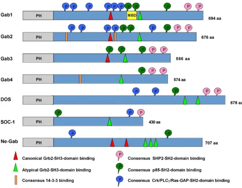
The Gab proteins are large scaffold or docking proteins of 50 to 100 kDa found in metazoans [11,12]. Functionally and/or structurally related proteins are: the docking pro-tein FRS2, an important signal transducer downstream of FGF receptors; the IRS proteins that have emerged as criti-cal signalling components regulating insulin action and sensitivity; and the proteins SLP-65 and SLP-76 that fulfil pivotal roles downstream of cell adhesion molecules and antigen receptors and in the haematopoietic system [13-18]. Vertebrates possess at least three paralogues, Gab1 to 3 [8,9,19,20]. In contrast to vertebrates, the genomes of the model organisms Drosophila and Caenorhabditis con-tain only one Gab gene [6,7,20-22]. However, compara-tive analyses of the amino acid (aa) sequences of these invertebrate Gab proteins with the vertebrate proteins, in particular for sequences outside of the highly conserved pleckstrin homology (PH) domain, suggest that SOC-1 probably represents an early divergent member of the Gab family. This issue will be further discussed below. As explained in detail in Fig. 1, all Gab proteins share a sim-ilar modular structure, including a PH domain at their N-terminus, proline-rich regions in the central part and mul-tiple phosphorylated tyrosine residues.
Recruitment of Gab proteins to their site of
action
Docking proteins of the Gab family use several different mechanisms to regulate their subcellular localization. Firstly, the PH domain confers recruitment of Gab pro-teins to plasma membrane patches enriched in specific phosphatidyl-inositol-phosphates (PIPs) [23-29]. In addition to the PH domain, Gab proteins use at least two
additional mechanisms for their recruitment to activated plasma membrane-associated receptors. The first mecha-nism appears unique to the c-Met/Gab1 receptor/trans-ducer system. Gab1 contains a specific c-Met binding domain (MBD), which encompasses aa residues 450 to 532 and confers the direct interaction between this dock-ing protein and the c-MET RTK followdock-ing its engagement by its ligand, hepatocyte growth factor (HGF) [17,30-32]. The MBD could be narrowed down to a sixteen amino acid motif (aa residues 486-501) called Met binding motif [31]. This direct interaction involves the activated kinase domain of c-MET and the MBD in Gab1 [17,31]. How-ever, c-MET also recruits Gab1 via a second mechanism involving the small adaptor protein Grb2, and this repre-sents the only mode of receptor interaction for Gab2 [33]. The significance of this indirect recruitment is under-scored by the observation that a c-Met receptor mutant selectively defective in Grb2 binding fails to induce branching morphogenesis in the Madin-Darby canine kidney (MDCK) cell line model system [34] and by the non-viable phenotype of knock-in mice expressing a Grb2 binding-deficient Gab1 mutant [32]. This indirect mode of recruitment (Figs. 2 and 3) appears to apply to all other receptors recruiting Gab proteins. Phospho-tyrosine resi-dues within the cytoplasmic tails of these receptors serve as docking sites for the SH2 and/or PTB domains of Grb2, which binds to the proline-rich regions in Gab1-3 via its C-terminal SH3 domain [11,33,35-40]. Shc proteins can serve as additional bridging adaptors between Grb2 and the tyrosine-phosphorylated receptors.
Indirect recruitment of Gab proteins with the help of Grb2 adaptors
Vertebrate Gab proteins possess at least two regions that are potentially involved in the recruitment of Grb2 or other proteins containing SH3 domains such as Mona/ Gads (Fig. 1; [11,41]). Such recruitment sites for SH3 domains were also identified in DOS and the Gab-like proteins identified in the sea squirt Ciona and the sea anemone Nematostella [42,43]. The small adaptor protein Grb2 contains a central SH2 domain flanked on each side by an SH3 domain [44]. Upon ligand binding, many cell surface receptors become tyrosine phosphorylated, which provides binding sites for the SH2 domain of Grb2 [3]. While being bound to the phosphorylated receptor, Grb2 can then use its two SH3 domains to recruit additional proteins to the activated receptor. For example, Grb2 binds to proline-rich stretches in the Ras-guanine nucle-otide exchange factor SOS via its N-terminal SH3 domain, while it uses its C-terminal SH3 domain to bind to two SH3 binding motifs within Gab proteins [42,45].
which occurs in Gab1/2/3, but not in SOC-1 and DOS, conforms to the canonical PXXP motif for SH3 domain binding [47]. In addition, both Gab1/2/3 as well as DOS and SOC-1 contain a so-called atypical Grb2 binding site with the recognition sequence PXXXR [46], which is also found in the SLP-76 and SLP-65/BLNK docking proteins [41,48]. Biochemical experiments by Lock et al. have dem-onstrated that both binding sites contribute to Grb2 bind-ing [46] and consequently most functional studies addressing the Gab/Grb2 interaction utilize Gab mutants in which both recruitment motifs are mutated (Grb2). However, these two sites may not be functionally
equiva-lent. Using crystallography, peptide arrays and isothermal calorimetry, Harkiolaki et al. recently provided new insights into the interaction between the C-terminal SH3 domain of Grb2 and Gab2. In this study they demon-strated that both Grb2 binding sites contain the core con-sensus motif RxxK (with x for any amino acid) [42]. However, they also determined that the individual bind-ing modes between the C-terminal SH3 domain of Grb2 and peptides derived from the typical and atypical Grb2 binding site differ significantly from each other. Conse-quently, this study provides a prime example of the flexi-bility of SH3 domains with regard to target recognition. Conserved structural features of Gab proteins
Figure 1
Since mutation/deletion experiments have clearly demon-strated the importance of the Gab2/Grb2 interaction for the activation of the various effector pathways controlled by this docking protein [39,40], important issues that remain to be resolved for many signalling systems are whether both recruitment sites are equally important, functionally redundant or are used in a stimulus-specific manner. In addition, it remains possible that the individ-ual sites are used sequentially during the Gab recruitment process. Feller et al. (2002) have addressed the first issue for DOS by showing that mutation of either of the two Grb2 binding sites impairs R7 photoreceptor cell develop-ment in a moderate manner, while simultaneous muta-tion abrogates R7 development completely [43]. Furthermore, Yamasaki et al. (2003) have shown that the atypical Grb2 binding site plays a dominant role in the Gads/Grb2-mediated recruitment of Gab2 to the LAT sig-nalling complex in the lipid rafts of T lymphocytes [41]. A final point of interest is that, although one might predict
the Gab/Grb2 interaction to be constitutive, time course experiments have revealed that the Grb2/Gab ratio is increased by extra-cellular signals such EGF or IL-3 stimu-lation [8,9,39,49,50]. It remains to be tested as to whether this increase is caused by a conformational change of Gab that facilitates Grb2 binding or if this reflects indirect recruitment of this adaptor into the Gab2 signalosome by other proteins. Such a scenario could involve SHP2, which interacts with Grb2 as well [51].
A more complex indirect recruitment mechanism is uti-lized by Gab1 in FGF receptor signalling [14,52]. Here, the activated FGFR recruits first the docking proteins FRS2/
, which are tethered to the plasma membrane by a myri-stylation anchor at their N-terminus and in addition con-tain a phosphotyrosine-binding (PTB) domain that mediates the direct interaction with FGF receptors [53]. Following interaction of FRS2/ with the activated receptor, phosphorylated tyrosine residues within the Recruitment of Gab proteins to activated receptors and the main effector arms of Gab signalling
Figure 2
FRS2 proteins serve as binding sites for the SH2 domain of Grb2 (Fig. 3). In turn, Grb2 binds with its C-terminal SH3 domain to Gab1, which is then recruited to the acti-vated FGFR complex and becomes tyrosine phosphor-ylated. This mechanism plays an important role in FGF-induced PI3K activation, since this is mediated via Gab1/ p85 interaction in the signalling complex [52]. It remains to be tested as to whether this mode of recruitment is also realized for Gab2 and Gab3 and for other receptor sys-tems, such as NGF/TRK receptors, which also employ FRS2 proteins as signalling platforms [54]. The different strategies employed by particular cell surface receptors to recruit Gab docking proteins are summarized in Fig. 3.
The Gab PH domain
Several PH domains are able to recognize specific phosph-oinositides such as phosphatidylinositol-3,4,5-trisphos-phate (PI3,4,5P3 or PIP3 in short), phosphatidylinositol-3,4-bisphosphate (PI3,4P2) as well as phosphatidylinosi-tol-4,5-bisphosphate (PI4,5P2) [55,56]. Interestingly, Gab1/2 belong to the few proteins, which bind preferen-tially to the PI3K product PI3,4,5P3, which is only found
within the plasma membrane, and less to PI3,4P2 and PI4,5P2 [23-25,40,57]. The PH domain plays an important role in the plasma membrane recruitment of Gab1 in cells stimulated via the EGF-, VEGF- or B cell antigen receptors (BCR) [23,26,27,58]. It is also required for recruitment of Gab1 to cell-cell contacts, and for the morphogenetic pro-gram triggered by the c-MET receptor [24,25]. In the case of Gab2, the PH domain mediates recruitment to phago-cytic cups induced by FcRI [28] and is required for bFGF-induced tyrosine phosphorylation of this docking protein in murine P19 teratocarcinoma cells [29]. In conclusion, these findings suggest that the PH domain might play an important role to localize or to concentrate Gab proteins to membrane areas where receptors are activated.
Recruitment via the PH domain or adaptors - which mechanism predominates?
The PH domain mediated recruitment of Gab proteins provides the opportunity to modulate their membrane residency by PI3K and lipid phosphatases in a very dynamic manner without the need to disrupt large signa-losomes such as the EGFR/Grb2/Gab complex that are potentially stabilised by various direct and indirect pro-tein-protein interactions. A recent study by Sampaio et al. (2008) reiterates the importance of the PH domain by showing that it is required for the EGF-triggered recruit-ment of Gab1 to the plasma membrane in the presence of low doses of EGF, while the recruitment of this docking protein by high doses of EGF relies on Grb2 [59]. The dependency on the PH domain could be explained by the fact that, in the presence of low EGF concentrations, fewer EGFR molecules are auto-phosphorylated and thereby have a reduced potential to recruit binding partners such as the SH2 domain of Grb2. However, the reason(s) as to why high concentrations of EGF induced lower tyrosine phosphorylation of a Gab1 mutant impaired in Grb2 binding than a low concentration of this growth factor is unclear, but might be explained by competition with other PH domain containing proteins [59].
If the PH domain were to play such an important role under low growth factor stimulation, one would expect that the membrane recruitment mechanisms reliant on protein-protein interactions such as the c-MET/Gab1 and Grb2/Gab interactions would be largely dispensable. In the following, we review several lines of evidence from various experimental settings indicating that the PH domain alone cannot confer long-term plasma-mem-brane residency or ensure adequate physiological Gab sig-nalling. For example, the MBD plays an important role in Gab1 recruitment under certain circumstances [33]. A strong interaction with particular activated receptors is mediated via the Grb2 binding sites, as indicated by vari-ous lines of evidence. Firstly, the tyrosine phosphoryla-tion of Gab1 is drastically reduced in mouse embryonic The Gab recruitment code
Figure 3
fibroblasts (MEFs) lacking Grb2 or expressing a function-ally impaired Grb2 protein in which its SH2 domain has been rendered non-functional by the E89K knock-in muta-tion [60]. The opposite experiment in which the Grb2 binding sites in Gab1 were mutated also resulted in an impaired tyrosine phosphorylation of Gab1 [46,59,61,62]. A similarly impaired tyrosine phosphoryla-tion of Gab1 was observed in Fr3T3 cells expressing a Grb2 binding deficient and transformation impaired mutant of the Tpr-Met oncoprotein [63]. Most impor-tantly, despite the presence of intact PH and MET-binding domains, knock-in mice that express a Gab1 mutant lack-ing the Grb2 bindlack-ing sites display an embryonic lethal phenotype and defects in liver, placenta and craniofacial development [32]. This finding underscores the impor-tance of the Gab1/Grb2 interaction. Furthermore, a Gab2 mutant lacking both typical and atypical Grb2 binding sites displays a reduced and short-lived tyrosine phospho-rylation in EGF-stimulated human mammary epithelial cells and in Fc RI-stimulated murine bone marrow derived mast cells (BMMCs) [39,40]. This suggests that the Grb2 binding sites, while not essential to achieve a cer-tain degree of tyrosine phosphorylation, are necessary to sustain tyrosine phosphorylation, in particular at time points at which PI3K levels have already returned to base-line levels due to the action of PIP3 hydrolysing phos-phatases such as SHIP and PTEN [64,65]. This notion is further supported by the plethora of receptors employing Grb2 as a recruitment device for Gab proteins (Fig. 3).
Overall, it appears that the relative roles played by these alternative recruitment mechanisms are context-depend-ent. The reports reviewed in this section invite for detailed future studies that not only take the amount and timing of the extra-cellular stimulus into account, but also consider the lineage and transformation status of the cell lines. Indeed, the Gab/Grb2 interaction might be more relevant in primary tissues or immortalized cell lines such as BMMCs and MCF-10A, than in particular tumour cell lines often used in signalling studies, e.g. Jurkat or MCF-7 cells, which display elevated PIP3 levels due to the loss of PTEN expression or PIK3CA mutations, respectively [65-68]. Lastly, it should be noted that even in the same cellu-lar setting, particucellu-lar Gab proteins may differ in their requirement for PH domain-mediated plasma membrane recruitment. For example, van den Akker et al. showed that the EPO-induced tyrosine phosphorylation of Gab2 is much more reliant on PI3K activity than that of Gab1 [69].
Positive regulation of Gab proteins and
downstream effectors
As bona fide signal transducers, Gab proteins not only pos-sess structural motifs for their receptor recruitment, but also contain features that are involved in the transduction,
localization and amplification of receptor-derived signals (Fig. 1). At the moment, the SHP2/Ras and the PI-3K/AKT pathways are considered as the two major effector arms of Gab proteins. However, a series of biochemical and genetic studies as well as yeast-two-hybrid (Y2H) screens have identified additional Gab effector proteins such as PLC-isoforms [8,70], adaptor proteins of the Shc [9,61,71] and Crk families [72-76], the lipid phosphatase SHIP [71], the Ras-GTPase activating protein RasGAP [77], GC-GAP [78] and transcriptional activators STAT3 and STAT5 [50,79-81]. In the following sections we will provide an update regarding recent insights into these effector pathways.
Tyrosine phosphorylation of Gab proteins
A fundamental mechanism for regulation of Gab-medi-ated signal transduction is site-specific tyrosine phospho-rylation of these docking proteins. Depending on the particular Gab family member, tyrosine phosphorylation may provide recruitment sites for the SH2 domains of the tyrosine phosphatase SHP2, adaptors of the Crk family, PLC and the regulatory subunit of PI3K, p85 [11,12]. However, the kinases and phosphatases controlling the phosphorylation status of these tyrosine residues are in many cases still ill-defined. While, at least in vitro, RTKs such as the EGFR are able to phosphorylate Gab1 directly [82], it is becoming increasingly evident that a variety of systems such as RTKs, antigen receptors, cytokine recep-tors and even the Bcr-Abl oncoprotein "sub-contract" PTKs of the Src-, Syk/ZAP-70 and JAK families to drive the tyrosine phosphorylation of Gab1/2 [37,83-94]. In some instances, a cascade of PTKs regulates Gab phosphoryla-tion, such as the Bcr-Abl/JAK2/Lyn pathway in human CML cells [91,95]. Also, it is possible that individual PTKs might target distinct tyrosine residues in Gab proteins. By recruiting various effectors with SH2 domains, Gab pro-teins mediate not only signal amplification, but, as a func-tion of the recruitment of distinct enzymatic activities, also channel the receptor-derived signals into pathways with distinct biological properties (Fig. 2). Thus, Gab pro-teins act as a nucleation core of an entire signalling sub-system, which we will dissect in the following sections.
The SHP2/Ras/ERK pathway
recruit-ment platforms, but also as allosteric activators. But what are the functional consequences of Gab-mediated SHP2 recruitment and activation? The best understood effect mediated by the Gab/SHP2 interaction is the sustained and/or increased activation of the ERK/MAPK pathway (Fig. 2). This effect occurs in response to a variety of stim-uli, including treatment of cells with EGF, VEGF, HGF and LPA [35,39,48,62,98-101]. However, in certain cellular contexts, the Gab/SHP2 complex also positively regulates other downstream pathways. These include c-Kit-induced Rac activation [102] as well as 1-integrin- and growth factor-induced PI3K activation [39,103]. The detailed mechanisms involved in Gab/SHP2-mediated regulation of Rac and PI3K have yet to be resolved. In cultured mam-malian cells, recruitment of SHP2 to particular Gab pro-teins regulates diverse biological endpoints, including PDGF-induced cytoskeletal organization and VEGF-induced migration in endothelial cells [62,104], cell adhe-sion and migration of Ba/F3 haematopoietic cells [103], epithelial morphogenesis in MDCK cells [99] and acinar growth of MCF-10A mammary epithelial cells [39]. These studies have been complemented by epistasis analyses in
Drosophila melanogaster and Caenorhabditis elegans
[7,21,105]. Recently, the physiological significance of the Gab/SHP2 interaction for Ras/ERK activation was under-scored by the generation of knock-in mice expressing a Gab1 mutant lacking the tyrosine residues involved in SHP2 recruitment (Gab1SHP2). These mice display impaired development of muscles and the placenta [32], an organ known to be extremely sensitive towards aber-rant ERK signalling [106].
Taken together, these findings demonstrate that SHP2 is an important positive modulator of ERK activation. How-ever, why the recruitment of SHP2 by Gab proteins is required for full activation of ERK signalling downstream of receptor tyrosine kinases is still not fully understood, but several mechanisms may contribute. Firstly, SHP2 de-phosphorylates binding sites for p120Ras-GAP on the acti-vated receptors for PDGF and EGF [107,108] and also on Gab1 [77] and thereby counteracts Ras inactivation. In the latter case, p120Ras-GAP is recruited via its SH2 domain to phosphorylated Y317 on Gab1, which is dephosphor-ylated by SHP2. Like Gab1, DOS is also de-phosphor-ylated by the SHP2 orthologue Corkscrew (CSW) resulting in enhanced Ras activation [6]. Secondly, Shp2 dephosphorylates recruitment sites for the Src-inactivat-ing kinase Csk on the transmembrane glycoprotein PAG/ Cbp [109] and paxillin [110], leading to enhanced activity of Src family kinases. These data are consistent with a report showing that the expression of a fusion protein consisting of the Gab1 PH domain and SHP2 does not only induce constitutive ERK pathway activation, but also enhances activation of Src [100].
The PI3K effector arm
Through the recruitment of PI3K to activated receptors, Gab proteins contribute to the initiation of signalling pathways promoting cellular growth, survival, migration and proliferation [11]. The generation of knock-in mice expressing a Gab1 protein defective in p85 recruitment (Gab1p85) demonstrated that the interaction between Gab1 and PI3K downstream of the EGFR is important in embryonic development for eyelid closure and for kerati-nocyte migration [32]. Nevertheless, their viable pheno-type also indicates that the Gab1/p85 interaction is, in comparison to the interactions of Gab1 with c-Met, Grb2 and SHP2, a relatively dispensable interaction during mouse development. While a Gab1 gene knock-out is embryonic lethal [61,111], Gab2 deficient mice are viable [112-114]. However, the essential role of Gab2 in IgE-mediated allergic responses is attributed to its function in coupling FcRI to PI3K activation [112]. The role of Gab2 in FcR-mediated phagocytosis also seems to be depend-ent on the recruitmdepend-ent of PI3K [11]. Some receptors recruit PI3K both via direct p85 binding sites and via Gab proteins, for example c-Kit and the NGF receptor [63,115]. A recent study showed that a splice variant of c-Kit that recruits Gab2 induces much stronger activation of the PI3K pathway than an isoform that does not bind Gab2 and recruits PI3K only directly [116]. Similarly, the B and T cell antigen receptors recruit PI3K via co-receptors and via Gab2 [27,117]. Thus, Gab proteins serve as ampli-fiers of PI3K signalling in many receptor systems, in par-ticular for those lacking direct p85 binding sites such as the IL-3 receptor. This receptor activates PI3K via a Shc/ Grb2/Gab2 complex and other cytokine receptors lacking direct PI3K binding sites might use the same pathway [38]. Lastly, it should be emphasised that Gab1-induced PI3K activation can amplify receptor signalling by gener-ating a positive-feedback loop, as described for the EGFR system by Rodrigues et al. (2000) [23].
Gab signalling to PLC
RANK/PLC2/Gab2 axis is supported by the observation that mice deficient in RANKL, RANK, PLC2 or Gab2 develop an osteopetrotic phenotype (see below). How-ever, while these studies identify particular PLC isoforms as important effectors or regulators of mammalian Gab proteins, a DOS protein lacking the putative PLC binding sites is able to rescue the phenotype of DOS-deficient flies [105], indicating that the DOS/PLC interaction does not play an essential role in this context.
Shc proteins - just companions of Grb2?
Another prominent component of immuno-purified Gab signalling complexes are the Shc adaptor proteins. In many cases, however, it is still unclear as to whether Shc interacts directly with Gab proteins or is recruited via Grb2. The latter mechanism has been demonstrated for Gab2 signalling complexes from EGF-stimulated mam-mary epithelial cells and from Fc RI- or stem cell factor (SCF)-stimulated mast cells [39,40,49,102]. Similarly, Liu et al. (2001) identified Shc in Gab2 complexes from M-CSF stimulated cells, but failed to purify Gab2 using GST fusion proteins bearing either the SH2 or PTB domain of Shc [120]. These data argue against a direct interaction. However, it should be noted that the Scansite program [121] predicts putative binding sites in Gab1 and Gab2 for the SH2 domain of Shc and Far Western blot analyses have demonstrated a direct interaction between the GST-Shc SH2 domain and tyrosine phosphorylated Gab1 puri-fied from BCR-stimulated B cells [122]. Consequently, Shc proteins might be able to interact with Gab proteins under certain circumstances. Be that as it may, the exact role of Shc proteins in the Gab signalosomes is still not completely resolved. Do they only serve as "bridging mol-ecules" (Fig. 2 and 3) or do they fulfil additional func-tions, e.g. by concentrating additional regulators of Gab signalosome components such as 14-3-3 proteins [123] or the SHIP lipid phosphatases [124,125]? Indeed, SHIP1 and 2 have been found in Gab signalosomes in a variety of settings, e.g. in Gab1 complexes purified from B cells stimulated either through the BCR alone or in co-cluster-ing experiments involvco-cluster-ing both BCR and the inhibitory FcRIIb [124,125]. Similarly, SHIPs have also been detected in Gab1/2 signalosomes isolated from EPO-stim-ulated UT-7 cells [71], a human pluripotent leukemia cell line, in FcRI-stimulated RBL-2H3 cells [126] and in M-CSF-stimulated FDCP1 cells, that represent mouse mye-loid progenitors [120]. Although a direct interaction between the SH2 domain of SHIP1 and Gab2 was demon-strated in Far-Western blot experiments [120], several studies suggest that these interactions are indirect and mediated via Shc [124,125]. The role of SHIPs in Gab sig-nalling complexes is still ill-defined, however, an attrac-tive idea is that they counteract the contribution of the Gab proteins to local PI3K signalling.
The role of Gab proteins in the activation of small GTPases
Crk proteins constitute another group of Gab interaction partners. These adaptor proteins consist of one N-termi-nal SH2 domain followed by one (CrkI) or two (CrkII) SH3 domains [127,128]. As shown in Fig. 1, both Gab1 and Gab2 as well as DOS contain multiple consensus binding sites (Y-X-X-P) for the SH2 domain of Crk pro-teins [127,129]. The interaction of Gab propro-teins with these adaptors has been observed in a variety of cell types and downstream of distinct receptor/transducer systems such as RTKs, antigen and certain cytokine receptors [74,127,129]. In turn, Crk proteins recruit particular effec-tors via their SH3 domains e.g. guanine nucleotide exchange factors for Rac and Rap-GTPases. Thereby, they potentially regulate cellular motility, adhesion and mor-phology [74,129]. Interestingly, Watanabe et al. have recently demonstrated that HGF/c-MET signalling in human synovial sarcoma cell lines induces sustained tyro-sine phosphorylation of Y307 on Gab1, which serves as a recruitment site for both Crk and PLC [129,130]. The recruitment of Crk to this residue is not only pivotal for downstream signalling events, e.g. Rac activation, and enhanced cell scattering, invasive behaviour and xenograft growth, but is also required for the sustained tyrosine phosphorylation of Gab1 itself [129]. In a follow-up study, the same grofollow-up demonstrated that Y307 is phos-phorylated by Src. This enhances cellular migration and contributes to the membrane recruitment of Gab1 in HGF-stimulated MDCK cells and the organisation of focal adhesion complexes [93]. Furthermore, other studies have shown that formation of the Gab1/Crk complex is a criti-cal event in c-Met induced activation of the JNK pathway, an event downstream of Rac activation and a prerequisite for several of the aforementioned morphological changes and efficient cellular transformation [72,131,132]. Inter-estingly, a recent report has demonstrated that the p85 and Crk binding sites in Gab1 play a pivotal role in the c-Met mediated entry of the intracellular bacterium Listeria monocytogenes, implicating the Gab1/Crk complex in pro-motion of cytoskeletal rearrangements required for path-ogen internalization [133].
sug-gests a direct interaction between both proteins, it should be noted that GC-GAP also interacts in mammalian cells with the N-terminal SH3 domain of Crk [78].
A recent study by Paliouras et al. (2009) has identified the Ser/Thr-kinase and Rac/Cdc42 effector PAK4 as a specific interaction partner of the Gab1 isoform [130]. PAK4 binds in a phosphorylation-dependent manner via its GEF-interacting domain to a region in Gab1 located between the PH domain and the first of the three Crk binding sites (aa 116-234). Interestingly, the authors could show that ectopically co-expressed Gab1 and PAK4 cooperate in HGF-induced epithelial cell scattering and invasiveness and that PAK4 knockdown or deletion of the PAK4 recruitment region impaired these biological responses.
The Jak/STAT pathway
Although the molecular details remain ill-defined, the Gab2 isoform is increasingly implicated in JAK/STAT sig-nalling. An early study demonstrated that in CD4-positive T cells derived from the rare human neoplasia mycosis fun-goides, tyrosine-phosphorylated Gab2 interacted with SHP2 and STAT5a in a IL-2- regulated fashion [79]. Later, Arnaud et al. (2004) uncovered a complex interplay between Gab2, SHP2 and STAT5 in IL-2 stimulated T cells [80]. Here, S623 becomes phosphorylated in a negative feedback loop by activated ERK, which in turn reduces the potential of Gab2 to interact with SHP2 via the phospho-rylated tyrosine residues Y614 and Y643. Interestingly, activation of the ERK pathway was blunted, as expected by other studies, by the Y614F mutation and slightly increased by the Gab2S623A mutant. In contrast, IL-2-induced STAT5 activation was enhanced by the SHP2 binding mutant Gab2Y614F and inhibited by Gab2S623A. These data indicate a potential role of STAT5, its interac-tion partners or its upstream kinases as SHP2 substrates.
Additional observations support the concept of a func-tional cooperation between STAT5 and Gab2. First, the murine gab2 gene is one of the top candidates on the modifier locus located on chromosome 7 that modulates the engraftment of hematopoietic stem cells (HSC) during steady-state haematopoiesis, a process dependent on intact cytokine signalling [134]. Second, two studies from the Gouilleux laboratory have shown that constitutively active mutants of STAT5 (caSTAT5) not only associate with Gab2, but also require this docking protein for the efficient induction of Ba/F3 cell proliferation via the Ras/ ERK and PI-3K/AKT pathways [50,135]. In this system, caSTAT5-induced cell proliferation, as well as ERK and Akt activation, is dependent on Gab2/p85 binding. Interest-ingly, the authors also demonstrate that the basal tyrosine phosphorylation of Gab2 is increased in caSTAT5-express-ing Ba/F3 cells [50]. This suggests that PTKs are recruited
to the Gab2 signalosome by caSTAT5 or that STAT5 pro-tects Gab2 against dephosphorylation by PTPs, e.g. Shp2. In support of the latter model, Gab2 is not associated with Shp2 in caSTAT5 expressing cells. However, it remains unclear at present whether the Gab2/STAT5 interaction is mediated via a direct interaction or via a mutual binding partner such as p85. Clearly, further work is required to characterize mechanisms underpinning the interplay between Gab2 and STAT5, and to determine how STAT5 antagonizes Shp2 recruitment to this docking protein.
In addition to the STAT5/Gab2 relationship, Ni et al. (2007) have demonstrated that murine and human Gab2 orthologues, but not Gab1, contains a canonical STAT3 binding motif (YXXQ). Using a Y194F substitution mutant, the authors could demonstrate that this site is indeed required for the recruitment of STAT3 and the effi-cient Friend erythroleukemia virus-mediated transforma-tion of murine hematopoietic progenitors [136]. It remains to be seen as to whether this site is also involved under more physiological circumstances and in the recruitment of other STAT proteins such as STAT5.
Gab proteins are ancient elements of the
metazoan signalling toolbox
contain the key residues for the interaction with the C-ter-minal SH3 domain of Grb2 [42]. It should be noted that the typical Grb2 binding site does not occur in the various DOS proteins and SOC-1 [20]. Importantly, our Scansite analysis revealed that NeGab also carries several tyrosine residues that match phosphorylation motifs involved in the recruitment of SH2-containing proteins such as the Gab signalosome components p85, CrkL and PLC (Fig. 1). Overall, these findings are consistent with the presence of orthologues of the Gab signalosome components Grb2, Shc, PLC and Crk in Nematostella [137] and data-base entries for SHP2- and p85-like proteins for another Cnidarian, Hydra magnipapillata (our unpublished obser-vations). However, it should be noted that, whilst there are also tyrosine residues in NeGab that align with those
involved in the recruitment of SHP2 to mammalian Gab proteins, the surrounding residues are not conserved and therefore do not constitute a bona fide recognition motif for both SH2 domains of mammalian SHP2 [141]). These findings suggest that either NeGab does not recruit the corresponding SHP2 orthologue or that the NeGab/SHP2 interaction takes place by other means. Nevertheless, according to a Scansite prediction [121], the SH2 binding motifs in NeGab align with one of the CrkL (Y266 in human Gab2) and p85 (Y584) recruitment sites in mam-malian Gab proteins. Taken together, these data suggest that at least the Gab/Crk, Gab/PLC and Gab/PI3K con-nections were already established in the precambrium and that a Gab-like docking protein was an early feature of the metazoan PTK signalling toolkit (Fig. 4A).
(A) Simplified phylogenetic tree illustrating the emergence of PTK signaling systems and the emergence of Gab/Dos proteins
Figure 4
So far, all sequenced genomes of invertebrate model organisms including those closer to the basis of verte-brates such as the chordates Ciona and Branchiostoma pos-sess only one Gab-like protein [42,142]. This suggests that the three Gab paralogues were generated by the genome-wide duplications that occurred with the emergence of vertebrates and have diversified subsequently [42,143]. However, the Gab family appears to expand further in mammals. An enigmatic entry in genomic databases rep-resents the GAB4 gene, for which entries at both the genomic DNA and transcript level have been recorded for humans and chimpanzees. The human GAB4 gene is located on chromosome 22q11.1 and its nucleotide sequence is most related to Gab2 (65% similarity: Fig. 4B), which is encoded on chromosome 11q13. GAB4 con-tains a bona fide exon/intron structure suggesting that this gene is not a retro-transposon-like element that has been derived from a Gab2 transcript. Furthermore, the detec-tion of Gab4 ESTs in testicular tissue, as well as sequence differences between Gab2 and Gab4, suggest that this gene is indeed actively transcribed and might give rise to a functional Gab4 protein [144]. While the putative Gab4 contains potential binding sites for SHP2, only one of the three p85 binding sites is conserved and it lacks the typical Grb2 binding site (Fig. 1). The expression pattern, signal-ling mechanisms and functional roles of the Gab4 protein remain to be characterized.
Physiological functions of Gab proteins
Various analyses have shown that most mammalian cell types express more than one Gab family member [19], suggesting that the individual proteins are not function-ally redundant. This hypothesis is supported by the phe-notypes of Gab1-3 gene knock-out mice. While Gab2 and Gab3 knock-out mice are viable and have a normal life span [112,115,145], Gab1 deficiency causes embryonic lethality due to severe defects in heart, placenta, liver, skin and muscle development [61,111]. In line with the spe-cific and intimate relationship between Gab1 and c-Met, it is perhaps not surprising that mice lacking Gab1 phen-ocopy many of the aspects of HGF- and c-MET-deficient mice, such as early embryonic lethality owing to placental defects, reduced liver size and defects in the migration of muscle precursor cells [32,61]. As already discussed above, the generation of knock-in-mice carrying mutations in either the SHP2 or the p85 binding sites of Gab1 revealed that these interactions play distinct roles in embryonic development [32]. Interestingly, enforced membrane localization of Gab2 through the addition of a myristoylation signal together with the introduction of the MBD from Gab1 is sufficient to confer a Gab1-like behaviour to Gab2 in HGF-stimulated MDCK cells [146]. These findings indicate that in the case of Gab1 and Gab2, differences in their subcellular compartmentalization,
rather than in coupling to effectors, leads to distinct bio-logical properties.
Using cardiomyocyte-specific Gab1/Gab2 double-defi-cient mice, Nakaoka et al. (2007) could show that both Gab1 and Gab2 play an important role in the postnatal maintenance of cardiac function [147]. Neuregulin-1 (NRG1 ), a paracrine factor produced from endothelium and a major ligand for the ErbB2/ErbB3 heterodimer, induces marked tyrosine phosphorylation of Gab1 and Gab2 leading to activation of ERK and AKT and the up-regulation of angiopoietin 1 in the heart. These responses were absent in Gab1/2-double deficient mice, which exhibited high postnatal mortality and various signs of cardiac insufficiency.
Gab2 plays an important albeit not essential role in the development of various haematopoietic lineages [113], except for NK cells [148]. Resulting defects in Gab2-defi-cient mice can be attributed to the reduced responsiveness of hematopoietic progenitors to early-acting cytokines [113]. Importantly, Gab2-deficient mast cells display a drastic phenotype. They fail to degranulate and to secrete cytokines following activation of the FcRI antigen recep-tor [112]. Consequently, allergic reactions including sys-temic anaphylaxis are markedly impaired in Gab2-/- mice. These mast cell activation defects reflect the pivotal role of Gab2 as an amplifier for FcRI induced PI3K activation. Similarly, knockdown of Gab2 expression with siRNA or antisense oligonucleotides in RBL-2H3 rat basophilic leu-kaemia cells, a widely used model system for mast cells, results in drastically impaired degranulation and cytokine production [149,150]. Furthermore, murine mast cell development is impaired, because of weakened c-Kit sig-nalling [115]. These findings suggest that Gab2, which is often up-regulated in inflammatory diseases [151], might be an important target for novel therapies against inflam-mation and allergy. However, both Gab1 and Gab2 are involved in the aggregation of platelets triggered by the collagen receptor GPVI [152]. Thus, there might be a cer-tain degree of redundancy between Gab1 and Gab2 for some functions within the hematopoietic system, which could be dissected further using inter-crosses between Gab2-deficient mice and a conditional Gab1 knock-out.
osteo-clast differentiation, which causes decreased bone resorption and a subsequent systemic increase in bone mass. Gab2 is tyrosine-phosphorylated in RANK ligand stimulated osteoclast progenitors and it interacts with the C-terminal domain of RANK. These reports are comple-mented by a study showing that Gab2 plays distinct roles in osteoclastogenesis in different phases of skeletal devel-opment. According to this study, Gab2-deficient mice dis-play enhanced bone formation at six weeks of age and reduced osteoclast differentiation at twelve weeks of age [153]. In addition to the RANK signalling pathway, EGFR signalling has also been implicated in osteoclast differen-tiation. Since Gab2 functions as signal transducer in both pathways, it has been suggested that the crosstalk between the two receptors might be mediated by Gab2. Indeed, an interaction of the EGFR, RANK and Gab2 could be shown. Moreover, stimulation of osteoclasts with RANKL induces tyrosine phosphorylation of the EGFR implying that the EGFR is transactivated by RANK [154]. Recently, the PTK Lyn has been shown to be recruited to the RANK/Gab2 signalling complex and to act as a negative regulator of osteoclast differentiation by inhibiting the tyrosine phos-phorylation of Gab2 [85]. This mechanism involves Lyn-mediated phosphorylation of the tyrosine phosphatase SHP-1. As a consequence, Lyn-deficient mice display bone loss due to increased osteoclastogenesis. Therefore, Lyn can either enhance [86,88,95] or attenuate [85] Gab2 tyrosine phosphorylation, depending on cellular context. These findings further illustrate how fragmentary our knowledge still is in respect to the mechanisms regulating Gab phosphorylation.
All three mammalian Gab proteins are expressed in neu-ronal tissues [10,19,29,155], however their precise role in the CNS remains to be elucidated. Several reports suggest that the individual Gab proteins exert important and potentially non-redundant roles in the nervous system. Firstly, Korhonen et al. (1999) reported that ectopic expression of Gab2 in PC12 cells increased NGF-inde-pendent neuronal differentiation and survival via PI3K-and MEK-dependent pathways [155]. Similarly, Gab2, but not Gab3 acts downstream of FGF receptors in order to ensure the survival of various stem cell models during retinoic acid-induced neuronal differentiation [29]. Inter-estingly, this study also demonstrated that the expression of Gab2 is strongly up-regulated during neuronal differen-tiation and that Gab2 requires its PH domain and p85 recruitment sites to confer bFGF-mediated survival.
So far, studies have not identified any specific roles for Gab3. Genetically-modified mice deficient for Gab3 are healthy and viable and despite the strong up-regulation of this protein during macrophage development, no obvious phenotype was identified in Gab3-deficient macrophages [145].
Negative regulation
Gab proteins fulfill critical roles in the communication between various receptor classes and several signalling pathways involved in the control of proliferation, cell death, migration and differentiation. Consequently, their expression, subcellular localisation and signalling compe-tent state must be strictly regulated. Although our knowl-edge about these processes is still very limited, it is becoming apparent that several layers of negative regula-tion are applied to Gab docking proteins, which we will now discuss.
Negative regulation by phosphatases
Firstly, as the PH domain plays an important role in mem-brane recruitment, Gab signalling is influenced by the bal-ance between the activities of PI3K and lipid phosphatases such as PTEN or SHIP1/2. As discussed above, the latter proteins are recruited into Gab signalosomes [156]. Simi-larly, "PH domain-only" proteins such as the recently-described p53 target and putative tumour suppressor gene product PHLDA3 may negatively influence the membrane recruitment of Gab proteins through direct competition for PI3K products [157]. Indeed, such a scenario is sup-ported by experiments in which the expression of the iso-lated PH domain of Gab1 suppressed EGF-induced ERK and AKT activation in breast cancer cell lines [158].
Secondly, tyrosine-phosphorylated Gab docking proteins recruit SHP2 and it is therefore highly likely that the phos-phorylation of certain tyrosine residues and their associ-ated downstream signalling events are directly regulassoci-ated by this protein tyrosine phosphatase (PTP). Indeed, stud-ies on both DOS and Gab1 have shown that they are dephosphorylated by CSW and SHP2, respectively [6,21,159]. Furthermore, the tyrosine residues implicated in the recruitment of p85 and RasGAP to Gab1 are sub-strates of SHP2 [77,160], which could explain as to why SHP2 mutants with impaired phosphatase activity pro-mote the interaction between Gab1 and a GST-p85 fusion protein [161]. Although this has not been proven so far, it is conceivable that a comparable mechanism can be applied to the Gab2 signalling complex and that the pres-ence of SHP2 in the Gab2 signalosome controls p85 recruitment and the extent of PI3K signalling. Indeed, such a scenario might explain as to why SHP2 recruitment dominates over p85 recruitment in the early phase of EGF-induced Gab2 activation [39] and, given the reports that p85 is accompanied by STAT5 into the signalosomes [135], why Shp2 recruitment is inversely correlated with STAT5 binding [80].
Regulation of Gab protein expression
the regulation of GAB gene promoters remains poorly-characterized, one study has shown that transcription of the GAB2 gene is induced by the transcription factor E2F [162]. Furthermore, Gab2 expression is estrogen-regu-lated in hormone-responsive breast cancer cells [163] and studies in various cellular systems have revealed that Gab2 and Gab3 are up-regulated during cellular differentiation processes [19,29,145,164].
Another study has demonstrated that Gab2 is subject to ubiquitin-mediated degradation in FcRI-stimulated RBL-2H3 basophilic leukaemia cells [165]. However, it remains to be seen as to whether this mode of negative regulation can be extended to other signalling systems and cell types.
The hidden layer of complexity - fine tuning of docking proteins by Ser/Thr-phosphorylation
A fourth and emerging mode of negative regulation of docking proteins is mediated by Ser/Thr-phosphoryla-tion, which is often correlated with their reduced tyrosine phosphorylation and/or changes in subcellular localisa-tion (see Refs. [15,97,166] for review). Indeed, early in Gab signalling research, the dramatic electrophoretic mobility shift displayed by these docking proteins upon growth factor or cytokine stimulation was attributed to phosphorylation events, although the sites and signalling pathways remained largely ill-defined until the recent advent of sensitive phospho-proteomics. Since then there is accumulating evidence that many docking proteins including those of the Gab family are targeted by several immediate early feedback loops involving various classes of protein Ser/Thr-kinases (Fig. 5; for review see Refs. [97,166,167]). Bioinformatic analyses, e.g. using the Net-Phos 2.0 algorithm, predict that Gab1 and Gab2 contain 47 and 76 potential Ser/Thr-phosphorylation sites, respectively [8,168]; our unpublished observations). Indeed, recent phospho-proteomic analyses on Gab2 and SLP-65 [49,169] have underscored our concept that dock-ing proteins are heavily phosphorylated in a dynamic manner and thereby act as the centre of entire signalling subsystems or hubs [170] as it is also depicted in Fig. 2. In the following section we will provide an overview of how this field has progressed over the last five years.
ERK mediated feedback phosphorylation of Gab1
The first reports on the feedback phosphorylation on Ser/ Thr-residues of Gab1 by the MAPK ERK were reported about ten years ago by the Cantley group and were subse-quently confirmed by others in a variety of experimental settings [90,168,171,172]. Six ERK-dependent phosphor-ylation sites (T312, S381, S454, T476, S581, S597) have been mapped on human Gab1 in assays in which recom-binant Gab1 was subject to an in vitro phosphorylation reaction using recombinant ERK1/2 [173]. All these sites
are located within putative MAPK phosphorylation motifs (Fig. 5). Interestingly, most studies pinpoint towards a negative role for ERK in Gab1 signalling as an increase in the Ser/Thr-phosphorylation content of Gab1 is corre-lated with a decrease in its tyrosine phosphorylation [90,168,171,174]. Although the molecular mechanisms involved in ERK-mediated inhibition of Gab1 tyrosine phosphorylation still remain ill-defined, it should be mentioned that four of these sites (S454, S581, S597, T476) are located within the vicinity of the YVPM motifs involved in p85 recruitment [171,173,174]. However, a positive role for the ERK-mediated feedback phosphoryla-tion of Gab1 has been also described [172]. Furthermore, Eulenfeld and Schaper (2008) have revealed that an addi-tional MAPK-dependent phosphorylation site in Gab1, S552, modulates the function of the PH domain in a pos-itive manner and thereby contributes to the IL-6-mediated recruitment of Gab1 to the plasma membrane [175]. Although the precise molecular mechanism remains to be elucidated, this study suggests that phosphorylation may regulate Gab proteins via conformational change. Lastly, Gab1 has been shown to be a substrate of the Ser/Thr-kinase ROK in vitro and possibly in vivo, although the sites of phosphorylation and the functional consequences of these phosphorylation events remain to be identified [176].
Gab2 is the target of several negative feedback loops
Gab2 is also subject to Ser/Thr-phosphorylation at multi-ple sites (Fig. 5). In this regard, we have recently identified 21 novel phosphorylation sites on Gab2 purified from growth factor-stimulated mammary epithelial cells [49]. The recent tally in Phosphosite http://www.phos phosite.org currently lists 10 tyrosine, 18 serine and 5 threonine bona fide phosphorylation sites indicating that Gab2 is a heavily phosphorylated protein.
ErbB2 transgene alone or in combination with an consti-tutively activated AKT transgene showed that the phos-phorylation of Gab2 at Y452 was dramatically reduced in the latter [178]. Although, the phosphorylation status of S159 was not addressed in this study, it is tempting to speculate that the aforementioned Akt-mediated feedback loop is responsible for the attenuation of Gab2 tyrosine phosphorylation. Another recent report has identified the Ser/Thr-phosphatase calcineurin as novel interaction part-ner of Gab2 that interacts with the serine-rich region
C-terminal of the PH domain [179]. This region includes S159 and ectopic expression of calcineurin results in reduced recognition of Gab2 by an anti-AKT substrate antibody, which appears to detect predominantly pS159. In line with the negative role of S159 [177], co-expression of Gab2 and a catalytically active, but not a phosphatase-dead, form of calcineurin enhanced IL-3 mediated activa-tion of a c-fos-reporter construct in a synergistic manner. As described later in this section, Gab2 is also subject to The Phosphomaps of human Gab1 and Gab2
Figure 5
additional PI3K-dependent negative feedback events on S210 and T391 [49].
Gab2 is also regulated by ERK-mediated negative feedback phosphorylation, as identified by Arnaud et al. in 2004 in IL-2 stimulated T lymphocytes [80]. Previously, ERK or an ERK-dependent kinase had been implicated in the phos-phorylation of Gab2 [177,180], however, the phosphor-ylation site(s) was not characterised. Arnaud et al (2004) identified S623 as the site of action of this ERK-mediated feedback loop and provided evidence that the Gab2/SHP2 interaction is enhanced by a S623A mutation.
Taken together, a series of studies over the last decade has shown that the tyrosine phosphorylation and signalling potential of docking proteins such as those of Gab, IRS, FRS and SLP families is counteracted by their Ser/Thr-phosphorylation, which usually represents the endpoint of feedback loops from cytoplasmic signalling cascades [13,15,49,81,169,177,181]. Consequently, major tasks for the future will be to characterize the spatiotemporal regulation of these phosphorylation events in response to
specific stimuli, the kinases and phosphatases involved and the mechanisms by which such modifications control signal output.
How is Gab function regulated by feedback phosphorylation?
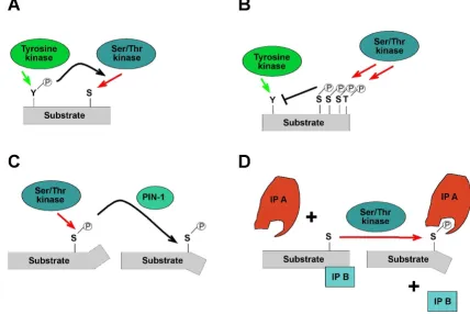
Potential mechanisms that may underpin the action of phosphorylation-dependent positive or negative feedback on Gab proteins are summarized in Fig. 6. Firstly, phorylation of a particular residue might affect the phos-phorylation of a nearby residue in either a positive or antagonistic fashion, due to phosphorylation-induced changes in protein conformation or simply changes in the electrostatic landscape of the substrate protein [182] (Fig. 6A/B). Secondly, phosphorylation-induced conforma-tional changes may alter the accessibility of key regions, such as the PH domain. These may occur due to electro-static repulsion/attraction between distinct protein moie-ties or phosphorylation-induced cis/trans peptidyl-prolyl-isomerisation (Fig. 6C). Although Gab proteins have not been identified as substrates of peptidyl-prolyl-isomerases such as PIN1 yet [183], the high number of
phosphoryla-Potential functional effects of Gab phosphorylation
Figure 6
tion sites preceding proline residues and the fact that Gab proteins are targeted by Pro-directed kinases such as ERK support the likelihood of this regulatory mechanism [80,173,175].
A third mechanism by which docking proteins can be neg-atively regulated by protein phosphorylation is via changes in their "social behaviour", specifically altera-tions in their ability to interact with crucial interaction partners or in their subcellular localisation (Fig. 6D; [4]). Key mediators of this kind of mechanism are 14-3-3 pro-teins, a highly- conserved and ancient group of eukaryotic adaptor proteins that bind to specific phospho-Ser/Thr-residues in their client proteins and thereby execute the effect of phosphorylation events, either by stabilizing cer-tain protein conformations or regulating intermolecular protein-protein interactions [2]. Several docking proteins such as KSR, SLP-76 and IRS proteins have been described as 14-3-3 client proteins [4,15,181] and we recently reported that Gab2 interacts with 14-3-3 proteins in a phosphorylation-dependent manner [49]. This interac-tion is mediated by two 14-3-3 binding motifs surround-ing S210 and T391 that flank the typical Grb2 bindsurround-ing site. Interestingly, while Akt phosphorylates Gab2 only at S159 [177], the phosphorylation of S210 and T391 is attenuated by PI3K and AKT inhibitors indicating that the responsible Ser/Thr-kinases are positively modulated by the PI3K-AKT axis and are therefore acting in negative feedback mode [49]. In support of this model, Gab2 mutants defective in 14-3-3 binding exhibit increased recruitment of Grb2 and consequently sustained associa-tion with the tyrosine phosphorylated EGFR and Shc. Fur-thermore, these Gab2 mutants promote cellular proliferation and transformation. Conversely, introduc-tion of constitutive 14-3-3-binding sites into Gab2 drasti-cally reduces its ability to recruit Grb2 and renders it refractory to receptor activation, demonstrating that site-selective binding of 14-3-3 proteins is sufficient to termi-nate Gab2 signalling. Based on these findings, we pro-posed a model in which signal attenuation occurs, because 14-3-3 promotes dissociation of Gab2 from Grb2, and thereby uncouples Gab2 from the receptor complex. As shown in Figs 2 and 3, the Gab2/Grb2 inter-action is pivotal for the recruitment of this docking pro-tein to most, if not all receptors and consequently this novel regulatory mechanism should have broad implica-tions for diverse signalling systems. Interestingly, the 14-3-3 recruitment motifs around S210 and T391 are con-served in Gab2 orthologues from bony fish to mammals, but are absent from Gab1 and Gab3 paralogues. Gab4 contains the 14-3-3 binding motif around S210, but lacks the motif around T391 and the typical Grb2 binding site, which is positioned in N-terminal vicinity of T391. Fur-thermore, these motifs are also absent from DOS and SOC-1 suggesting that the 14-3-3 interaction is a
verte-brate-specific regulatory layer for Gab2. However, Scansite [121] predicts three potential mode I 14-3-3 binding sites in NeGab (S162, S328 and T516), which also flank the equivalent of the typical Grb2 binding site in NeGab (359-366). Although this remains purely speculative, this observation could indicate that the Gab/14-3-3 interac-tion is an ancestral feature that was modified or lost dur-ing the evolution of the SOC-1, DOS and Gab1/3 proteins.
Gab docking proteins in human pathologies
Gab proteins and tumourigenesis
Given their pivotal role in many physiological processes, it is perhaps not surprising that Gab proteins are impli-cated in a variety of human diseases. In particular, Gab proteins contribute to aberrant PTK signalling in certain malignancies, reflecting their functions as signal amplifi-ers. Only a few mutations have been reported in human Gab proteins so far [184] and COSMIC database) and due to their low frequency it is unclear whether the corre-sponding mutant Gabs represent real drivers or merely passengers of tumourigenesis. However, it is well-estab-lished that Gab proteins promote tumourigenesis by func-tioning as 'accomplices' of certain oncoproteins or by amplifying signalling upon their overexpression. In the following sections we will provide an update regarding their identified roles in hematopoietic disorders and solid tumours.
Haematological neoplasia
The first evidence for the critical involvement of Gab2 in leukemogenesis was the groundbreaking finding that myeloid progenitors from Gab2-deficient mice are resist-ant to transformation by Bcr-Abl [185]. The latter repre-sents a leukemogenic fusion protein generated as a consequence of a chromosomal translocation found in more than 90% of patients with chronic myeloid leukae-mia (CML). Phosphorylation of Y177 within the Bcr moi-ety leads to recruitment of the Grb2/Gab2 complex and downstream signalling via SHP2 and PI3K, which is cru-cial for enhanced proliferation and survival. Similarly, the oncogenic Bcr-FGFR1 fusion protein, which is also the product of a chromosomal translocation (Table 1) and consists of a Bcr-derived moiety and the tyrosine kinase domain of the fibroblast growth factor receptor 1 (FGFR1), drives the tyrosine phosphorylation of Gab2 in murine bone marrow cells and their malignant transfor-mation through phospho-Y177 mediated Grb2 associa-tion [186]. These data strongly suggest that Grb2-mediated recruitment of Gab2 to oncogenic fusion pro-tein tyrosine kinases is a critical event for the induction of a CML-like disease.
silencing of endogenous Gab2 inhibits proliferation and colony formation of CD34+ cells from CML patients, but not their counterparts isolated from healthy donors [187]. However, the role of Gab2 in CML might be more com-plex than just driving proliferation and survival through the PI3K and SHP2/Ras pathways. Indeed, through the recruitment of SHP2, Gab2 tightly controls ERK/MAPK signalling, which will, if it exceeds a certain threshold, drive the terminal differentiation rather than the prolifer-ation of Bcr-Abl transformed myeloid progenitors. Indeed, Gab2 over-expression induces increased ERK acti-vation and megakaryocytic differentiation of the CML cell line K562 [188]. This suggests that the expression levels and signalling competence of Gab2 needs to be tightly controlled in Bcr-Abl+ CML in order to drive proliferation and to repress differentiation at the same time, raising the possibility that modulation of Gab2 signalling might rep-resent a strategy to control this disease.
Despite the great clinical success of the PTK inhibitor imatinib in the therapy of CML, imatinib resistance, due to acquired mutations in the Bcr-Abl oncogene or subse-quent alterations in the cellular signalling network, remains a serious clinical challenge [189]. Interestingly, imatinib resistance in the absence of detectable Bcr-Abl kinase mutation is often mediated by persistent activation of the Src family kinase Lyn, which tyrosine phosphor-ylates Gab2 leading to activation of its downstream effec-tors. Lyn inhibition silences Gab2 and Bcr-Abl tyrosine phosphorylation and restores imatinib sensitivity [86].
Another kinase implicated as a key component of the Bcr-Abl signalling network is Jak2 that in turn activates Lyn leading to Gab2 phosphorylation. Consequently, phar-macological or siRNA-mediated inhibition of Jak2 or Lyn reduces tyrosine phosphorylation of Gab2 in CML cells. Taken together, these findings identify Jak2 and Lyn as additional drug targets in CML and further highlight the important role of tyrosine-phosphorylated Gab2 as a driver of CML [86,91,95].
After the pivotal role of Gab2 in Bcr-Abl-mediated trans-formation had been established, its involvement in the pathogenesis of several other leukemias was discovered (Table 1). The oncogenic fusion kinases Abl and Tel-Jak2 engage Gab2 in a similar manner to Bcr-Abl [190,191]. Tyrosine 314 is crucial for the recruitment of the Grb2/Gab2 complex to Tel-Abl and presumably to Tel-Jak2 as well [192]. Consequently, a Tel-AblY314F mutant exhibits reduced fibroblast transforming capacity and fails to induce a CML-like disease in mice [191]. It should be emphasised that the common denominator of the structurally unrelated Bcr and Tel fusion partners is their potential to recruit Grb2/Gab2 complexes, which underscores again the significance of Gab2 as an amplifier of dysregulated signalling by Abl, Jak2 and FGFR1.
The role of Gab proteins in JMML and NCFC syndromes
Juvenile myelomonocytic leukemia (JMML) and the neuro-cardio-facious cutaneous syndromes (NCFC) are human pathologies caused by aberrant Ras/ERK
signal-Table 1: Oncogenic events in human and murine leukemias involving Gab2
Genetic aberration Leukemic disease Involvement of Gab2 References
Bcr-Abl
translocation t(9;22)
CML, B-ALL Recruitment of Grb2/Gab2 complex to Y177 of Bcr-Abl
Y177 and Gab2 are essential for Bcr Abl-mediated transformation and leukemogenesis
[185,187]
Bcr-FGFR1 t(8;22) CML-like disease Recruitment of Grb2 and presumably Gab2 to Y177 of Bcr-Abl. Increased Gab2 tyrosine phosphorylation
[186]
Tel-Abl translocation t(9;12)
B-ALL, T-ALL, CML Recrutiment of Grb2/Gab2 complex to Y314 of Tel-Abl
Y314 is essential for Tel Abl-mediated transformation and leukemogenesis
[191]
Tel-Jak2 translocation t(9,12) ALL Some isoforms of Tel-JAK2 recruit the Grb2/Gab2 complex via Y314
[190]
Npm-Alk translocation t(2;5)
Anaplastic large cell lymphomas Gab2, SHP2 and Grb2 form a complex with Npm-Alk
[242]
SHP2 E76K point mutation
JMML E76K mutation confers enhanced catalytic activity to SHP2 and requires Gab2 for transformation
[200]
Sf-Stk Friend's virus-induced erythroleukemia in mice Recruitment of the Grb2/Gab2 complex to Sf-Stk is essential for erythroid transformation by Friend virus,
this involves the direct binding of STAT3 to Gab2
[36,81]
Amplification of MLL locus AML/MDS Gab2 is frequently co-amplified with the mixed lineage leukaemia (MLL) gene
ling. The NCFC syndromes comprise neurofibromatosis (NF) and the Noonan (NS), Costello (CS), LEOPARD (LS) and cardio-facious-cutaneous (CFC) syndromes, which are correlated with autosomal-dominant germ-line mutations within either the core components (Ras, B-Raf, Raf-1, MEK) or modulators of the Ras/ERK pathway (NF1, SHP2, SOS and Spred). The resulting mutant proteins dis-play aberrant activities and consequently disturb the over-all fine-tuning of the Ras/ERK pathway and to a certain degree the Ras/PI3K pathway [35,193]. As the ERK path-way steers both proliferation and differentiation, many processes underlying normal human development and organ homeostasis are perturbed and give rise to the vari-ous clinical symptoms, which range from cardiac defects, skin and cranio-facial abnormalities to growth and men-tal retardation [194-196]. Importantly, some NCFC syn-dromes predispose affected individuals to neoplastic diseases [196]. Indeed, the discovery of germline missense mutations in the SHP2-encoding PTPN11 gene in ~50% of NS cases led to the identification of PTPN11 as the most common target of somatic mutations in JMML, a rare, albeit aggressive myelo-proliferative disorder occurring in children, where PTPN11 mutation rates of up to 35% have been reported [196-199]. The most frequently JMML-associated mutation, E76K, confers enhanced cata-lytic activity to SHP2 and requires Gab2 for transforma-tion of primary murine myeloid progenitors [200]. However it should be noted that the nature of somatic JMML-associated PTPN11 mutations differ from the germline mutations identified in Noonan syndrome in that JMML-associated PTPN11 alleles usually encode stronger gain-of-function mutant proteins [200,201]. Nevertheless, this finding demonstrates that Gab2 is an important player in JMML and suggests that NS-associated SHP2 mutants may require Gab proteins as recruitment devices in a similar manner. Indeed, co-expression exper-iments in COS-7 cells revealed that NS-associated SHP2 mutants exhibit a stronger and more sustained interaction with Gab1 than SHP2wt. Importantly, co-expression of Gab1SHP2 in this system blocks the EGF-induced increase in the phosphatase activity of the NS-associated SHP2 mutants and consequently abolishes their positive effect on EGF-induced ERK phosphorylation [202].
While NS patients carry mostly gain-of-function muta-tions in SHP2, this phosphatase often contains dominant-negative mutations in LS patients [197,203]. Interestingly, expression of LS-associated SHP2 mutants with impaired catalytic activity in cells strongly enhances the EGF-induced interaction between Gab1 and p85 [161], which suggests that these mutant proteins, while acting in dom-inant negative fashion on the Ras/ERK pathway, may pro-mote aberrant PI3K activation by protecting the p85 recruitment sites against SHP2wt. Taken together, these studies identify Gab proteins as important "accomplices"
of NCFC-associated SHP2 mutants and suggest that a bet-ter knowledge of Gab signalling will contribute to an improved understanding and treatment of these syn-dromes. Furthermore, the close relationship between SHP2 and Gab proteins and the important role of Gab proteins as modulators of Ras signalling also raise the question as to whether the Gab genes themselves are awaiting their identification as novel "NCFC" alleles.
Aberrant activation and/or expression of Gab proteins in solid tumours
Dysregulated Gab signalling is also increasingly recog-nised as an important contributor to the biology of solid tumours. Firstly, the signalling potential of Gab1 needs to be considered in tumours with aberrant expression or mutations of c-MET [17,204]. As discussed above several studies have demonstrated the close collaboration between c-Met and Gab1 and, in contrast to many other RTKs that merely induce a transient tyrosine-phosphor-ylation of Gab1, c-MET induces a very sustained tyrosine phosphorylation of this docking protein [24,129]. Fur-thermore, a recent study has revealed Gab1 not only as a convergence point between c-MET and EGFR pathways, but also suggests that Gab1 cooperates with MET amplifi-cation in lung cancer cells, which have become resistant towards the EGFR inhibitor gefitinib [204]. A correlation between the tyrosine phosphorylation status of Gab1 and the progression of ErbB2-transgene driven murine mam-mary tumours has also been reported, indicating that Gab1 needs to be considered as an important downstream effector of this oncogenic RTK as well [205]. Lastly, it should be mentioned that somatic missense mutations resulting in conversion of the amino acid residues Y83, T387 and R498 into C, N and M, respectively, have been identified in the human GAB1 gene in human breast and lung cancers, albeit at very low frequencies (for details see the COSMIC database at http://www.sanger.ac.uk/genet ics/CGP/cosmic/ and Ref. [184]). However, it remains to be tested as to whether these mutations alter the signalling properties of Gab1, and so it is unclear at this stage whether they represent real drivers or merely passengers of tumourigenesis.