R E S E A R C H
Open Access
HECTD1 controls the protein level of
IQGAP1 to regulate the dynamics of
adhesive structures
Xiaoli Shen
1,5, Zanhui Jia
1,6, Donato D
’
Alonzo
1, Xinggang Wang
1, Elisabeth Bruder
2, Fabienne Hélène Emch
3,
Christian De Geyter
1,3and Hong Zhang
1,4*Abstract
Background:Cell migration including collective cell movement and individual cell migration are crucial factors in embryogenesis. During the spreading/migration of cells, several types of adhesive structures physically interacting with the extracellular matrix (ECM) or with another cell have been described and the formation and maturation of adhesion structures are coordinated, however the molecular pathways involved are still not fully understood.
Results:We generated a mouse embryonic fibroblast line (MEF) from homozygous mutant (Hectd1R/R,
Hectd1Gt(RRC200)) mouse of the E3 ubiquitin ligase for inhibin B receptor (Hectd1). Detailed examination of cell motion on MEF cells demonstrated that loss of Hectd1 resulted in accelerated cell spreading and migration but impaired directionality of migration. InHectd1R/Rcells paxillin and zyxin were largely mis-localized, whereas their expression levels were unchanged. In addition the formation of focal adhesions (FAs) was impaired and the focal complexes (FXs) were increased. We further identified HECTD1 as a key regulator of IQGAP1. IQGAP1 co-localized together with HECTD1 in the leading edge of cells. HECTD1 interacted with IQGAP1 and regulated its degradation through ubiquitination. Over-expression of IQGAP1 in control MEF phenocopied the spreading and migration defects of
Hectd1R/Rcells. In contrast, siRNA-mediated knockdown of IQGAP1 rescued the defects in cellular movement of
Hectd1R/Rcells.
Conclusions:The E3 ligase activity of Hectd1 regulates the protein level of IQGAP1 through ubiquitination and therefore mediates the dynamics of FXs including the recruitment of paxillin and actinin. IQGAP1 is one of the effectors of HECTD1.
Keywords:HECTD1, Cell movement, Cell spreading, Focal adhesions, IQGAP1, Ubiquitination
Background
Cell migration including collective cell movement and individual cell migration are crucial factors in embryo-genesis [1, 2], as best exemplified in neurulation [3, 4]. Generally, cell migration has been conceptualized as a cyclic process [5], in which a spreading phase is followed by migration involving actin polymerization and myosin contraction. Various mechanisms have been proposed for the regulation of cell spreading/migration, including
active C-terminal Src kinase (CSK) remodeling [6], activation of focal adhesion kinase (FAK) and APR 2/3 [7], actin polymerization and the development of con-tractile forces [8, 9].
During the spreading/migration of cells in culture sev-eral types of adhesive structures physically interacting with the extracellular matrix (ECM) or with another cell have been described [10]. Owing to their highly dynamic nature and size, nascent adhesive structures and FXs typically are sized smaller than 1 μm2 [11]. As cells migrate, these structures either disappear or develop to mature FAs, which are large in size (>5 μm2). Although it is clear that the formation and maturation of adhesion * Correspondence:[email protected]
1
Department of Biomedicine, University Hospital, University of Basel, Basel, Switzerland
4Department of Biomedicine, University of Basel, Hebelstra. 20, CH-4031
Basel, Switzerland
Full list of author information is available at the end of the article
structures are coordinated, the molecular pathways in-volved are still not fully understood [12].
EULIR was first identified as an E3 ubiquitin ligase for the putative inhibin B receptor in our laboratory [13], but international nomenclature later renamed EULIR to HECTD1. Sarkar and Zohn suggested that HSP90 is a binding partner of HECTD1 and that increased secretion of HSP90 in the cranial mesenchyme of HECTD1-mutants is in part responsible for the altered organization and behavior of these cells [14]. Tran and coworkers suggested that HECTD1 promotes the inter-action of the adenomatous polyposis coli (APC) protein with Axin to negatively regulate Wnt signaling through Lys-63 polyubiquitination [15]. We found that knock-down of HECTD1 expression by siRNAs increased the migration velocity and membrane ruffling of HeLa cells. However during the course of our studies, Sarkar and Zohn demonstrated that opmmice increased the cranial mesenchyme cell migration [16, 17] but the findings from Li and coworkers showed that knockdown of HECTD1 inhibits the migration of breast cancer MDA-MB-231 cells [18]. To resolve this contradictory issue, we have used the Hectd1 homozygous mutant (Hectd1R/
R
) mouse embryonic fibroblasts (MEF) generated from a gene-trap mouse embryonic stem (ES) cell line RRC200 (BayGenomics, San Francisco, CA, USA), for cell migra-tion studies.
IQGAP1 belongs to the IQGAPs family of scaffold proteins. Despite the homology of amino-acid sequence with GAP, IQGAP1 does not exert any GTP hydrolysis activity [19–21]. In eukaryotic cells, IQGAP1 localizes to actin-containing structures such as lamellipodia, mem-brane ruffles and cell-to-cell adhesions. As such, IQGAP1 is involved in regulating cellular motility and morphogenesis [22]. Under normal conditions, through its coordinating with small GTPase, Rac1, RhoA and CDC42, IQGAP1 supports cell movement via regulating adherens junctions, actin filaments and microtubules. Initially, IQGAP1 was identified as a target of Rac1 and CDC42. In addition, activation of Rac and CDC42 in re-sponse to stimulation signals leads to the recruitment of IQGAP1, APC and CLIP-170, forming a complex which connects to the actin cytoskeleton and microtubules promoting cell polarization and directional cell migra-tion [23–25]. Another mechanism proposed that IQGAP1 requires PIPKIγ for targeting to the leading edge of migrating cells and be activated specifically by PIP2 to promote actin polymerization and cell migration [26]. In contrast, IQGAP1 may also negatively impact on cell migration. One study demonstrated that IQGAP1 suppresses TβRII- and TGF-β-dependent myofibroblas-tic differentiation in tumors thereby inhibiting tumor growth [27]. Besides, anti-GTPase activity of IQGAP1 sustains the amount of GTP-bound Rac1 at sites of
cell-to-cell contact, resulting in stable adhesion [28]. Re-cently, IQGAP1 was found to localize in FAs [29, 30] and in FXs together with integrin-linked kinase ILK [31]. Schiefermeier and coworkers reported that IQGAP1 in-teracts with FA proteins [32]. However, whether IQGAP1 is directly involved in regulation of the dynam-ics of FAs is still not known, neither is there anything known about its regulation. Through screening various ECMs and a number of adhesion proteins, we found that the stability of IQGAP1 is regulated by HECTD1.
We here propose a novel molecular mechanism explaining the role of Hectd1 in cell movement. Defi-ciency in Hectd1 results in failure to recruit phaxillin and zyxin to FAs thereby promoting rapid cell migration. Taking all data together, our results demonstrate that Hectd1 contributes to morphogenesis through the regu-lation of cell migration.
Methods
Aminals and mating scheme of mutant mouse
To generate Hectd1 mutation mice [33], the gene-trap mouse embryonic stem (ES) cell line RRC200 on a 129 background (129P2/OlaHsd) obtained from (BayGe-nomics, San Francisco, CA, USA) was selected since the insertion site of the gene trap (β-geo) was mapped onto the intron 26 of theHectd1gene, which includes the en-tire open reading frame but lacking the HECT1-domain (Additional file 1: Figure S1A). The ES cells were micro-injected into blastocysts (C57BL/6NCrl × 6 J). Resulting agouti chimeric male mice were crossed with C57BL/6 female mice. Then F1 mice were intercrossed to gener-ate more Hectd1Gt(RRC200)Byg mice for more than 10 generations.
Generation and culture of mouse embryonic fibroblast (MEF) cells
CO2. MEF cells were sub-cultured when they reached 80–90% confluence.
Cell culture and transfection
MEF cells were maintained in high glucose DMEM medium (HeLa cells in low glucose medium) with 10% FBS, 1% of Sodium Pyruvate, 1% of L-Glutaminate and 1% of Penicillin-Streptomycin. Cells were grown in a humidified incubator at 5% CO2at 37 °C. MEF or HeLa cells used for transfection were pre-seeded 24 h in culture vessels. On the day of transfection, the conflu-ence was 50–80%. Transfection of MEF or HeLa cells with plasmid DNA using Effectene reagent according to the protocol of Qiagen.
Fibronectin coating
For cell spreading and migration assay, 24- well plates were coated with 2 μg/ml fibronectin (R&D, 1030-FN) in PBS overnight. For immunohistochemistry staining, glass coverslips were used for coating.
Cell spreading assay
Cells were seeded on 6-well plates and incubated at 37 ° C for 24 h before serum starvation overnight. Starved cells were counted and seeded on fibronectin pre-coated 24-well plates. The plate was immediately sent to time-lapse microscopy (Nikon IX81) pre-warmed to 37 °C and maintaining the CO2level at 5%. Quickly adjusting the positions, the focus, the time interval and total time by CellSens software, the programme was initiated. Dur-ation of spreading was analyzed from attachment to formation of leading protrusion. Cell spreading area was quantified by Image J software.
Wound-healing assay
In monolayer wound-healing assays, 4 × 10⁴ cells were collected and plated in 24-well plate for 24 h. Cells were washed twice with PBS and continuously cultured for 24 h in growing medium containing 0.5% FBS, then cells were starved in serum free medium supplemented with 1 μM aphidicolin overnight. Then, cells were scratched with a 200 μl pipette tip, washed twice with PBS and placed into a complete medium containing 10% FBS and aphidicolin. The plate was immediately sent to time-lapse microscopy (Nikon IX81) pre-warmed to 37 °C and with 5% CO2. Migration images were taken at 10 min intervals for a period of 24 h with a 4× lens. Cell trajectories were measured by tracking the position of the cell over time using “Manual Tracking” plugin (Image J, v 2.0) and the cell velocity and straightness were determined by“Chemotaxis Tool” plugin (Image J, v 2.0). Cells that proliferate or that failed to migrate during the experimental period were not evaluated.
The percentage of MTOC orientated towards the wound was determined at 10 h post wounding. Cells were fixed with 4% paraformaldehyde then co-stained with acety-lated alpha tubulin and Giantin antibodies. Bar, 50 μm. The percent of cells at the wound edge having their Golgi apparatus in the forward-facing 120° sector was measured after wounding. Over 600 cells from 3 inde-pendent experiments were analyzed. Orientation of the Golgi apparatus with respect to the wound edge corre-sponds to percent on the ordinate. *,P< 0.05.
Immunocytochemistry
Cells were seeded on glass coverslips pre-coated with fibronectin for defined time intervals. After that, cells were washed with PBS, then fixed with 4% paraformal-dehyde for 10 min, and permeabilized with 0.15% Tri-ton-×100 in PBS for 15 min and blocked with 5% BSA in PBS for 1 h at room temperature. Primary antibody diluted in PBS was added to the coverslips and incu-bated at 4 °C for overnight. Primary antibodies were used as follows: rabbit anti-paxillin (N-term) (1: 300, epi-tomics, Burlingame, USA), Rabbit anti-paxillin (phospho Y118) (1: 300, Abcam, Cambridge, UK), rabbit anti-zyxin (1:200, Epitomics, Burlingame, USA), Mouse anti-α-Actinin, clone BM-75.2, (1: 150,Sigam-Aldrich, St. Louis, USA), Mouse anti-Src (active), clone28 (1: 500,MBL international corporation),Rabbit anti-IQGAP1 (H-109) (1: 800, Santa Cruz Biotech, Dallas, USA), Rabbit anti-HECTD1(M03), clone 1E10 (1: 100, Abnova, Taipei, Taiwan), Mouse anti-Giantin (1:1000, a gift from Prof. Martin Spiess, Biozentrum, University of Basel). After washing the cells with PBS for 5 times with PBS, the sec-ondary antibody (goat anti-rabbit-FITC, 1:1000; goat anti-mose-FITC, 1:1000; goat anti-mose-546, 1:1000, Invitrogen, Carlsbad, USA) tagged with fluorescent dye was added and incubated for 1 h in the dark at room temperature. After washing, cells were incubated in DAPI in PBS for 3 min at room temperature for counter staining. After washing, cells were mounted with Prolong® Gold Antifade Reagent and stored in 4 °C pro-tected from light. The fluorescent pictures were made with the Nikon Confocal microscope.
Western blot
(1: 300, epitomics, Burlingame, USA), Rabbit anti-paxillin (phospho Y118) (1: 300, Abcam, Cambridge, UK), rabbit anti-zyxin (1:200, Epitomics, Burlingame, USA), Rabbit anti-IQGAP1 (H-109) (1: 800, Santa Cruz Biotech, Dallas, USA), Rabbit GAPDH (14C10) (1:3000, Cell Signalling, Danvers, USA). After 3 times washing in TTBS, the blots were incubated in secondary antibody (goat anti-rabbit-HRP, 1:1000; goat anti-mose-HRP, 1:1000, Invitrogen, Carlsbad, USA) for 1 h at room temperature. To remove the unspecific bound antibody, the blots were washed in TTBS for 3 times. Bands were detected by ECL substrates, visualized by an infrared-based laser scanner (LiCor) and quantified using Image Lab software (Bio-Rad). The band intensity of wild-type cells of no stimulation was normalized with GAPDH as control and the other results were recorded as fold changes compared to control.
Immunoprecipitation
Cell pellets were lysed with IP lysis buffer (20 mM Tris-HCl, PH 8.0, 137 mM NaCl, 1% NP40 and 2 mM EDTA supplemented with 1% protease inhibi-tor cocktail) on ice for 20 min and vortexed in be-tween. Cellular débris was removed by centrifugation at 14,000 g for 5 min and the supernatant was trans-ferred to pre-cooled fresh tubes. The protein amount was equilibrated with the IP buffer. 2 μl primary anti-bodies (Mouse anti-GFP GF28R, Thermo scientific, Waltham, USA) was added per 500 μg protein sam-ples and incubated for overnight at 4 °C. The lysates were then incubated with prewashed protein A/G agarose beads (20 μl/500 μg protein) and rocked for 1 h at 4 °C. Beads were washed three times with IP buffer, 6000 rpm, 3 min. After washing, the beads were heated for 5 min at 95 °C in 2× Laemmli sam-ple buffer. Target proteins were detected by western blot by using specific antibodies. Antibodies were used as: Rabbit anti-HECTD1 (M03), clone 1E10 (1:1000, Abnova, Taipei, Taiwan), Rabbit anti-PIP5K1A (1:1000, Cell Signaling, Danvers, USA), Rabbit anti-β-Catenin (D10A8) (1:1000, Cell Signaling, Danvers, USA).
In vivo ubiquitination
MEF cells were transfected with plasmids DNA for HA-ubiquitin and GFP-IQGAP1 at ratio of 1:1. Twenty four hours after transfection, the cells were washed twice with PBS and changed to serum-free medium supplemented with 1 nM MG132 or DMSO, then incubated for overnight at 37 °C. For endogen-ous ubiquitination assay, MEF cells were seeded for 24 h and directly treated for starvation. Starvated cells were harvested as pellets and re-suspended in serum-free medium. Half of the pellets were spinned
down and lysed with ubiquitination lysis buffer (50 mM Tris, pH 7.5, 1 mM EDTA, 150 mM NaCl, 0.1% Triton X-100, complete protease inhibitor cock-tail, 100 μM MG132 and 100 μM N-ethylmalemide) on ice for 15 min followed with centrifugation (12,000 g, 5 min) at 4 °C. The other half was seeded on fibronectin pre-coated plates and cultivated in 37 °C for 60 min, after that, the plates were placed on ice, washed with pre-cooled PBS and lysed with lysis buffer (as previously) 15 min on ice before cen-trifugation. The supernatant was collected and then we continued with the protein concentration assay. Equal amount of protein was immune-precipitated with target protein and detection of ubiquitin by Western blot. Ubiquitination of target proteins were normalized by the protein amount in MEF cells.
Statistical analysis
All data analyzed using the statistical software package SPSS 13.0 for Windows 7 (SPSS Inc., Chicago, Ill, USA). Normally distributed data was analyzed for statistical dif-ferences using the t-test (paired comparisons) or ANOVA (Analysis of Variance). For data not normally distributed, non-parametric ANOVA and the Mann-Whitney U test were used. All values are reported as means ± SEM. Differences are considered statistically significant with P< 0.05, highlighted with *. For each particular experiment, statistical analysis is presented in the figure legend.
Results
Loss of Hectd1 results in accelerating cell spreading/ migration and impairs directional migration of cells
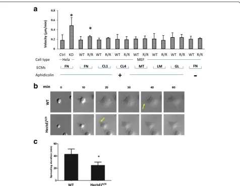
Knockdown of HECTD1 by siRNAs in HeLa cells in-creased the rate of migration (Fig. 1a), to confirm this result we generated a mutant mouse of the E3 ubiquitin ligase for inhibin B receptor (Hectd1). We found that
Hectd1 homozygous mutant embryos display defective
of neural tube closure with excencephaly (Additional file 1: Figure S1 and D’Alonzo et al., manuscript in preparation). We used mouse embryonic fibroblast (MEF) cells obtained from matched wild-type and
Hectd1R/R mouse to analyze the time period from cell
attachment to migration by time-lapse microscopy on various extracellular matrices, such as fibronectin (FN), collagen type I (CL1) or IV (CL4), matrigel (MT), lam-inin (LM) and gelatin (GL). There were significant dif-ferences in cell spreading and migration between the adhesion of wild-type and Hectd1R/R cells on FN but not or to much less extent on other ECMs (Fig. 1a), suggesting that HECTD1 regulates cell migration through only certain subtypes of integrin receptors.
started to form leading edges within 40 min while this process occurred approximately 10 min earlier in Hectd1Hectd1R/Rcells (Fig. 1b and c). We further exam-ined the migration/directionality of cells in wound heal-ing assays (Fig. 2). Loss of Hectd1 results in acceleratheal-ing cell migration (Fig. 2a and time-lapse images were shown in Additional file 2: Figure S2A and Additional file 3: Figure S2B). The velocity (total distance/time) of Hectd1Hectd1R/R cells was to 0.25 ± 0.07μm/min com-pared to 0.19 ± 0.05 μm/min in wild-type cells (P< 0.05) (Fig. 2b), agreed to the results found in HeLa cells (Fig. 1a). Wild-type cells migrated in a cohesive fashion with little dispersion and with aligned displacement paths. In contrast, the trajectories of Hectd1 Hectd1R/R
cells was more scattered (Fig. 2c).
The straightness (Euclidean distance/Accumulated distance) was 0.60 ± 0.14 in Hectd1R/R cells versus 0.78 ± 0.09 in wild-type cells (P< 0.05) (Fig. 2c), indicating that the directed migration of cells was impaired. To further confirm the results, both wild-type and Hectd1R/R cells were stained with acetylated α tubulin and giantin (Fig. 2d), which are cell direc-tional markers since the microtubule-organizing center (MTOC) and the Golgi matrix are reorient and toward leading edges during cell migration or wound healing [34–36]. The percentage of cells with giantin and acetylated α tubulin oriented to the wound was 61.29 ± 15.33% in wild-type cells, whereas this percentage dropped to 40.67 ± 11.25% in
Hectd1R/R cells.
a
b
c
Loss of Hectd1 impairs the subcellular localization of adhesion proteins
To dissect the molecular mechanism involved in causing the observed changes in cell migration in Hectd1R/R
cells, we examined functional molecules in integrin sig-naling.α5β1 is the major FN receptor in fibroblasts but we did not observe differences in expression and localization of subunitsα5 and β1, as well as β3 in con-trast to the expression of α-actinin (Additional file 4: Figure S3 and data not shown), these results suggested that Hectd1 functions downstream of the receptors.
The expression and localization of talin and vinculin, which have been shown to be incorporated into adhesive structures at early stage [37], did not significantly differ in both cell types when cultured on FN (data not shown). When both cells were cultured on FN, the total proteins of paxillin and zyxin were equally expressed (Fig. 3a) and the total focal adhesion area for paxillin did not have significantly different (Fig. 3b). However in
Hectd1R/R cells, the proteins show a decrease of size
distribution at the leading edges (Fig. 3d). Furthermore, paxillin-Y118, one of the FN-stimulated paxillin phos-phorylation, became located in FAs where it was associ-ated with stress fibers in WT. The expression of paxillin-Y118 was mostly located at the cell leading edges as FXs with disperse distribution in cytoplasm in
Hectd1R/Rcells (Fig. 3e). These results indicate that pax-illin and zyxin were mislocalized in the adhesions of
Hectd1R/Rcells.
It has been suggested thatα-actinin acts as a bridge to connect adhesion structures with the actin-cytoskeleton [38]. At the leading edges of wild-type cells activated by FN for 30, 60 and 90 min, α-actinin was mainly co-localized together with paxillin and zyxin in wild-type cells, while this co-localization was not present in
Hectd1R/R cells. 60 min after spreading of Hectd1R/R
cells, we could barely detect any patches of α-actinin at the cellular periphery. As the Hectd1R/R cells continued to migrate, some patches of α-actinin became visible at the leading edges, but still fewer than in wild-type cells
a
c
d
b
(Fig. 4 and Additional file 5: Figure S4). These data indi-cate that Hectd1 exerts its function at adhesion sites.
The formation of FAs but not FXs is impaired inHectd1R/R
cells
FXs are characterized as small punctate adhesions with <1 μm2 surface area lying close to the cell periphery, whereas FAs are classified as larger structures with their surface area varying between 5 and 20μm2[11]. Having allowed cell spreading for 30 min on FN, we started to analyze the dynamics of early (paxillin-only) versus late (paxillin-and-zyxin) adhesions. As shown in Fig. 5a, sig-nificantly more paxillin-containing FXs developed in
Hectd1R/Rcells (P< 0.05) than in wild-type cells. In con-trast, FAs were more prominent in wild-type cells than
in Hectd1R/R cells. However, zyxin, being a late-stage
marker in adhesion formation, was similarly present in the FXs of both cell types (Fig. 5b). In migrating wild-type cells, both paxillin and zyxin showed similar distri-bution patterns in FAs after 60 min and after 90 min, whereas in Hectd1R/R cells the dominant cell adhesion structures consisted of FXs. Our results suggest that the defects in assembly of FAs at cell leading edges were caused by differences in the accumulation or transporta-tion of proteins rather than by differences in the synthe-sis of the proteins.
One of the main kinases thought to be responsible for tyrosine phosphorylation of FA molecules is Src [39, 40]. Fig. 5c showed that there was no statistically significant difference in the expression level and activity of c-Src betweenHectd1R/Rand in wild-type cells after FN stimu-lation (P> 0.05).
a
c
e
b
d
Localized activation of Rac and Rho regulate adhesion dynamics during migration. Using the RhoA activation assay, we found that the activities of RhoA were signifi-cantly (P< 0.05) enhanced in Hectd1R/R cells 60 min after FN stimulation as compared that of wild-type cells (Fig. 5d), in which the total level of Rac1 and RhoA were not significant altered (Fig. 5e).
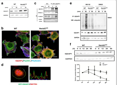
IQGAP1 interacts and co-localizes with HECTD1
We found that IQGAP1 is a protein component of Hectd1 complexes [30] involved in formation of in-tegrin adhesome and membrane ruffling. It has been demonstrated that IQGAP1 is an important factor in regulation of cell migration [26, 41]. As shown in Fig. 6a, the protein level of IQGAP1 was higher in
Hectd1R/R cells than wild-type cells (P< 0.05). Consist-ent with this result, we observed that IQGAP1 is not only expressed in the leading edge of the Hectd1R/R
cells but also heavily present in entire cytoplasm (Fig. 6b). Thus, we further focus on the functional re-lationship between IQGAP1 and HECTD1 in cell migration.
To confirm the interaction between HECTD1 and IQGAP1, we transfected GFP-IQGAP1 plasmids into HEK293 cells for immunoprecipitation. As shown in Fig. 6c, immunoprecipitation of endogenous HECTD1 resulted in the co-immunoprecipitation with GFP-IQGAP1 and co-immunoprecipitation was enhanced after 60 min of stimulation with FN. Next, we per-formed co-localization assays to verify the protein-protein interaction of IQGAP1 with HECTD1. HeLa cells transfected GFP-IQGAP1 were plated on FN coated plates for 60 min. Similar to the presence of HECTD1 in the cell, IQGAP1 was mainly localized in the cytoplasmic of the cells, but was enriched at the
leading edge of cells. The Pearson’s correlation coeffi-cient of GFP-IQGAP1 and HECTD1 at the cell lead-ing edge was 0.65 ± 0.19 (Fig. 6d), suggestlead-ing that they co-localized with each other.
Ubiquitination of IQGAP1 is regulated by HECTD1 and the half-life of IQGAP1 is increased inHectd1R/Rcells
To evaluate whether IQGAP1 is ubiquitinated by HECTD1 we first examined the ubiquitination level of IQGAP1. We treated cells with the proteasome inhibitor MG132 to block the ubiquitin-proteasome degradation pathway. Compared to DMSO-treated control cells the overall ubiquitination level of IQGAP1was increased after treatment with MG132. The degree of ubiquitination of IQGAP1 after treat-ment with MG132 was more pronounced in wild-type cells than in Hectd1R/R cells 60 min after stimulation with FN (Fig. 6e).
We then verified whether the half-life of IQGAP1 varies accordingly in wild-type and in Hectd1R/R
cells. We tested the degradation profile of IQGAP1 using cycloheximide (CHX-chase experiment). The CHX-chase experiments showed that the IQGAP1 level remained largely unchanged after up to 30 h in
Hectd1R/R cells, whereas in wild-type cells this level
decreased to near 50% within 12 h (Fig. 6f ), suggest-ing that HECTD1 is involved in the degradation of IQGAP1.
Overexpression of GFP-IQGAP1 in wild-type cells induces defects of FAs
As IQGAP1 can be ubiquitinated by HECTD1 and degraded and as IQGAP1 has been reported to regulate FAs and cell migration [28], we speculated that the elevated protein level of IQGAP1 in
Hectd1R/R cells were the direct cause of the im-paired formation of FAs. In order to examine this hypothesis, we overexpressed GFP-IQGAP1 in wild-type cells, then performed immunostaining for paxil-lin and zyxin and measured the average number FXs and FAs per cell at different time points using paxil-lin or zyxin as markers. Interestingly, regardless whether paxillin or zyxin was chosen as the marker, the expression of FAs was dramatically decreased in the cells overexpressing GFP-IQGAP1 compared to non-transfected wild-type cells (Fig. 7a). In contrast, the expression of FAs in GFP expressing cells remained no change (Fig. 7b). In wild-type cell the ratio of FAs to FXs was 1/2, while the ratio of FAs to FXs decreased to around 1/8 in GFP-IQGAP1-overexpressed cells (Fig. 7c).
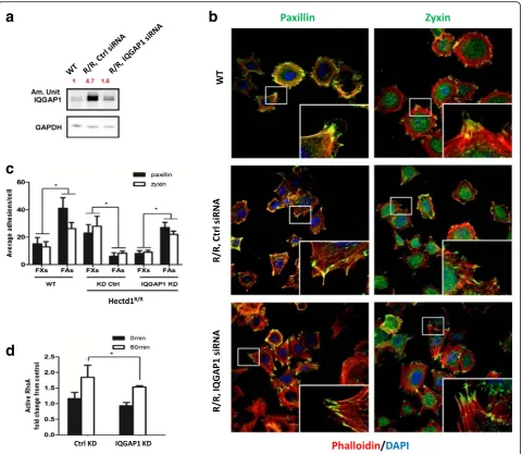
Knockdown of IQGAP1 rescues the dynamics of FAs, the duration of cell spreading and directional cell migration inHectd1R/Rcells
To further test our hypothesis whether overexpression of IQGAP1 is involved in dysfunctional cell adhesion, spreading and migration in Hectd1R/R cells, we trans-fected Hectd1R/R cells with IQGAP1-siRNA (siIQ) or with control-siRNA. As a result the protein level of IQGAP11 inHectd1R/R-siIQ-transfected cells was knock-down (am. Unit 4.7 to 1.6 as compared to 1 in the wt cells, Fig. 8a).
Subsequently, after IQGAP1-knockdown in Hectd1R/R
cells we analyzed the cytoskeleton and the FAs by immunostaining for actin, paxillin and zyxin. As shown in Fig. 8b, different structures of actin could be clearly distinguished, including stress fibers in the cell body,
a
b
c
d
e
Fig. 5Loss of HECTD1 impairs formation of focal adhesions. Equal amounts of wild-type andHectd1R/RMEF cells were seeded on FN coated coverslips for 30, 60 or 90 min. The cells were fixed and co-stained with phalloidin and anti-paxillin (a) or anti-zyxin (b), respectively. Average adhesions/cell and protein intensity were quantified by Lab Image software from 3 independent experiments, according to the sizes (FXs < = 1μm2[11], FAs > 2–5μm2.
cortical F-actin enriched at the periphery and well-organized lamellipodia structures at the leading edge in wild-type cells. In contrast, in control siRNA-treated
Hectd1R/R cells, stress fibers were less prominent than
in wild-type cells and lamellipodia were difficult to de-tect. Importantly, the formation of lamellipodia was rescued by down-regulation of IQGAP1-siRNA in
Hectd1R/Rcells.
In line with our previous results, taking paxillin and zyxin as cell adhesion markers, the ratio of FAs to FXs was about twice in wild-type cells, while in control
siRNA-treated Hectd1R/R cells, the average ratio of the number of FAs/FXs fell to around 1/3. The ratio of FXs to FAs was rescued by IQGAP1-siRNA knockdown in
Hectd1R/R cells, in which the FAs accounted for the
majority of cell adhesions and the ratio of FAs to FXs per cell again became threefold (Fig. 8c). Moreover, ac-tivity of RhoA was also evidently increased in control siRNA Hectd1 mutant MEFs after FN stimulation for 60 min (Fig. 5d), whereas RhoA activity could be signifi-cantly inhibited by IQGAP1 siRNA scilencing in
Hectd1R/R MEFs (P< 0.05) (Fig. 8d). These results
a
c
e
f
b
d
Fig. 6IQGAP1 interacts and colocalizes with HECTD1 in cell leading edge, and its ubiquitination is regulated by HECTD1.aLysates of wild-type and Hectd1R/RMEF cells were harvested immediately or plated on FN coated culture dishes for 60 min at 37 °C and were analyzed by anti-IQGAP1 and GAPDH blotting.bWild-type andHectd1R/RMEF cells were stained with paxillin (green), IQGAP1 (red) and phalloidin (blue).cIQGAP1 interacts with HECTD1. HEK293 cells were stably transfected with GFP-IQGAP1 and seeded on fibronectin-coated dishes for 60 min, protein lysates were harvested and immunoprecipitated (IP) by GFP antibody. The lP lysates and whole cell lysates were used for detecting HECTD1, PIP5K1A andβ-catenin by western blot. CUGBP1 served as a negative control.dHela cell stably express His-HECTD1 was transiently transfected with GFP-IQGAP1 for 24 h. Cells were starved overnight and plated on FN-coated slides for 60 min, followed by fixation and staining with HECTD1. Note the site of colocalization shown in intensity profiles (white arrows). Pearson’s correlation coefficient was analyzed by Image J software. *P< 0.05.eEndogenous ubiquitination of IQGAP1. Wild-type andHectd1R/Rcells were treated with the proteasome inhibitor MG132 1μg/ml or DMSO in serum starvation medium for overnight. Cell were lysed immediately or after 60 min seeded on FN coated dishes. The ubiquitination of IQGAP1 was further verified by
suggest that in the absence of Hectd1, the activation of RhoA correlated with increased protein levels of IQGAP1.
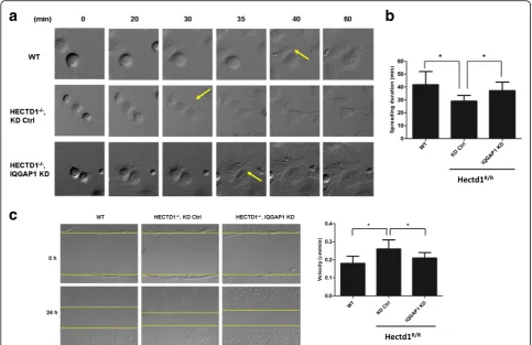
Furthermore we found that the spreading duration time shortened to (29.03 ± 4.48 min) inHectd1R/Rcells
(Hectd1R/R control group) in contrast with (41.80 ±
10.19 min) in wild-type cells, and the spreading duration time in IQGAP1-silenced cells (37.23 ± 6.60 min) was partly rescued as compared to control siRNA-transfected cells (P< 0.05, P< 0.05, resp.) (Fig. 9a and b).
Next, in order to further investigate whether down-regulation of IQGAP1 in Hectd1R/R cells would also affect directional cell migration, confluent cell layers of wild-type cells and Hectd1R/R cells with down-regulated IQGAP1 through siRNA and Hectd1R/R cells with con-trol siRNA were scratched and wound closure was re-corded by time lapse microscopy. The migration speed of control-siRNA treated cells was 0.97 ± 0.14 μm/min, as compared with 0.86 ± 0.17μm/min in wild-type cells, which was consistent with our previous results. The mi-gration defect was rescued by siRNA-mediated down-regulation of IQGAP1 (0.94 ± 0.14 μm/min). Similarly, as compared with wild-type cells the straightness of dir-ectional cell migration was impaired in control-siRNA MEFs, whereas of siRNA-mediated knockdown of IQGAP1 compensated the defect (Fig. 9c).
Discussion
Although the eminent role of HECTD1 in embryogen-esis, including neural tube formation, placenta formation
and embryonic growth, has been clearly demonstrated in at least two transgenic mouse models, limited informa-tion has been collected so far to uncover the regulatory mechanisms involved. Moreover, the involvement of HECTD1 in regulating cell migration during organogen-esis has as yet remained unexplored. We observed that loss of HECTD1 induced earlier cell spreading and en-hanced cell migration through controlling IQGAP1 and adhesion proteins. Our study proposes a new mechan-ism of HECTD1 in maintaining accurate cell movement during embryogenesis.
HECTD1 is a selective effector of ECM-integrin signaling
The complexity of the molecular signaling respon-sible for ECM selective guidance is associated with various ligand-binding possibilities for integrin sub-types [42–45]. Our first observation was that the mi-gration patterns of Hectd1R/R cells is significantly different to that of wild-type cells on various ECM when these cells were incubated in culture medium lacking serum. These results indicate that factors in serum may compensate the loss of HECTD1 through yet unknown signaling pathways. In addition, the localization of paxillin and zyxin but not of talin and vinculin was different during migration of these cells on FN. Furthermore, more FAs formed in wild-type cells whereas more FXs developed in mutant cells. These differences were not apparent when the cells were cultured on collagen type I and on gelatin.
The adhesion proteins are known to have a specific binding relationship with integrins. For instance, paxillin
a
b
c
is normally binds to ɑ5β1, ɑ4β1, ɑ5β3 and ɑ4β3integrins [46–49], while zyxin binds to the integrin subtypes ɑ5β1 and ɑ5β5[50, 51]. Besides, the integrin motifs expressed in different cell types may vary considerably. For ex-ample, in fibroblasts ɑ1β1, ɑ2β1, ɑ3β1 and ɑ5β1integrins were predominantly identified [52]. Thus, our results suggest that the regulation of the composition of FAs and FXs by HECTD1 in fibroblasts depends on specific integrin receptor subtypes, probably via coordinating withɑ5β1.
The involvement of IQGAP1 in regulating adhesion dynamics is mediated by HECTD1
IQGAP1 has been widely reported to be involved in regulating FAs dynamics and cell migration. We confirmed the interaction of HECTD1 with IQGAP1 and their co-localization through co-immunoprecipitation and double-labeled immunocytochemistry, respectively. We observed that loss of HECTD1, being an E3-ubiquitin lig-ase, enhances the protein level of IQGAP1 through de-creased ubiquitination. When IQGAP1 was overexpressed
a
c
d
b
Fig. 8Knockdown of IQGAP1 rescue focal adhesion formation inHectd1R/Rcells.aHectd1R/Rcells were transfected with control siRNA or IQGAP1 siRNA. 36 h after transfection, cell pellets were harvested and the level of IQGAP1 was detected by western blot. GAPDH was taken as
in wild-type cells, it reduced the formation of FAs as de-termined by differences in the expression of paxillin and zyxin. Moreover, siRNA knockdown of IQGAP1 in
Hectd1R/Rcells compensated the defects in the formation
of cell adhesions, in cell spreading and migration. Taken all these results together, IQGAP1 has now been demon-strated to be regulated through degradation by HECTD1. We therefore conclude that HECTD1 regulates cell adhe-sion and controls cell spreading and migration via IQGAP1.
High FXs-FAs ratio inHectd1R/Rcells contributes to higher motility
We have demonstrated that the mutation of HECTD1 results in altered cell spreading and migration, in which the velocity of Hectd1R/R cells was increased with impaired directionality. In our assay, we used aphidicolin to ensure that proliferation did not inter-fere with cell migration. We also showed that HECTD1 ablation did not influence cell migration speed in the presence of 10% FBS without aphidicolin in MEF cells on FN. This result is consistent with Li’s
result [18], in which 10 ng/ml of EGF was used in breast cancer cells. We therefore, used the same set-ting for the Hectd1/IQGAP1 double knockout/down experiments. Instead of measuring total adhesion structures we differentiated FXs from FAs in cells. Interestingly, when compared to wild-type cells, the average total number of small adhesions in Hectd1R/R
cells is increased. Moreover, FXs are evidently associ-ated with fewer FAs, which are bigger in size than FXs
inHectd1R/Rcells than in their wild-type counterparts.
Maturation of adhesions occurs along an α-actinin-actin template that elongates centripetally from nas-cent adhesions. We found that α-actinin is co-localized with paxillin or zyxin at the leading edge of wild-type cells, but not in Hectd1R/R cells. These re-sults suggest that in Hectd1R/R cells, FXs including paxillin fail to reassemble or/and cannot mature to FAs.
Since the presence of FXs and nascent adhesions is a marker of highly motile cells, their quick appear-ance and turnover correlate directly with protrusion and cell movement. The higher number of small
a
b
c
paxillin patches in Hectd1R/R cells strongly correlates with their increased motility and fast spreading. In motile cells, the recruitment of the adhesion proteins into FXs occurs sequentially, so that composition of the specific proteins relies on their age. Moreover, using double color staining, time-lapse assay, one study demonstrated that the transition from paxillin-rich FXs to zyxin-containing FAs takes place after the leading edge stops advancing or retracts [37]. Generally, zyxin has been thought to be a component of FA plaques and is absent from FXs [37, 53]. Although these three types of adhesions are distinguishable, there is always a continuum between types and many of the same adhesion proteins have been identified in each [54].
Consistent with our findings, in highly motile cells such as melanoma cells, glioma cells and growing neurons [55] the dynamic adhesions most similar to FXs are enriched in the leading edge of cells and act as common features of rapid cell movement [56]. Therefore, we conclude that the accu-mulation of paxillin and zyxin in the lamellipodia of FXs is a major hallmark of highly motile cells. Thompson has also proved that decreased size of FAs is related to higher vel-ocity and impaired directionality of cells, and vice versa [57]. Increased numbers of the adhesions are accompanied with a lesser motility [58, 59]. Here, we show that the dynamics of cell adhesion are responsible for the velocity of cells during migration.
We propose that 30 min spreading is too early for the recruitment of abundant zyxin into FAs, so that the
presence of zyxin is not enough to distinguish the differ-ence inHectd1R/Rand wild-type cells.
Model for the role of HECTD1 in regulating cell movement
Our data revealed that FXs in Hectd1R/R cells failed to recruit enough adhesion proteins (such as paxillin and zyxin) to mature into FAs. Therefore, the alteration in number and/or size of FXs is expected to influence cell motility. Thus, we propose the following model for the role of HECTD1 in cell movement (Fig. 10).
During cell spreading and early migration the cell receives stimulating signals from its extracellular en-vironment, such as FN in the extracellular matrix, which activates relevant integrin receptors and Src in the cell leading edge. The recruitment of paxillin results in phosphorylation of paxillin at Y118. With this event the initiation of focal complexs formation becomes complete. The activation signals are passed to small GTPases, such as Rac1 and RhoA via IQGAP1 recruitment. Together with filamin-A, IQGAP1 inhibits Rac1 activity [60]. Subsequently re-moval of IQGAP1 from Focal complexs together with high RhoA activities triggers the maturation of focal adhesions by recruiting more paxillin and zyxin.
As an E3 ubiquitin ligase, HECTD1 regulates the level of IQGAP1 through ubquitination. Loss of HECTD1 prolonges the half-life of IQGAP1 and thereby reduces
the recruitment of paxillin and zyxin. As a result, FXs failed to mature into FAs through a deficiency of paxillin and zyxin. HECTD1 is a key regulator of IQGAP1 and through this interaction HEDTD1 impacts on cellular adhesion and movement.
Conclusions
By generating a knockout mouse of the E3 ubiquitin lig-ase for inhibin B receptor (HECTD1), we reveal a mo-lecular mechanism in which IQGAP1 is one of the effectors of HECTD1. HECTD1 interacted with IQGAP1 and regulated its degradation through ubiquitination. In-creased expression of IQGAP1 inHectd1R/Rcells lead to the mis-localized paxillin and zyxin so that the forma-tion of focal adhesions (FAs) was impaired, resulting in accelerated cell spreading and migration but impaired directionality of migration.
Additional files
Additional file 1: Fiugre S1. Generation ofHectd1mutant animals. (PPTX 142 kb)
Additional file 2: Figure S2.Time lapse images for cell spreading and directional migration ofHectd1R/Rcells. (AVI 36637 kb)
Additional file 3: Figure S2.Time lapse images for cell spreading and directional migration ofHectd1R/Rcells. (AVI 36086 kb)
Additional file 4: Figure S3.Subcellular localization of adhesion proteins inHectd1R/Rcells. (PPTX 1221 kb)
Additional file 5: Figure S4.Loss of HECTD1 leads to mislocalization of
α-actinin and paxillin/zyxin. (PPTX 1571 kb).
Abbreviations
APC:Adenomatous polyposis coli; CL1: Collagen type I; CL4: Collagen type IV; CSK: C-terminal Src kinase; ECM: Extracellular matrix; FAK: Focal adhesion kinase; FAs: Focal adhesions; FN: Fibronectin; FXs: Focal complexes; GL: Gelatin; LacZ:β-galactosidase activity; LM: Laminin; MEF: Mouse embryonic fibroblasts; MT: Matrigel; MTOC: Microtubule-organizing center; PIP5K: Phosphatidylinositol 4-phosphate 5-kinase
Acknowledgements
XL. Shen and XG. Wang are recipients of China Scholarship Council Stipendia of the P.R. China. ZH. Jia was supported by ESKAS, a Swiss Federal
Scholarship.
Funding
This research was funded by University of Basel, Switzerland and a grant of the Repronatal Foundation, Basel, Switzerland.
Availability of data and materials
The datasets supporting the conclusions of this article are included within the article and its additional files.
Authors’contributions
XLS, ZHJ, CDG and HZ conceived and designed the experiments. XLS, ZHJ, DDA, XGW, EB and FHE performed the experiments. XLS, CDG and HZ analyzed the data and wrote the paper. All authors read and approved the final manuscript.
Competing interests
The authors declare that they have no competing interests.
Not applicable.
Ethics approval and consent to participate
The mice used in the study were obtained from the animal facility of department DBM, university Basel. The animals were treated in accordance with the National Research Council’s Guide for the Care and Use of Laboratory Animals (USA).
Author details
1Department of Biomedicine, University Hospital, University of Basel, Basel,
Switzerland.2Pathologie, Universitätsspital Basel, Schönbeinstrasse 40, CH-4031 Basel, Switzerland.3Clinic of Gynecological Endocrinology and
Reproductive Medicine, University Hospital, University of Basel, Basel, Switzerland.4Department of Biomedicine, University of Basel, Hebelstra. 20,
CH-4031 Basel, Switzerland.5Present Address: Chongqing Reproductive and Genetics Institute, 64 Jing Tang STYu Zhong District, Chongqing 400013, China.6Present Address: 2nd hospital of Jilin University, Changchun, China.
Received: 25 May 2016 Accepted: 6 December 2016
References
1. Tada M, Heisenberg CP. Convergent extension: using collective cell migration and cell intercalation to shape embryos. Development. 2012; 139(21):3897–904.
2. Dzamba BJ, Jakab KR, Marsden M, Schwartz MA, DeSimone DW. Cadherin adhesion, tissue tension, and noncanonical Wnt signaling regulate fibronectin matrix organization. Dev Cell. 2009;16(3):421–32. 3. Theveneau E, Mayor R. Cadherins in collective cell migration of
mesenchymal cells. Curr Opin Cell Biol. 2012;24(5):677–84.
4. Weijer CJ. Collective cell migration in development. J Cell Sci. 2009;122(Pt 18):3215–23.
5. Lauffenburger DA, Horwitz AF. Cell migration: a physically integrated molecular process. Cell. 1996;84(3):359–69.
6. Chamaraux F, Ali O, Keller S, Bruckert F, Fourcade B. Physical model for membrane protrusions during spreading. Phys Biol. 2008;5(3):036009. 7. Serrels B, Serrels A, Brunton VG, Holt M, McLean GW, Gray CH, et al. Focal
adhesion kinase controls actin assembly via a FERM-mediated interaction with the Arp2/3 complex. Nat Cell Biol. 2007;9(9):1046–56.
8. Loosli Y, Luginbuehl R, Snedeker JG. Cytoskeleton reorganization of spreading cells on micro-patterned islands: a functional model. Philos Trans A Math Phys Eng Sci. 2010;368(1920):2629–52.
9. Wakatsuki T, Wysolmerski RB, Elson EL. Mechanics of cell spreading: role of myosin II. J Cell Sci. 2003;116(Pt 8):1617–25.
10. Izzard CS, Lochner LR. Cell-to-substrate contacts in living fibroblasts: an interference reflexion study with an evaluation of the technique. J Cell Sci. 1976;21(1):129–59.
11. Galbraith CG, Yamada KM, Sheetz MP. The relationship between force and focal complex development. J Cell Biol. 2002;159(4):695–705.
12. Zaidel-Bar R, Itzkovitz S, Ma’ayan A, Iyengar R, Geiger B. Functional atlas of the integrin adhesome. Nat Cell Biol. 2007;9(8):858–67.
13. Zhang H. Homo sapiens E3 ligase for inhibin receptor mRNA, complete cds http://www.ncbi.nlm.nih.gov/nuccore/AY254380.1. Accessed 22 Apr 2003. 14. Sarkar AA, Zohn IE. Hectd1 regulates intracellular localization and secretion
of Hsp90 to control cellular behavior of the cranial mesenchyme. J Cell Biol. 2012;196(6):789–800.
15. Tran H, Bustos D, Yeh R, Rubinfeld B, Lam C, Shriver S, et al. HectD1 E3 ligase modifies adenomatous polyposis coli (APC) with polyubiquitin to promote the APC-axin interaction. J Biol Chem. 2013;288(6):3753–67. 16. Zohn IE, Anderson KV, Niswander L. The Hectd1 ubiquitin ligase is required
for development of the head mesenchyme and neural tube closure. Dev Biol. 2007;306(1):208–21.
17. Sarkar AA, Nuwayhid SJ, Maynard T, Ghandchi F, Hill JT, Lamantia AS, et al. Hectd1 is required for development of the junctional zone of the placenta. Dev Biol. 2014;392(2):368–80.
19. Brill S, Li S, Lyman CW, Church DM, Wasmuth JJ, Weissbach L, et al. The Ras GTPase-activating-protein-related human protein IQGAP2 harbors a potential actin binding domain and interacts with calmodulin and Rho family GTPases. Mol Cell Biol. 1996;16(9):4869–78.
20. Hart MJ, Callow MG, Souza B, Polakis P. IQGAP1, a calmodulin-binding protein with a rasGAP-related domain, is a potential effector for cdc42Hs. EMBO J. 1996;15(12):2997–3005.
21. Kuroda S, Fukata M, Kobayashi K, Nakafuku M, Nomura N, Iwamatsu A, et al. Identification of IQGAP as a putative target for the small GTPases, Cdc42 and Rac1. J Biol Chem. 1996;271(38):23363–7.
22. Mateer SC, Wang N, Bloom GS. IQGAPs: integrators of the cytoskeleton, cell adhesion machinery, and signaling networks. Cell Motil Cytoskeleton. 2003; 55(3):147–55.
23. Fukata M, Watanabe T, Noritake J, Nakagawa M, Yamaga M, Kuroda S, et al. Rac1 and Cdc42 capture microtubules through IQGAP1 and CLIP-170. Cell. 2002;109(7):873–85.
24. Watanabe T, Wang S, Noritake J, Sato K, Fukata M, Takefuji M, et al. Interaction with IQGAP1 links APC to Rac1, Cdc42, and actin filaments during cell polarization and migration. Dev Cell. 2004;7(6):871–83. 25. Brown MD, Sacks DB. IQGAP1 in cellular signaling: bridging the GAP. Trends
Cell Biol. 2006;16(5):242–9.
26. Choi S, Thapa N, Hedman AC, Li Z, Sacks DB, Anderson RA. IQGAP1 is a novel phosphatidylinositol 4,5 bisphosphate effector in regulation of directional cell migration. EMBO J. 2013;32(19):2617–30.
27. Liu C, Billadeau DD, Abdelhakim H, Leof E, Kaibuchi K, Bernabeu C, et al. IQGAP1 suppresses TbetaRII-mediated myofibroblastic activation and metastatic growth in liver. J Clin Invest. 2013;123(3):1138–56.
28. Noritake J, Watanabe T, Sato K, Wang S, Kaibuchi K. IQGAP1: a key regulator of adhesion and migration. J Cell Sci. 2005;118(Pt 10):2085–92.
29. Kuo JC, Han X, Hsiao CT, Yates 3rd JR, Waterman CM. Analysis of the myosin-II-responsive focal adhesion proteome reveals a role for beta-Pix in negative regulation of focal adhesion maturation. Nat Cell Biol.
2011;13(4):383–93.
30. Schiller HB, Friedel CC, Boulegue C, Fassler R. Quantitative proteomics of the integrin adhesome show a myosin II-dependent recruitment of LIM domain proteins. EMBO Rep. 2011;12(3):259–66.
31. Wickstrom SA, Lange A, Hess MW, Polleux J, Spatz JP, Kruger M, et al. Integrin-linked kinase controls microtubule dynamics required for plasma membrane targeting of caveolae. Dev Cell. 2010;19(4):574–88.
32. Schiefermeier N, Scheffler JM, de Araujo ME, Stasyk T, Yordanov T, Ebner HL, et al. The late endosomal p14-MP1 (LAMTOR2/3) complex regulates focal adhesion dynamics during cell migration. J Cell Biol. 2014;205(4):525–40. 33. Stanford WL, Cohn JB, Cordes SP. Gene-trap mutagenesis: past, present and
beyond. Nat Rev Genet. 2001;2(10):756–68.
34. Euteneuer U, Schliwa M. Mechanism of centrosome positioning during the wound response in BSC-1 cells. J Cell Biol. 1992;116(5):1157–66. 35. Schaar BT, McConnell SK. Cytoskeletal coordination during neuronal
migration. Proc Natl Acad Sci U S A. 2005;102(38):13652–7.
36. Farhan H, Wendeler MW, Mitrovic S, Fava E, Silberberg Y, Sharan R, et al. MAPK signaling to the early secretory pathway revealed by kinase/ phosphatase functional screening. J Cell Biol. 2010;189(6):997–1011. 37. Zaidel-Bar R, Ballestrem C, Kam Z, Geiger B. Early molecular events in the
assembly of matrix adhesions at the leading edge of migrating cells. J Cell Sci. 2003;116(Pt 22):4605–13.
38. Maruyama K, Ebashi S. Alpha-actinin, a new structural protein from striated muscle. II. Action on actin. J Biochem. 1965;58(1):13–9.
39. Frame MC. Newest findings on the oldest oncogene; how activated src does it. J Cell Sci. 2004;117(7):989–98.
40. Schlaepfer DD, Mitra SK. Multiple connections link FAK to cell motility and invasion. Curr Opin Genet Dev. 2004;14(1):92–101.
41. Jacquemet G, Humphries MJ. IQGAP1 is a key node within the small GTPase network. Small GTPases. 2013;4(4):199–207.
42. Doughman RL, Firestone AJ, Wojtasiak ML, Bunce MW, Anderson RA. Membrane ruffling requires coordination between type Ialpha
phosphatidylinositol phosphate kinase and Rac signaling. J Biol Chem. 2003; 278(25):23036–45.
43. Dall’Armi C, Devereaux KA, Di Paolo G. The role of lipids in the control of autophagy. Curr Biol. 2013;23(1):R33–45.
44. Barczyk M, Carracedo S, Gullberg D. Integrins. Cell Tissue Res. 2010;339(1): 269–80.
45. Levental KR, Yu H, Kass L, Lakins JN, Egeblad M, Erler JT, et al. Matrix crosslinking forces tumor progression by enhancing integrin signaling. Cell. 2009;139(5):891–906.
46. Crowe DL, Ohannessian A. Recruitment of focal adhesion kinase and paxillin to beta1 integrin promotes cancer cell migration via mitogen activated protein kinase activation. BMC Cancer. 2004;4:18.
47. Laukaitis CM, Webb DJ, Donais K, Horwitz AF. Differential dynamics of alpha 5 integrin, paxillin, and alpha-actinin during formation and disassembly of adhesions in migrating cells. J Cell Biol. 2001;153(7):1427–40.
48. Schaller MD, Otey CA, Hildebrand JD, Parsons JT. Focal adhesion kinase and paxillin bind to peptides mimicking beta integrin cytoplasmic domains. J Cell Biol. 1995;130(5):1181–7.
49. Schaller MD, Parsons JT. pp125FAK-dependent tyrosine phosphorylation of paxillin creates a high-affinity binding site for Crk. Mol Cell Biol. 1995;15(5): 2635–45.
50. Bianchi-Smiraglia A, Kunnev D, Limoge M, Lee A, Beckerle MC, Bakin AV. Integrin-beta5 and zyxin mediate formation of ventral stress fibers in response to transforming growth factor beta. Cell Cycle. 2013;12(21):3377–89. 51. Mise N, Savai R, Yu H, Schwarz J, Kaminski N, Eickelberg O. Zyxin is a
transforming growth factor-beta (TGF-beta)/Smad3 target gene that regulates lung cancer cell motility via integrin alpha5beta1. J Biol Chem. 2012;287(37):31393–405.
52. Mineur P, Guignandon A, Lambert Ch A, Amblard M, Lapiere Ch M, Nusgens BV. RGDS and DGEA-induced [Ca2+]i signalling in human dermal fibroblasts. Biochim Biophys Acta. 2005;1746(1):28–37.
53. Zaidel-Bar R, Cohen M, Addadi L, Geiger B. Hierarchical assembly of cell-matrix adhesion complexes. Biochem Soc Trans. 2004;32(Pt3):416–20. 54. Parsons JT, Horwitz AR, Schwartz MA. Cell adhesion: integrating cytoskeletal
dynamics and cellular tension. Nat Rev Mol Cell Biol. 2010;11(9):633–43. 55. Gatlin JC, Estrada-Bernal A, Sanford SD, Pfenninger KH. Myristoylated,
alanine-rich C-kinase substrate phosphorylation regulates growth cone adhesion and pathfinding. Mol Biol Cell. 2006;17(12):5115–30. 56. Estrada-Bernal A, Gatlin JC, Sunpaweravong S, Pfenninger KH. Dynamic
adhesions and MARCKS in melanoma cells. J Cell Sci. 2009;122(Pt 13):2300–10. 57. Thompson O, Moore CJ, Hussaina SA, Kleino I, Peckham M, Hohenester E, et
al. Modulation of cell spreading and cell-substrate adhesion dynamics by dystroglycan. J Cell Sci. 2010;123(1):118–27.
58. Lu ZM, Jiang GQ, Blume-Jensen P, Hunter T. Epidermal growth factor-induced tumor cell invasion and metastasis initiated by dephosphorylation and downregulation of focal adhesion kinase. Mol Cell Biol. 2001;21(12): 4016–31.
59. Nagano M, Hoshino D, Koshikawa N, Akizawa T, Seiki M. Turnover of focal adhesions and cancer cell migration. Int J Cell Biol. 2012;2012:310616. 60. Jacquemet G, Morgan MR, Byron A, Humphries JD, Choi CK, Chen CS, et al.
Rac1 is deactivated at integrin activation sites through an IQGAP1-filamin-A-RacGAP1 pathway. J Cell Sci. 2013;126(Pt 18):4121–35.
• We accept pre-submission inquiries
• Our selector tool helps you to find the most relevant journal
• We provide round the clock customer support
• Convenient online submission
• Thorough peer review
• Inclusion in PubMed and all major indexing services
• Maximum visibility for your research
Submit your manuscript at www.biomedcentral.com/submit