Volume 26, Issue 1 • Winter 2016
ABstrAct
Red blood cell (RBC) transfusions are vital for many patients with chronic anemias associated with oncologic/hematologic disorders. However, repeated transfusions over time can lead to iron overload, which, if left untreated, can increase the risk of further malignancy and end-organ damage.
Nurses and other health care professionals may not be aware of the significant implications of RBC transfusions and iron over-load in patients with hematological/oncological disorders. This article was developed by a group of Canadian nurse practitioners and specialized oncology nurses to help improve health care pro-fessionals’ understanding of iron overload in oncology patients and its associated risks, as well as provide a practical guide for the management of patients receiving treatment for this potentially serious condition.
Key words: oncology, malignant hematology, iron overload,
iron chelation therapy, nursing practice
iNtrODuctiON
A
s new treatments, transplant options and supportive care strategies become available for hematologic/oncologic disorders, patients with these conditions are now living lon-ger (Fenaux et al., 2009). Given the ineffective hematopoiesis associated with many of these disorders, a significant propor-tion of these patients will require chronic red blood cell (RBC) transfusions, which can result in transfusion-related iron over-load. If left unrecognized and untreated, iron overload can lead to an increased risk of infection, malignancy, and end-organ damage.Through patient advocacy organizations, patients with hematologic/oncologic disorders are now well-informed about iron overload and its associated risks, and many mis-takenly believe that all patients receiving transfusions should be monitored and treated for iron overload. However, moni-toring for iron overload is not considered a “standard of care” for all patients and, therefore, nurses must be able to respond to patient concerns and queries and provide appropriate guidance. Often, such guidance can be as simple as advising patients that the need for monitoring iron status and treat-ing iron overload varies from patient to patient and that this should be discussed further with their respective physicians.
In some Canadian centres, nurse practitioners are respon-sible for providing supportive care for patients with hemato-logic/oncologic disorders, including the provision of blood transfusions and monitoring of blood counts. These nurses, in particular, must be well-informed to ensure that they work collaboratively with physicians to identify and manage patients who fulfill the criteria for iron overload screening and/or treatment.
This article was developed by a group of Canadian nurse practitioners and specialized hematology/oncology nurses to provide health care professionals with a review of normal iron physiology, the consequences of transfusional iron overload, and current Canadian iron chelation guidelines, as well as a practical guide for managing treatment-related complications.
WHAt is tHe NOrMAl PHYsiOlOGY OF
irON?
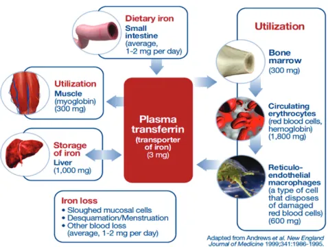
Under normal conditions, the human body stores approxi-mately 3.5 g of iron. Most of this iron is distributed within the RBC (a component of hemoglobin), with the remainder being stored in muscle fibres, macrophages, the liver, and bone marrow. On average, 1–2 mg of iron is absorbed daily via the duodenum, with the same amount being excreted through sloughing of mucosal cells, desquamation of epithelial cells,
Management of iron overload in the Canadian
hematology/oncology population: Implications for
nursing practice
by Cindy Murray, Tammy De Gelder, Nancy Pringle, J. Colleen Johnson and Mary Doherty
ABOut tHe AutHOrs
Cindy Murray, MN, NP (Adult), Nurse Practitioner, Blood and Marrow Disorder Program – Transfusion Outpatient Clinic, University Health Network (UHN) Princess Margaret Cancer Centre, Toronto, ON
Tammy De Gelder, MN, NP (Adult), CON(C), Nurse Practitioner, Hamilton Health Sciences, Juravinski Hospital and Cancer Centre, Hamilton, ON Nancy Pringle, RN, Specialized Oncology Nurse, Leukemia Clinic, UHN Princess Margaret Cancer Centre, Toronto, ON
J. Colleen Johnson, MN, NP (Adult),CON(C), Nurse Practitioner, Red Blood Cell Disorders Clinic, UHN, Toronto General Division, Toronto, ON
Mary Doherty, MN, NP-PHC, Nurse Practitioner, Blood and Marrow Disorder Program – Transfusion Outpatient Clinic, University Health Network (UHN) Princess Margaret Cancer Centre, Toronto, ON
Address for correspondence: Cindy Murray, MN, NP (Adult), 610 University Ave., Toronto, ON M5G 2C4
Phone: (416) 946-4501 ext. 5919; Email: Cindy.Murray@uhn.ca
Conflicts of interest: The authors have no conflicts of interest to declare regarding the content of this manuscript.
and blood loss during menstruation (Figure 1) (Andrews, 1999; Stein, Hartmann, & Dignass, 2010). Because the same amount of iron is absorbed and eliminated on a daily basis, the iron pool represents a closed system (Shah et al., 2012). Therefore, the human body has no effective means of natu-rally disposing of excess iron.
Although iron is physiologically critical, excess iron is toxic. Due to its ability to donate and accept electrons, iron can cat-alyze the conversion of hydrogen peroxide into free radicals if it is left unbound within cells. These free radicals, in turn, can cause damage to a number of cellular structures and pro-cesses, leading to cell death. To prevent this free radical for-mation, iron must be bound to proteins (Andrews, 1999) (see Table 1 for definitions of the most important iron-binding pro-teins). It is important to note that 80% of the body’s daily iron needs are used for the production of new RBC, which requires only 20–25 mg of iron (Shah et al., 2012).
HOW DOes trANsFusiON DePeNDeNce
leAD tO irON OVerlOAD?
RBC transfusions are vital for many patients with chronic anemias, including those associated with β-thalassemia, myel-odysplastic syndromes (MDS), and, to a lesser extent, sickle cell disease (SCD). However, multiple or repeated transfusions over time can lead to iron overload. Each unit of packed RBC contains approximately 200–250 mg of iron which, as dis-cussed earlier, the body is unable to eliminate (Porter, 2001). After approximately 20 transfusions, an individual who is not hemorrhaging will have accumulated approximately 5 g of unexcretable iron—nearly double the amount of iron nor-mally found in the body (List, 2010). As per normal iron phys-iology, this excess iron attempts to bind to transferrin to be transported to cells and stored as ferritin. However, if a person continues to have transfusions for conditions other than hem-orrhaging, the capacity for transferrin to bind iron is exceeded (or saturated) resulting in “free” or non-transferrin-bound iron (NTBI). As stated earlier, this “free”, unbound iron is toxic to cells, leading to inflammation, fibrosis and organ dysfunction (Leitch, 2011). As blood levels of NTBI increase, tissue absorp-tion of toxic NTBI results in iron deposits in various organs including the heart, liver, and endocrine glands. Elevated lev-els of iron in the liver leads to an increased risk of hepatic dysfunction and cirrhosis, while excess iron in the heart is associated with an increased risk of cardiac-related events including myocardial infarction, congestive heart failure, and arrhythmias. Excess iron in the endocrine system can lead to gonadal and thyroid dysfunction, as well as diabetes (Shah et al., 2012; Porter, 2001).
WHAt Are tHe cliNicAl cONseQueNces
OF trANsFusiON-relAteD irON
OVerlOAD iN PAtieNts WitH
HeMAtOlOGic/ONcOlOGic DisOrDers?
In patients with thalassemia, it has been well-documented that transfusion-related iron overload leads to cardiac iron deposi-tion, heart failure, and reduced overall survival (Zurlo et al.,
1989; Schafer et al., 1981), and that long-term treatment with iron-chelating agents (drugs that trap and remove excess iron, which is then excreted in the stool or urine, depending on the iron chelator prescribed in these patients) reduces cardiac and hepatic complications and improves survival (Zurlo et al., 1989; Gabutti & Piga, 1996; Borgna-Pignatti et al., 2004).
Although chronic transfusion therapy is the primary cause of iron overload in MDS patients, the clinical importance of iron overload and its management in this patient population is somewhat controversial since data in this area has primarily been derived from retrospective studies. Further compound-ing this issue is the fact that age-related comorbidities are common in MDS, making it difficult to determine whether organ damage is due to transfusion therapy or these comor-bid conditions. Nonetheless, findings from retrospective stud-ies suggest that iron overload may have clinical consequences in MDS, such as organ dysfunction and reduced leukemia-free and overall survival (Schafer et al., 1981; Malcovati et al., 2005; Sanz et al., 2008; Takatoku et al., 2007). Prospective trials are required to confirm these findings.
There is also growing evidence that iron overload prior to hematopoietic stem cell transplantation (HSCT) may affect transplant outcomes. A number of primarily retrospective
Figure 1: Iron homeostasis under normal conditions.
Table 1: Iron-binding proteins
Heme: • Found in red blood cells (RBC)
Ferritin: • Intracellular protein that functions as the primary form of iron storage
• Provides an indirect measure of the amount of iron stored in the body
Transferrin: • Responsible for iron transport • Provides a measure of iron circulation Ferroportin: • Transmembrane protein that transports iron
studies have found high pre-transplantation serum ferritin lev-els to be associated with reduced overall survival and increased complications after HSCT, particularly in the MDS and leu-kemia populations (Pullarkat, 2010). A prospective study of HSCT patients also found serum ferritin values ≥1,000 µg/L to be associated with a significant increase in blood stream infections (Pullarkat et al., 2008). At present, however, the role of iron chelation therapy in the pre-transplant population is unknown, and further prospective studies in this area are required.
It is important to note that iron overload has also been associated with both impaired immunity and heightened microbial virulence. Specifically, iron overload has been found to inhibit inflammatory markers and nitric oxide, and impair macrophage, neutrophil and T-cell function (Álvarez, Fernández-Ruiz, & Aguado, 2013; Pieracci & Barie, 2005; Ibrahim et al., 2011).
HOW DO We Best iDeNtiFY irON
OVerlOAD?
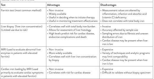
Although many tests are available to assess iron overload (see Table 2), serum ferritin level (which provides an indi-rect estimate of iron overload) is the most commonly used since it is widely available, affordable, and easy to perform repeatedly, which can provide trends over time in patients with variable transfusion intervals. Normal values for serum ferritin are usually in the range of 12–300 µg/L for men and 12–150 µg/L for women. Serum ferritin levels greater than 1,000–2,500 µg/L indicate iron overload (Malcovati et al., 2007).
Serum ferritin has been shown to correlate with the number of RBC transfusion units received, with a value of 1,000 µg/L often reached after as few as 20 units (Malcovati et al., 2005). A major disadvantage of the ferritin test is that results are affected by inflammation, infection, liver disease and ascorbic acid (vitamin C) deficiency. Therefore, the assess-ment of “trends” in ferritin levels over time is most useful for monitoring iron overload (Malcovati et al., 2007). In general, it is recommended that serum ferritin be assessed at the time of diagnosis and repeated every three months in transfusion-de-pendent patients.
The transferrin saturation (TS) test is another sensitive and relatively inexpensive biochemical measure of iron overload that can be performed along with serum ferritin (TS levels >50% are considered elevated). In clinical practice, however, the TS test is not often used in the assessment of transfu-sion-related iron overload (Gattermann, 2009).
The most precise method and, therefore, the current gold standard for determining body iron is the measurement of liver iron concentration (LIC). However, its assessment requires a percutaneous liver biopsy, which may not be feasible in some patients (Malcovati, 2007). Magnetic resonance imaging (MRI) can also be used to measure tissue iron content. However, this modality is expensive and not yet widely available for the assess-ment of iron overload in the oncology population. Nonetheless, it has been researched and is the standard of care for the assess-ment of iron overload in patients with RBC disorders receiving transfusions (Carson & Martin, 2014).
In addition to the above mentioned tests, it is impera-tive that patients be instructed to keep track of the number
Table 2: Tests for iron overload
Test Advantages Disadvantages
Ferritin test (most common method) • Non-invasive • Widely available
• Useful in deciding when to initiate therapy • Useful in monitoring treatment effectiveness
• Measurement values are altered by inflammation, infection and absorbic acid (vitamin C) deficiency
• Does not correlate with total body iron Liver biopsy (liver iron concentration)
(Limited use due to risk) • Correlates well with total body iron burden• Allows for assessment of liver histology • High levels predict risk for cardiac disease,
endocrine complications and death
• Invasive
• Accuracy affected by sample size
• Sampling errors due to fibrosis and uneven distribution of iron
• Cardiac disease may be present when liver iron is low
MRI (used to evaluate abnormal liver enzymes in patients with elevated ferritin)
• Non-invasive • More widely available
• Correlates well with liver iron concentration by biopsy
• Expensive
• Variety of techniques and analytic programs may limit comparability
• Cardiac disease may be present when liver iron is low
Cardiac iron loading by MRI (used primarily to evaluate cardiac symptoms in patients with elevated ferritin)
• Non-invasive
• Correlates with risk for cardiac disease • Expensive• Difficult to validate without biopsy specimen
of transfusions they have had since this will assist health care professionals in assessing the patient’s overall risk of iron overload. Excellent tracking tools for patients are avail-able through the MDS Foundation at www.mds-founda-tion.org (see Building Blocks of Hope: Strategies for Patients & Caregivers Living with MDS and Anemia, Blood Transfusions, Iron Overload, and Myelodysplastic Syndromes: A Handbook for Adult MDS Patients).
WHAt Are tHe curreNt cANADiAN
GuiDeliNe recOMMeNDAtiONs FOr tHe
iDeNtiFicAtiON AND MANAGeMeNt OF
irON OVerlOAD?
Definitive evidence has shown that iron overload negatively impacts overall survival and morbidity, and that iron chelation therapy improves both survival and complication rates in the thalassemia population. As a result, iron chelation therapy has been incorporated into the Canadian guidelines for thalassemia management (Anemia Institute for Research & Education, Thalassemia Foundation of Canada, 2009) and is a standard of care in current clinical practice for this patient population. Although similar iron overload guidelines have been in exis-tence since 2008 for patients with MDS (Wells et al., 2008), their integration into the clinical management of these patients has not yet been fully adopted. Therefore, we have focused this section on these latter recommendations to help increase awareness and utilization of these Canadian consensus guide-lines, which are summarized in Table 3 (Wells et al., 2008). As can be seen in the table, these guidelines recommend that iron chelation therapy be considered in transfusion-dependent MDS patients with a good prognosis (as indicated by an IPSS score of low or int-1), a serum ferritin level >1,000 µg/L, a life expec-tancy > 1 year, or in those who are candidates for allogeneic hematopoietic stem cell transplantation (Wells et al., 2008). In general, the Canadian consensus recommendations for iron overload in MDS are consistent with other published guidelines as identified through a MEDLINE search (Gattermann, 2008;
NCCN 2015) in terms of: the subgroups of MDS patients who should be considered for iron chelation therapy; the serum fer-ritin level that defines iron overload; the use of chelating agents; and the monitoring of chelation therapy (Wells et al., 2008).
As mentioned earlier, another patient population that may be negatively affected by iron overload are those who have had, or who are scheduled to undergo HSCT. Although several publications on the impact of iron overload in these patients have been noted in the literature (Meyer et al., 2013; Trottier et al., 2013; Pullarkat 2010; Lee et al., 2009), we were unable to locate any guidelines through a MEDLINE search that specifi-cally addressed this patient population.
Are tHe curreNt cANADiAN
GuiDeliNes FOr irON OVerlOAD
BeiNG useD cONsisteNtlY iN cliNicAl
PrActice?
As a group of nurses working with MDS patients in two major Canadian cancer centres, we continue to witness incon-sistent use of guideline recommendations for identifying and managing iron overload and hypothesize that this is due, at least in part, to the low level of evidence supporting these rec-ommendations. We refer the readers to an interesting article by Steensma (2011), which highlights the controversy sur-rounding the importance of iron chelation and the lack of strong evidence supporting guideline recommendations.
Even the authors of the Canadian guidelines have acknowl-edged that their recommendations are based, for the most part, upon a low level of evidence (i.e., evidence obtained from expert committee reports or opinions and/or clinical experi-ences of respected authorities) and extrapolation from data in other diseases, particularly thalassemia major. Specifically, the patient selection criteria for iron chelation therapy and moni-toring recommendations are based on the least desirable level of evidence (IV) and the lowest grade (C) according to the British Committee for Standards in Hematology. The recom-mendations for chelation therapy, however, are based on stron-ger evidence (level IIb, grade B). The guideline authors further highlight that although prospective randomized studies of the effects of iron chelation therapy are needed, these are unlikely to be conducted in the near future given the expense of such trials and the ethical considerations associated with a “no che-lation” arm. Nonetheless, attempts are currently being made to validate and strengthen the Canadian guideline recommen-dations. A study comparing the effects of iron chelation (with deferasirox) to placebo on cardiac and liver function and mor-tality in patients with low-risk MDS and transfusion-related iron overload (ClinicalTrials.gov Identifier: NCT00940602 – Myelodysplastic Syndromes (MDS) Event Free Survival With Iron Chelation Therapy Study) was initiated in 2010, with com-pletion expected in 2018. There is also a Canadian MDS Patient Registry that has been collecting health information, such as serum ferritin levels and number of RBC transfusions, since 2010. This registry, which continues to collect data, will provide valuable information about iron overload in the Canadian MDS population in the near future.
Table 3: Canadian guidelines for the identification and
management of iron overload in patients with MDS (Wells et al., 2008)
Serum ferritin level
representing iron overload: >1,000 µg/L
Patient selection criteria: • Transfusion-dependent • Good prognosis • Life expectancy > 1 year • Transplant candidate
Target serum level: • Decrease ICT when < 2,000 µg/L • Discontinue ICT when
< 1,000 µg/L Recommended monitoring
techniques for iron status: • Serum ferritin
WHAt irON-cHelAtiNG AGeNts
Are curreNtlY AVAilABle FOr tHe
MANAGeMeNt OF irON OVerlOAD?
There are currently three iron-chelating agents available for the treatment of iron overload: deferoxamine (Desferal), deferasirox (Exjade) and deferiprone (Ferriprox) (see Table 4). Introduced more than 40 years ago, most clinical experience is associated with deferoxamine. However, because it must be administered via parenteral infusions over eight to 24 hours, three to seven days per week, maintaining treatment with deferoxamine is often suboptimal.
Deferasirox is an oral iron chelator that was approved in Canada in 2006 for the treatment of chronic iron overload in patients with transfusion-dependent anemias and non-trans-fusion-dependent thalassemia syndromes. The drug is admin-istered once daily and should be dissolved (dispersed) in non-carbonated water or juice (except grapefruit juice) prior to ingestion. Deferasirox has been associated with greater patient satisfaction and continuation with therapy than deferoxamine (Taher et al., 2010) given its ease of administration.
Deferiprone is another oral iron chelator that has been available for more than 25 years in several countries. Because its iron-binding efficiency (drug-to-iron ratio of 3:1) is less than that of deferoxamine and deferasirox, it is generally
used as second-line therapy in patients who have had subop-timal responses to prior iron chelation therapies. In Canada, deferiprone has recently been approved for the treatment of iron overload in patients with some thalassemia syndromes who have had an inadequate response to previous iron chela-tion therapy.
It is important to note that access to these iron-chelat-ing agents can be difficult for many patients without private drug insurance coverage, since government-assisted coverage varies from province to province. In fact, this may represent yet another barrier to implementation of current Canadian guideline recommendations for the management of iron overload. Drugcoverage.ca is an excellent online resource that provides information on drug reimbursement across Canada.
WHAt Are tHe siDe eFFects AssOciAteD
WitH irON cHelAtiON tHerAPY AND
HOW cAN Nurses HelP MANAGe tHese
ADVerse eVeNts?
Careful selection and identification of patients who will clearly benefit from iron chelation therapy must be performed on a case-by-case basis and be continuously re-evaluated against the potential for chelation-associated adverse events (Shah et al., 2012).
Table 4: Overview of iron chelating agents available for the treatment of iron overload (Shah et al., 2012; Bring et al., 2008). Iron-Chelating Agent
Deferoxamine (Desferal) Deferasirox (Exjade) Deferiprone (Ferriprox)*
Route of administration Subcutaneous, intramuscular or
intravenous Oral Oral
Dosage 25–50 mg/kg/day over 8–24 hours 20–30 mg/kg/day 75 mg/kg/day
Frequency of administration 3–7 days/week Once daily Three times daily
Efficiency of chelation
(drug: iron ratio) 1:1 2:1 3:1
Oral bioavailability Poor (due to large size) High High
Half-life 10-20 min 8-16 hrs 2-3 hrs
Time to peak 4 hrs (SC infusion) 1.5 – 4 hrs 1 hr
Target site for iron removal Plasma and liver Liver and kidneys Liver and heart
Routes of iron excretion Urine, bile Feces Urine
Adverse effects • Injection-site reactions
• Hearing and vision abnormalities • Skeletal abnormalities
• Yersinia infections
• Skin rash
• Mild GI symptoms
• Non-progressive serum creatinine elevations
• Mild aminotransferase elevations • Hearing and vision abnormalities
• Agranulocytosis • Transient neutropenia • Arthralgias/arthropathy • Mild GI symptoms • Mild aminotransferase
elevations
*In Canada, deferiprone is only approved for the treatment of iron overload in patients with some thalassemia syndromes who have had an inadequate response to previous iron chelation therapy.
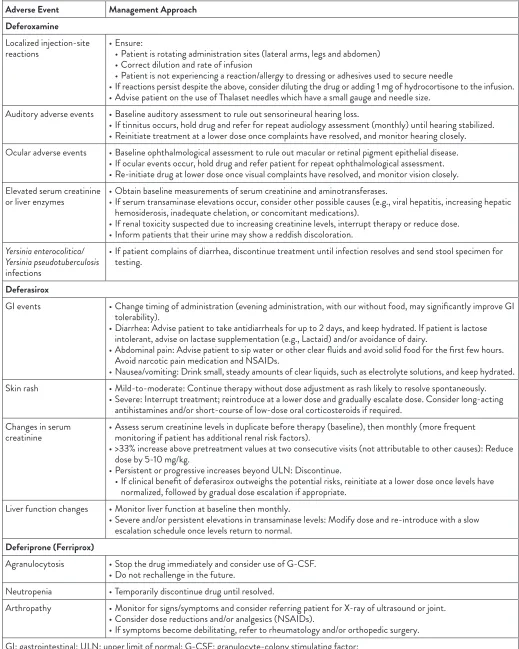
Table 5: Strategies for the management of adverse events associated with iron chelation therapy (Vichinsky, 2008; UHN, 2012) Adverse Event Management Approach
Deferoxamine
Localized injection-site
reactions • Ensure: • Patient is rotating administration sites (lateral arms, legs and abdomen) • Correct dilution and rate of infusion
• Patient is not experiencing a reaction/allergy to dressing or adhesives used to secure needle
• If reactions persist despite the above, consider diluting the drug or adding 1 mg of hydrocortisone to the infusion. • Advise patient on the use of Thalaset needles which have a small gauge and needle size.
Auditory adverse events • Baseline auditory assessment to rule out sensorineural hearing loss.
• If tinnitus occurs, hold drug and refer for repeat audiology assessment (monthly) until hearing stabilized. • Reinitiate treatment at a lower dose once complaints have resolved, and monitor hearing closely. Ocular adverse events • Baseline ophthalmological assessment to rule out macular or retinal pigment epithelial disease.
• If ocular events occur, hold drug and refer patient for repeat ophthalmological assessment. • Re-initiate drug at lower dose once visual complaints have resolved, and monitor vision closely. Elevated serum creatinine
or liver enzymes • Obtain baseline measurements of serum creatinine and aminotransferases. • If serum transaminase elevations occur, consider other possible causes (e.g., viral hepatitis, increasing hepatic hemosiderosis, inadequate chelation, or concomitant medications).
• If renal toxicity suspected due to increasing creatinine levels, interrupt therapy or reduce dose. • Inform patients that their urine may show a reddish discoloration.
Yersinia enterocolitica/ Yersinia pseudotuberculosis
infections
• If patient complains of diarrhea, discontinue treatment until infection resolves and send stool specimen for testing.
Deferasirox
GI events • Change timing of administration (evening administration, with our without food, may significantly improve GI tolerability).
• Diarrhea: Advise patient to take antidiarrheals for up to 2 days, and keep hydrated. If patient is lactose intolerant, advise on lactase supplementation (e.g., Lactaid) and/or avoidance of dairy.
• Abdominal pain: Advise patient to sip water or other clear fluids and avoid solid food for the first few hours. Avoid narcotic pain medication and NSAIDs.
• Nausea/vomiting: Drink small, steady amounts of clear liquids, such as electrolyte solutions, and keep hydrated. Skin rash • Mild-to-moderate: Continue therapy without dose adjustment as rash likely to resolve spontaneously.
• Severe: Interrupt treatment; reintroduce at a lower dose and gradually escalate dose. Consider long-acting antihistamines and/or short-course of low-dose oral corticosteroids if required.
Changes in serum
creatinine • Assess serum creatinine levels in duplicate before therapy (baseline), then monthly (more frequent monitoring if patient has additional renal risk factors).
• >33% increase above pretreatment values at two consecutive visits (not attributable to other causes): Reduce dose by 5-10 mg/kg.
• Persistent or progressive increases beyond ULN: Discontinue.
• If clinical benefit of deferasirox outweighs the potential risks, reinitiate at a lower dose once levels have normalized, followed by gradual dose escalation if appropriate.
Liver function changes • Monitor liver function at baseline then monthly.
• Severe and/or persistent elevations in transaminase levels: Modify dose and re-introduce with a slow escalation schedule once levels return to normal.
Deferiprone (Ferriprox)
Agranulocytosis • Stop the drug immediately and consider use of G-CSF. • Do not rechallenge in the future.
Neutropenia • Temporarily discontinue drug until resolved.
Arthropathy • Monitor for signs/symptoms and consider referring patient for X-ray of ultrasound or joint. • Consider dose reductions and/or analgesics (NSAIDs).
• If symptoms become debilitating, refer to rheumatology and/or orthopedic surgery. GI: gastrointestinal; ULN: upper limit of normal; G-CSF: granulocyte-colony stimulating factor;
The adverse events associated with the currently avail-able iron chelation therapies are reviewed in this section, and practical strategies for their management are summarized in Table 5. It is important to bear in mind that the side-effect profile and management of adverse events with chelators has been well researched in hemoglobinopathy patients (thalas-semia and sickle cell anemia). The following nursing man-agement considerations are based on research and experience with this population, since there is a lack of research on the side-effect profile in oncology patients.
Deferoxamine (Desferal)
Localized injection-site reactions are common with deferox-amine use. It is important for nurses to ensure patients are rotating the site of administration (lateral arms, legs and abdo-men) and that the correct dilution and rate of infusion are being utilized (Eckes, 2011). Patients should also be advised on the use of Thalaset needles, which may be less likely to cause localized reactions given their small gauge and needle size.
Ocular and auditory adverse events have also been observed with deferoxamine, as well as deferasirox use (Wells et al., 2008; Eckes, 2011), and tend to occur at lower serum ferritin levels (<1,000 µg/L). Therefore, prior to treatment, patients require baseline ocular and audiology testing (note that the consult form/letter should specifically state that these tests are being ordered as baseline assessments prior to the initiation of deferoxamine or deferasirox therapy). If the patient complains of tinnitus and/or visual loss while on treatment, it is import-ant to hold deferoxamine and refer the patient for repeat oph-thalmologic or auditory assessment. Once these complaints have resolved, deferoxamine can be re-initiated at a lower dose.
Elevations in serum creatinine levels and liver enzymes have also been noted in patients taking deferoxamine and, therefore, baseline measurements should be obtained prior to treatment. If transaminase increases occur following treat-ment initiation, other possible causes of altered liver func-tion should be considered. If renal toxicity is suspected due to increasing creatinine levels, deferoxamine should be inter-rupted or the dose reduced. Patients using deferoxamine should also be informed that their urine may reveal a reddish discoloration.
Iron overload increases susceptibility to the Gram-negative bacterial infections Yersinia enterocolitica and Yersinia pseudotuberculosis, which result in gastrointestinal (GI) and tuberculosis-like symptoms. In rare instances, treatment with deferoxamine has enhanced this susceptibility, resulting in generalized infections. In patients complaining of diarrhea, a stool specimen should be sent for testing and deferoxamine should be discontinued until the infection resolves.
Deferasirox (Exjade)
Transient GI symptoms, such as abdominal pain, diarrhea, nausea and vomiting, are the most common adverse events associated with deferasirox therapy. Many of these GI distur-bances can be avoided or minimized if patients are initiated on a low dose, with gradual dose increases every 3-5 days until the full dose is achieved. Although the prescribing informa-tion for deferasirox states that the drug should be taken in the
morning on an empty stomach, it is important that patients experiencing GI events try administering deferasirox at dif-ferent times of the day, with orwithout food intake (e.g., two hours after the evening meal), since this has been associated with improved GI tolerability (Vichinsky et al., 2008; Goldberg et al., 2013). Also, altering the timing of administration does not impact drug efficacy.
In patients experiencing diarrhea with deferasirox, ade-quate hydration should be maintained and the administration of antidiarrheals for up to two days when initiating treatment is advised (Vichinsky, 2008). Patients experiencing abdom-inal pain should be advised to avoid solid food for the first few hours after ingestion, and to avoid narcotic pain medica-tion and non-steroidal anti-inflammatory drugs (NSAIDs). Although skin rash can occur in some patients treated with deferasirox, it tends to resolve spontaneously without the need for treatment interruptions or dose adjustments (Vichinsky et al., 2008).
Elevations in serum creatinine levels and proteinuria have also been observed in patients receiving deferasirox, and these tend to be dose-related and non-progressive in nature. Management should be individualized based on the magni-tude of the patient’s elevations (see Table 5) (Vichinsky, 2008). Liver enzyme elevations have also been reported in a small proportion of patients treated with deferasirox. If severe or per-sistent elevations occur, treatment interruption or dose mod-ifications should be considered and a hepatologist should be consulted (Vichinsky et al., 2008).
Deferiprone (Ferriprox)
Transient GI side effects have also been noted in patients treated with deferiprone. Although less common, more seri-ous side effects, such as agranulocytosis, neutropenia, arthrop-athy and arthralgias, have also been observed in patients treated with this agent (Eckes, 2011; Hoffbrand, Taher, & Cappellini, 2012). In cases of agranulocytosis and neutro-penia, deferiprone should be held until neutropenia resolves. Treatment with granulocyte-colony stimulating factor (G-CSF) can be considered if neutropenia is not resolving. Dose reduc-tions and the use of analgesics/NSAIDs should be considered for those with arthropathy/arthralgias.
WHAt NON-PHArMAcOlOGicAl
iNterVeNtiONs Are AVAilABle FOr
irON OVerlOAD?
HOW sHOulD resPONse tO irON
cHelAtiON tHerAPY Be MONitOreD AND
WHeN cAN treAtMeNt Be DiscONtiNueD?
To monitor response to iron chelation therapy, it is gen-erally recommended that serum ferritin be monitored every three months following treatment initiation (Table 6). However, since ocular and auditory adverse events have been associated with lower ferritin levels in patients using deferox-amine, consideration should be given to monitoring ferri-tin monthly, or even more frequently in patients using this agent, particularly if they are experiencing any hearing or ocu-lar issues. If available, annual cardiac MRI T2* and R2 liver MRI (Ferriscan) are also recommended for monitoring patient response to iron chelation therapy (Wells et al., 2008).
In patients using deferasirox, alanine transaminase (ALT), creatinine and urinalysis should be assessed monthly given the potential for renal- and liver-related adverse events (Table 6). In patients with underlying renal function issues, creati-nine and urinalysis should be performed weekly during treat-ment initiation. In patients using deferoxamine, monthly assessment of ALT and creatinine is recommended, as well as annual ophthalmological and audiology assessments. Patients on this agent should also be instructed to immediately report any vision or hearing issues. For patients prescribed deferiprone, a weekly complete blood count (CBC) is essential during the first six to 12 months of therapy (Table 6), since this is the time when most episodes of neutropenia and agranulo-cytosis occur (Wells et al., 2008; UHN, 2012).
According to current Canadian guidelines, iron chela-tion dose reducchela-tions can be considered when ferritin falls below 2,000 µg/L, and treatment can be discontinued when a ferritin level of < 1,000 µg/L is reached (Wells et al., 2008). However, given that ferritin values are affected by inflamma-tory markers, it is more important to focus on trends in ferri-tin measurements over time rather than a single ferriferri-tin value.
WHAt is tHe rOle OF tHe Nurse tO
Assist PAtieNts iN selF-MANAGiNG
tHeir irON cHelAtiON tHerAPY?
Following the recommended oral administration instruc-tions for iron chelation therapy is essential for the effective management of iron overload. Altering the chelation regimen can lead to the development or worsening of iron overload in target organs, which will not only necessitate the intensifica-tion of chelaintensifica-tion therapy and further commitment from the patient (such as the need to insert a portacath or PICC line for continuous IV deferoxamine infusion), but will also require treatment for the complications associated with iron overload.
Table 6: Baseline assessment and monitoring recommendations for patients on iron chelation therapy (Wells et al., 2008; UHN, 2012)
Baseline Monitoring
Deferoxamine
(Desferal) • Ferritin• ALT, Cr
• Cardiac MRI T2*, R2 Liver MRI (Ferriscan®) • Ophthalmology and audiology assessments
• Ferritin every 3 months • ALT, Cr every 3 months
• Cardiac MRI T2*, R2 Liver MRI (Ferriscan®) annually • Screen for ocular/auditory events at every visit • Ophthalmology and audiology assessment annually Deferasirox
(Exjade) • Ferritin• ALT, Cr, urinalysis
• Cardiac MRI T2*, R2 Liver MRI (Ferriscan®)
• Ferritin every 3 months
• ALT, Cr, urinalysis monthly (weekly initially if underlying issues with renal function)
• Cardiac MRI T2*, R2 Liver MRI (Ferriscan®) annually Deferiprone
(Ferriprox)* • Ferritin• CBC, ALT, Cr
• Cardiac MRI T2*, R2 Liver MRI (Ferriscan®)
• Ferritin every 3 months • CBC weekly
• ALT, Cr every 3 months
• Cardiac MRI T2*, R2 Liver MRI (Ferriscan®) annually *In Canada, deferiprone is only approved for the treatment of iron overload in patients with some thalassemia syndromes who have had an inadequate response to previous iron chelation therapy.
ALT: alanine transaminase; Cr: creatinine; MRI: magnetic resonance imaging; CBC: complete blood count
Table 7: Factors contributing to difficulties in following administration recommendations
Practical • Subcutaneous infusion over many hours • Frequent treatment
• Time-consuming
• Variety of equipment required • Restriction of activities • Training required
• Infusion site pain and adverse effects Psychological • Complacency and lack of motivation • Reluctance to accept need for therapy • Negative body image
• Effect on family and personal relationships • Effect on social activity
• Constant reminder of underlying illness Other • Lack of knowledge about disease
• Lack of understanding about the consequences of iron overload
In general, the more disruptive the treatment is to activities of daily living, the less likely the patient is to follow administra-tion guidelines. This tends to be a greater issue in those taking deferoxamine given the requirement for parenteral admin-istration and long infusion times. However, even patients treated with deferasirox can find it difficult to continue therapy given the associated GI side effects, particularly early in the course of therapy. Factors contributing to difficulties in follow-ing administration recommendation for iron chelation therapy are summarized in Table 7 (Cappellini, 2005).
The nurse plays a pivotal role in helping patients self-man-age their therapy through patient education and using a col-laborative patient-focused approach in the prevention, early recognition, and management of the side effects associated with iron chelation therapy. Minimizing the impact of chela-tion therapy on quality of life is key and necessitates not only a thorough understanding of the pharmacology of the avail-able chelators, but also frequent communication and feedback between the nurse and the patient/caregiver(s) (Eckes et al.,
2011). Examples of questions that can be utilized when collab-orating with patients are shown in Table 8. It is also import-ant to note that utilizing newer modes of communication for patient contact, including web-based instant messaging and mobile phone SMS text messaging, may be more effective in engaging certain patient groups, particularly teenagers and adolescents, in their management regimen. A patient support program for deferasirox is also available in Canada, which can complement the information, support and motivation pro-vided by nurses to patients prescribed this agent (please go to drugcoverage.ca for more information on this program).
cONclusiONs
Although essential for many patients with chronic anemias, repeated RBC transfusions can lead to iron overload and sub-sequent end-organ damage. Therefore, the management of iron overload is vital for many transfusion-dependent patients, and it is essential that nurses responsible for the care of these patients have a thorough understanding of iron homeostasis, the pathophysiology of iron overload and its consequences, and the available iron chelation therapeutic options. Furthermore, nurses play a central role in educating patients and caregivers about iron overload and iron chelation therapy, empowering patients to manage the adverse events associated with chela-tion therapy and, ultimately, ensuring long-term patient con-tinuation of therapy and improved patient outcomes.
reFereNces
Álvarez, F., Fernández-Ruiz, M., & Aguado, J.M. (2013). [Iron and invasive fungal infection]. Revista Iberoamericana de Micologia, 30(4), 217–225.
Andrews, N.C. (1999). Disorders of iron metabolism. New England
Journal of Medicine, 341(26), 1986–1995.
Anemia Institute for Research & Education, Thalassemia Foundation of Canada. (2009). Guidelines for the Clinical Care of Patients with
Thalassemia in Canada. Available at http://www.thalassemia.ca/
wp-content/uploads/Thalassemia-Guidelines_LR.pdf
Borgna-Pignatti, C., Rugolotto, S., De Stefano, P., Zhao, H., Cappellini, M.D., Del Vecchio, G.C., Romeo, M.A., … Cnaan A. (2004). Survival and complications in patients with thalassemia major treated with transfusion and deferoxamine. Haematologica, 89(10), 1187–1193. Bring, P., Partovi, N., Ford, J.A., Yoshida, E.M. (2008). Iron overload
disorders: Treatment options for patients refractory to or intolerant of phlebotomy. Pharmacotherapy, 28(3), 331–342.
Cappellini, M.D. (2005). Overcoming the challenge of patient compliance with iron chelation therapy. Semin Hematol, 42(2 Suppl 1), S19–21.
Carson, S.M., & Martin, M.B. (2014). Effective iron chelation practice for patients with β-thalassemia major. Clinical Journal of Oncology
Nursing, 18(1), 102–111.
Eckes, E.J. (2011). Chelation therapy for iron overload: nursing practice implications. Journal of Infusion Nursing, 34(6), 374–380. Fenaux, P., Mufti, G.J., Hellstrom-Lindberg, E., Santini, V., Finelli,
C., Giagounidis, A., Schoch, R., … Silverman, L.R. (2009). Efficacy of azacitidine compared with that of conventional care regimens in the treatment of higher-risk myelodysplastic syndromes: A randomised, open-label, phase III study. Lancet Oncology, 10(3), 223–232.
Gabutti, V., & Piga, A. (1996). Results of long-term iron-chelating therapy. Acta Haematologica, 95(1), 26 –36.
Gattermann, N. (2008). Overview of guidelines on iron chelation therapy in patients with myelodysplastic syndromes and transfusional iron overload. International Journal of Hematology, 88(1), 24–29.
Gattermann, N. (2009). The treatment of secondary
hemochromatosis. Deutsches Ärzteblatt International, 106(30), 499–504.
Goldberg, S.L., Giardina, P.J., Chirnomas, D., Esposito, J., Paley, C., & Vichinsky, E. (2013). The palatability and tolerability of deferasirox taken with different beverages or foods. Pediatric Blood & Cancer, 60(9), 1507–1512.
Hoffbrand, A.V., Taher, A., & Cappellini, M.D. (2012). How I treat transfusional iron overload. Blood, 120(18), 3657–3669.
Ibrahim, A.S., Gebremariam, T., Luo, G., Fu, Y., French, S.W., Edwards, J.E. Jr., & Spellberg B. (2011). Combination therapy of murine mucormycosis or aspergillosis with iron chelation, polyenes, and echinocandins. Antimicrobial Agents &
Chemotherapy, 55(4), 1768–1770.
Lee, J.W., Kang, H.J., Kim, E.K., Kim, H., Shin, H.Y., & Ahn, H.S. (2009). Effect of iron overload and iron-chelating therapy
on allogeneic hematopoietic SCT in children. Bone Marrow
Transplantation, 44(12), 793–797.
Leitch, H.A. (2011). Controversies surrounding iron chelation therapy for MDS. Blood Reviews, 25, 17–31.
List, A.F. (2010). Iron overload in myelodysplastic syndromes: diagnosis and management. Cancer Control, 17(Suppl), 2–8. Table 8: Dialoguing with patients about their iron chelation therapy
Open-Ended Questions:
• What medications are you taking? • Why are you taking them?
• How are you taking your iron chelating therapy?
Malcovati L., Porta, M.G., Pascutto, C., Invernizzi, R., Boni, M., Travaglino, E., Passamonti, F., … Cazzola M. (2005). Prognostic factors and life expectancy in myelodysplastic syndromes classified according to WHO criteria: A basis for clinical decision making. Journal of Clinical Oncology, 23(30), 7594–7603.
Malcovati, L. (2007). Impact of transfusion dependency and secondary iron overload on the survival of patients with myelodysplastic syndromes. Leukemia Research, 31(Suppl 3), S2–S6.
MDS Foundation. (2011). Anemia, Blood Transfusions, Iron Overload, and Myelodysplastic Syndromes: A Handbook for Adult MDS
Patients. Retrieved from http://www.mds-foundation.org/pdf/
AnemiaBloodTransfusionsIronOverloadAndMDS-5-16-11.pdf Meyer, S.C., O’Meara, A., Buser, A.S., Tichelli, A., Passweg, J.R., &
Stern, M. (2013). Prognostic impact of posttransplantation iron overload after allogeneic stem cell transplantation. Biology of Blood
and Marrow Transplantation, 19(3), 440–444.
National Comprehensive Cancer Network. (2015). NCCN Clinical
Practice Guidelines in Oncology: Myelodysplastic Syndromes. Version
1.2015. Available at: http://www.nccn.org/professionals/physician_ gls/pdf/mds.pdf
Pieracci, F.M., & Barie, P.S. (2005). Iron and the risk of infection.
Surgical Infections, 6(Suppl 1), S41–S46.
Porter, J.B. (2001). Practical management of iron overload. British
Journal of Haematology, 115, 239–252.
Pullarkat, V. (2010). Iron overload in patients undergoing hematopoietic stem cell transplantation. Advances in Hematology, 2010.
Pullarkat, V., Blanchard, S., Tegtmeier, B., Dagis, A., Patane, K., Ito, J., & Forman, S.J. (2008). Iron overload adversely affects outcome of allogeneic hematopoietic cell transplantation. Bone Marrow
Transplantation, 42, 799–805.
Sanz, G., Nomdedeu, B., Such, E., Bernal, T., Belkaid, M., Ardanaz, M.T., … Cervera, J. (2008). Independent impact of iron overload and transfusion dependency on survival and leukemic evolution in patients with myelodysplastic syndrome [Abstract 640]. Blood, 112, 238–239.
Schafer, A.I., Cheron, R.G., Dluhy, R., Cooper, B., Gleason, R.E., Soeldner, J.S., & Bunn, H.F. (1981). Clinical consequences of acquired transfusional iron overload in adults. New England
Journal of Medicine, 304(6), 319–24.
Shah, J., Kurtin, S.E., Arnold, L., Lindroos-Kolqvist, P. & Tinsley, S. (2012). Management of transfusion-related iron overload in patients with myelodysplastic syndromes. Clinical Journal of
Oncology Nursing, 16(Suppl), 37–46.
Steensma, D.P. (2011). The relevance of iron overload and the appropriateness of iron chelation therapy for patients with myelodysplastic syndromes: A dialogue and debate. Current
Hematologic Malignancy Reports, 6(2), 136–144.
Stein, J., Hartmann, F., & Dignass, A.U. (2010). Diagnosis and management of iron deficiency anemia in patients with IBD.
Nature Reviews Gastroenterology & Hepatology, 7(11), 599–610.
Taher, A., Al Jefri, A., Elalfy, M.S., Al Zir, K., Daar, S., Rofail, D., Baladi, J.F., Habr, D., Kriemler-Krahn, U., & El-Beshlawy, A. (2010). Improved treatment satisfaction and convenience with deferasirox in iron-overloaded patients with beta-Thalassemia: Results from the ESCALATOR Trial. Acta Haematologica, 123(4), 220–225. Takatoku, M., Uchiyama, T., Okamoto, S., Kanakura, Y., Sawada, K.,
Tomonaga, M., Nakao, S., … Japanese National Research Group on Idiopathic Bone Marrow Failure Syndromes. (2007). Retrospective nationwide survey of Japanese patients with transfusion-dependent MDS and aplastic anemia highlights the negative impact of iron overload on morbidity/mortality. European Journal
of Haematology, 78(6), 487–494.
Trottier, B.J., Burns, L.J., DeFor, T.E., Cooley, S. & Majhail, N.S. (2013). Association of iron overload with allogeneic hematopoietic cell transplantation outcomes: A prospective cohort study using R2-MRI-measured liver iron content. Blood, 122(9), 1678–84.
University Health Network (UHN). (2012). Guidelines for the
Care of Patients in the UHN Red Blood Cell Disorders Program. Available at: http://lgdata.s3-website-us-east-1.amazonaws.com/ docs/1099/501764/UHN_RBCDP_Guidelines_For_Care_2012.pdf Vichinsky, E. (2008). Clinical application of deferasirox: practical
patient management. American Journal of Hematology, 83(5), 398–402.
Wells, R.A., Leber, B., Buckstein, R., Lipton, J.H., Hasegawa, W., Grewal, K., Yee, K., … Tinmouth, A. (2008). Iron overload in myelodysplastic syndromes: A Canadian consensus guideline.
Leukemia Research, 32(9), 1338–1353.