R E S E A R C H A R T I C L E
Open Access
Calculation of iris-claw IOL power for
correction of late in-the-bag IOL complex
dislocation
Valentín Huerva
1,2*, Francisco J. Ascaso
3,4, Isabel Caral
1,2and Andrzej Grzybowski
5,6Abstract
Background:To assess the constants and formula for aphakia correction with iris-claw IOLs to achieve the best refractive status in cases of late in-the-bag IOL complex dislocation.
Methods:A literature search was performed. The following data were obtained: Iris-claw IOL model, Iridal or retroiridal enclavation, A-constant, ultrasound or optical biometry, formula employed and refractive outcomes. Acceptable emmetropia was considered if the resulting spherical equivalent (SE) was within ±1.00 D.
Results:The majority of the studies used SRK/T formula (66.6%). The 88.9% of the reports obtained a SE within ±1. 00 D. Using A-115 for ultrasound biometry and A-115.7 for optical biometry and SRK/T formula, the emmetropia (±1.00 D) of SE, was able to get near 100% of reported cases over the pupil implantation. However, the emmetropia decreased to 80% when the enclavation is retropupilar using the same formula. The A-constant can vary from 116.7 to 117.5 for retropupilar enclavation.
Conclusions:Using A-115 for ultrasound biometry and A-115.7 for optical biometry and SRK/T formula, ±1.00 D of SE, is able to get near 100% of cases. Nevertheless, ±1.00 D of SE decreased to 80% of the cases when the enclavation is retropupilar.
Keywords:IOL-in-the bag dislocation, Iris-claw IOL, Late cataract surgery complication, Artisan aphakia, Verysise
Background
Decentration or luxation of a posterior chamber intraocu-lar lens (IOL) is an uncommon problem following cataract surgery. The phenomenon late in-the-bag IOL complex dislocation after an uneventful cataract surgery can occur many years postoperatively, due to previous progressive zonular disintegration and capsular shrinkage [1–6].
Pseudoexfoliation syndrome is present in more than 50% of the reported cases [6]. Other risk factors such as advanced age, high myopia, uveitis, trauma, previous vitreoretinal surgery, diabetes mellitus and connective tis-sue disorders may be also associated [6]. Complications during cataract surgery and advanced status of the cata-ract also increased the risk of late in-the-bag IOL disloca-tion [7]. No significant differences have been found
following extracapsular cataract extraction when com-pared with phacoemulsification [8]. However, a long phacoemulsification time may be also a risk factor [9]. Although, the cumulative risk of IOL dislocation remains low after cataract surgery, an increased occurrence of late in-the-bag IOL dislocation can be expected in postopera-tive years due to life expectancy [5]. On the other hand, high myopia may be the main risk factor in other studies [10]. Many myopic refractive procedures have been per-formed by means of clear lens extraction. After a 10-years follow-up a 0.6% of cases may require a surgical procedure for a dislocated IOL [11]. Until now, it is not clear if this percentage could increase in cases operated of myopia in future years.
The mean time after cataract extraction and a sec-ondary surgery for late in-the-bag IOL dislocation is approximately 8.5 years [12–14]. When in-the-bag IOL dislocation occurs, due to the risk of further decentration, it is usually better to perform surgery while an anterior * Correspondence:[email protected]
1Department of Ophthalmology, Universitary Hospital Arnau de Vilanova,
Avda. Alcade Rovira Roure 80, 25198 Lleida, Spain
2IRB Lleida, Lleida, Spain
Full list of author information is available at the end of the article
© The Author(s). 2017Open AccessThis article is distributed under the terms of the Creative Commons Attribution 4.0
approach is still possible. Iris-claw lens insertion has been shown their utility in acquired aphakia conditions in the overwhelming majority of cases, with little evidence of post-operative problems such as uveitis, glaucoma, or hyphema [15–17]. The exchange of the dislocated IOL-bag complex and implantation of an iris-claw IOL for aphakia correction during the same time may be a safe and predictable technique with minimal complications [18]. Many reports with iris-claw IOL implantation for correction of this syndrome have been reported. Never-theless, there is no consensus about the A-constant and the employed formula for determining IOL power. In the majority of studies, the authors do not mention about A constant and employed formula [19]. This fact constitutes a challenge for achieving an emmetropia when the extrac-tion and implantaextrac-tion of an iris-claw IOL would be at the same time.
The aim of the study is to assess readers in the best A-constant and formula employed in different studies report-ing aphakia correction with iris-claw IOLs to achieve the best refractive status following surgery in cases of late in-the-bag IOL complex dislocation.
Methods
A literature search was performed using Pubmed and Google Scholar until February 2017. The keywords used were: late in-the-bag IOL dislocation, IOL luxation, cata-ract and pseudoexfoliation syndrome, iris-claw IOL and aphakia, Artisan aphakia, Verisyse aphakia. References cited in the identified reports were also reviewed. Only the reports that have reference to iris-claw IOL implantation in cases of in-the-bag IOL dislocation in the same time were considered. From the articles identified, the following data were obtained if were referenced: Iris-claw IOL model, Iridal (over the pupil) or retroiridal (retropupillary) enclavation, A-constant, ultrasound or optical biometry, formula employed and refractive outcomes. Reports that do not refer the majority above mentioned data in the ma-terial and methods were discarded. From the reports that have reference to this situation only the cases operated in the same time dislocated complex IOL bag extraction and implantation of iris-claw in the same time were choosen for the study. Acceptable emmetropia was considered if the resulting spherical equivalent (SE) was within ±1.00 D. The A-constant provided by the manufacturer of the iris-claw IOLs (Opthec, 9700 AJ Groningen, The Netherlands) were also consulted [20].
Results
Data provided by the manufacturer
Regarding refractive iris-claw IOLs in phakic conditions keratometry and anterior chamber depth (ACD) were taken into account for the power IOL calculation by means of a calculator [20]. The van der Heijde formula
is usually employed in phakic conditions [21]. van der Heijde formula is a theoretical formula of first generation. The problem with this formula is the need to predict from the preoperative data the position that the IOL will take within the eye in a pseudophakic eye. Do not confuse pseudophakic anterior chamber depth (ACD) with pre-operative phakic ACD.
The A-constant provided by the manufacturer is 115.0 (ultrasound) and 115.7 (optical biometry) for Artisan® aphakia IOL (model 205, Ophtec, Groeningen, The Neederlands) for the SRK/T formula. There are available powers from +2.00 to +30.00 D [22]. Verisyse® IOL for aphakia model VRS54 from Abott (Abott Laboratories, Santa Clara, US) is the same IOL that Artisan® aphakia model 205 from Ophtec aproved by the FDA in United States [23]. The A-constant is the same. Refractive models for myopia correction may be employed in special situations because there is not a power lower than +2.00 D for Artisan® aphakia IOL [24].
Data of the revised manuscripts
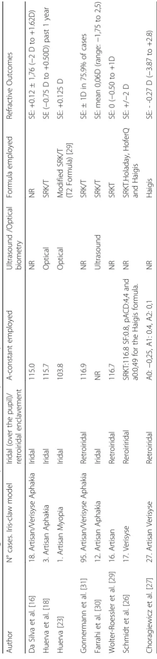
Many reports treat the beneficial outcomes after an iris-claw IOL implantation to resolve the aphakia. However, there are few reports that have reference to the employed biometry, constant and final spherical out-comes after dislocated IOL-bag extraction and iris-claw implantation. Many consulted studies did not report the A-constant and formula employed and, therefore, they were discarded. Table 1 summarizes the eight reviewed reports with the number of cases relating the procedure in the same time and refractive outcomes after the iris-claw IOL enclavement in this situation. Many of them report the iris-claw IOLs implant in other situations.
reports the enclavation was retroiridal [23, 26–29, 31] and the SRK/T formula was employed in the 80% of the reports [23, 26, 28, 29]. With this formula almost 60% of cases could achieved the emmetropia within ±1.00 D. However, differences on the A-constant employed are notable between all reports. A-116.9 for optical biometry may be the mean for the SRK/T for-mula in large reported series that employer retroiridal enclavement [23, 32].
Discussion
Surgical correction of the aphakia results in a challen-ging decision and approach. One solution may be the reposition and suture of the IOL to the scleral wall [33]. The advantage of repositioning and suturing the IOL is that it may avoid a large incision and astigma-tism induction. Nevertheless, in some cases this tech-nique is not possible because either IOL is dislocated into the vitreous cavity or vitreous appears into the anterior chamber. An exchange of the IOL-bag com-plex may be preferable in these situations. Angle supported IOLs or iris-claw IOLs may resolve the aphakia at the same time of surgery or in a second time. However, anterior chamber angle supported IOLs are not popular because they may produce cor-neal decompensation in compromised corneas [34]. Another possibility is the scleral-fixated PC-IOLs, but they may produce lens tilting and decentration among others [34–36].
There are few reports about treatment of aphakia in the late in-the-bag dislocation with an iris claw IOL. Description about the formula and A-constant employed are summarized in Table 1. Retroiridal enclavation is re-ported more frequently in the last time. The theoretical advantage of this location is to avoid the endothelial cell loss [37]. However, the largest series reported show no significant endothelial cells loss after over the pupil enclavation [38, 39]. A study showed that there were no differences in endothelial cells count between three tech-niques for aphakia correction: anterior fixated iris claw, posterior iris fixated IOL and scleral-fixated IOL [40]. The percentage of endothelial cells loss can vary in the different studies between 5.5 and 11.7% according to their follow-up [37, 41]. The majority conclude that the major loss occurs during the first year, due to the surgi-cal trauma during the implantation as it occurs with the implantation of phakic iris claw IOLs [42]. According to these results, anterior or posterior enclavation of the iris claw depend of the decision, ability and preference of the surgeon.
Most authors report the implantation of an iris claw IOL to resolve the refractive status after aphakia, trauma, vitrectomy or complicated cataract surgery. However, an iris-claw IOL may be implanted at the same time of the
extraction of the dislocated IOL-bag complex. Because of this, it is necessary to observe the reported refractive out-comes with the use of iris-caw IOLs for aphakia.
We observe eight studies reporting cases with ex-plantation of the dislocated IOL-bag complex and im-plantation of an iris-claw at the same time [16, 18, 23, 24, 26–30]. Since many refractive procedures have been performed by means of clear lens extraction and IOL implantation in myopic population, it is probable in a higher frequency of late in-the-bag IOL dislocation cases in the future. The duration of surgery and pseu-doexfoliation syndrome may increase the possibility of this syndrome about 8.5 years later [9–11, 43, 44]. In this sense, it is possible to perform the extraction of the dislocated complex and implantation of an iris claw IOL due the above mentioned advantages and get close to emmetropia.
Emmetropia can be achieved within ±1.00 D of SE near of 100% of cases using the SRK/T formula in cases over the pupil enclavement using the A-constants of the manufacturer [16, 18, 24, 30] and also near 80% of cases with the same formula for posterior enclavement. Nevertheless, for posterior enclavation there are no clear consensus for A-constant employed in this study [23, 26, 28, 29]. Although, with an A-constant of 116.9 the emmetropia can be achieved within ±1.00 D of SE in other study about retroiridal enclavation of an iris-claw IOL [32]. For the cases out of ±1.00 D of SE the corneal curvature and axial length should be considered and optimized constants for SRK/T, Haigis, Hoffer Q, and Holladay 1 formulas as for primary IOL implant-ation during cataract surgery or refractive lens ex-change should be necessary to minimize the deviation between postoperative refraction and the target refrac-tion. The revised studies have employed mostly the SRK/T. An alternative may be the Holladay 2 formula used in a study with megalocornea [45] or modification of SRK/T employed in another one in cases of high my-opia [23]. The great SE deviation is observed in the re-port that use all formula [26]. When the Haigis formula is used emmetropia can be achieved; however, a great standard deviation is observed [27].
Conclusions
Abbreviations
ACD:Anterior chamber depth; D: Diopter; FDA: Food and drug
administration; IOL: Intraocular lens; PC-IOLs: Posterior chamber intraocular lenses; SE: Spherical equivalent
Acknowledgements
Not applicable.
Funding
The authors have not received any funding or fees for the study and preparation of the report.
Availability of data and materials
The datasets used and/or analysed during the current study are available from the corresponding author on reasonable request. All data presented in the study have been referenced in the manuscript.
Authors’contributions
VH is responsible for acquisition of clinical information and data, preparation of the manuscript and criticism with the data and final approvation. FJA: Is responsible in the interpretation of the data, preparation of the manuscript, analysis of the data and final approvation. IC is responsable of the acquisicion of the data and prepatation of the manuscript and final approvation. AG is responsible of the intrepretation of the data, critic analysis of the data, preparation of the manuscripst and final approval.
Ethics approval and consent to participate
Not applicable. The report is a review.
Consent for publication
Not applicable.
Competing interests
The authors declare that they have no competing interest in any material or IOls reported in the study.
Publisher’s Note
Springer Nature remains neutral with regard to jurisdictional claims in published maps and institutional affiliations.
Author details
1Department of Ophthalmology, Universitary Hospital Arnau de Vilanova,
Avda. Alcade Rovira Roure 80, 25198 Lleida, Spain.2IRB Lleida, Lleida, Spain. 3
Department of Ophthalmology, Hospital Clínico Universitario“Lozano Blesa”, Zaragoza, Spain.4Instituto de Investigación Sanitaria de Aragón (IIS Aragón),
Zaragoza, Spain.5Department of Ophthalmology, PoznańCity Hospital,
Poznań, Poland.6University of Warmia and Mazury, Olsztyn, Poland.
Received: 18 April 2017 Accepted: 6 July 2017
References
1. Davison JA. Capsule contraction syndrome. J Cataract Refract Surg.
1993;19:582–9.
2. Auffarth GU, Tsao K, Wesendahl TA, Sugita A, Apple DJ. Centration and
fixation of posterior chamber intraocular lenses in eyes with
pseudoexfoliation syndrome. An analysis of explanted autopsy eyes. Acta
Ophthalmol Scand. 1996;74:463–7.
3. Jehan FS, Mamalis N, Crandall AS. Spontaneous late dislocation of
intraocular lens within the capsular bag in pseudoexfoliation patients.
Ophthalmology. 2001;108:1727–31.
4. Gross JG, Kokame GT, Weinberg DV. In-the-bag intraocular lens dislocation.
Am J Ophthalmol. 2004;137:630–5.
5. Gimbel HV, Condon GP, Kohnen T, Olson RJ, Halkiadakis I. Late in-the-bag
intraocular lens dislocation: incidence, prevention, and management. J
Cataract Refract Surg. 2005;31:2193–204.
6. Ascaso FJ, Huerva V, Grzybowski A. Epidemiology, Etiology, and prevention
of late IOL-capsular bag complex dislocation: review of the literature. J Ophthalmol. 2015;2015:805706.
7. KrėpštėL, KuzmienėL, Miliauskas A, JanulevičienėI. Possible predisposing
factors for late intraocular lens dislocation after routine cataract surgery.
Medicina (Kaunas). 2013;49:229–34.
8. Pueringer SL, Hodge DO, Erie JC. Risk of late intraocular lens dislocation
after cataract surgery, 1980-2009: a population-based study. Am J
Ophthalmol. 2011;152:618–23.
9. Dabrowska-Kloda K, Kloda T, Boudiaf S, Jakobsson G, Stenevi U. Incidence
and risk factors of late in-the-bag intraocular lens dislocation: evaluation of
140 eyes between 1992 and 2012. J Cataract Refract Surg. 2015;41:1376–82.
10. Fernández-Buenaga R, Alio JL, Pérez-Ardoy AL, Larrosa-Quesada A,
Pinilla-Cortés L, Barraquer R, Alio JL 2nd, Muñoz-Negrete FJ. Late in-the-bag intraocular lens dislocation requiring explantation: risk factors and
outcomes. Eye (Lond). 2013;27:795–801.
11. Mönestam EI. Incidence of dislocation of intraocular lenses and
pseudophakodonesis 10 years after cataract surgery. Ophthalmology.
2009;116:2315–20.
12. Ostern AE, Sandvik GF, Drolsum L. Positioning of the posterior intraocular
lens in the longer term following cataract surgery in eyes with and without
pseudoexfoliation syndrome. Acta Ophthalmol. 2014;92:253–8.
13. Lorente R, de Rojas V, Vazquez de Parga P, Moreno C, Landaluce ML,
Domínguez R, Lorente B. Management of late spontaneous in-the-bag intraocular lens dislocation: retrospective analysis of 45 cases. J Cataract
Refract Surg. 2010;36:1270–82.
14. Scherer M, Bertelmann E, Rieck P. Late spontaneous in-the-bag intraocular
lens and capsular tension ring dislocation in pseudoexfoliation syndrome. J
Cataract Refract Surg. 2006;32:672–5.
15. Lett KS, Chaudhuri PR. Visual outcomes following artisan aphakia iris claw
lens implantation. Eye (Lond). 2011;25:73–6.
16. De Silva SR, Arun K, Anandan M, Glover N, Patel CK, Rosen P. Iris-claw
intraocular lenses to correct aphakia in the absence of capsule support. J
Cataract Refract Surg. 2011;37:1667–72.
17. Chen Y, Liu Q, Xue C, Huang Z, Chen Y. Three-year follow-up of secondary
anterior iris fixation of an aphakic intraocular lens to correct aphakia. J
Cataract Refract Surg. 2012;38:1595–601.
18. Huerva V, Soldevila J, Sanchez MC. Iris-claw intraocular lens for treatment of
late in-the-bag IOL dislocations associated with pseudoexfoliation
syndrome. J Emmetropia. 2015;6:73–7.
19. Kristianslund O, Råen M, Østern AE, Drolsum L. Late in-the-bag intraocular
lens dislocation: a randomized clinical trial comparing lens repositioning
and lens exchange. Ophthalmology. 2017;124:151–9.
20. https://calculations.ophtec.com/professional/en/refractive-surgery/articalc.
Accessed 04 Apr 2017.
21. van der Heijde. Some optical aspects of implantation of an IOL in a myopic
eye. Eur J Implant Refract Surg. 1989;1:254–48.
22. http://www.ophtec.com/professional/en/cataract-surgery/intra-ocularlenses/
artisan-aphakia/artisan-aphakia-ref-205. Accessed 27 Oct 2016.
23. Gicquel JJ, Langman ME, Dua HS. Iris claw lenses in aphakia. Br J
Ophthalmol. 2009;93:1273–5.
24. Huerva V. Myopic iris-claw intraocular lens for late intraocular lens-capsular
bag complex dislocation using the T2 formula. JCRS Online Case Reports.
2014;2:e38–40.
25. Sheard RM, Smith GT, Cooke DL. Improving the prediction accuracy of the
SRK/T formula: the T2 formula. J Cataract Refract Surg. 2010;36:1829–34.
26. Schmidt I, Langenbucher A, Moussa S, Schirra F, Seitz B, Eppig T.
Retroiridal implantation of a Verisyse™iris claw lens: refractive
outcome and individualized intraocular lens constants. Ophthalmologe.
2015;112(3):261–5.
27. Choragiewicz T, Rejdak R, Grzybowski A, Nowomiejska K, Moneta-WielgośJ,
Ozimek M, Jünemann AG. Outcomes of Sutureless iris-claw lens implantation. J Ophthalmol. 2016;2016:7013709.
28. Galvis V, Tello A, Carreño NI, Revelo ML. Retropupillary iris-claw intraocular
lens in aphakic eyes. J Cataract Refract Surg. 2013;39:970–1.
29. Wolter-Roessler M, Küchle M. Correction of aphakia with retroiridally fixated
IOL. Klin Monatsbl Augenheilkd. 2008;225:1041–4.
30. Farrahi F, Feghhi M, Haghi F, Kasiri A, Afkari A, Latifi M. Iris claw versus
Scleral fixation intraocular lens implantation during pars Plana Vitrectomy. J
Ophthalmic Vis Res. 2012;7:118–24.
31. Gonnermann J, Klamann MK, Maier AK, Rjasanow J, Joussen AM, Bertelmann
E, Rieck PW, Torun N. Visual outcome and complications after posterior iris-claw aphakic intraocular lens implantation. J Cataract Refract Surg.
32. Häberle H, Schiedel S, Pham DT. Retroiridal iris claw lens as routine
procedure. Klin Monatsbl Augenheilkd. 2014;231:995–8.
33. Drolsum L, Ringvold A, Nicolaissen B. Cataract and glaucoma surgery
in pseudoexfoliation syndrome: a review. Acta Ophthalmol Scand.
2007;85:810–21.
34. Evereklioglu EH, Bekir NA, Borazan M, Zorlu F. Comparison of secondary
implantation of flexible open-loop anterior chamber and scleral-fixated
posterior chamber intraocular lenses. J Cataract Refract Surg. 2003;29:301–8.
35. Dadeya S, Kamlesh, Kumari Sodhi P. Secondary intraocular lens (IOL)
implantation: anterior chamber versus scleral fixation long-term comparative
evaluation. Eur J Ophthalmol. 2003;13:627–33.
36. Kwong YY, Yuen HK, Lam RF, Lee VY, Rao SK, Lam DS. Comparison of
outcomes of primary scleral-fixated versus primary anterior chamber intraocular lens implantation in complicated cataract surgeries.
Ophthalmology. 2007;114:80–5.
37. Gonnermann J, Amiri S, Klamann M, Maier AK, Joussen AM, Rieck PW, Torun
N, Bertelmann E. Endothelial cell loss after retropupillary iris-claw intraocular
lens implantation. Klin Monatsbl Augenheilkd. 2014;231:784–7.
38. Güell JL, Verdaguer P, Elies D, Gris O, Manero F, Mateu-Figueras G, Morral M.
Secondary iris-claw anterior chamber lens implantation in patients with
aphakia without capsular support. Br J Ophthalmol. 2014;98:658–63.
39. Güell JL, Verdaguer P, Mateu-Figueras G, et al. Unilateral iris-claw
intraocular lens implantation for Aphakia: a paired-eye comparison.
Cornea. 2016;35:1326–32.
40. Hazar L, Kara N, Bozkurt E, Ozgurhan EB, Demirok A. Intraocular lens
implantation procedures in aphakic eyes with insufficient capsular support
associated with previous cataract surgery. J Refract Surg. 2013;29:685–91.
41. Xue K, Hildebrand GD. Retropupillary artisan intraocular lens implantation in
very young children with aphakia following penetrating eye injuries. J
AAPOS. 2013;17:428–31.
42. Menezo JL, Cisneros AL, Rodriguez-Salvador V. Endothelial study of iris-claw
phakic lens: four year follow-up. J Cataract Refract Surg. 1998;24:1039–49.
43. Liu E, Cole S, Werner L, Hengerer F, Mamalis N, Kohnen T. Pathologic
evidence of pseudoexfoliation in cases of in-the-bag intraocular lens
subluxation or dislocation. J Cataract Refract Surg. 2015;41:929–35.
44. Steeples LR, Jones NP. Late in-the-bag intraocular lens dislocation in
patients with uveitis. Br J Ophthalmol. 2015;99:1206–10.
45. Saffra N, Rakhamimov A, Masini R, Rosenthal KJ. Anterior chamber iris claw
lens for the treatment of Aphakia in a patient with Megalocornea. Case Rep
Ophthalmol. 2015;6:164–9.
• We accept pre-submission inquiries
• Our selector tool helps you to find the most relevant journal
• We provide round the clock customer support
• Convenient online submission
• Thorough peer review
• Inclusion in PubMed and all major indexing services
• Maximum visibility for your research
Submit your manuscript at www.biomedcentral.com/submit