Li Pan, Jian Peng Wen and Lian Ting Ma
*Abstract
In the past, the cavernous dural arteriovenous fistula was categorized as spontaneous cavernous carotid fistula [1] due to
the lack of knowledge and limitation of imaging equipment. In the recent time, with the accumulation of knowledge of
DAVF
’
s etiology, mechanism, physiology, clinical symptoms and imaging data, the diagnostic methods and treatment
have achieved novel understandings and progresses. In fact, it
’
s a specific type of dural arteriovenous fistula
——
cavernous dural arteriovenous fistula. The purpose of this paper is to tell the difference between cavernous dural
arteriovenous fistula and traumatic carotid cavernous fistula, and to redefine cavernous dural arteriovenous fistula from
the aspects of etiology, mechanism, pathology, clinical symptoms, DSA characteristic and therapy. DAVF is an
independent disease. The Cavernous dural arteriovenous fistula can not be classified as spontaneous CCF any
longer, but a specific type of DAVF
—
cavernous DAVF.
Keywords:
Traumatic carotid cavernous fistula, Cavernous dural arteriovenous fistula, Differentiation
Etiology
TCCF is mainly caused by trauma and skull base
fracture.
CS-DAVF is caused by cerebral venous sinus thrombosis,
venous sinus hypertension, inflammation, trauma or
endo-crine diseases, etc.
Mechanism
TCCF: Skull base fracture injures the internal carotid artery
and its branches, which gives rise to the abnormal
commu-nication between ICA and cavernous sinus.
CS-DAVF: Multiple reasons lead to the
communica-tion between dural neoformative multi-branch arteriole
and cavernous sinus through the dural microfistulae.
Pathology
TCCF: The feeding artery is cavernous segment of
internal carotid artery or its branch-vessels; In few cases,
the feeding artery is the external carotid artery; Normally
single fistulous orifice; The draining veins are superior
ophthalmic vein, superior petrosal sinus, inferior petrosal
sinus,basal veins, cortex veins (labbe, Tralend veins) or
mixed-type draining veins (two, three or four veins of the
four draining veins mentioned above).
CS-DAVF: In most cases, the feeding arteries are the
ascending pharyngeal artery, sphenopalatine artery and
the middle meningeal artery of external carotid artery.
There are also the tiny branch arteries from the
menin-gopituitary trunk feeding the CS-DAVF; Normally
mul-tiple microporous fistulous orifices. The draining veins
are mainly superior ophthalmic vein, superior petrosal
sinus and inferior petrosal sinus. Basal vein and cortex
veins are not frequently seen. The contralateral internal
(external) carotid artery are often feeders when bilateral
cavernous sinuses are involved by the lesion; Stealing
symptom is not manifest (Figs.
1
and
2
).
Clinical symptoms
TCCF: intracranial bruit;exophthalmos; obvious hyperemia
and edema of bulbar conjuctive and eyelid; elevation of
intraorbital pressure; restriction of occular motility;
some-times intracranial hemorrhage or steal syndrome.
CS-DAVF: mild and rare intracranial bruit; rare
hyperemia and edema of bulbar conjuctive and eyelid;
* Correspondence:[email protected]Department of Neurosurgery, Wuhan General hospital of Guangzhou Military, Wuhan 430061, Hubei Province, China
Fig. 1The anastomosis of cavernous region [3] and illustration of ICA-cavernous fistula.aBranches of cavernous segment of Internal carotid artery (1. meningopituitary trunk arter,including:①inferior hypophyseal artery,②dorsal meningeal artery,③tentorial artery; 2. inferior cavernous sinus artery; 3. capsular arteries; 4. Internal carotid artery; 5. Ophthalmic artery;6. pituitary);bCavernous sinus (internal basal surface of skull; including: 1. Superior ophthalmic vein, 2. Anterior intercavernous sinus; 3. pituitary; 4. Cavernous sinus; 5. Posterior intercavernous sinus; 6. Plexus basilaris; 7. Inferior petrosal sinus; 8. Glossopharyngeal nerve; 9. Vagus nerve; 10. accessory; 11. Hypoglossal nerve; 12. tentoriumcerebelli; 13. Sphenoparietal sinus; 14. Optic nerve; l5. Internal carotid artery; 16. Posterior clinoid process; 17. Oculomotor nerve; 18. Trochlear nerve; 19. Trigeminal nerve; 20. Abducent nerve; 21. Superior petrosal sinus; 22. Facial nerve、acoustic nerve; 23. sigmoid sinus; 24. great cerebral vein; 25. sinus rectus);cCavernous sinus (coronal section; 1. pituitary; 2. Internal carotid artery; 3. Aducent nerve; 4. Sphenoid sinus; 5. Sphenoid bone; 6. Oculomotor nerv; 7. Trochlear nerve; 8. Ophthalmic branch of trigeminal nerve);dTopography of cavernous sinus (relationship between ICA and cerebral nerve; 1. Superior wall of cavernous sinus; 2. Oculomotor nerve; 3. Trochlear nerve; 4. Aducent nerve; 5. ophthalmic branch of trigeminal nerve; 6. Trigeminal semilunar ganglion; 7. Mandibular branch of trigeminal nerve; 8. Internal carotid artery; 9. Ophthalmic artery; 10. optic nerve; 11. Superior optical fissure; 12. Maxillary branch of tigeminal nerve;eCarotid-cavernous fistula (1. ICA;2. Fistulous orifice;3. Cavernous sinus)
Fig. 2Five types of venous draining of cavernous fistula.aArterial blood is drained to facial vein from cavernous sinus through superior ophthalmic vein and angular vein;barterial blood is drained to superior sagittal sinus from cavernous sinus through trolard frontal parietal anastomotic vein.carterial blood is drained to basilar vein from cavernous sinus through an anastomotic vein, and together with Vein of Galen , drained to straight sinus.darterial blood is drained to internal carotid vein from cavernous sinus through superior petrosal vein, inferior petrosal vein, basilar vein and plexus pterygoideusta.eA mixed type: Two kinds of draining together of the four mentioned above. 1. cavernous sinus; 2. basilar vein; 3. straight sinus; 4. superior petrosal sinus; 5. inferior petrosal sinus; 6. plexus pterygoideusta’7. Trolard frontal parietal anastomotic vein; 8.superior sagittal sinus; 9. superior ophthalmic vein; 10. facial vein
Fig. 3The DSA and clinical symptoms before and after the embolization of right ICA cavernous sinus fistula.aRight carotid cavernous sinus; Arrow shows misembolization of internal carotid artery in other hospitals.bArrow shows angiography of left internal carotid artery: right cavernous sinus fistula is opacified through anterior communicating artery.cAngiography of left internal carotid artery, right cavernous sinus is opacified through ACA. Arrow shows dilated draining of superior ophthalmic vein.dDirectly puncture to the fistulous orifice through superior ophthalmic vein and inject the NBCA to occlude the fistula. Arrow shows the catheter.eAngiography of left internal carotid artery after embolization. The right cavernous sinus fistula is not opacified. Arrow shows the fistulous orifice is occluded.fAngiography of left internal carotid artery after embolization. The right cavernous sinus fistula is not opacified. Arrow shows superior ophthalmic vein is vanished.gThe ophthalmic symptoms before embolization.hThe ophthalmic symptoms are gone 2 weeks after embolization.i. Follow-up 5 years after embolization
rare intracranial hemorrhage or steal syndrome;
easy-ily misdiagnosed as conjuctivitis.
DSA characteristic
The feeding artery of TCCF is the cavernous segment of
internal carotid artery, mostly the horizontal segment.
Sin-gle fistulous orifice, mostly on the side near skull base.
The draining veins are mainly superior ophthalmic vein,
superior petrosal sinus, inferior petrosal sinus, sometimes
cortex veins and basilar vein (Figs.
3a
-
i
,
4a
-
h
,
5a
-
d
).
CS-DAVF: The feeding arteries are the branches of
ex-ternal carotid artery. In some few cases, the branches of
meningopituitary trunk feed the contralateral
synonym-ous vessels. The fistulsynonym-ous orifices on the dural mater are
multi-microporous, which are naked-eye invisible. The
draining veins are mainly superior ophthalmic vein,
superior petrosal vein and inferior petrosal vein, which
can communicates to the contralateral vessels through
intercavernous sinus (Figs.
6a
-
f
,
7a
-
j
,
8a
-
d
).
Therapy
TCCF: Endovascular embolization is the prior therapy
and internal carotid artery is the prior approach. But
when internal carotid artery approach can not be
per-formed, superior ophthalmic vein, superior petrosal vein
and inferior petrosal vein are also optimal approaches.
Detachable ballon is the prior embolic material, but in
some special cases, coils and onyx glue are also available.
Surgical therapy is barely considered. Our hospital has
treated TCCF in a total of more than 900 cases. All
patients are completely cured. No disability or death is
observed.
Fig. 5TCCF.aThe static DSA image in orthophoria view before treatment. Red arrow shows the fistulous orifice;bThe static DSA image in lateral view before treatment. Red arrow shows the fistulous orifice.cThe static DSA image in orthophoria view after treatment.dThe static DSA image in lateral view before treatment
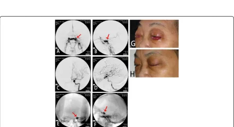
Fig. 6The DSA before and after CS-DAVF embolization.a,bThe DSA of left internal carotid artery in orthophoria and lateral view before embolization. Arrow shows the fistulous orifice.c,dThe DSA of left internal carotid artery in orthophoria and lateral view after embolization. eThe DSA of left external carotid artery in orthophoria and lateral view before embolization. Arrow shows the fistulous orifice.fThe DSA of left external carotid artery in orthophoria and lateral view after embolization
CS-DAVF:Endovascular treament is the first choice for
cavernous DAVF [
2
]. We can choose arterial approach,
venous approach or combined approach. Coils plus onyx
glue, silk line segments or grubran glue can be chosen
as embolic materials. As there are multiple feeding
arter-ies and fistulous orifices, the remaining fistulous orifices
can be cured by pressing the CCA postoperatively (2
–
3 months, 2 times a day, keep pressing CCA for 30 min
every time).
The treatment of CS-DAVF is rather more difficult
than TCCF. However, when choosing the proper
treat-ment, CS-DAVF can be cured and zero death can be
reached with extremely low complication rate. Because
dangerous anastomasis exists between branches of
Fig. 8CS-DAVF.aThe static DSA image in orthophoria view before treatment. Red arrow shows the fistulous orifice;bThe static DSA image in lateral view before treatment. Red arrow shows the fistulous orifice.cThe static DSA image in orthophoria view after treatment.dThe static DSA image in lateral view before treatment
external carotid artery and ophthalmic artery,
vertebro-basilar artery, embolic materials would cause
misemboli-zation through dangerous anastomasis when arterial
embolizaion is performed. Over-packing with coils
would cause ocular muscle paralysis or reflux of liquid
embolic agent to artery and their correspondent
com-plications. Our hospital has treated cavernous CS-DAVF
in a total of 200 cases since 1900. The longest follow-up is
25 years. No death occurred and only 1 case of right eye
blindness was observed.
Conclusion
Based on the above knowledge, we suggest that the term
of spontaneous internal carotid cavernous sinus should
not be used. DAVF is an independent disease. Do not
con-fuse CS-DAVF with TCCF and categorize CS-DAVF as
cavernous type of DAVF when we write papers or books.
Abbreviations
CCA:Common carotid artery; CCF: Cavernous carotid fistula; CS-DAVF: Cavernous dural arteriovenous fistula; DAVF: Dural arteriovenous fistula; DSA: Digital subtraction angiography; ICA: Internal carotid artery; TCCF: Traumatic carotid cavernous fistula
Acknowledgements
We acknowledge the support of Wuhan General hospital of Guangzhou military.
Availability of data and materials
All data generated or analysed during this study are included in this published article.
Authors’contributions
LTM and LP participated in collecting data, and drafted the manuscript. All authors read and approved the final manuscript.
Ethics approval and consent to participate Not applicable.
Competing interests
The authors report no potential conflicts of interest concerning the materials or methods used in this study or the findings presented.
Received: 8 June 2017 Accepted: 14 May 2018
References
1. Barrow DL, Spector RH, Braun IF, et al. Classification and treatment of spontaneous carotid-cavernous sinus fistulas. J Neurosurg. 1985;62(2):248–56. 2. Harris FS, Rhoton AL. Anatomy of the cavernous sinus. A microsurgical
study. J Neurosurg. 1976;45(2):169–80.
3. Borden JA, Wu JK, Shucart WA. A proposed classification for spinal and cranial dural arteriovenous fistulous malformations and implications for treatment. J Neurosurg. 1995;82(2):166.