R E S E A R C H A R T I C L E
Open Access
Serum neutrophil gelatinase-associated lipocalin
and recovery of kidney graft function after
transplantation
Maria E Hollmen
1*, Lauri E Kyllönen
2, Jussi Merenmies
3and Kaija T Salmela
2Abstract
Background:Neutrophil gelatinase-associated lipocalin (NGAL) is a marker for acute kidney injury. We studied whether serum NGAL predicts delayed graft function (DGF) and recovery of kidney function after transplantation.
Methods:Serum NGAL was analyzed using commercial ELISA and point-of-care (POC) (Triage®, Biosite) methods. Serum samples were collected from 176 consecutive, deceased-donor kidney recipients just before transplant surgery and on day 1 and 14 after transplantation. The first 132 samples were analyzed with both methods and the remaining samples with the POC method.
Results:The correlation between the ELISA and POC methods was 0.89, p < 0.0001 and hence the POC method was used for the remaining analyses. DGF was seen in 66/176 patients. Day 1 sNGAL was significantly higher in DGF (588 ng/ml, SD 189.6) compared to early graft function (355 ng/ml, SD 166.2, p < 0.0001) and this difference persisted on day 14. Day 1 sNGAL predicted DGF with an area under the curve (AUC) of 0.853 (CI 0.792-0.914, p < 0.0001). At the optimal cutoff level of 423 ng/ml the sensitivity was 87% and the specificity 77%. In a multivariate analysis, day 1 sNGAL emerged as an independent predictor of DGF. The sNGAL also predicted DGF lasting longer than 14 days with an AUC of 0.825 (CI 0.751-0.899, p < 0.0001). At the optimal cutoff level of 486 ng/ml, the sensitivity was 80% and specificity 75%.
Conclusion:Serum NGAL predicts clinically significant DGF and is useful in the care of kidney transplant recipients.
Keywords:Kidney transplantation, Delayed graft function, Serum NGAL, Point-of-care analysis
Background
Neutrophil gelatinase-associated lipocalin (NGAL) is a small, positively charged iron-carrier protein expressed at low levels in various epithelial cells (e.g. kidney, gastro-intestinal tract, lungs) [1,2]. Due to its size and charge, NGAL is freely filtered through the glomerulus. Normal, steady state serum and urine NGAL concentration is ap-proximately 20 ng/ml [3]. NGAL is markedly increased in the serum and urine of patients with acute kidney injury (AKI) [4-7]. Delayed graft function (DGF) is a form of AKI and is associated with complicated posttransplant recovery and an inferior 1-year outcome [8-15]. Prolonged DGF has been reported to severely impair the long-term prog-nosis of kidney transplantation [8,12]. The diagprog-nosis of
DGF is currently made on clinical grounds days after transplantation. However, we, and others, have shown that urine NGAL also predicts DGF after deceased-donor kid-ney transplantation [16-18]; moreover, there is data sug-gesting that measuring serum/plasma NGAL soon after transplantation is additionally valuable in predicting DGF [19-24]. NGAL has also been shown to predict kidney in-jury in liver transplant patients [25-27]. Despite these promising results, the use of NGAL has not yet been adopted in clinical transplantation.
The gold standard for NGAL analyses is immunoblotting. However, this method is time consuming and not readily available in clinical settings. Urine NGAL can also be mea-sured using ELISA or a chemiluminescent microparticle immonoassay (ARCHITECT®, Abbott). When testing NGAL in the blood, ELISA or the recently introduced point-of-care (POC) fluorescence-based immunoassay (Triage®, Bio-site) can be used. This POC test enables fast, bedside * Correspondence:maria.hollmen@helsinki.fi
1
Department of Medicine, Division of Nephrology, Helsinki University Hospital, Haartmaninkatu 4, PL 372, Helsinki 00029, HUS, Finland Full list of author information is available at the end of the article
measuring of NGAL. However, data concerning the use of the POC method in NGAL analyses is still scarce.
Therefore, the aim of this study was to analyze how the POC method correlates with the ELISA method in measuring serum NGAL (sNGAL) and whether sNGAL predicts the occurrence and duration of DGF.
Methods
Study design and patient population
This study is parallel to our previous work on urine NGAL in the prediction of DGF [16]. We recruited 176 consecutive, adult, dialysis-dependent, deceased-donor kidney transplant recipients between August 2007 and August 2008. Here, we analyzed NGAL using the pro-spectively collected serum samples from this patient population. The Department of Surgery and the Ethics Committee at Helsinki University Hospital approved the study protocol. Written informed consent was obtained from the recipients before enrolment.
The primary outcome variable was the onset of graft function after transplantation, defined here, as it is most widely defined, as the need for dialysis during the first week after transplantation. However, in this classification, patients requiring one dialysis due to fluid overload or high potassium levels are also included in the DGF group. Conversely, there are DGF patients with fluent urine out-put from their native kidneys needing dialysis only after the first week and they are thus excluded from the DGF group. Therefore, to assess in more detail NGAL’s poten-tial to predict DGF, we additionally performed a receiver operating characteristic (ROC) analysis using the DGF definition published by Halloran et al. consisting of plasma creatinine concentration >500μmol/l throughout the first week, or oliguria of less than 1000 ml/24 hours for more than two days, or more than one dialysis session needed during the first week (28).
We collected the clinical data from the patients’ med-ical records and the Finnish Kidney Transplant Registry database, as previously described [16].
Sample collection and NGAL analyses
A pretransplant (day 0) blood sample was taken upon ar-rival to the transplant unit. This sample was used to assess serum NGAL concentrations in patients with end-stage renal disease and as a reference value for the later time points. Posttransplant samples were collected in the morning following the transplant surgery (day 1) and 14 days after the transplantation (day 14). The samples were immediately processed and stored at−70°C.
We used a commercial ELISA kit (BioPortoDiagnostics, Gentofte, Denmark), as recommended by the manufac-turer, for the NGAL analyses. The day 0, day 1, and day 14 samples were also analyzed using a point-of-care (POC) fluorescence immunoassay NGAL kit and device (Triage®
Biosite, San Diego, California, USA), as recommended by the manufacturer. All measurements were performed in duplicate and blinded to the sample sources and clinical outcomes.
Statistical analyses
SPSS software version 20.0 (SPSS Inc., Chicago, Illinois, USA) was used for the statistical analyses. All analyzed variables were tested for distribution.T-test and ANOVA were used for samples with normal distribution, and the Mann–Whitney U and Kruskal-Wallis tests for analyses of samples with skewed distribution. Chi-square and Fisher’s exact tests were employed in analyses of contingency tables. Bivariate correlations were analyzed using the Spearman correlation coefficient for non-parametric, and the Pearson correlation coefficient for parametric, measures of statistical dependence. To assess DGF predictors, a multivariate ana-lysis was used. A ROC anaana-lysis was performed to assess NGAL’s potential to predict DGF and prolonged DGF. A p-value <0.05 was considered significant.
Results
Pretransplant serum samples from 132 kidney recipients and day 1 samples from 128 recipients were analyzed with both ELISA and POC. The corresponding ELISA and POC sNGAL values are shown in Figure 1. The mean sNGAL was 506 ng/ml (SD 188.7) measured with ELISA, and 536 ng/ml (SD 238.4) measured with POC. Their cor-relation was 0.89, p < 0.0001. Since the corcor-relation was good we decided to use the sNGAL values measured using the more practical POC method for the clinical analyses in this study.
In recipients with no residual diuresis from their native kidneys, the mean pretransplant sNGAL was significantly higher (822 ng/ml, SD 193.5) than that of recipients with residual diuresis: the mean pre-transplant sNGAL was 697 ng/ml (SD 166.5) if the diuresis was 100–1000 ml/ 24 hours and 561 ng/ml (SD 164.4) if the diuresis was more than 1000 ml/24 hours (p < 0.0001). There was no signifi-cant difference between hemodialysis and peritoneal dialysis patients in this respect (data not shown). Pretransplant NGAL was not affected by recipient age, gender, underlying kidney disease, number of previous transplantations, mode or length of pretransplant dialysis, or pretransplant plasma creatinine (data not shown).
After transplantation, recipients with a urine output on day 1 of more than 1000 ml had significantly lower sNGAL levels (356 ng/ml, SD 168.1) compared to those with a urine output of less than 1000 ml (593 ng/ml, SD 185.7, p < 0.0001). Recipients with a decrease in plasma creatinine on day 1 compared to their pretransplant level had significantly lower sNGAL levels (382 ng/ml, SD 196.0) compared to those with no change or an increase in plasma creatinine on day 1 (547 ng/ml, SD 186.7, p < 0.0001). There were no significant differences in the other parameters studied (data not shown).
The patient and transplantation characteristics ac-cording to the onset of graft function are shown in Table 1. DGF was seen in 66/176 patients and their oli-guria lasted a mean of 12.1 days (SD 7.0). When com-pared to the early graft function (EGF) group, the DGF group’s donors were older, expanded criteria donors [28] were more common, CIT longer, pretransplant
hemodialysis more common than peritoneal dialysis, and time on pretransplant dialysis longer.
Day 1 sNGAL was significantly higher in patients with DGF (588 ng/ml, SD 189.6) compared to those with EGF (355 ng/ml, SD 166.2, p < 0.0001) and this difference per-sisted on day 14 (Figure 2). Day 1 sNGAL correlated with the duration of DGF (R = 0.70, p < 0.0001). Day 1 sNGAL correlated with day 1 plasma creatinine (R = 0.64, p < 0.0001), day 3 plasma creatinine (R = 0.76, p < 0.0001), day 7 plasma creatinine (R = 0.69 p < 0.0001), day 1 urine out-put (R = 0.65, p < 0.0001), day 3 urine outout-put (R = 0.51, p < 0.0001), and day 7 urine output (R = 0.39, p < 0.0001).
A ROC analysis was performed to assess the potential of day 1 sNGAL in predicting DGF. The area under the curve (AUC) was 0.853 (CI 0.792-0.914, p < 0.0001) (Figure 3). At the optimal cutoff level of 423 ng/ml, the sensitivity was 87% and the specificity 77% (the sensitivities and specific-ities for different cutoff levels are shown in Table 2). In comparison, urine output on day 1 predicted DGF with an AUC of 0.962 (CI 0.922-0.979, p < 0.0001). At the optimal cutoff level of 1035 ml/24 h, the sensitivity was 92% and the specificity 81%. Moreover, day 1 plasma creatinine pre-dicted DGF with an AUC of 0.785 (CI 0.718-0.852, p < 0.0001). At the optimal cutoff level of 523μmol/l, the sensi-tivity was 74% and the specificity 71%. The change in plasma creatinine from the pretransplant level to the day 1 level predicted DGF with an AUC of 0.769 (CI 0.696-0.843, p < 0.0001). At the optimal cutoff level of 68 μmol/l, the sensitivity was 76% and specificity 55%.
Additionally, we repeated the ROC analysis for sNGAL on day 1 after classifying the patients according to the
Table 1 Patient and transplantation characteristics
Early graft function (EGF) n = 110 (62.5%)
Delayed graft function (DGF) n = 66 (37.5%)
p-value
Mean recipient age years (SD) 50.4 (12.8) 54.4 (13.2) NS
Gender, male (%) 65 (59.1) 45 (68.2) NS
Re-transplantation (%) 8 (7.3%) 7 (10.6%) NS
Mode of dialysis, hemodialysis (%) 63 (57.3%) 51 (77.3%) 0.009
Time on dialysis before transplantation, days (SD) 788 (570.3) 953 (608.7) 0.029
Mean donor age years (SD) 49.1 (14.6) 56.2 (11.5) 0.001
Expanded criteria donors 33 (30.0%) 36 (54.5%) 0.001
Mean cold ischemia time, hours (SD) 21.3 (3.5) 22.9 (3.7) 0.003
Day 1 mean plasma creatinine,μmol/L (SD) 464 (218.4) 207 (114.4) <0.0001
Day 1 mean urine output, mL/24 hours (SD) 2504 (1514.8) 502 (538.5) <0.0001
Number of rejections 5 (4.5%) 5 (7.5%) NS
Mean eGFR, mL/min at 1 year (SD) 66.2 (20.1) 54.8 (20.5) <0.0001
1-year patient survival 99.1% 98.5% NS
1-year graft survival 98.2% 89.4% 0.015
criteria for DGF by Halloran et al. [29]; the AUC for day 1 sNGAL in predicting DGF was 0.908 (CI 0.860-0.955, p < 0.0001) (Figure 4). At the optimal cutoff level of 426 ng/ml, the sensitivity was 91% and the specificity 83%. The sensi-tivities and specificities for different cutoff levels are shown in Table 3.
Altogether, 10 patients were classified differently into the DGF and EGF groups depending on whether the conventional or the Halloran criteria for DGF were used (Table 4). All of the patients who otherwise had prompt function but required one dialysis (classified as DGF by conventional criteria and EGF by the Halloran criteria) had day 1 sNGAL less than 400 ng/ml. On the other hand, those with poorly declining plasma creatinine but
with no dialysis performed (classified as EGF by conven-tional criteria and DGF by the Halloran criteria) had day 1 sNGAL higher than 400 ng/ml.
In a multivariate analysis, we assessed the effect on the occurrence of DGF of: donor age, donor creatinine, donor eGFR, expanded criteria donor status, CIT, recipient age, mode and length of dialysis before transplantation, change in plasma creatinine from day 0 to day 1, day 1 urine put, and day 1 sNGAL. Day 1 sNGAL and day 1 urine out-put emerged as independent predictors of DGF, irrespective of the DGF definition used (Tables 5 and 6).
The day 1 serum samples were drawn the morning after transplant surgery, a mean 11.8 hours (SD 5.2) after the re-perfusion of the graft. The time interval between reperfu-sion and this day 1 sample varied from 2 hours to 24 hours; it was 6 hours or less in 18 cases, 7 to 12 hours in 82 cases, and more than 12 hours in 70 cases. DGF was predicted by sNGAL well in all time interval groups, but the predictive power was best in the group were the sample was drawn less than 6 hours after reperfusion (Table 7).
Figure 2The sNGAL concentration at day 0, day 1, and day 14 in early graft function (EGF) and delayed graft function (DGF) groups measured using the point-of-care (POC) method.The results are expressed as means (+standard deviation, SD). DGF is defined as the need for dialysis during the first week after transplantation.
Figure 3The ROC-curve in predicting delayed graft function (DGF) defined as the need for dialysis during the first week after transplantation.AUC, area under the curve; CI, confidence interval.
Table 2 The sensitivities and specificities at different cutoff levels for day 1 sNGAL in predicting delayed graft function (DGF)
Day 1 sNGAL ng/ml Sensitivity Specificity
71 1.00 0.00
199 0.97 0.11
302 0.95 0.41
423 0.87 0.77
607 0.40 0.93
852 0.11 0.98
1131 0.00 1.00
sNGAL = serum neutrophil gelatinase-associated lipocalin. Delayed graft function defined according to the conventional definition: i.e. the need for dialysis during the first week after transplantation.
Figure 4The ROC-curve in predicting delayed graft function (DGF) defined using the Halloran et al. criteria for DGF [29].
To assess whether sNGAL predicted a prolonged period of posttransplant oliguria, we divided the patients according to the onset of graft function: <14 days (n = 150) or≥14 days (Prolonged DGF) (n = 26). There were significantly more expanded criteria donors, day 1 creatinine was higher, and day 1 urine output lower in the prolonged DGF group com-pared to the group with onset of function <14 days (Table 8). The mean day 1 sNGAL was significantly higher in the prolonged DGF group (655 ng/ml, SD 217.6), com-pared to those with the onset of graft function before day 14 (404 ng/ml, SD 183.2, p < 0.0001). Day 1 sNGAL pre-dicted prolonged DGF with an AUC of 0.825 (CI 0.751-0.899, p < 0.0001). At the optimal cutoff of 486 ng/ml, the sensitivity was 80% and the specificity 75%. In multivariate analysis, day 1 urine output and day 1 sNGAL emerged as independent predictors for prolonged DGF (Table 9).
Discussion
We have recently shown that NGAL measured in the urine of deceased organ donors is an independent risk factor for prolonged DGF, suggesting that donor urine
NGAL levels reflect the quality of the donor kidney and its ability to tolerate ischemia-reperfusion injury during transplantation [30]. Additionally, we, and others, have found the urine NGAL of kidney transplant recipients to be an early marker of DGF.
Serum/plasma NGAL has also been reported to predict DGF in various studies. In one of these studies, NGAL levels were analyzed using the POC method [21], whereas the other studies had used the ELISA or the ARCHITECT method. The first study by Kusaka et al. found that NGAL levels were significantly higher in DGF patients than those with immediate graft function, and in cases requiring hemodialysis after transplantation the serum NGAL levels were >400 ng/ml [19]. The study by Lebkowska et al. found Table 3 The sensitivities and specificities at different cut
off levels for Day 1 sNGAL in predicting delayed graft function (DGF) using the Halloran definition for DGF
Day 1 sNGAL ng/ml Sensitivity Specificity
71 1.00 0.00
201 0.99 0.14
323 0.97 0.51
426 0.91 0.83
568 0.51 0.92
794 0.15 0.98
1131 0.00 1.00
sNGAL = serum neutrophil gelatinase-associated lipocalin. Delayed graft function defined according to the Halloran criteria [29].
Table 4 Subgroup of 10 kidney transplantations categorized differently depending on the used DGF definition
Patient DGF definition Conventional UOP 1d Δcreatinine d0-d1 sNGAL 1d
Halloran
1 EGF DGF 1750 −98 374
2 EGF DGF 1710 −264 198
3 EGF DGF 1670 −60 221
4 DGF EGF 430 −272 604
5 DGF EGF 2090 −74 722
6 DGF EGF 2060 −319 759
7 DGF EGF 1330 −67 971
8 DGF EGF 100 −83 657
9 DGF EGF 2350 −11 451
10 DGF EGF 1030 +88 706
EGF = early graft function, DGF = delayed graft function, UOP = urine output,Δcreatinine d0-d1 = change in plasma creatinine from pretransplant level to day 1, sNGAL = serum neutrophil gelatinase-associated lipocalin.
Table 5 Multivariate analysis of risk factors for delayed graft function (DGF) defined using the conventional definition
p-value
Donor age (years) 0.115
Donor plasma creatinine (μmol/L) 0.232
Donor eGFR (mL/min) 0.348
Expanded criteria donors 0.434
Cold ischemia time (hours) 0.364
Recipient age (years) 0.504
Mode of dialysis (hemodialysis or peritoneal dialysis) 0.058
Time on dialysis before transplantation (days) 0.488
Change in plasma creatinine from pretransplant to day 1 0.792
Recipient day 1 urine output (mL) <0.0001
Recipient day 1 sNGAL (ng/mL) 0.019
that NGAL correlated with serum creatinine, cystatin C, and urine output. Serum NGAL levels were and remained significantly higher after transplantation in the DGF group (mean 419 ng/ml) compared to the EGF group (mean 308 ng/ml) [20]. Bataille et al. found that pretransplant plasma NGAL was high in all patients (mean 453 ng/ml). After transplantation, plasma NGAL was significantly higher in the DGF group (mean 571 ng/ml) compared to the transplant patients with immediate graft function (mean 242 ng/ml). Indeed, plasma NGAL predicted DGF at 12 hours after transplantation with an AUC of 0.97. NGAL concentration >400 ng/ml predicted DGF with a sensitivity of 94% and specificity of 89% [21].
Furthermore, Lee et al. found that serum NGAL was higher in transplant patients with DGF and that day 1 serum NGAL predicted DGF with an AUC of 0.86, sensi-tivity of 79% and specificity of 50% [22]. In their more re-cent study, Kusaka et al. found that sNGAL measured using the ELISA method correlated well with sNGAL measured using the ARCHITECT method. The sNGAL
concentration decreased markedly and rapidly after trans-plantation in patients with immediate or slow graft func-tion, but not in those with DGF or prolonged DGF. They found that day 1 sNGAL 500 ng/ml predicted the need for dialysis with an AUC of 0.99, sensitivity of 91% and speci-ficity of 97%. Day 1 sNGAL of 700 ng/ml predicted pro-longed DGF with an AUC of 0.93, sensitivity of 82%, and specificity of 83%. In their study, day 1 sNGAL correlated significantly with urine output, duration of DGF, and serum creatinine on day 5 [23]. Mahdavi-Mazdeh et al. found that serum NGAL levels were significantly higher in the DGF and slow graft function groups compared to the early graft function group. NGAL was able to identify pa-tients with slow graft function from early graft function unlike creatinine. They found that NGAL measured at 24 hours after transplantation best predicted DGF with an AUC of 0.82 [24]. The findings in these studies, although examining relatively small (n = 16 to 67) and heteroge-neous kidney transplant patient populations (deceased-donor, living (deceased-donor, donation after cardiac death), are in line with our results: serum NGAL predicts DGF and an NGAL level of >400 ng/ml gives the best sensitivity and specificity.
The ELISA test is well established and routinely used for research. However, it is time consuming and prone to hu-man error. When we began this project, only the ELISA test was available; this test was thus used for the NGAL analyses of the first 132 study patients. During the course of the study a commercial POC kit for analyzing NGAL levels from blood became available and was subsequently used for the NGAL analyses of all the 176 study patients. As the correlation between the NGAL measurements using these two methods was acceptable and the POC method is better suited for clinical work, we used the POC test results in the evaluation of serum NGAL as a predictor of DGF. Pretransplant sNGAL was measured to investigate the serum NGAL levels in dialysis-dependent patients, and these values were used as a reference for sNGAL after transplantation. The only variable signifi-cantly affecting pretransplant sNGAL was residual diur-esis. Anuric or severely oliguric patients had substantially Table 6 Multivariate analysis of risk factors for delayed
graft function (DGF) defined using the definition of Halloran et al.
p-value
Donor age (years) 0.707
Donor plasma creatinine (μmol/L) 0.622
Donor eGFR (mL/min) 0.751
Expanded criteria donors 0.929
Cold ischemia time (hours) 0.063
Recipient age (years) 0.994
Mode of dialysis (hemodialysis or peritoneal dialysis) 0.102
Time on dialysis before transplantation (days) 0.721
Change in plasma creatinine from pretransplant to day 1 0.586
Recipient day 1 urine output (mL) <0.0001
Recipient day 1 sNGAL (ng/mL) <0.0001
eGFR = estimated glomerular filtration rate calculated using the MDRD equation, sNGAL = serum neutrophil gelatinase-associated lipocalin. Delayed graft function defined according to Halloran et al. [28]. Expanded criteria donor status defined according to Port et al. [28].
Table 7 Time from reperfusion to day 1 serum sampling
Sample time after reperfusion
Mean sNGAL ng/ml EGF (SD)
Mean sNGAL ng/ml DGF (SD)
p-value DGF vs EGF
ROC-analysis AUC, p-value, CI optimal cut-off value, sensitivity, specificity
<6 hours n = 9 n = 9 0.002 1.00, p = 0.001, CI 1.00-1.00
278 (78.8) 687 (239.4) 436 ng/ml, 100%, 100%
7 to 12 hours n = 46 n = 36 <0.0001 0.864, p < 0.0001, CI 0.783-0.946
365 (159.5) 594 (169.2) 420 ng/ml, 94%, 73%
>12 hours n = 47 n = 23 <0.0001 0.920, p < 0.0001, CI 0.838-0.999
321 (125.0) 526 (187.9) 420 ng/ml, 91%, 83%
higher sNGAL levels than patients with ample diuresis. It is probable that, as the tubules no longer function, their uptake of NGAL is limited and thus it is excreted in the urine, causing the sNGAL concentration to decrease.
Furthermore, day 1 sNGAL predicted DGF well in the ROC analysis. It also correlated with the length of DGF and predicted prolonged DGF well. In the multivariate analysis, day 1 sNGAL and day 1 urine output emerged as independent predictors of DGF. In fact, day 1 urine output predicted DGF better than sNGAL. Usually, diur-esis is the easiest, and cheapest, method to assess the onset of graft function. However, there are patients who have copious diuresis and still experience DGF; in con-trast, there are recipients with only moderate diuresis but rapidly decreasing plasma creatinine without the need for posttransplant dialyses.
Analysis of day 1 sNGAL enables a diagnosis of DGF and prolonged DGF days before it is possible clinically. Al-though there is no specific treatment for DGF, recognizing the patients who have an increased risk of developing DGF helps clinicians to optimize postoperative care and avoid nephrotoxic treatments and interventions.
Our study does have certain limitations. First, it was a single center study. Further, we had not standardized the timing of the day 1 sampling, and the number of blood samples taken very soon after reperfusion was low. The confounding effects of surgery, dialysis, anesthesia, and dif-ferent medications on sNGAL are also not known. Serum, instead of the recommended plasma/whole blood, was used for the POC analyses and we only studied one biomarker instead of a panel of biomarkers. However, our study also had several strengths. It is a nationwide study of 176 con-secutive, adult, deceased-donor kidney recipients and, so far, this is the largest study reporting sNGAL levels in kid-ney transplant recipients. We analyzed the sNGAL levels at the most significant time point after transplantation (day 1) using two different methods and we analyzed the data using two different, widely acknowledged definitions of DGF.
Conclusions
Our study clearly demonstrated that day 1 sNGAL pre-dicted DGF well, even in cases where such a diagnosis would be impossible on the basis of clinical signs alone. Furthermore, day 1 sNGAL predicted prolonged DGF, which affects the long-term success of transplantation. The best performance of sNGAL in predicting DGF was seen in the small group of cases with a timeframe of six hours or less from reperfusion to blood sampling. In the future, this finding should be confirmed with standardized blood sampling. Therefore, on the basis of the results of this study, sNGAL clearly has a place in the postoperative surveillance of kidney transplant patients.
Table 8 Patient and transplantation characteristics according to the prolonged onset of graft function
Before 14 days At/later 14 days p-value
N 150 26
Mean age, years (SD) 51.8 (12.7) 52.6 (15.5) NS
First transplantation 138/150 (92.0%) 24/26 (92.3%) NS
Mode of dialysis, hemodialysis 95 (22.9%) 19 (73.1%) NS
Mean time on dialysis, days (SD) 842 (614.1) 899 (410.2) NS
Donor age, years (SD) 51.0 (14.3) 56.3 (SD 10.9) NS
CIT, hours (SD) 21.7 (3.7) 23.2 (3.4) NS
ECD donor status 53 (35.3%) 16 (61.5%) 0.008
Day 1 creatinine,μmol/L (SD) 508 (219.4) 676 (224.6) 0.002
Day 1 urine output, mL (SD) 1983 (1594.0) 392 (446.6) <0.0001
1-year graft survival 148 (98.7%) 19/26 (73.1%) <0.0001
1-year patient survival 149 (99.3%) 25 (96.2%) NS
SD = standard deviation, CIT = cold ischemia time, ECD = expanded criteria donor, defined according to Port et al. [28].
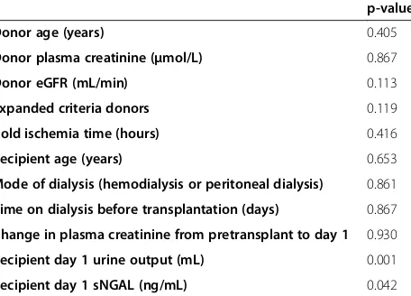
Table 9 Multivariate analysis of risk factors for prolonged delayed graft function (DGF)
p-value
Donor age (years) 0.405
Donor plasma creatinine (μmol/L) 0.867
Donor eGFR (mL/min) 0.113
Expanded criteria donors 0.119
Cold ischemia time (hours) 0.416
Recipient age (years) 0.653
Mode of dialysis (hemodialysis or peritoneal dialysis) 0.861
Time on dialysis before transplantation (days) 0.867
Change in plasma creatinine from pretransplant to day 1 0.930
Recipient day 1 urine output (mL) 0.001
Recipient day 1 sNGAL (ng/mL) 0.042
Abbreviations
AKI:Acute kidney injury; AUC: Area under the curve; CIT: Cold ischemia time; DGF: Delayed graft function; EGF: Early graft function; eGFR: Estimated glomerular filtration rate; NGAL: Neutrophil gelatinase-associated lipocalin; sNGAL: Serum neutrophil gelatinase associated lipocalin; POC: Point-of-care; SD: Standard deviation.
Competing interests
The authors declare that they have no competing interests.
Authors’contributions
MH performed the NGAL analyses, collected the data, analyzed the data, and wrote the manuscript. LK participated in designing the study and analyzing data. JM participated in designing the study, and revised the manuscript. KS participated in designing the study, acquired funding, and participated in writing the manuscript. All authors read and approved the final manuscript.
Acknowledgements
This study has been supported by The Finnish Cultural Foundation, The Finnish Medical Society Duodecim, The Paulo Foundation, The Helsinki University Hospital Research Funds, and The Finnish Kidney Foundation.
Author details
1Department of Medicine, Division of Nephrology, Helsinki University
Hospital, Haartmaninkatu 4, PL 372, Helsinki 00029, HUS, Finland. 2Department of Surgery, Division of Transplantation, Helsinki University Hospital, Helsinki, Finland.3Clinical Laboratory, Finnish Red Cross Blood Service, Helsinki, Finland.
Received: 31 December 2013 Accepted: 3 July 2014 Published: 28 July 2014
References
1. Axelsson M, Bergenfeldt M, Ohlsson K:Studies of the release and turnover of a human neutrophil lipocalin.Scand J Clin Lab Invest1995, 55:577–588.
2. Cowland JB, Borregaard N:Molecular characterization and pattern of tissue expression of the gene for neutrophil gelatinase-associated lipocalin for humans.Genomics1997,45:17–23.
3. Paragas N, Qiu A, Hollmen M, Nickolas TL, Devarajan P, Barasch J: NGAL-Siderocalin in kidney disease.Biochim Biophys Acta1823, 2012:1451–1458.
4. Mishra J, Ma Q, Prada A, Mitsnefes MM, Zahedi K, Yang J, Barasch J, Devarajan P:Identification of neutrophil gelatinase-associated lipocalin as a novel early urinary biomarker for ischemic renal injury.J Am Soc Nephrol2003,14:2534–2543.
5. Mishra J, Dent C, Tarabishi R, Mitsnefes MM, Ma Q, Kelly C, Ruff SM, Zahwedi K, Shao M, Bean J, Mori K, Barasch J, Devarajan P:Neutrophil gelatinase-associated lipocalin (NGAL) as a biomarker for acute renal injury following cardiac surgery.Lancet2005,365:1231–1238. 6. Mori K, Lee HT, Rapaport D, Drexler IR, Foster K, Yang J, Schmidt-Ott
KM, Chen X, Li JY, Weiss S, Mishra J, Cheema FH, Markowitz G, Suganami T, Sawai K, Mukoyama M, Kunis C, D’Agati V, Devarajan P, Barasch J:Endocytic delivery of lipocalin-siderophore-iron complex rescues the kidney from ischemia-reperfusion injury.J Clin Invest 2005,115:610–621.
7. Schmidt-Ott KM, Mori K, Li JY, Kalandadze A, Cohen DJ, Devarajan P, Barasch J: Dual action of neutrophil gelatinase-associated lipocalin.J Am Soc Nephrol 2007,18:407–413.
8. Giral-Classe M, Hourmant M, Cantarovich D, Dantal J, Blancho G, Daguin P, Ancelet D, Soulilou JP:Delayed graft function of more than six days strongly decreases long-term survival of transplanted kidneys.Kidney Int 1998,54:972–978.
9. Kyllönen LE, Salmela KT, Eklund BH, Halme LE, Höckerstedt KA, Isoniemi HM, Mäkisalo HJ, Ahonen J:Long-term results of 1047 cadaveric kidney transplantations with special emphasis on the initial of graft function and rejection.Transpl Int2000,13:122–128.
10. Sola R, Alarcon A, Jimenez C, Osuna A:The influence of delayed graft function.Nephrol Dial Transplant2004,19:iii32–iii37.
11. Humar A, Johnson EM, Payne WD, Wrenshall L, Sutherland DE, Najarian JS, Killingham KJ, Matas AJ:Effect of initial slow graft function on renal allograft rejection and survival.Clin Transplant1997,11:623–627. 12. Dominguez J, Lira F, Rebolledo R, Troncoso P, Aravena C, Ortiz M, Gonzalez R:
Duration of delayed graft function is an important predictor of 1-year serum creatinine.Transplant Proc2009,41:131–132.
13. Yarlagadda SD, Coca SG, Formica RN Jr, Poggio ED, Parikh CR:
Association between delayed graft function and allograft and patient survival: a systemic review and meta-analysis.Nephrol Dial Transplant 2009,24:1039–1047.
14. Johnston O, O’Kelly P, Spencer S, Donohoe J, Walshe JJ, Little DM, Hickey D, Conlon PJ:Reduced graft function (with or without dialysis) vs immediate graft function–a comparison of long-term renal allograft survival.Nephrol Dial Transplant2006,21:2270–2274.
15. Humar A, Ramcharan T, Kandaswamy R, Gillingham J, Payne WD, Matas AJ: Risk factors for slow graft function after kidney transplants; a multivariate analysis.Clin Transplant2002,16:425–429. 16. Hollmen ME, Kyllönen LE, Inkinen KA, Lalla ML, Salmela KT:Urine
Neutrophil gelatinase-associated lipocalin is a marker of graft recovery after kidney transplantation.Kidney Int2011,79:89–98.
17. Hall IE, Yarlagadda SG, Coca SG, Wang Z, Doshi M, Devarajan P, Han WK, Marcus RJ, Parikh CR:IL-18 and Urinary NGAL predict dialysis and raft recovery after kidney transplantation.J Am Soc Nephrol2010, 21:189–197.
18. Parikh CR, Jani A, Mishra J, Ma Q, Kelly C, Barasch J, Edelstein CL, Devarajan P:Urine NGAL and IL-18 are predictive biomarkers for delayed graft function following kidney transplantation.Am J Transplant2006,6:1639–1645.
19. Kusaka M, Kuroyanagi Y, Mori T, Nagaoka K, Sasaki H, Maruyama T, Hayakawa K, Shiroki R, Kurahashi H, Hoshinaga K:Serum neutrophil gelatinase-associated lipocalin as a predictor of organ recovery from delayed graft function after kidney transplantation from donors after cardiac death.Cell Transplant2008,17:129–134.
20. Lebkowska U, Malyszko J, Lebkowska A, Koc-Zorawska E, Lebkowski W, Malyszko JS, Kowalewski R, Gacko M:Neutrophil geltainase-associated lipocalin and cystatin c could predict renal outcome n patients undergoing kidney allograft transplantation.Transplant Proc2009, 41:154–157.
21. Bataille A, Abbas S, Semoun O, Bourgeois E, Marie O, Bonnet F, Resche-Rigon M, Abboud I, Losser MR, Jacob L:Plasma neutrophil gelatinase-associated lipocalin in kidney transplantation and early renal function prediction.
Transplantation2011,92:1024–1030.
22. Lee EY, Kim MS, Park Y, Kim HS:Serum neutrophil gelatinase-associated lipocalin and interleukin-18 as predictive biomarkers for delayed graft function after kidney transplantation.J Clin Lab Anal 2012,26:295–301.
23. Kusaka M, Iwamatsu F, Kuroyanagi Y, Nakaya M, Ichino M, Marubashi S, Nagano H, Shiroki R, Kurahashi H, Hoshinaga K:Serum neutrophil associated lipocalin during the early postoperative period predicts the recovery of graft function after kidney transplantation from donors after cardiac death.J Urol2012,187:2261–2267.
24. Mahdavi-Mazdeh M, Amerian M, Abdollahi A, Hatmi ZM, Khatami MR: Comparison of serum neutrophil gelatinase-associated lipocalin (NGAL) with serum creatinine in prediction of kidney recovery after renal transplantation.Int J Org Transplant Med2012,3:176–182.
25. Portal AJ, McPhail MJ, Bruce M, Coltart I, Slack A, Sherwood R, Heaton ND, Shawcross D, Wendon JA, Heneghan MA:Neutrophil gelatinase-associated lipocalin predicts acute kidney injury in patients undergoing liver transplantation.Liver Transpl2010,16:1257–1266.
26. Feldkamp T, Bienholz A, Kribben A:Urinary neutrophil gelatinase-associated lipocalin (NGAL) for the detection of acute kidney injury after orthopic liver transplantation.Nephrol Dial Transplant2011, 5:1456–1458.
27. Wagener G, Minhaz M, Mattis FA, Kim M, Emond JC, Lee HT:Urinary neutrophil gelatinase-associated lipocalin as a marker of acute kidney injury after orthopic liver transplantation.Nephrol Dial Transplant2011, 26:1717–1723.
29. Halloran PF, Aprile MA, Farewell V, Ludwin D, Smith EK, Tsai SY, Bear RA, Cole EH, Fenton SS, Cattran DC:Early function as the principal correlate of graft survival. A multivariate analysis of 200 cadaveric renal transplants treated with a protocol incorporating antilymphocyte globulin and cyclosporine.Transplantation1988,46:223–228.
30. Hollmen ME, Kyllönen LE, Inkinen KA, Lalla ML, Merenmies J, Salmela KT: Deceased donor neutrophil gelatinase-associated lipocalin and delayed graft function after kidney transplantation.Crit Care2011,15:R121.
doi:10.1186/1471-2369-15-123
Cite this article as:Hollmenet al.:Serum neutrophil gelatinase-associated lipocalin and recovery of kidney graft function after transplantation.BMC Nephrology201415:123.
Submit your next manuscript to BioMed Central and take full advantage of:
• Convenient online submission
• Thorough peer review
• No space constraints or color figure charges
• Immediate publication on acceptance
• Inclusion in PubMed, CAS, Scopus and Google Scholar
• Research which is freely available for redistribution