R E S E A R C H A R T I C L E
Open Access
Lower serum magnesium is associated with
vascular calcification in peritoneal dialysis
patients: a cross sectional study
Amber O. Molnar
1, Mohan Biyani
1, Ian Hammond
2,3, John Paul Harmon
4, Susan Lavoie
1, Brendan McCormick
1,
Manish M. Sood
1,5, Jessica Wagner
4, Elena Pena
2,3and Deborah L. Zimmerman
1,5,6*Abstract
Background:Coronary artery calcification (CAC) is highly prevalent among dialysis patients and is associated with increased cardiovascular and all cause mortality. Magnesium (Mg) inhibits vascular calcification in animal and in-vitro studies but whether the same effect occurs in humans is uncertain.
Methods:A single centre cross-sectional study of 80 prevalent peritoneal dialysis (PD) patients; on PD only for a minimum of 3 months. A radiologist blinded to patient status calculated their abdominal aortic calcification (AAC) scores on lateral lumbar spine radiographs, a validated surrogate for CAC.
Results:Eighty patients provided informed consent and underwent lumbar spine radiography. The mean serum Mg was 0.8 mmol/L (standard deviation 0.2) and mean AAC score 8.9 (minimum 0, maximum 24). A higher serum Mg level was associated with a lower AAC score (R2= 0.06, unstandardized coefficient [B] =−7.81,p= 0.03), and remained after adjustment for age, serum phosphate, serum parathyroid hormone, low-density lipoprotein cholesterol, smoking history, and diabetes (model adjustedR2= 0.36, serum Mg and AAC score B =−11.44,p= 0.00). This translates to a 0.1 mmol/L increase in serum Mg being independently associated with a 1.1-point decrease in AAC score.
Conclusions:Our findings suggest that Mg may inhibit vascular calcification. If this association is replicated across larger studies with serial Mg and vascular calcification measurements, interventions that increase serum Mg and their effect on vascular calcification warrant further investigation in the PD population.
Keywords:Magnesium, Peritoneal dialysis, Vascular calcification
Background
The mortality rate of patients on dialysis is in excess of 20% per year, with approximately one half of deaths attrib-utable to cardiovascular disease [1, 2]. Dialysis patients have a high prevalence of traditional cardiac risk factors and experience further risk due to abnormal mineral me-tabolism [1]. Studies in prevalent hemodialysis (HD) pa-tients have found that 90% of such papa-tients have coronary artery calcification (CAC), which is associated with in-creased all cause and cardiovascular mortality [2–4]. Hy-percalcemia, hyperphosphatemia, and hyperparathyroidism
have received the most attention; several studies have dem-onstrated their association with accelerated vascular calcifi-cation [5]. However, there has been comparatively little exploration of the association of serum magnesium (Mg) with vascular calcification. Both in-vitro and animal studies have found that the addition of Mg to vascular smooth muscle cells inhibits the increase in mineralization associ-ated with an osteoblastic phenotype, increases the expres-sion of anti-calcification proteins, and down regulates pathways necessary for the development of vascular calcifi-cation [6–12]. The transient receptor potential melastin (TRPM)7 cation channel as well as the Wnt/β-catenin pathway are proposed as being essential to Mg regulating vascular calcification [7, 11]. Small observational studies and pilot studies administering Mg containing phosphate binders in dialysis populations have found a significant
* Correspondence:dzimmerman@toh.on.ca
1
Division of Nephrology, Department of Medicine, McMaster University, Hamilton, ON, Canada
5Kidney Research Centre, Ottawa Hospital Research Institute, Ottawa, ON,
Canada
Full list of author information is available at the end of the article
association between lower serum Mg levels and the pro-gression of CAC, peripheral arterial calcification, mitral an-nular calcification, and atherosclerosis of the common carotid artery [13–19]. A recent study demonstrated that a higher serum Mg significantly decreased the mortality risk associated with hyperphosphatemia in HD patients [20] As well, a lower serum Mg level has been found to be associ-ated with increased mortality in both HD and PD patients [21–27]. These cumulative results suggest a possible asso-ciation between hypomagnesemia and CAC. The majority of studies have used poorly validated surrogates for CAC, [3, 28] and only one study included PD patients, [16] who are at highest risk for hypomagnesemia due to the low Mg concentration of commonly used PD solutions [29, 30]. To better characterize the relationship between serum Mg and CAC in PD patients, we performed a cross sectional study using the degree of abdominal aortic calcification (AAC) seen on a lateral lumbar spine radiograph, a validated and inexpensive surrogate for CAC [28, 31].
Methods
Dialysis prescription
Patients in the Ottawa Hospital Home Dialysis Program in Ottawa, Ontario, Canada were recruited from 2012 to 2014. These individuals are assessed routinely in a multi-disciplinary clinic every six weeks and recruitment was performed at a regular clinic visit. Included patients had to be on PD for a minimum of three months and had to be capable of providing informed consent. Pa-tients on hybrid therapy (combined HD and PD) were excluded. Because the study was of cross-sectional de-sign, all variables were measured once upon patient en-rollment. Our Home Dialysis Program exclusively used PD solutions provided by Baxter Healthcare Corporation (Deerfield, Illinois) during the study period. Solutions used included Dianeal, Extraneal, and rarely Physioneal. Information on these solutions and their composition is available at: http://www.baxter.com/healthcare_profes-sionals/products/index.html#Renal. Patients are offered a choice between continuous ambulatory peritoneal dia-lysis and continuous cyclic peritoneal diadia-lysis. The dialy-sis programs are adjusted to deliver a minimum weekly Kt/V urea of 1.7 as per Canadian Society of Nephrology guidelines [32].
Biochemical assays
At the time of enrolment, a patient’s serum calcium (Ca), phosphate (PO4), parathyroid hormone (iPTH), albumin, Mg and a non-fasting cholesterol profile were measured. All samples were analyzed in the Hospital Laboratory in accordance with the Hospital Laboratory Guidelines. Serum Ca and PO4 were analyzed with the Siemens Vista 1500 analyzer (Munich, Germany) (coefficient of variation (CV) for Ca 2.63% at 1.42 mmol/L and 2.1% at 2.5 mmol/
L; CV for PO4 3.4% at 0.6 mmol/L and 2.4% at 1.3 mmol/ L). iPTH was analyzed using the Beckman Coulter Im-munoassay (Brea, California) (CV 6.9% at 2.6 pmol/L, 6.9% at 19.9 pmol/L and 5.8% at 59.3 pmol/L). Serum al-bumin was analyzed with the Dimension Vista (Siemens, Munich, Germany) system using an adaptation of the bro-mocresol purple dye binding method (CV 2.5% at 3.2 g/ dL). Serum Mg was analyzed with the Dimension Vista system using a modification of the methylthymol blue complexometric procedure (CV 3% at 0.78 mmol/L and 2% at 1.93 mmol/L). Serum total cholesterol, triglycerides and high-density lipoprotein cholesterol (HDL) were mea-sured using the Dimension Vista system (CHOL, TRIG and HDLC methods respectively). Low-density lipoprotein cholesterol (LDL) was calculated using Friedewald’s for-mula. Residual renal function (RRF) was calculated from a 24-h urine collection. Urine creatinine and urine urea were measured; the average of the creatinine clearance and urea clearance was taken to calculate the estimated glomerular filtration rate (eGFR) in mL/min.
Vascular calcification
The AAC score was calculated using a lateral lumbar spine radiograph as described by Kauppila et al. from their assessment of 617 Framingham heart study participants [31]. The extent of the calcification of the anterior and posterior aortic wall was graded at each vertebral level from L1 to L4 on a 0–3 scale, yielding three different com-posite scores. Of these, the antero-posterior severity score, which ranges from 0 to 24 and has the highest inter-rater correlation (intra-class correlation (ICC) 0.93–0.96), was used in our study [31]. This method of determining ab-dominal aortic calcification has been found to be a valid surrogate for assessing CAC (area under the curve 0.78) [28, 31]. Each patient had one radiograph performed shortly after his or her enrolment. A radiologist who was blinded to each patient’s status interpreted all radiographs. A second independent, blinded radiologist interpreted the abdominal radiographs to confirm the findings of the first radiologist (ICC = 0.99 (95% confidence interval 0.98– 0.99)). The ICC was calculated using a two-way mixed ef-fects model with an absolute agreement definition.
Statistical analysis
a p-value ≤0.2 on univariate regression were adjusted for in the multiple linear regression model. We also performed a multiple linear regression analysis where all pre-specified variables were maintained in the model. Five patients had missing LDL cholesterol values (n= 1, no test was performed; n= 4, triglycer-ides were too high to calculate LDL). Multiple imput-ation (SPSS automatic imputimput-ation method) was used to impute missing values for LDL in the multiple linear re-gression analysis. SPSS automatically chooses an imput-ation method that is most appropriate for the data and uses linear regression to impute missing continuous vari-ables. Five imputed datasets were created and the esti-mates from each dataset were pooled.
We performed a sensitivity analysis where patients with an aortic calcification score of 0 were removed from the analysis. Patients with very little or no vascular calcifica-tion do not tend to develop vascular calcificacalcifica-tion over time and seem to represent a different subgroup from the general end stage renal disease population [33, 34]. For this reason, we performed a separate analysis to determine the impact upon the overall results. All analyses were per-formed using SPSS Statistics version 24. The reporting of this study follows the STROBE guidelines for observa-tional studies [35].
Results
Baseline characteristics
Eighty six patients provided informed consent; 80 patients completed a lateral lumbar X-ray and were included in the primary analysis. Patient selection is outlined in Fig. 1. The baseline characteristics of included patients are
outlined in Table 1. The mean age of patients was 62.8 years, 56 patients (70%) were male, 34% were diabetic and 33% were lifelong non-smokers. Mean serum Mg was 0.84 mmol/L (normal range 0.74 to 1.03 mmol/L) and mean AAC score 8.9 (minimum score 0, maximum score 24). The mean serum PO4 level was 1.70 mmol/L and mean LDL cholesterol 2.02 mmol/L. The median time on dialysis was 1.3 years (IQR 0.6 to 3.0). Twenty six patients (32.5%) had a low serum Mg (defined by <0.74 mmol/L), 42 patients (52.5%) had a normal serum Mg and 12 pa-tients (15.0%) had a high serum Mg (defined as >1.03 mmol/L). Most patients (77.5%) were on ambulatory peritoneal dialysis (APD). Included patients used the following PD fluid combinations: 1. Dianeal only in 19 (23.8%) patients, 2. Extraneal only in 4 (5.0%) patients, 3. Dianeal and Extraneal in 54 (67.5%) patients, 4. Physioneal and Extraneal in 3 (3.8%) patients.
Univariate regression
On univariate regression, age, serum phosphate, serum iPTH, LDL cholesterol, smoking history, diabetes, and Fig. 1Patient selection. *These patients consented to participate
but then failed to complete the required lumbar x-ray despite multiple reminders
Table 1Baseline characteristics
TotalN= 80
Age 62.8 (12.8) Sex (male) 56 (70.0) BMI (kg/metre squared) 28.33 (5.6)a iPTH (pmol/L) 34.0 (28.6) Serum Calcium (mmol/L) 2.3 (0.2) Serum Phosphate (mmol/L) 1.7 (0.4) LDL cholesterol (mmol/L) 2.0 (0.8)b Time on dialysis (years) 1.3 (0.6, 3.0)c Lifelong non-smoker 26 (32.5) Diabetes 27 (33.8) Serum Mg (mmol/L) 0.84 (0.2) Number of patients with low serum Mg 26 (32.5) Serum Albumin (g/L) 32.4 (4.5) nPCR (g/kg/day) 0.8 (0.2) RRF (mL/min) 4.7 (3.8) Number of patients on a cholesterol lowering medication (statin or ezetemibe)
55 (68.8) Number of patients on APD 62 (77.5) Aortic calcification score 8.9 (6.9)
Continuous measurements presented as mean (standard deviation (SD)), other variables presented as N (%) unless otherwise specified
BMIbody mass index,iPTHparathyroid hormone,LDLlow density lipoprotein, Mgmagnesium,nPCRnormalized catabolic rate,RRFResidual renal function, APDautomated peritoneal dialysis (the remainder of patients were on continuous ambulatory peritoneal dialysis)
a
Mean value for 79 patients (1 patient had a missing value)
b
Mean value for 75 patients (5 patients had values that were missing or could not be calculated)
c
serum Mg had a pvalue≤0.2. Older age, a higher serum PO4, a lower serum Mg and a history of diabetes were significantly associated with an increased AAC score (p< 0.05). Higher LDL cholesterol was associated with a decreased AAC score (p= 0.03) (Table 2).
Multiple linear regression
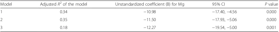
After adjustment for age, serum phosphate, serum iPTH, LDL cholesterol, smoking history, and diabetes, a lower serum Mg level was independently associated with a higher AAC score (Unstandardized coefficient [B] =−10.98, p= 0.000). This statistic translates to a 0.1 mmol/L increase in serum Mg being associated with a 1.1-point decrease in AAC score. When all pre-specified variables (Table 2) were maintained in the model regardless of the univariate analysispvalue, the results were similar (B for Mg =−11.50, p= 0.000). When patients with an AAC score of 0 were excluded, n= 68 patients included in the analysis, the results were similar (B for Mg =−12.27, p= 0.001) (Table 3). Testing assumptions of the linear regression model can be found in Additional file 1.
Discussion
Our data demonstrate that in PD patients, a lower serum Mg is independently associated with an increased AAC score. We found that a 0.1 mmol/L increase in serum Mg is associated with a 1.1-point decrease in AAC score. This suggests that Mg may act as a possible inhibitor of vascu-lar calcification.
Our study results are consistent with previously pub-lished observational and pilot studies in the dialysis and chronic kidney disease populations [9, 13–19]. A recent co-hort study with a maximum follow up of 10.8 years found that a lower serum Mg was associated with increased mor-tality in PD patients [23]. Several other studies have demon-strated an association between lower serum Mg and mortality among dialysis patients [21, 22, 24–27] This asso-ciation could potentially be attributable to low serum Mg causing accelerated vascular calcification. Increasing data from in-vitro and animal studies support the assertion that Mg acts as an inhibitor of vascular calcification [6–12]. The addition of Mg to vascular smooth muscle cells increases the expression of anti-calcification proteins, such as matrix G1a, bone morphogenetic protein-7 and osteopontin, and
Table 2Univariate linear regression
Variable R2 Unstandardized coefficient (B) 95% confidence interval (CI) Pvalue
Age 0.17 0.22 0.11, 0.33 0.00
Sex 0.005 −1.04 −4.41, 2.33 0.54
iPTH 0.02 0.03 −0.02, 0.09 0.23
Calcium 0.00 0.04 −8.55, 8.63 0.99 Phosphate 0.07 4.52 0.91, 8.13 0.02 LDLa 0.07 −2.19 −4.09,−0.29 0.03 Time on dialysis 0.02 −0.40 −1.10, 0.30 0.26 Smoking history 0.03 2.53 −0.73, 5.79 0.13 Diabetes 0.06 3.51 0.33, 6.68 0.03 Albumin 0.004 −0.10 −0.45, 0.24 0.56 RRF 0.001 −0.05 −0.46, 0.37 0.83 Magnesium 0.06 −7.81 −15.01,−0.61 0.03
Smoking history (ex or current vs non-smoker) iPTHintact parathyroid hormone
LDLlow-density lipoprotein RRFResidual renal function
a
75 patients included in the analysis for LDL (5 patients had values that were missing or could not be calculated). 80 patients included for all other variables
Table 3Multiple linear regression: the independent association of serum Mg with vascular calcification
Model AdjustedR2of the model Unstandardized coefficient (B) for Mg 95% CI Pvalue 1 0.34 −10.98 −17.40,−4.56 0.000 2 0.35 −11.50 −17.93,−5.06 0.000 3 0.18 −12.27 −19.54,−5.00 0.001
Model 1: Adjusted for age, serum phosphate, LDL cholesterol, iPTH, smoking history, and diabetes Model 2: All pre-specified variables in Table2were included in the model
Model 3: Adjusted for the variables in model 1; patients with an aortic calcification score of 0 (N= 12) excluded from the analysis
inhibits an osteoblastic transformation [7, 11]. As well, the addition of Mg to vascular smooth muscle cells down-regulates the Wnt/β-catenin pathway. This pathway is es-sential for the osteogenic transformation of pluripotent mesenchymal cells and is activated during the development of vascular calcification [11]. The mechanism by which Mg regulates vascular calcification may involve the transient re-ceptor potential melastin (TRPM)7 cation channel as inhib-ition of TRPM7 negates the anti-calcification effects of Mg [7, 11]. Mg may also inhibit vascular calcification by sup-pressing PTH, which has been found in animal models to increase vascular calcification [36, 37]. Among PD patients, an inverse correlation between PTH and serum Mg, inde-pendent of Ca concentration, has been demonstrated in several studies [38–41]. However, in our cohort of PD pa-tients, we did not find a significant, independent association between serum Mg and PTH (data not shown). To our knowledge, our study is the largest thus far in the PD popu-lation (previously published PD study n= 44; outcome of peripheral arterial calcification [16]), and the largest study with a vascular calcification outcome that is a validated sur-rogate for CAC.
AAC is a valid surrogate marker as it correlates with CAC and is associated with increased all-cause and cardio-vascular mortality [28, 42, 43]. Among diabetic patients, re-ported hazard ratios for all-cause and cardiovascular mortality were 1.7 and 1.9 respectively when the AAC score was examined as a continuous variable [42]. Cardiovascular mortality has also been found to increase in a graded fash-ion with increasing tertile of AAC score [43].
Our study population had a mean serum Mg of 0.84 mmol/L, and over 32% of our cohort had low serum Mg (defined as <0.74 mmol/L). The mean serum Mg was similar in a study by Fein et al. (0.8 mmol/L) examining the association of serum Mg with mortality in PD patients [23]. Comparatively, studies examining serum Mg in an HD population found a higher mean Mg level of 1.14 mmol/L [24], 0.92 mmol/L [26], and 0.86 mmol/L [21]. The high prevalence of hypomagnesemia in our cohort of PD pa-tients could be due to the continuous nature of PD, coupled with the low magnesium concentration of com-monly used PD solutions (Mg concentration ranging from 0.25 to 0.75 mmol/L). Patients on automated PD overnight using a PD solution containing 0.25 mmol/L of Mg along with a day time dwell of an icodextrin so-lution, (the most commonly used regimen at our insti-tution), have an overall transperitoneal Mg loss of 3.26 mmol per 24 h [29]. Magnesium losses in the di-alysate are compounded by the significant restrictions of a renal diet. Taken together, this highlights the unique risk of hypomagnesermia in PD patients, mak-ing them an ideal population for testmak-ing interventions targeted at increasing serum Mg and examining the ef-fect on vascular calcification.
Our study has some important limitations. Due to the cross-sectional nature of the study, we can only determine association and not causation. Residual confounding is pos-sible. However, we were able to adjust for important con-founders associated with vascular calcification, such as serum PO4 and age [3], and our findings were consistent across unadjusted, adjusted and sensitivity analyses. A low serum Mg may be a marker of generalized malnutrition [44, 45] and inflammation, which are both associated with increased vascular calcification [46, 47]. We did not directly measure any inflammatory markers; however, albumin is a recognized surrogate for inflammation and malnutrition. On univariate analysis, albumin was not associated with the AAC score, and the results of the multivariate analysis were not attenuated upon adjustment for albumin. We only measured serum Mg at one time point; it is possible that a single measurement may not be reflective of an individual’s overall Mg status. The imaging technique used in our study did not allow us to reliably differentiate between medial (AIM) and intimal calcification (AIC). Both types of vascular calcification occur commonly in dialysis pa-tients, often co-existing in the same patient, and are as-sociated with increased mortality. However, among dialysis patients, AIC has been found to be associated with worse survival when compared to AIM. As well, the clinical consequences of AIC and AIM differ. While AIC represents advanced atherosclerosis that is associ-ated with the development of plaques and occlusive dis-ease, AIM causes arterial stiffness, increased pulse pressure and left ventricular hypertrophy [4, 48, 49].
Conclusions
In conclusion, our results support the assertion that Mg may inhibit vascular calcification, a condition that is highly prevalent in the dialysis population and is associated with increased mortality [2–4]. If our re-sults can be duplicated in large observational studies with repeated serum Mg and vascular calcification measurements, interventions, such as Mg supplemen-tation in hypomagnesemic patients, the use of Mg based phosphate binders, or the use of PD solutions with a higher Mg concentration and their effect on vascular calcification warrant testing in the PD popu-lation. Such interventions would be easy to administer and would carry minimal side effects.
Additional file
Additional file 1:Testing assumptions of the linear regression model. (DOCX 101 kb)
Abbreviations
PD: Peritoneal dialysis; PO4: Phosphate; TRPM7: The transient receptor potential melastin
Acknowledgements
AOM received salary support from the KRESCENT Foundation. AOM, BM and DZ receive salary support form the University of Ottawa Department of Medicine.
The results presented in this paper have not been published previously in whole or part, except in abstract form.
Funding
None to declare.
Availability of data and materials
The data used and/or analyzed during the current study are available from the corresponding author upon reasonable request.
Authors’contributions
M.B. and S.L. contributed to revising the article and the final approval of the version to be published. I.H. read patient radiographs, revised and contributed to the article and had final approval of the version to be published. EPF read patient radiographs, revised and contributed to the article and had final approval of the version to be published. B.M. revised the article, provided intellectual content and had final approval of the version to be published. M.M.S. helped with the analysis and interpretation of the data, revising the article and provided final approval of the version to be published. J.W. and J.P.H. helped with the collection of data, revising the manuscript and final approval of the version to be published. D.L.Z. and A.O.M. helped with the conception, design, analysis and interpretation of the data, drafting and revising of the article, providing intellectual content and final approval of the version to be published.
Competing interests
BM has the following disclosures that are unrelated to this submission: Baxter-consulting, speaker board, research grant; Concert Pharmaceuticals-consulting; Sanofi- Advisory board. DZ has the following disclosure unrelated to this submission: Baxter- research grant. All other authors declare that they have no competing interests.
Consent for publication
Not applicable.
Ethics approval and consent to participate
Our study was conducted according to a pre-specified protocol approved by the Institutional Review Board at the Ottawa Hospital
Research Institute (20120175-01H)and followed the Declaration of Helsinki. All patients provided informed consent.
Publisher’s Note
Springer Nature remains neutral with regard to jurisdictional claims in published maps and institutional affiliations.
Author details
1Division of Nephrology, Department of Medicine, McMaster University,
Hamilton, ON, Canada.2Department of Radiology, University of Ottawa,
Ottawa, ON, Canada.3Department of Medical Imaging, The Ottawa Hospital Ottawa, Ottawa, ON, Canada.4Division of Nephrology, Department of
Medicine, Northern Ontario School of Medicine, Sudbury, ON, Canada.
5Kidney Research Centre, Ottawa Hospital Research Institute, Ottawa, ON,
Canada.6The Ottawa Hospital, Riverside Campus 1967 Riverside Drive, Ottawa, ON, CanadaK1H 7W9.
Received: 8 December 2015 Accepted: 3 April 2017
References
1. Block GA, Klassen PS, Lazarus JM, Ofsthun N, Lowrie EG, Chertow GM. Mineral metabolism, mortality, and morbidity in maintenance hemodialysis. J Am Soc Nephrol. 2004;15:2208–18.
2. Parfrey PS, Foley RN. The clinical epidemiology of cardiac disease in chronic renal failure. J Am Soc Nephrol. 1999;10:1606–15.
3. Ketteler M, Biggar PH. Review article: getting the balance right: assessing causes and extent of vascular calcification in chronic kidney disease. Nephrology (Carlton). 2009;14:389–94.
4. London GM, Guerin AP, Marchais SJ, Metivier F, Pannier B, Adda H. Arterial media calcification in end-stage renal disease: impact on all-cause and cardiovascular mortality. Nephrol Dial Transplant. 2003;18:1731–40. 5. Kalpakian MA, Mehrotra R. Vascular calcification and disordered mineral
metabolism in dialysis patients. Semin Dial. 2007;20:139–43. 6. Inagaki O, Syono T, Nakagawa K, Nishian Y, Takenaka Y, Takamitsu Y.
Influence of magnesium deficiency on concentration of calcium in soft tissue of uremic rats. Ren Fail. 1996;18:847–54.
7. Montezano AC, Zimmerman D, Yusuf H, Burger D, Chignalia AZ, Wadhera V, van Leeuwen FN, Touyz RM. Vascular smooth muscle cell differentiation to an osteogenic phenotype involves TRPM7 modulation by magnesium. Hypertension. 2010;56:453–62.
8. Kircelli F, Peter ME, Sevinc Ok E, Celenk FG, Yilmaz M, Steppan S, Asci G, Ok E, Passlick-Deetjen J. Magnesium reduces calcification in bovine vascular smooth muscle cells in a dose-dependent manner. Nephrol Dial Transplant. 2012;27:514–21.
9. Salem S, Bruck H, Bahlmann FH, Peter M, Passlick-Deetjen J, Kretschmer A, Steppan S, Volsek M, Kribben A, Nierhaus M, et al. Relationship between magnesium and clinical biomarkers on inhibition of vascular calcification. Am J Nephrol. 2012;35:31–9.
10. Louvet L, Buchel J, Steppan S, Passlick-Deetjen J, Massy ZA. Magnesium prevents phosphate-induced calcification in human aortic vascular smooth muscle cells. Nephrol Dial Transplant. 2013;28:869–78.
11. Montes de Oca A, Guerrero F, Martinez-Moreno JM, Madueno JA, Herencia C, Peralta A, Almaden Y, Lopez I, Aguilera-Tejero E, Gundlach K, et al. Magnesium inhibits Wnt/beta-catenin activity and reverses the osteogenic transformation of vascular smooth muscle cells. PLoS One. 2014;9:e89525. 12. Xu J, Bai Y, Jin J, Zhang J, Zhang S, Cui L, Zhang H. Magnesium modulates
the expression levels of calcification-associated factors to inhibit calcification in a time-dependent manner. Exp Ther Med. 2015;9:1028–34.
13. Ishimura E, Okuno S, Kitatani K, Tsuchida T, Yamakawa T, Shioi A, Inaba M, Nishizawa Y. Significant association between the presence of peripheral vascular calcification and lower serum magnesium in hemodialysis patients. Clin Nephrol. 2007;68:222–7.
14. Tzanakis I, Pras A, Kounali D, Mamali V, Kartsonakis V, Mayopoulou-Symvoulidou D, Kallivretakis N. Mitral annular calcifications in haemodialysis patients: a possible protective role of magnesium. Nephrol Dial Transplant. 1997;12:2036–7. 15. Tzanakis I, Virvidakis K, Tsomi A, Mantakas E, Girousis N, Karefyllakis N,
Papadaki A, Kallivretakis N, Mountokalakis T. Intra- and extracellular magnesium levels and atheromatosis in haemodialysis patients. Magnes Res. 2004;17:102–8.
16. Meema HE, Oreopoulos DG, Rapoport A. Serum magnesium level and arterial calcification in end-stage renal disease. Kidney Int. 1987;32:388–94. 17. Spiegel DM, Farmer B. Long-term effects of magnesium carbonate on
coronary artery calcification and bone mineral density in hemodialysis patients: a pilot study. Hemodial Int. 2009;13:453–9.
18. Turgut F, Kanbay M, Metin MR, Uz E, Akcay A, Covic A. Magnesium supplementation helps to improve carotid intima media thickness in patients on hemodialysis. Int Urol Nephrol. 2008;40:1075–82. 19. Tzanakis IP, Stamataki EE, Papadaki AN, Giannakis N, Damianakis NE,
Oreopoulos DG. Magnesium retards the progress of the arterial calcifications in hemodialysis patients: a pilot study. Int Urol Nephrol. 2014;46:2199–205.
20. Sakaguchi Y, Fujii N, Shoji T, Hayashi T, Rakugi H, Iseki K, Tsubakihara Y, Isaka Y, Committee of Renal Data Registry of the Japanese Society for Dialysis T. Magnesium modifies the cardiovascular mortality risk associated with hyperphosphatemia in patients undergoing hemodialysis: a cohort study. PLoS One. 2014;9, e116273.
21. Li L, Streja E, Rhee CM, Mehrotra R, Soohoo M, Brunelli SM, Kovesdy CP, Kalantar-Zadeh K. Hypomagnesemia and mortality in incident hemodialysis patients. Am J Kidney Dis. 2015;66:1047–55.
22. de Roij van Zuijdewijn CL, Grooteman MP, Bots ML, Blankestijn PJ, Steppan S, Buchel J, Groenwold RH, Brandenburg V, van den Dorpel MA, Ter Wee PM, et al. Serum magnesium and sudden death in European hemodialysis patients. PLoS One. 2015;10, e0143104.
24. Ishimura E, Okuno S, Yamakawa T, Inaba M, Nishizawa Y. Serum magnesium concentration is a significant predictor of mortality in maintenance hemodialysis patients. Magnes Res. 2007;20:237–44.
25. Joao Matias P, Azevedo A, Laranjinha I, Navarro D, Mendes M, Ferreira C, Amaral T, Jorge C, Aires I, Gil C, et al. Lower serum magnesium is associated with cardiovascular risk factors and mortality in haemodialysis patients. Blood Purif. 2014;38:244–52.
26. Lacson Jr E, Wang W, Ma L, Passlick-Deetjen J. Serum magnesium and mortality in hemodialysis patients in the United States: a cohort study. Am J Kidney Dis. 2015;66:1056–66.
27. Cai K, Luo Q, Dai Z, Zhu B, Fei J, Xue C, Wu D. Hypomagnesemia is associated with increased mortality among peritoneal dialysis patients. PLoS One. 2016;11, e0152488.
28. Bellasi A, Ferramosca E, Muntner P, Ratti C, Wildman RP, Block GA, Raggi P. Correlation of simple imaging tests and coronary artery calcium measured by computed tomography in hemodialysis patients. Kidney Int. 2006;70:1623–8. 29. Eddington H, Hurst H, Ramli MT, Speake M, Hutchison AJ. Calcium and
magnesium flux in automated peritoneal dialysis. Perit Dial Int. 2009;29:536–41. 30. Ejaz AA, McShane AP, Gandhi VC, Leehey DJ, Ing TS. Hypomagnesemia in
continuous ambulatory peritoneal dialysis patients dialyzed with a low-magnesium peritoneal dialysis solution. Perit Dial Int. 1995;15:61–4. 31. Kauppila LI, Polak JF, Cupples LA, Hannan MT, Kiel DP, Wilson PW. New indices
to classify location, severity and progression of calcific lesions in the abdominal aorta: a 25-year follow-up study. Atherosclerosis. 1997;132:245–50.
32. Blake PG, Bargman JM, Brimble KS, Davison SN, Hirsch D, McCormick BB, Suri RS, Taylor P, Zalunardo N, Tonelli M, et al. Clinical practice guidelines and recommendations on peritoneal dialysis adequacy 2011. Perit Dial Int. 2011;31:218–39.
33. Bellasi A, Kooienga L, Block GA, Veledar E, Spiegel DM, Raggi P. How long is the warranty period for nil or low coronary artery calcium in patients new to hemodialysis? J Nephrol. 2009;22:255–62.
34. Block GA, Spiegel DM, Ehrlich J, Mehta R, Lindbergh J, Dreisbach A, Raggi P. Effects of sevelamer and calcium on coronary artery calcification in patients new to hemodialysis. Kidney Int. 2005;68:1815–24.
35. von Elm E, Altman DG, Egger M, Pocock SJ, Gotzsche PC, Vandenbroucke JP, Initiative S. The Strengthening the Reporting of Observational Studies in Epidemiology (STROBE) statement: guidelines for reporting observational studies. Lancet. 2007;370:1453–7.
36. Coen G. Calcimimetics, parathyroid hormone, and vascular calcification in chronic kidney disease. Kidney Int. 2008;74:1229–31.
37. Neves KR, Graciolli FG, dos Reis LM, Graciolli RG, Neves CL, Magalhaes AO, Custodio MR, Batista DG, Jorgetti V, Moyses RM. Vascular calcification: contribution of parathyroid hormone in renal failure. Kidney Int. 2007;71:1262–70. 38. Navarro JF, Mora C, Garcia J, Macia M, Gallego E, Chahin J, Mendez ML,
Rivero A. Hypermagnesemia in CAPD. Relationship with parathyroid hormone levels. Perit Dial Int. 1998;18:77–80.
39. Navarro JF, Mora C, Macia M, Garcia J. Serum magnesium concentration is an independent predictor of parathyroid hormone levels in peritoneal dialysis patients. Perit Dial Int. 1999;19:455–61.
40. Saha HH, Harmoinen AP, Pasternack AI. Measurement of serum ionized magnesium in CAPD patients. Perit Dial Int. 1997;17:347–52. 41. Wei M, Esbaei K, Bargman JM, Oreopoulos DG. Inverse correlation between
serum magnesium and parathyroid hormone in peritoneal dialysis patients: a contributing factor to adynamic bone disease? Int Urol Nephrol. 2006;38:317–22. 42. Cox AJ, Hsu FC, Agarwal S, Freedman BI, Herrington DM, Carr JJ, Bowden
DW. Prediction of mortality using a multi-bed vascular calcification score in the Diabetes Heart Study. Cardiovasc Diabetol. 2014;13:160.
43. Wilson PW, Kauppila LI, O'Donnell CJ, Kiel DP, Hannan M, Polak JM, Cupples LA. Abdominal aortic calcific deposits are an important predictor of vascular morbidity and mortality. Circulation. 2001;103:1529–34.
44. Martin-del-Campo F, Batis-Ruvalcaba C, Gonzalez-Espinoza L, Rojas-Campos E, Angel JR, Ruiz N, Gonzalez J, Pazarin L, Cueto-Manzano AM. Dietary micronutrient intake in peritoneal dialysis patients: relationship with nutrition and inflammation status. Perit Dial Int. 2012;32:183–91. 45. Ye H, Zhang X, Guo Q, Huang N, Mao H, Yu X, Yang X. Prevalence and
factors associated with hypomagnesemia in Southern Chinese continuous ambulatory peritoneal dialysis patients. Perit Dial Int. 2013;33:450–4. 46. An WS, Son YK. Vascular calcification on plain radiographs is associated with
carotid intima media thickness, malnutrition and cardiovascular events in dialysis patients: a prospective observational study. BMC Nephrol. 2013;14:27.
47. Wang AY. Vascular and valvular calcification in chronic peritoneal dialysis patients. International journal of nephrology. 2011;2011:198045. 48. Nakamura S, Ishibashi-Ueda H, Niizuma S, Yoshihara F, Horio T, Kawano Y.
Coronary calcification in patients with chronic kidney disease and coronary artery disease. Clin J Am Soc Nephrol. 2009;4:1892–900.
49. Amann K. Media calcification and intima calcification are distinct entities in chronic kidney disease. Clin J Am Soc Nephrol. 2008;3:1599–605.
• We accept pre-submission inquiries
• Our selector tool helps you to find the most relevant journal
• We provide round the clock customer support
• Convenient online submission
• Thorough peer review
• Inclusion in PubMed and all major indexing services
• Maximum visibility for your research
Submit your manuscript at www.biomedcentral.com/submit