W
Wiinntteerr22001166,,VVooll55,,NNoo11 D
DOOII::1100..2222008888//aaccaaddppuubb..BBUUMMSS..55..11..1199
The Genotoxic and Cytotoxic Effects of Bisphenol-A (BPA) in
MCF-7 Cell Line and Amniocytes
Seyed Mohsen Aghajanpour-Mir1,2, Ebrahim Zabihi1, Haleh Akhavan-Niaki1, Elahe Keyhani2, Iman
Bagherizadeh3, Sajjad Biglari2, Farkhondeh Behjati2,3
1.Cellular & Molecular Biology Research Center, Health Institute, Babol University of Medical Sciences,
Babol, Iran.
2. Genetics Research Center, University of Social Welfare and Rehabilitation Sciences, Tehran, Iran.
3. Department of Medical Genetics and Sarem Cell Research Center (SCRC), Sarem Women’s Hospital, Tehran, Iran.
Bisphenol-A (BPA) is an industrial xenoestrogen used widely in our living environment. Recently, several
studies suggested that BPA has destructive effects on DNA and chromosomes in normal body cells via estrogen
receptors (ER). Therefore, BPA could be considered as an important mediator in many diseases such as cancer.
However, there are still many controversial issues which need clarification. In this study, we investigated the
BPA-induced chromosomal damages in MCF-7 cell line, ER-positive and negative amniocyte cells. Cytotoxicity
and genotoxicity effects of BPA were also compared between these three cell groups. Expression of estrogen
receptors was determined using immunocytochemistry technique. The cell cytotoxicity of BPA was measured by
MTT assay. Classic cytogenetic technique was carried out for the investigation of chromosome damage. BPA, in
addition to cytotoxicity, had remarkable genotoxicity at concentrations close to the traceable levels in tissues or
biological fluids. Although some differences were observed in the amount of damages between ER-positive and
negative fetal cells, interestingly, these differences were not significant. The present study showed that BPA
could lead to chromosomal aberrations in both ER-dependent and independent pathways at some concentrations
or in cell types yet not reported. Also, BPA could probably be considered as a facilitator for some predisposed
cells to be cancerous by raising the chromosome instability levels. Finally, estrogen receptor seems to have a
different role in cytotoxicity and genotoxicity effects.
Key words: Bisphenol-A (BPA), estrogen receptor, MCF-7, amniocyte, chromosome abnormality, classic
cytogenetics
Correspondence: Genetics Research Center, University of Social Welfare and Rehabilitation Sciences, Tehran, Iran. Email: f_behjati@uswr.ac.ir
isphenol-A (BPA) is an industrial
xeno-estrogene which is widely used in the
production of polycarbonate plastics, drink
containers, baby bottles, epoxy resin lining of food
containers, medical devices and dental sealants.
BPA is an organic colorless solid compound, with 8
billion pounds yearly production and one hundred
tones releasing in atmosphere in 2010 which is
increased to 15 billion pounds yearly production
and probably more than 200 tones releasing in
B
Submmited 31 May 2015; Accepted 6 September 2015; Published 18 March 2016
atmosphere per year during recent years (1-2). BPA
has also been detected in a variety of environmental
samples, including water, dust, sewage, indoor and
outdoor air samples (3). In the last decade, several
studies investigated the hazardous effects of BPA
which has probably been associated with diabetes,
cardiovascular disease, neurobehavioral disorders,
recurrent miscarriages, abnormal karyotypes,
poly-cystic ovarian syndrome, reproductive
impair-ments and cancer (4-14). In 1960, the first
chromosomal abnormality associated with cancer
was reported using cytogenetics techniques in
patients with chronic myeloid leukemia (15).
Genomic instability and chromosomal
abnorma-lities are well-known common features of cancer
(16-17).
Recent studies have strongly suggested that
DNA damage induced by xenoestrogens and
estrogen is dependent on estrogen receptors (ERs)
(3, 18-19). An in vitro study has indicated the effect
of estradiol on radiation-induced chromosome
aberrations in human peripheral lymphocytes (20).
BPA is considered as an estrogenic endocrine
disrupting chemical which exhibits estrogen-like
activity (21). BPA binds to ERs that could promote
breast cancer (22).
To date many studies have indicated
controversial issues concerning chromosomal
aberrations induced by BPA. Although some
studies suggested that BPA cannot have a genotoxic
effect, some others suggested that BPA exposure
can lead to chromosomal abnormalities such as
aneuploidy through disruption of meiotic process
(23-24) and also genomic structural aberrations like
DNA breakage (25). Recent studies have
demonstrated that BPA impairs the double-strand
break repair machinery in the germline and causes
chromosome abnormalities (26). Furthermore, BPA
induces synaptic defects, such as end-to-end
chromosome associations and asynapsis (27).
Nevertheless, it seems that there are no strong
evidences to favor BPA as a genotoxic agent in low
concentrations which are probably traceable in
human biologic fluids or tissues.
Investigating direct genotoxic effects of BPA
on chromosomes, necessitates the exclusion of
secondary genotoxic effects of BPA which may
occur subsequent to its cytotoxic effects such as
apoptosis or necrosis. For this purpose, we used
classic cytogenetics method. As cells affected by
high cytotoxicity could not be prepared to enter the
metaphase stage, then, these kinds of cells will
automatically be removed from genotoxicity
evaluation of BPA.
To date, different results have been obtained
from the study of BPA toxic effects on different cell
groups (28). In the present study, we selected
MCF-7 cell line which seems to be a suitable
representative cell line from breast as one of the
main target tissues of BPA. MCF-7 is an ER
positive cancerous epithelial cell, with immortal
features and high proliferation potential like other
cancerous cells (29). These features as well as its
high endurance potential to the toxic agents make
this cell line a good monitoring system to detect
chromosomal aberrations.
The effects of environmental pollutants on
fetuses are an important health issue and
amniocytes seem to be accessible and suitable
representative of fetal cells. Amniocytes are less
differentiated cells with higher proliferation
potential compared to differentiated cells.
For the investigation of BPA effects on
normal cell population, we have selected ER
negative and positive amniocytes, derived from
human male and female fetal amnion cells,
respectively (30). To the best of our knowledge,
there is no other similar study on amniocytes.
Materials and methods
Human amniocytes and MCF-7 cell culture
MCF-7 cell line was obtained from Pasteur
institute, Tehran, Iran. These cells were cultured in
RPMI-1640 medium (PAA, Austria) supplemented
with 10% fetal bovine serum and 1% antibiotics
(penicillin/streptomycin) (Invitrogen, USA) in a
humidified atmosphere containing 5% CO2 at 37
°C. Cytogenetically normal human amniocytes
were obtained from discardable cell cultures
belonging to individuals referred to Sarem Hospital
(Tehran, Iran), for some prenatal diagnosis tests.
The names and specifications of the subjects were
not available for the authors. This project has been
approved by the ethical committee of University of
Social welfare and Rehabilitation Sciences.
Cell culture on cover slip and
immunocytoche-mistry (ICC)
Immunocytochemistry (ICC) was performed
in order to check the expression of estrogen
receptors in amniocytes. MCF-7 cells were used as
an ER-positive control sample.
For performing ICC staining, about 3×10⁴
male and female amniocytes and also MCF-7 were
cultured on sterile coverslips. Cells were then fixed
and permeabilized with acetone and incubated with
mouse anti human estrogen receptor primary
antibody (Clone 1D5, Dako, Denmark) for 1 h at
room temperature. After that, cells were washed
three times with PBS and then incubated with
secondary antibodies which was conjugated
with Horse Radish Peroxidase (HRP) (Real
envision Dako, Denmark), for 30 min at room
temperature. Coverslips were washed and covered
with chromogen and 3,3'-Diaminobenzidine
(DAB) solution (DAKO, Denmark). Hematoxylin
was used for counterstaining followed by
alcohol and xylene for dehydration. Finally, the
coverslips were mounted on slides using mounting
media.
Cell cytotoxicity evaluation using MTT assay
MTT
(3-(4,5-dimethylthiazol-2-yl)-2,5-diphe-nyltetrazolium bromide) assay was done in order to
determine the half maximal inhibitory
concentration (IC50) of BPA in each cell type and
hence the determination of suitable BPA
concentrations for the exposure of cell cultures.
MCF-7 and amniocyte cells were seeded in
complete medium in a 96-well plate at a density of
8x10³ and 1x10⁴ cells per well, respectively. After
24 h incubation in 5% CO2 at 37 °C, culture media
were replaced by new media containing different
concentrations of BPA (0 or control, 0.4, 1, 4, 40, 100 and 400 μg/ml) and cells were incubated for 48 h. After this time period, 100 μl of 5 mg/ml concentrated MTT or tetrazolium salt was added to
each well and then the plates were incubated in 5%
CO2 at 37 oC for 4 h. Acidic isopropanol was used
for dissolving the blue crystals derived from yellow
MTT in live mitochondria which lead to a color in
each well. The spectrophotometric absorbance of
the samples was measured using the micro titer
plate (ELISA) reader at the wavelength of 550 nm.
Preparation of metaphase chromosomes
Six different concentrations of BPA (0 or
control, 0.4, 1, 4, 40, and 100 μg/ml equivalent to 0, 1.75, 4.37, 17.5, 175, 437 μmol, respectively) were used in this study. Acetonitrile was used as the
solvent. The final concentration of acetonitrile in
cell cultures was 0.6%.
Cells were seeded in 25 cm3 flasks at an
initial density of 1.5x10⁴ cells. RPMI 1640 with
10% fetal bovine serum (FBS) and 1%
penicillin/streptomycin was used for MCF-7 cell
culture. Amniomax media containing 10% fetal
bovine serum (FBS) and 1%
penicillin/strep-tomycin was used for the amniocytes. After the
cells reached 50-60% confluency, the old medium
was replaced with the new one containing different
concentrations of BPA and was incubated for
further 48 h. Cells were arrested at metaphase stage
by adding 50 μl colcemid (Gibco, USA) for 2 h
prior to the harvest. Cells were detached with
0.025% trypsin-EDTA (Sigma, Germany). The cell
suspension was centrifuged at 1200 X g for 10 min
and the pellet was resuspended in 10 ml of
hypotonic potassium chloride and incubated for 15
min at 37 oC followed by centrifugation at 1200 X
g for 10 min. The plate was resuspended in fixative
Fig. 2. Cytotoxicity evaluation of BPA on MCF-7 cell line using MTT assay, male (A.F-M) and female (A.F-F) amniocytes. The IC50 for MCF-7, male and female amniocytes were about 100, 40 and 4 μg/ml BPA, respectively.
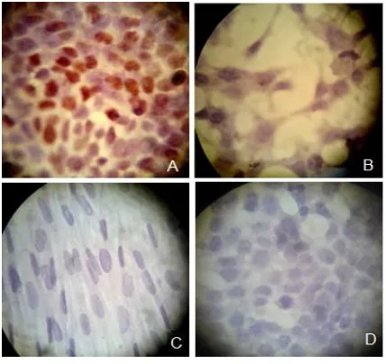
Fig. 1. Immunocytochemistry staining of the estrogen receptors in the cells. MCF-7 as the positive control (A), female amniocytes (B), male amniocytes (C) and MCF-7 cells without the estrogen receptor primary antibodies as the negative control (D).
solution of 3:1 mixture of methyl alcohol and
glacial acetic acid. The fixed cells were dropped
onto a clean glass slide and then were aged for 24 h
at 72 oC.
Solid staining and G-banding
Solid staining has been performed as a
standard method for investigation of chromosome
and chromatid gaps and breaks. To this end, slides
were covered with 10% giemsa stain for 5 min. For
GTG banding, slides were rinsed in the pancreatin
enzyme solution (Sigma-Aldrich, USA) followed
by giemsa stain for 5 min. Then, all the slides were
dried and sealed with Entellan mounting medium
and were covered by coverslips.
Results
Confirmation of the existence of estrogen
receptors in MCF-7 and female amniocytes
In order to evaluate the presence of estrogen
receptors, ICC was used for all three cell groups.
For this purpose, MCF-7 was used as a positive
control which clearly expressed ERs (Figure 1- A).
As it was expected, results showed that unlike the
male amniocytes, the females expressed ER (Figure
1- B, C). To evaluate the specificity of this method
we used MCF-7 without anti estrogen receptor as a
negative control (Figure 1- D).
Cell cytotoxicity evaluation of BPA
For all three cell groups, the related IC50 have been
determined. Results of MTT showed that the half
maximal inhibitory concentration for MCF-7, male
and female amniocytes were about 100, 40 and 4 μg/ml, respectively (Figure 2).
The obtained results suggest that cytotoxicity
effects of BPA are dose dependent and different
cells with different ER pattern showed completely
different susceptibility to BPA. To the best of our
knowledge, there was not any similar study on the
amniocytes. To determine the minimal genotoxic
concentrations of BPA in in vitro, it was decided to use the IC50 concentrations (4, 40 and 100 μg/ml) for all three cell groups as well as 0.4 and 1 μg/ml
BPA which also was suggested by other studies
(28).
Induction of chromosomal aberrations in the
cells exposed to BPA
Assessments of the chromosomal
abnormali-ties in terms of structural and numerical aberrations
have been done to investigate the effects of BPA on
three cell types with relatively high proliferation
potential that make them more sensitive to the toxic
agents.
For MCF-7 which is a cancer cell line with
many chromosomal abnormalities, we assessed the
abnormalities in the untreated or control group of
MCF-7 cells and then compensated it with the other
concentrations to exclude it's in born abnormalities.
Only structural chromosome abnormalities
inclu-ding fragments, chromosome gaps, chromosome
breaks, chromatid gaps, chromatid breaks, and
chromosome rearrangements such as triradials were
scored. The numerical abnormalities were not
scored as there was a wide range of chromosome
aneuploidies in cells obtained from the MCF-7 cell
line. Thus, just the tendency to decrease or increase
the chromosomal numbers was considered.
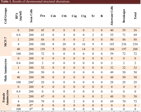
Structural aberrations
The complete data of chromosomal structural
aberrations are provided in table 1. Some of
chromosomal structural aberrations are shown in
figure 3.
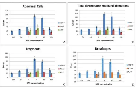
A significant increase of abnormal cells (cells
with at least one structural aberration) was observed
at 1 μg/ml of BPA for all three cell groups.
Althou-gh in MCF-7 cells, a notable increase of abnormal
cells (19% increase compared to normal control
group) was obvious at the lowest concentration, but
it was non-significant (figure 4A).
As it is clear in figure 4B, an unexpected
increase of structural aberrations was seen in amniocytes at a dose of 1 μg/ml but not at 0.4 μg/ml of BPA. For MCF-7 cell line, total structural aberrations increased from 0.4 μg/ml, but it was not significant at 0.4 and 1 μg/ml of BPA.
Table 1. Results of chromosomal structural aberrations
T o ta l B re a ka g es Aberr a nt Cells R Tr Ctg Csg Ctb Csb Fra Seen Cells B P A (µg /ml ) Cell G ro up s 56 59 40 0 3 0 0 8 0 45 200 0 M CF -7 69 71 55 0 2 0 0 4 0 63 200 0.4 88 86 80 0 0 2 0 0 0 86 200 1 234 236 185 9 9 16 0 20 0 180 200 4 208 197 168 3 0 14 0 28 5 158 200 40 72 72 59 0 0 0 0 0 0 72 200 100 0 0 0 0 0 0 0 0 0 0 200 0 M a le Amni o cy tes 2 2 2 0 0 0 0 0 0 2 200 0.4 32 32 32 0 0 0 0 0 0 32 200 1 50 50 49 0 0 0 0 0 0 50 200 4 50 50 48 0 0 0 0 0 0 50 200 40 28 28 28 0 0 0 0 0 0 28 200# 100 0 0 0 0 0 0 0 0 0 0 200 0 F ema le Amni o cy
tes 0.4 200 0 0 0 0 0 0 0 0 0 0
50 51 48 2 0 0 1 0 0 47 200 1 72 70 69 0 0 0 2 0 0 70 200 4 0 0 0 0 0 0 0 0 0 0 0* 40 0 0 0 0 0 0 0 0 0 0 0* 100 #
: Because of high BPA cytotoxicity in this concentration, the analyzed metaphases were less than 200 which were extrapolated to 200; *: Because of high BPA cytotoxicity in this concentrations, no analyzable metaphase was observed; Fra: fragments; DM: double minute; M: marker chromosome; Csb: chromosomal breakage; Ctb: chromatid breakage; Csg: chromosomal gap; Ctg: chromatid gap; Tr: triaradial; R: ring chromosome.
Fig. 3. Some structural aberrations. A1&2: chromatid gap and chromatid break, respectively in MCF-7; B1&2: fragment and chromosome gap, respectively in MCF-7; C: fragment in female amniocyte.
Fig. 4. Average amounts of abnormal cells. A: cells with at least one chromosomal structural aberration; B: total structural abnormalities; C,D: two of main structural abnormalities in different concentrations of BPA (μg/ml). AFM: male amniocytes, AFF: female amniocytes, *: p< 0.001, ¥: p< 0.005, £: p< 0.05.
Chromosomal fragments were the most
frequent structural aberrations induced by BPA in
this study. The patterns of fragments and breakages
induction were similar to the pattern of total
structural aberrations (figures 4C, D).
As it is obvious in figure 4, the number of
abnormal cells and also structural aberrations
decreased in higher doses. For example no
analyzable female amniocyte metaphase was found at doses 40 and 100 μg/ml which could be due to cytotoxic effects of BPA and cell arrest at these
doses.
Chromosomal rearrangements
The karyotypes of the chromosomal spreads in
MCF-7 cells and amniocytes were normal using
GTG banding technique.
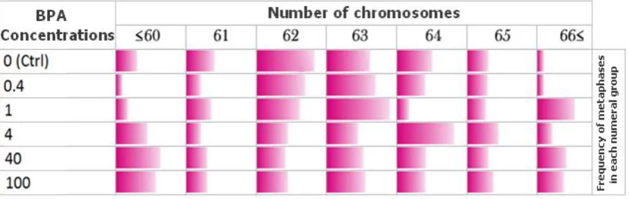
Fig. 5. Chromosomal numerical variations in BPA exposed MCF-7 cell line. The color spectrum magnitudes indicate the relative frequency of metaphases in each chromosomal numerical group (each column).
Numerical aberrations
Although, the numerical variations in BPA
exposed MCF-7 cells were completely observable,
but it didn't follow any particular pattern of
chromosomes number variation after exposure to
different concentrations of BPA (Table 2).
Table 2.Numerical aberrations in cells exposed to different concentrations of BPA
Cell type Concentrations The percentage of MCF-7 metaphases observed in different chromosome number group
≤60 61 62 63 64 65 ≥66
MCF-7
Negative (Ctrl) 8.8 13 30.3 21.7 17.4 8.8 -
0.4 - 5 25 25 13 8.5 -
1 3.7 11.1 22.3 33.3 3.7 7.4 18.5
4 15 5 15 15 30 15 5
40 22.6 9 13.6 18.1 13.6 9 13.5
100 20 9 15 19 13 12 12
The percentage of amniocyte metaphases observed in different chromosome number group
≤43 44 45 46 47 48 ≥49
Male Amniocytes
Negative ( Ctrl ) - - - 100 - - -
0.4 - - - 100 - - -
1 - - 3 97 - - -
4 - - - 96 4 - -
40 - - - 93 7 - -
100# - - 12 88 - - -
Negative ( Ctrl ) - - - 100 - - -
Female Amniocytes
0.4 - - - 100 - - -
1 - - 8 88 4 - -
4 - - 8 53* 28 8 -
40≠ - - - -
100≠ - - - -
#: Because of high BPA cytotoxicity in this concentration, the analyzed metaphases were less than 200 which were
extended to 200 and percentages were calculated from 200 cells; ≠: Because of high BPA cytotoxicity in this
concentrations, no analyzable metaphase was observed;
*:
p< 0.05Discussion
Despite the availability of information on the
toxicity effects of BPA on reproductive system as
an endocrine disruptor, there are still many
remaining questions concerning the genotoxic
effects of BPA (31-35). BPA binds to ERs that
could increase breast cancer risk (22). In the present
study we investigated the effects of BPA on DNA
damage in ER-positive MCF-7 cell line as well as
ER-positive and negative amniocytes. We have
used ICC in order to confirm the presence of ERs in
female amniocytes and lack of its expression in
male amniocytes. The results were obtained as
expected and are in agreement with the findings of
Chen et al. (30).
Cytotoxicity effects of BPA have been
evaluated by MTT assay in MCF-7 cells as well as
female and male amniocytes. Results expectedly
showed that the amniocytes with positive ER are
more sensitive to BPA (3, 18, 19). Despite the
presence of ER on MCF-7 cells, this cell line
showed a high tolerance to BPA compared to
amniocytes that could be attributed to the cancerous
features of MCF-7. The MCF-7 cell line could
continue its cell cycle despite the presence of
chromosome aberrations induced by relatively high
BPA dosage. This result confirmed our supposition
that MCF-7 cell line is a good monitoring system
for genotoxic evaluation of BPA as a xenoestrogen
compound.
Because of high cytotoxicity effects of BPA,
the ER-positive (female) amniocytes were not able
to survive at concentrations of 40 and 100 μg/ml of
BPA (figure 4). As a result, the comparison of
induced structural aberrations between ER-positive
and negative cells were not possible at these concentrations of BPA. But, at 1 μg/ml of BPA a notable increase of structural aberrations
(geno-toxicity) in both ER-positive and negative
amniocytes were seen (interestingly, with no
significant difference between these two cell
groups) (Figure 4). This data is in agreement with
the study of Pfeifer et al. (36) and suggests that
BPA not only can cause structural aberrations in
amniocytes through other pathway(s) in addition to
ER, but ER-mediated pathway has a smaller share.
In other words, increase of structural aberrations
(genotoxicity) in both ER-positive and negative amniocytes at 1 μg/ml of BPA suggest that BPA exerts most of its genotoxic effects through
ER-independent pathway(s). At the other hand,
dramatic increase of cell arrest or death just for
ER-positive amniocytes (cytotoxicity effects of BPA) at 40 and 100 μg/ml of BPA showed that the ER-mediated pathway is more important for cytotoxic
effects of BPA and probably other xenoestrogens.
To the best of our knowledge, this is the first report
which demons-trates the contribution of ER in
cytotoxic or genotoxic effects of xenoestrogens.
Details of this contribution need to be further
clarified.
In contrast to MCF-7, there was almost no structural aberration in amniocytes at 0.4 μg/ml BPA (Figure 4). It shows that amniocytes as normal
body cells with a perfect DNA repair system can
endure this concentration of BPA in in vitro
situation. On the other hand, a cancerous cell, like
MCF-7 with genomic instabilities and defective
DNA repair system (37, 38), showed, although not
significant, a notable increase (about 19%) of
structural aberrations at this concentration of BPA
(Table 1). This result suggests that BPA probably
has more effects on cells which show genomic
instability for various reasons or patients with
cancer predisposition syndromes.
According to the study of Alard et al., the notable increase of structural aberrations at 1 μg/ml of BPA or more (especially in amniocytes) could be
the result of impaired DNA repair system due to
genotoxic effects of BPA (26).
Chromosome aneuploidy or numerical
aberra-tion effects of BPA in amniocytes and MCF-7 cell
line at different concentrations of BPA was clearly
observable (Table 2). As it is obvious in figure 5,
compared with the control group, the frequency of
counted MCF-7 metaphases became nearly equal
between all chromosomal numerical aberration groups, after 48 h exposure to 100 μg/ml of BPA. It suggests that chromosome numerical variations in
MCF-7 cells were completely random, without any
clear variation pattern and with no dose
depen-dency. After exposure to different concentrations of
BPA, the number of amniocytes with normal 46
chromosomes decreased. A significant decrease has
been observed at 4 µg/ml of BPA in female
amnio-cytes but not in males.
The genotoxic investigation of BPA after
exposure of ER negative and positive cells,
indicates that this component could be more
harmful for predisposed ER-positive cells and also
has more effects in numerical aberrations and
cytotoxicity, rather than chromosomal structural
aberrations in these cell types. Thus, depending on
the entry way of BPA, the cells may have a
different toxic destination.
Different amounts of BPA level have been
measured in human biological fluids and tissues in
developed countries. BPA has been detected in the
majority of populations in these countries. In a
study by Vandenberg et al., 0.1 µg BPA per gram
of placenta tissue was obtained (3). Hormann et al.
showed that short-term exposure to bills or receipts
printed by thermal printers can be a cause of
multifold increase of BPA concentration in human
plasma (39). According to our unpublished data, 1
µg BPA per ml of plasma was extracted from mice
after 5 µg/kg oral administration of BPA for 35
days.
Although, the toxic levels of BPA were not
reported in the human body fluid in the literature
yet, but the potential risk of BPA should not be
overlooked. However, the data concerning the BPA
levels in different populations is not available in
developing as well as underdeveloped countries.
Because of low quality of plastics industry as well
as lack of efficient recycling system in these
countries, it is expected that in different populations
of these countries, BPA levels should be higher
than in the developed ones. The same applies to
workers of plastics industry and other highly
exposed people. On the other hand, many types of
xenoestrogens exist in human living environments
and the cumulative effects of these components in
in vivo situation, may have the same effects as the
examined toxic levels of BPA in the present in vitro
investigation.
Acknowledgments
This research project was conducted with the
collaboration of Genetics Research Centre of
University of Social Welfare and Rehabilitation
Sciences in Tehran, Cellular and Molecular Biology
Research Center of Babol University of Medical
Sciences, and Cytogenetics laboratory of Sarem Women’s Hospital in Tehran.
Conflict of interests
The authors declared no conflict of interests.
References
1. Vandenberg LN, Chahoud I, Heindel JJ, et al. Urinary,
circulating, and tissue biomonitoring studies indicate widespread
exposure to bisphenol A. Environ Health Perspect
2010;118:1055-70.
2. GrandViewResearch. Global bisphenol A (BPA) market by
appliation (appliances, automotive, consumer, construction,
electrical & electronics) expected to reach USD 20.03 billion by
2020 [database on the Internet] 2015. http://
www.digitaljournal.com/pr/2009287, Accessed June 24, 2014.
3. Vandenberg LN, Hauser R, Marcus M, et al. Human exposure
to bisphenol A (BPA). Reprod Toxicol 2007;24:139-77.
4. Mirmira P, Evans-Molina C. Bisphenol A, obesity, and type 2
diabetes mellitus: genuine concern or unnecessary
preoccupation? Transl Res 2014;164:13-21.
5. Washam C. Exploring the roots of diabetes: Bisphenol a may
promote insulin resistance. Environmental health perspectives
2006;114:A48-A9.
6. Gao X, Wang HS. Impact of bisphenol a on the cardiovascular
system - epidemiological and experimental evidence and
molecular mechanisms. Int J Environ Res Public Health
2014;11:8399-413.
7. Haighton L, Card JW, Lynch B, et al. Bisphenol A and infant
neonatal neurobehavior. Environ Health Perspect
2012;120:A102; author reply A-3.
8. Saili KS, Corvi MM, Weber DN, et al. Neurodevelopmental
low-dose bisphenol A exposure leads to early life-stage
hyperactivity and learning deficits in adult zebrafish. Toxicology
2012;291:83-92.
9. Sugiura-Ogasawara M, Ozaki Y, Sonta S, et al. Exposure to
bisphenol A is associated with recurrent miscarriage. Hum
Reprod 2005;20:2325-9.
10. Yamada H, Furuta I, Kato EH, et al. Maternal serum and
amniotic fluid bisphenol A concentrations in the early second
trimester. Reprod Toxicol 2002;16:735-9.
11. Fernandez M, Bourguignon N, Lux-Lantos V, et al. Neonatal
exposure to bisphenol a and reproductive and endocrine
alterations resembling the polycystic ovarian syndrome in adult
rats. Environ Health Perspect 2010;118:1217-22.
12. Kang IJ, Yokota H, Oshima Y, et al. Effects of bisphenol a
on the reproduction of Japanese medaka (Oryzias latipes).
Environ Toxicol Chem 2002;21:2394-400.
13. Yang M, Ryu JH, Jeon R, et al. Effects of bisphenol A on
breast cancer and its risk factors. Arch Toxicol 2009;83:281-5.
14. Ho SM, Tang WY, Belmonte de Frausto J, et al.
Developmental exposure to estradiol and bisphenol A increases
susceptibility to prostate carcinogenesis and epigenetically
regulates phosphodiesterase type 4 variant 4. Cancer Res
2006;66:5624-32.
15. Nowell PC, Hungerford DA. Chromosome studies on normal
and leukemic human leukocytes. J Natl Cancer Inst
1960;25:85-109.
16. Negrini S, Gorgoulis VG, Halazonetis TD. Genomic
instability--an evolving hallmark of cancer. Nat Rev Mol Cell
Biol 2010;11:220-8.
17. Thompson SL, Compton DA. Chromosomes and cancer
cells. Chromosome Res 2011;19:433-44.
18. Miller K. Estrogen and DNA damage: the silent source of
breast cancer? J Natl Cancer Inst 2003;95:100-2.
19. Mobley JA, Brueggemeier RW. Estrogen receptor-mediated
regulation of oxidative stress and DNA damage in breast cancer.
Carcinogenesis 2004;25:3-9.
20. Kanda R, Hayata I. Effect of estradiol on radiation-induced
chromosome aberrations in human lymphocytes. Journal of
radiation research 1999;40:95-100.
21. Tabb MM, Blumberg B. New modes of action for
endocrine-disrupting chemicals. Molecular Endocrinology 2006;20:475-82.
22. Alonso-Magdalena P, Ropero AB, Soriano S, et al.
Bisphenol-A acts as a potent estrogen via non-classical estrogen
triggered pathways. Molecular and cellular endocrinology
2012;355:201-7.
23. Machtinger R, Combelles CM, Missmer SA, et al.
Bisphenol-A and human oocyte maturation in vitro. Hum Reprod
2013;28:2735-45.
24. Liu C, Duan W, Li R, et al. Exposure to bisphenol A disrupts
meiotic progression during spermatogenesis in adult rats through
estrogen-like activity. Cell Death Dis 2013;4:e676.
25. Fic A, Zegura B, Sollner Dolenc M, et al. Mutagenicity and
DNA damage of bisphenol A and its structural analogues in
HepG2 cells. Arh Hig Rada Toksikol 2013;64:3-14.
26. Allard P, Colaiacovo MP. Bisphenol A impairs the
double-strand break repair machinery in the germline and causes
chromosome abnormalities. Proc Natl Acad Sci U S A
2010;107:20405-10.
27. Susiarjo M, Hassold TJ, Freeman E, et al. Bisphenol A
exposure in utero disrupts early oogenesis in the mouse. PLoS
genetics 2007;3:e5.
28. Bisphenol A [MAK Value Documentation, 2011] [database
on the Internet]. Wiley Online Library. 2011.
29. Holliday DL, Speirs V. Choosing the right cell line for breast
cancer research. Breast Cancer Res 2011;13:215.
30. Chen CP, Lai TC, Chern SR, et al. Proteome differences
between male and female fetal cells in amniotic fluid. OMICS
2013;17:16-26.
31. Herath CB, Jin W, Watanabe G, et al. Adverse effects of
environmental toxicants, octylphenol and bisphenol A, on male
reproductive functions in pubertal rats. Endocrine
2004;25:163-72.
32. Toyama Y, Suzuki-Toyota F, Maekawa M, et al. Adverse
effects of bisphenol A to spermiogenesis in mice and rats. Arch
Histol Cytol 2004;67:373-81.
33. Al-Hiyasat AS, Darmani H. In vivo effects of BISGMA-a
component of dental composite-on male mouse reproduction and
fertility. J Biomed Mater Res A 2006;78:66-72.
34. Aarab N, Lemaire-Gony S, Unruh E, et al. Preliminary study
of responses in mussel (Mytilus edilus) exposed to bisphenol A,
diallyl phthalate and tetrabromodiphenyl ether. Aquat Toxicol
2006;78 Suppl 1:S86-92.
35. Ortiz-Zarragoitia M, Cajaraville MP. Biomarkers of
exposure and reproduction-related effects in mussels exposed to
endocrine disruptors. Arch Environ Contam Toxicol
2006;50:361-9.
36. Pfeifer D, Chung YM, Hu MC. Effects of Low-Dose
Bisphenol A on DNA Damage and Proliferation of Breast Cells:
The Role of c-Myc. Environ Health Perspect 2015;123:1271-9.
37. Acu ID, Liu T, Suino-Powell K, et al. Coordination of
centrosome homeostasis and DNA repair is intact in MCF-7 and
disrupted in MDA-MB 231 breast cancer cells. Cancer Res
2010;70:3320-8.
38. Peddi P, Francisco DC, Cecil AM, et al. Processing of
clustered DNA damage in human breast cancer cells MCF-7
with partial DNA-PKcs deficiency. Cancer Lett
2008;269:174-83.
39. Hormann AM, Vom Saal FS, Nagel SC, et al. Holding
thermal receipt paper and eating food after using hand sanitizer
results in high serum bioactive and urine total levels of bisphenol
A (BPA). PLoS One 2014;9:e110509.