Hoi I. Cheong, … , Sathyamangla V. Naga Prasad, Serpil C. Erzurum
JCI Insight. 2016;1(21):e90240. https://doi.org/10.1172/jci.insight.90240.
Life-sustaining responses to low oxygen, or hypoxia, depend on signal transduction by HIFs, but the underlying mechanisms by which cells sense hypoxia are not completely understood. Based on prior studies suggesting a link between the β-adrenergic receptor (β-AR) and hypoxia responses, we hypothesized that the β-AR mediates hypoxia sensing and is necessary for HIF-1α accumulation. Beta blocker treatment of mice suppressed hypoxia induction of renal HIF-1α accumulation, erythropoietin production, and erythropoiesis in vivo. Likewise, beta blocker treatment of primary human endothelial cells in vitro decreased hypoxia-mediated HIF-1α accumulation and binding to target genes and the downstream hypoxia-inducible gene expression. In mechanistic studies, cAMP-activated PKA and/or GPCR kinases (GRK), which both participate in β-AR signal transduction, were investigated. Direct activation of cAMP/PKA pathways did not induce HIF-1α accumulation, and inhibition of PKA did not blunt HIF-1α induction by hypoxia. In contrast,
pharmacological inhibition of GRK, or expression of a GRK phosphorylation–deficient β-AR mutant in cells, blocked hypoxia-mediated HIF-1α accumulation. Mass spectrometry–based quantitative analyses revealed a hypoxia-mediated β-AR phosphorylation barcode that was different from the classical agonist phosphorylation barcode. These findings indicate that the β-AR is fundamental to the molecular and physiological responses to hypoxia.
Research Article Vascular biology
Find the latest version:
Conflict of interest: The authors have declared that no conflict of interest exists.
Submitted: August 29, 2016 Accepted: November 8, 2016 Published: December 22, 2016 Reference information: JCI Insight. 2016;1(21):e90240. doi:10.1172/jci.insight.90240.
Hypoxia sensing through
β
-adrenergic
receptors
Hoi I. Cheong,1,2 Kewal Asosingh,1 Olivia R. Stephens,1,2 Kimberly A. Queisser,1 Weiling Xu,1
Belinda Willard,3 Bo Hu,4 Josephine Kam Tai Dermawan,5 George R. Stark,6
Sathyamangla V. Naga Prasad,7 and Serpil C. Erzurum1,8
1Department of Pathobiology, Lerner Research Institute, Cleveland Clinic, Cleveland, Ohio, USA. 2Department of Molecular
Medicine, Cleveland Clinic Lerner College of Medicine, Case Western Reserve University, Cleveland, Ohio, USA.
3Proteomics and Metabolomics Laboratory, Lerner Research Institute, 4Department of Quantitative Health Sciences, 5Pathology & Laboratory Medicine Institute, 6Department of Cancer Biology, Lerner Research Institute, 7Department of
Molecular Cardiology, Lerner Research Institute, and 8Respiratory Institute, Cleveland Clinic, Cleveland, Ohio, USA.
Introduction
Oxygen delivery to tissues is essential for life. Under hypoxia, adaptive responses take place over the course of hours and days to generate erythrocytes and create new blood vessels. These responses have been identified as the downstream effects of HIFs (1–4). HIF-1 is a master transcription factor for
oxy-gen sensing. It is composed of an oxyoxy-gen-regulated α subunit (HIF-1α) and a constitutively expressed β
subunit (HIF-1β) (5–7). Under hypoxia, HIF-1α is stabilized, dimerizes with HIF-1β, and translocates
to the nucleus to promote transcription of genes, such as erythropoietin (EPO) and vascular endothelial growth factors (VEGF), to orchestrate greater capacity of oxygen delivery to tissues (1). However, the
determinants of HIF-1α regulation are incompletely understood in the complex physiologic
environ-ment of cells (8–10).
More than 40 years ago, long before the discovery of HIF and erythropoietin, β-adrenergic receptor
(β-AR) blockade was shown to diminish the erythropoietic response to hypoxia in animal models, but
the mechanisms of these effects have been unexplored (11, 12). Since that time, there have been a wealth of studies that have detailed the downstream signal transduction events that result from activation of the
β-ARs. β-ARs belong to a large family of GPCRs. β-ARs are ubiquitously expressed throughout the body,
with β1-AR as the major subtype expressed in the heart and β2-AR in the lung, vascular endothelium, and
kidneys (13–15). When an agonist, such as the endogenous ligands noradrenaline and adrenaline, binds
to β-AR, the receptor is stabilized in an active conformation and couples to the heterotrimeric G proteins,
resulting in dissociation of Gα and Gβγ subunits. These molecular events rapidly propagate complex
intra-cellular signals. For instance, the dissociation of the Gα subunit initiates the release of cAMP-dependent
PKA (16), while Gβγ subunits recruit GPCR kinases (GRKs). Although both PKA and GRK participate
in β-AR phosphorylation, the level of phosphorylation at distinct PKA and GRK sites depends on the type
and concentrations of agonists (17).
Phosphorylation ultimately diminishes β-AR responsiveness to agonist stimulation, also known as
receptor desensitization, and modulates receptor trafficking and turnover. The diminished responsiveness to an overload of noradrenaline is the cardinal feature of congestive heart failure (18–20). Effective ther-apies with beta blockers, such as carvedilol, have resulted in this class of drug being widely prescribed for
left and right heart failure (21, 22). Elevation of HIF-1α levels is also implicated in the pathophysiology
of the failing heart (23). We hypothesized that the β-AR is part of the pathway for hypoxia sensing and
necessary for HIF-1α accumulation. We tested this hypothesis using in vivo models; genomic,
pharma-cologic, and molecular approaches; and detailed barcoding of the phosphorylation status of the β-AR.
Results
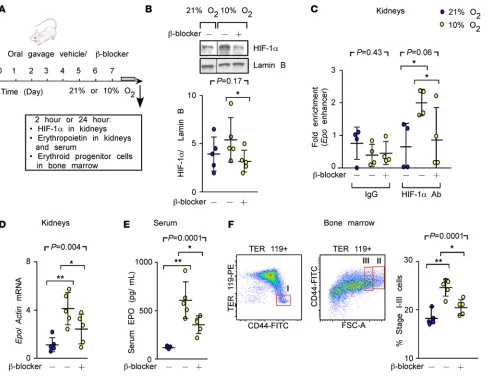
Decreased hypoxia responses in mice with beta blocker treatment. To determine whether β-AR blockade prevents
hypoxia-mediated erythropoietic responses via HIF-1α in vivo, mice were orally administered vehicle or the
beta blocker carvedilol, followed by exposure to hypoxia (10% oxygen) (Figure 1A). Under
hypoxia, HIF-1α accumulated in kidneys and
was significantly decreased by carvedilol
(Fig-ure 1B). In addition, HIF-1α–specific binding
to the Epo gene increased and was blocked by carvedilol (Figure 1C). Moreover, hypoxia increased erythropoietin mRNA and protein
(24), both of which were suppressed by β-AR
blockade (Figure 1, D and E). Since erythro-poietin is known to induce the proliferation of erythroid progenitor cells (25), we quantified the percentages of proerythroblasts and basophilic and polychromatic erythroblasts in the bone marrow with flow cytometry (26). Under hypoxia, the erythroid progenitor cell population increased by 35%, and
the effect was blunted by carvedilol (Figure 1F). These findings indicate that β-AR blockade by carvedilol
suppresses the classical hypoxia erythropoietic responses through HIF-1α.
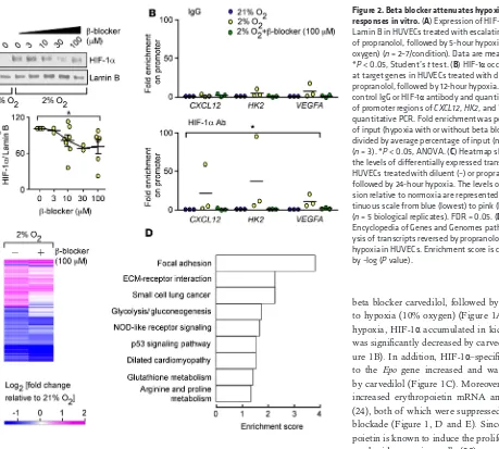
Decreased HIF responses in hypoxic cells with beta blocker treatment. Next, we tested the in vitro effects of
β-AR blockade on HIF-1α accumulation in hypoxic human endothelial cells isolated from umbilical cord
veins (HUVECs). HUVECs were exposed to escalating doses of propranolol and then exposed to hypoxia
(2% oxygen). Western blot analyses of nuclear proteins revealed that hypoxia increased HIF-1α levels when
compared with normoxia and propranolol dose-dependently decreased HIF-1α (Figure 2A). To confirm
β-AR blockade inhibitory effects on HIF-1α, ChIP studies were performed to test whether β-AR blockade
decreases the binding of HIF-1α to the promoters of its classical target genes, such as chemokine stromal
cell–derived factor-1 (SDF-1 or CXCL12), hexokinase 2 (HK2), and VEGFA. Quantitative PCR revealed that
hypoxia significantly increased HIF-1α–specific binding to promoter sequences, an effect that was blocked
by propranolol (Figure 2B).
To evaluate the entire spectrum of signaling pathways affected by beta blockers under hypoxia, we performed transcriptome-profiling studies that tested 47,000 probes representing well-characterized genes, gene candidates, and splice variants. HUVECs were exposed to diluent or propranolol alone and then exposed to 24-hour hypoxia (data are accessible at GEO, accession GSE86793). Hypoxia induced changes in RNA expression levels relative to normoxia, corresponding to 1,247 probes, 434 of which were reversed by propranolol (at a FDR of 0.05) (Figure 2C and Supplemental Table 1). Pathway analyses and
func-tional annotation clustering of the identified transcripts revealed the biological significance of β-AR in
regulating hypoxia-inducible phe-notypes, including focal adhesion, glycolysis, p53 signaling pathways, angiogenesis, and cell migration (Figure 2D, Supplemental Table 2, and refs. 27, 28).
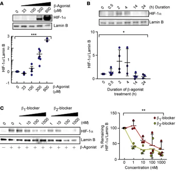
β-Agonist increases HIF-1α
accumulation under normoxia. To
investigate if β-AR activation can
increase HIF-1α levels,
dose-re-sponse and time-course studies
with the β-agonist isoproterenol
were performed in HUVECs under normoxia. Isoproterenol increased
nuclear HIF-1α in a dose- and time-dependent manner, with a maximum response at 2 hours (Figure 3, A
and B). To test the possibility that stabilization of HIF-1α is mediated via a specific β-AR subtype (β1-AR
or β2-AR), HUVECs were exposed to CGP-20712A (a beta 1 blocker) or ICI-118551 (a beta 2 blocker),
fol-lowed by isoproterenol stimulation. Isoproterenol stabilization of HIF-1α was dose-dependently decreased
by both beta 1 and beta 2 blockers (Figure 3C). This suggests that isoproterenol stabilization of HIF-1α can
be mediated by β1-AR or β2-AR and excludes off-target effects.
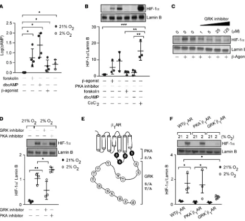
HIF-1α accumulation under hypoxia relies on GRK phosphorylation of β-AR. To establish the mechanistic
link between β-AR and HIF-1α, we investigated classical downstream signal pathways of β-ARs. Agonist
binding to β-AR stabilizes the receptor in an active conformation and leads to coupling to the
heterotri-meric G proteins. This results in production of cAMP by adenylate cyclase that activates PKA, which,
in turn, phosphorylates the β-AR (13). To test if hypoxia activates the β-AR canonical downstream signal
transduction, we measured the intracellular cAMP level in HUVECs exposed to hypoxia (2% oxygen) for 10 to 15 minutes. Similar to isoproterenol, forskolin (which activates adenylate cyclase), and dibutyryl (a cAMP analog), hypoxia increased cAMP levels (Figure 4A). However, forskolin and dibutyryl did not lead
to HIF-1α accumulation (Figure 4B). Further, the PKA inhibitor H89 did not affect HIF-1α accumulation
by isoproterenol (Figure 4B). Thus, hypoxia can activate β-AR signaling, but activation of the PKA/cAMP
pathway does not lead to HIF-1α accumulation.
GRKs are the other classical pathway known to phosphorylate β-ARs following receptor activation (13,
29–31). Evaluation of the GRK phosphorylation pathway using pharmacological inhibition of GRK2 in
HUVECs revealed a dose-dependent decrease in β-agonist–mediated HIF-1α accumulation (Figure 4C).
Like-wise, GRK inhibition, but not PKA, significantly attenuated hypoxia-induced HIF-1α levels (Figure 4D). The
results suggest that hypoxia-mediated HIF-1α accumulation is dependent on GRK phosphorylation of β-AR.
To test this idea, we evaluated the hypoxia response of human embryonic kidney cells (HEK293), which
over-express wild-type β1-AR (WTβ1-AR) or mutant β1-ARs lacking either the phosphorylation target sites of PKA
Figure 3. β-Agonist promotes HIF-1α accumulation under normoxia. (A) Expression of HIF-1α and Lamin B in HUVECs treated with diluent or isoproterenol (33–900 μM) for 2 hours (n = 3–6/condition). Data are mean ± SEM. (B) Expression of HIF-1α and Lamin B in HUVECs treated with 300
(PKA–β
1-AR) or GRK (GRK–β1-AR) (Figure 4E and ref. 32). Hypoxia significantly induced HIF-1α levels in
WTβ1-AR and PKA–β1-AR cells but not in GRK–β1-AR cells (Figure 4F). These results indicate that HIF-1α
accumulation occurs in response to hypoxia in a manner that requires β-AR–GRK signal transduction.
Hypoxia-specific β-AR phosphorylation barcode. Because agonist binding to β-AR results in unique receptor phosphorylation patterns that correlate with distinct downstream signals (33), we investigated whether hypoxia imparts a specific receptor phosphorylation signature, i.e., a unique barcode. HEK293
cells overexpressing β2-AR were exposed to the β-agonist isoproterenol (10 μM) or hypoxia (2%
oxy-gen). β2-ARs from the plasma membranes were enriched via binding to alprenolol and subjected to
mass spectrometry analyses, which successfully identified 8 key phospho-peptide species of the receptor (Table 1 and Supplemental Table 3).
Figure 4. Hypoxia or β-agonist–mediated HIF-1α accumulation depends on phosphorylation of β-adrenergic receptor (β-AR) by GPCR kinase (GRK). (A) Intracellular cAMP in HUVECs 10–15 minutes after stimulation with factors shown (n = 3–6). (B) Expression of HIF-1α and Lamin B in HUVECs treated with 300 μM β-agonist isoproterenol with or without 10 μM of the PKA inhibitor H89 or forskolin or dibutyryl cAMP (dbcAMP) for 2 hours. Cobalt chloride (CoCl2) shown as positive control (n = 3). (C) Expression of HIF-1α and Lamin B in HUVECs exposed to GRK inhibitor (1–125 μM) for 45 minutes followed by 2-hour isoproterenol. The blot is representative of 2 independent experiments. (D) Expression of HIF-1α and Lamin B in HUVECs exposed to a GRK (125 μM) or PKA inhibitor H89 (10 μM), followed by 5-hour hypoxia (2% oxygen) (n = 3). (E) Map of β-AR serine and threonine residues that are phosphorylated by PKA and GRK, mutated to alanine. (F) Expression of HIF-1α and Lamin B in human embryonic kidney cells (HEK293) overexpressing β1-AR (WTβ1-AR) or mutants lacking PKA (PKA–β
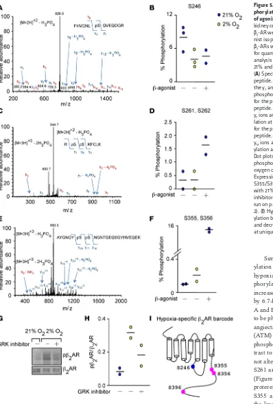
Similar to canonical phosphor-ylation seen with isoproterenol,
hypoxia decreased β2-AR
phos-phorylation at S246 by 2-fold and increased phosphorylation at S396 by 6.7-fold (Table 1 and Figure 5,
A and B). S246 on β2-AR is known
to be phosphorylated by ataxia tel-angiectasia mutated protein kinase (ATM) and S396 is known to be phosphorylated by GRK2. In con-trast to isoproterenol, hypoxia did
not alter β2-AR phosphorylation at
S261 and S262, known PKA sites (Figure 5, C and D). Although iso-proterenol and hypoxia increased S355 and S356 phosphorylation, the level of increase was substan-tially different between the two (hypoxia, 1.6-fold versus isoproterenol, 153-fold) (Figure 5, E and F). These findings are consistent with Western blot analysis of protein extracts from hypoxic HUVECs. Hypoxia increased S355 and S356 phosphorylation, an effect that was blunted by GRK inhibition (Figure 5, G and H). Altogether, these
findings identify a distinct hypoxia-specific β2-AR phosphorylation signature, which provides a
defini-tive biochemical link between hypoxia sensing and activation of the β-AR (Figure 5I).
Figure 5. Hypoxia induces a phos-phorylation barcode in the absence of agonist binding. Human embryonic kidney cells (HEK293) overexpressing
β2-AR were exposed to the β -ago-nist isoproterenol or 5-hour hypoxia.
Discussion
Physiologic responses are rapidly initiated within minutes of hypoxia. Catecholamines activate the β1
-AR in the cardiac muscles, resulting in increased cardiac output (34–36), while the peripheral vasculature dilates to increase the perfusion of blood to oxygen-limited tissues (37). Secondary to these immediate events, adaptive responses take place over the course of hours and days to generate erythrocytes and create new blood vessels in order to provide greater oxygen delivery to tissues. These late responses have been identified as the downstream effects of HIFs (1, 2). HIF-1 is the primary intracellular mediator of the myr-iad physiological processes for adaptation. This study provides a biochemical connection between the cat-echolamine pathway and hypoxia responses. Here, we indicate that hypoxia, like catcat-echolamine agonists,
can activate the β-AR and lead to HIF accumulation and adaptive responses. Prior work has suggested a
close relationship between HIF and β-AR, i.e., both are hydroxylated by prolyl hydroxylase 3 under
hypox-ia (38). However, to our knowledge, this is the first connection of hypoxhypox-ia to activation of β-AR.
In early studies of beta blockers in the 1970s, intraperitoneal injection of rabbits with the subtype-non-specific beta blocker propranolol prior to an 18-hour exposure to hypobaric hypoxia resulted in decreased erythropoietic responses (11). In another study, rats preinjected with metipranolol (another subtype-non-specific beta blocker) had less erythropoiesis (12). There were some discrepant findings based on type of
beta blocker used, but the studies suggested the possibility that β-AR may regulate hypoxia-inducible
eryth-ropoiesis. Many years after these studies, Semenza et al. identified HIF-1 in the molecular mechanisms that lead to production of erythropoietin and the subsequent erythropoiesis associated with hypoxia (2).
However, the effect of β-AR on HIF-1 accumulation under hypoxia has been untested until now.
In this study, mice were orally administered the readily absorbed beta blocker carvedilol, which has a longer half-life (6–10 hours) as compared with propranolol (3–6 hours) (39). The mice given carvedilol had
lower levels of renal HIF-1α and consequently less binding of the transcription factor to Epo in vivo.
Eryth-ropoietin mRNA in the kidneys, the principal site of production, and serum erythEryth-ropoietin levels were sig-nificantly lower with carvedilol treatment. Consistent with these results, mice with carvedilol treatment had
fewer erythroid progenitor cells in the bone marrow. Importantly, the beta blocker decreased HIF-1α levels
and its binding to target genes and reversed approximately 30% (equivalent to 434 identified transcripts) of the hypoxia-inducible changes in expression. These transcripts mapped to known signal transduction
pathways that contribute to cancer biology and dilated cardiomyopathy. In this context, HIF-1α levels are
increased in heart tissues obtained from patients with ischemic cardiomyopathy (23). It is interesting to
speculate that benefits of beta blockers in heart failure may be related to suppression of HIF-1α.
To define the molecular mechanisms that link β-AR and HIF-1α, the cAMP/PKA pathway was
eval-uated using pharmacologic approaches and genetic mutants. Under normoxia, simply increasing the
intra-cellular cAMP levels in cells could not cause HIF-1α accumulation. These findings are slightly in variance
from a recent study that suggests that isoproterenol can increase HIF-1α protein levels (40). In that study,
treatment of human cardiomyocytes with the β-agonist isoproterenol or cervical cancer cells with forskolin
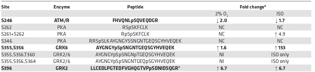
Table 1. Quantitative analyses of β2-AR phosphorylation sites
Site Enzyme Peptide Fold changeA
2% O2 ISO
S246 ATM/R FHVQNLpSQVEQDGR ↓ 2.0 ↓ 1.7
S262 PKA RSpSKFCLK NC NC
S261+S262 PKA RpSpSKFCLK NC ↑ 4.9
S346 PKA RRSpSLKAYGNGYSSNGNTGEQSGYHVEQEK NC NC
S355,S356 GRK6 AYGNGYpSpSNGNTGEQSGYHVEQEK ↑ 1.6 ↑ 153
S355,S356,T360 GRK2/6 AYGNGYpSpSNGNpTGEQSGYHVEQEK NI ISO only
S355,S356,S364 GRK2/6 AYGNGYpSpSNGNTGEQpSGYHVEQEK NI ISO only
S396 GRK2 LLCEDLPGTEDFVGHQGTVPpSDNIDSQGRB ↑ 6.7 ↑ 6.7
AFold change was normalized to untreated 21% O
increased HIF-1α protein abundance, but forskolin did not increase
HIF-1α transcriptional activity (40). In this study, pharmacological inhibition of
PKA in primary human endothelial cells, or the expression of a PKA
phos-phorylation–deficient β-AR mutant in human kidney embryonic cells, does
not alter hypoxia-mediated HIF-1α accumulation. This is consistent with the
study in which a stable knockdown of the catalytic subunit of PKA did not
decrease hypoxic accumulation of HIF-1α (40). Unlike PKA,
pharmacolog-ical inhibition of GRK, or genetic mutation of the β-AR that impairs GRK
phosphorylation, profoundly decreased hypoxia-mediated HIF-1α
accumu-lation in this study. These results support the idea that hypoxia-mediated
HIF-1α accumulation is dependent on GRK phosphorylation of β-AR. In
a study by Baloğlu, et al., preexposure to 24 hours of hypoxia decreased
β-agonist induction of cAMP in alveolar epithelial cells (41). Here,
10-min-ute hypoxia exposure of human endothelial cells rapidly increased cAMP.
These findings suggest that under hypoxia the β-AR produces higher basal levels of cAMP but is overall less
responsive to agonist stimulation. Another possibility for the different findings in this study is that cAMP responses to hypoxia are dependent on specific cell types and/or times of exposure.
Carvedilol and propranolol were both employed in experiments performed in this study, and distinc-tions should be noted in these beta blockers. Carvedilol, but not propranolol, induces phosphorylation at
GRK6 sites within β2-AR and initiates β-arrestin–mediated signaling (33, 42). Thus, carvedilol might exert
some of its effects independent of receptor antagonism. Shenoy et al. previously found that β-arrestin 1
interacts with HIF-1α, which could be one mechanistic link between the hypoxia barcode and HIF-1α
stabi-lization (43). Studies are needed to test the possible roles of β-arrestins, receptor internalization, and other
mechanisms through which GRK phosphorylation may induce HIF.
Human β1-AR and β2-AR protein sequences share 54% sequence identity and contain phosphorylation
sites that are targeted by PKA and GRK; but they have different expression patterns and activate different
signaling pathways (18, 32). One limitation of the current study is that β1-AR mutants were used in
evalua-tion of hypoxia signal transducevalua-tion, but β2-AR was used to investigate effects of hypoxia on the
phosphor-ylation barcode. Effects of β2-AR PKA and GRK mutants were not studied. Likewise, the phosphorylation
barcodes of β1-AR or its GRK/PKA mutant forms were not studied. In addition, expression levels of
β1-AR or β2-AR were not quantitated in vitro or in vivo. Studies delineating differences and similarities
between β1-AR and β2-AR under hypoxia will be important to understanding mechanisms and cell-specific
effects. Abundance and membrane localization of the β1-AR and β2-AR under hypoxia will also be
import-ant to understanding regulatory mechanisms of the receptors under hypoxia.
Quantitative mass spectrometry analyses of β2-AR peptides, enriched from alprenolol binding, indicate
highly differential percentages of phosphorylation of known serine and threonine amino acid residues, rang-ing from 0.045% at S396 to 99% at S262 (Supplemental Table 3). Our method identified 9 amino acids in the
β2-AR phosphorylated with isoproterenol treatment, compared with 13 amino acids that were previously
reported using stable isotope labeling with amino acids in cell culture with phosphopeptide enrichment (33). Despite differences in methodologies, we identified an overall similar level of phosphorylation change from isoproterenol stimulation at S246 (1.7-fold decrease compared with previously reported 2-fold), at S396 (6.7-fold increase compared with previously reported 3.5-(6.7-fold), and at S261 and S262 (4.9-(6.7-fold compared with 15.5-fold). Strikingly, hypoxia alone induces decreased phosphorylation at S246 and increased phosphoryla-tion at S396 at a comparable level to isoproterenol. No change in the phosphorylaphosphoryla-tion level at PKA sites is
observed with hypoxia. These findings point to a unique hypoxia-specific β2-AR phosphorylation signature,
perhaps mediating a different pattern of signal transduction for HIF-1α regulation.
Our results collectively support a model in which hypoxia activates a β-AR phosphorylation barcode
that allows for distinct downstream signals, which are essential for HIF-1α accumulation (Figure 6). The
agonist-independent hypoxia-driven β-AR activation is mediated by GRK phosphorylation, which results
in a unique phosphorylation signature that mediates HIF-1α stabilization.
Although β-ARs were first identified for their roles in the fight-or-flight response as part of the
sym-pathetic nervous system, this study identifies an unsuspected essential role of β-AR in hypoxia sensing in
renal hypoxia-induced accumulation of HIF in vivo and in endothelial and epithelial cells in vitro. Howev-er, beta blockers are used principally in the context of cardiovascular disease, such as heart failure. Further
work is needed to define whether β-AR–dependent hypoxic signaling is operative in the heart and in a
clinically relevant model, such as ischemic heart failure.
Methods
Animal model. Eight-week-old female balb/c mice from The Jackson Laboratory were orally
adminis-tered with vehicle or carvedilol (C3993, Sigma-Aldrich) dissolved in 20% DMSO/water at 15 mg/kg/d
for 7 days, followed by exposure to hypoxia (10% O2). Heart rates were monitored consciously with the
MouseOx pulse oximeter.
Flow cytometry. Bone marrow was isolated from the femur, tibia, and vertebrae. Nonspecific binding
sites were blocked by incubation of cells with 10% normal goat serum in PBS for 15 minutes at room tem-perature. Cells were washed with 1% BSA in PBS and incubated with APC-conjugated anti-CD45
(eBio-science) at 1:100 (2 μg/ml), PE-conjugated TER119 (eBioscience) at 1:50 (4 μg/ml), and FITC-conjugated
anti-CD44 (eBioscience) at 1:100 (5 μg/ml) for 30 minutes on ice. All antibodies were diluted in 1% BSA
in PBS. Cells were washed twice with 1% BSA in PBS and resuspended in FACS Flow containing the dead cell marker 7-AAD (BD Biosciences) at 1:200. Samples were run on a LSRII flow cytometry (Becton Dick-inson), and data were analyzed using Flowjo vX.0.7 (Tree Star Inc.). Aggregates, cell debris, and dead cells were gated out. Leukocytes were excluded using CD45. Three populations were defined based on size and
CD44 and TER119 levels. Proerythroblasts were defined as CD44hiTER119lo, and basophilic and
polychro-matic erythroblasts were defined as TER119hi and selected based on decreasing size and CD44 expression.
Proerythroblasts and basophilic and polychromatic erythroblasts were defined such that the percentage of cells in each population fit the expected ratio of 1:2:4 (26).
ELISA. Blood obtained from cardiac puncture was clotted at 37°C for at least 30 minutes. Serum was
isolated after centrifugation at 10,000 g for 10 minutes. The Mouse Erythropoietin Quantikine ELISA Kit
(MEP00B, R&D Systems) was used to quantify serum erythropoietin concentration.
Cell culture. HUVECs were cultured in Endothelial Basal Medium-2 basal medium, supplemented with
the Endothelial Cell Grow Medium-2 bulletkit (Lonza). They were cultured up to passage 6 for experiments. All kidney embryonic cells (HEK293) were cultured in minimum essential media with 10% heat-inactivated fetal bovine serum and 1% penicillin/streptomycin. Cells were serum starved for 4 hours or overnight prior
to any experiment. For hypoxia studies, cells were incubated in a sealed chamber at 37°C with 2% O2, 5%
CO2, balanced with 93% N2. For treatment of cells, propranolol (P0884, Sigma-Aldrich), β-ARK1/GRK2
inhibitor (125 μM) (182200, EMD Millipore), or PKA inhibitor H89 (10 μM) (B1427, Sigma-Aldrich) was
added to cells for 45 minutes prior to normoxia or hypoxia. HUVECs were gifts from F. Terenzi and P. Fox at
Cleveland Clinic; HEK293 cells overexpressing β1-AR (WTβ1-AR) or mutants lacking PKA (PKA–β
1-AR) or
GRK (GRK–β
1-AR) sites were from Howard Rockman at Duke University, Durham, North Carolina, USA;
and HEK293 cells overexpressing β2-AR were from Robert Lefkowitz at Duke University.
Western blot. For HIF-1α protein quantification, nuclear protein extracts were prepared according to the manufacturing protocol of the nuclear extraction kit (Affymetrix). Proteins were subjected to SDS-PAGE in 4%–15% or 18% Tris-HCl gel (Bio-Rad) and transferred to polyvinylidene difluoride membranes.
Antibodies for human HIF-1α (610959, BD Biosciences), mouse HIF-1α (sc-10790, Santa Cruz
Biotech-nology Inc.), phosphorylated β2-AR (sc-22191-R, Santa Cruz Biotechnology Inc.), β2-AR (sc-569, Santa
Cruz Biotechnology Inc.), and Lamin B (sc6216, Santa Cruz Biotechnology Inc.) were used for detection.
Isolation of RNA. Cells were lysed using QIAzol buffer. RNA was isolated according to the
manu-facturing protocol of the miRNeasy Mini Kit (Qiagen). The quality of RNA was assessed with the 2100 BioAnalyzer (Agilent Technologies). Samples with an RNA Integrity number higher than 7 were used for genome-wide expression assay.
Genome-wide expression assay. The HumanHT-12 v4 Expression BeadChip (BD-103-0204, Illumina) was
used. The experiment was performed on 5 biological replicates. Data have been deposited in the GEO repository (accession GSE86793).
ChIP — quantitative PCR. HUVECs were treated with propranolol, isoproterenol, or hypoxia for 12
Immuno-precipitation Assay Kit (17295, EMD Millipore). Rabbit anti-HIF-1α antibody (sc10790, Santa Cruz
Bio-technology Inc.) was used to pull down chromatin bound by HIF-1α; rabbit IgG (sc2027, Santa Cruz
Biotechnology Inc.) was used as control. DNA primers used for quantitative PCR to quantify HIF-1α–
bound promoters are as follows: SDF-1 forward, 5′-TCTAACGGCCAAAGTGGTTT-3′; SDF-1 reverse,
5′-GCCACCTCTCTGTGTCCTTC-3′; HK2 forward, 5′-CACATTGTTGCATGAAACTCC-3′; HK2
reverse, 5′-GACCTCTCCGATTCACAGG-3′; VEGFA forward, 5′
-TCTTCGAGAGTGAGGACGTGT-GT-3′; VEGFA reverse, 5′-AAGGCGGAGAGCCGGAC-3′ (44); Epo forward, 5′
-TCTCTTCATGACT-GTACACACC-3′; Epo reverse, 5′-TGACAGCCACATAGAATAAAAG-3′ (45). Quantitative PCR was
performed using iQ SYBR Green Supermix (170-8880, Bio-Rad).
Measurements of cAMP. cAMP was measured according to the manufacturing protocol of the
Catch-Point cAMP Fluorescent Assay Kit (R8089, Molecular Devices).
Reverse transcription–quantitative PCR. mRNA was prepared according to manufacturing protocol of the
miRNeasy kit (217004, Qiagen). mRNA was reverse transcribed using the SuperScript First-Strand Syn-thesis System (11907, Invitrogen). DNA primers used for quantitative PCR to quantify EPO and ACTIN
cDNA were as follows: EPO forward, 5′-GAGGCAGAAAATGTCACGATG-3′; EPO reverse, 5′
-CTTC-CACCTCCATTCTTTTCC-3′; ACTIN forward, 5′-CTAGGCACCAAGGTGTGAT-3′; ACTIN reverse,
5′-CAAACATGATCTGGGTCATC-3′.
Pathway analysis and annotation clustering. DAVID v6.7 (Database for Annotation, Visualization and
Integrated Discovery) was used for Kyoto Encyclopedia of Genes and Genomes pathways analysis and annotation clustering of genes that show significant differential expression in cells treated with hypoxia, isoproterenol, and hypoxia with propranolol pretreatment.
β2-AR alprenolol bead purification. Kidney embryonic kidney cells which overexpress β2-AR (HEK293 β2
-AR) were serum starved for at least 4 hours prior to exposure to isoproterenol (10 μM) for 5 minutes or
hypoxia (2% oxygen) for 5 hours. After cell lysis with hypotonic solution, plasma membranes were
solubi-lized with 1% n-Dodecyl β-D-maltoside supplemented with Halt Protease and Phosphatase Inhibitor
Cock-tail (78440, Thermo Fisher Scientific). Alprenolol beads (CM29101, CellMosaic Inc.) (46) were incubated with plasma membranes at 4°C overnight. Receptors were eluted with loading buffer for gel electrophoresis.
Mass spectrometry. The protein samples were subjected to in-gel digestion in which the bands were cut
from the gels and washed in 50% ethanol, 5% acetic acid. The gel pieces were then dehydrated in
acetoni-trile, dried in a Savant Speedvac (ThermoFisher Scientific), and digested by adding 5 μl 10 ng/μl of trypsin
or chymotrypsin, in 50 mM ammonium bicarbonate, followed by incubation overnight. The peptides were extracted into two portions of 30 μL aliquots of 50% acetonitrile, 5% formic acid. The combined extracts
were evaporated to <10 μl in a Speed-vac and then resuspended in 1% acetic acid to make up a final volume
of approximately 30 μl for liquid chromatography–mass spectrometry (LC-MS) analysis. The LC-MS
sys-tem was a Finnigan LTQ-Obitrap Elite hybrid mass spectrometer syssys-tem. The HPLC column was a Dionex
15 cm × 75 μm id Acclaim Pepmap C18, 2 μm, 100 Å reversed-phase capillary chromatography column.
Five μl volumes of the extract were injected, and the peptides — eluted from the column in an acetonitrile,
0.1% formic acid gradient at a flow rate of 0.25 μl/min — were introduced online into the ion source of
the mass spectrometer.
The digest was analyzed in both a survey manner and a targeted manner. The survey experiments were performed using the data-dependent multitask capability of the Orbitrap Elite mass spectrometry system (ThermoFisher Scientific) acquiring full-scan mass spectra to determine peptide molecular weights and product ion spectra to determine amino acid sequence in successive instrument scans. The LC-MS/MS data were searched with the programs Mascot and Sequest against both the full human reference sequence
database and specifically against the sequence of β2-AR. The parameters used in this search include a
pep-tide mass accuracy of 10 ppm, fragment ion mass accuracy of 0.6 Da, carbamidomethylated cysteines as a constant modification, and oxidized methionine and phosphorylation at S, T, and Y as a dynamic modifi-cation. The results were filtered based on Mascot ion scores and Sequest XCorr scores. All positively iden-tified phosphopeptides were manually validated. The targeted experiments involve the analysis of specific
β2-AR peptides, including the phosphorylated and unmodified forms of several tryptic and chymotryptic
peptides. The chromatograms for these peptides were plotted based on known fragmentation patterns and the peak areas of these chromatograms were used to determine the extent of phosphorylation.
Statistics. JMP PRO 10 was used for analysis. ANOVA or Student’s 1-tailed t test was used to test the
chosen for statistical significance was 0.05. For mouse kidney ChIP, data outside 95% confidence interval were
excluded from analysis. For genome-wide expression array, the raw expression data were log2 transformed with
force-positive background correction and were then quantile normalized. Differential expression analysis was carried out between controls and the hypoxia, propranolol, or hypoxia with propranolol pretreatment groups. For each 2-group comparison, unexpressed and unannotated probes were first excluded from differential anal-ysis. Probes with differential expression levels were identified using the “limma” package in R. FDR was used to account for multiple comparisons. Significant probes were identified as those with a FDR of less than 0.05. All analyses were conducted using R-3.1.3 (https://cran.r-project.org/).
Study approval. All animal experiments were approved by the Cleveland Clinic Institutional Animal
Care and Use Committee at the Lerner Research Institute in Cleveland, Ohio.
Author contributions
HIC performed research; collected, analyzed and interpreted data; and wrote the manuscript. KA, ORS, and KAQ collected data and analyzed data for the animal model. BW collected and analyzed data for mass spectrometry. BH contributed to statistical analysis. WX and JKTD contributed to data collection and analysis. GRS and SVNP provided research design and directions. SCE designed and performed research; analyzed and interpreted data; and wrote the manuscript.
Acknowledgments
This work is supported by NIH grants (HL115008, HL103453, HL081064, and HL60917). HIC is in the Molecular Medicine PhD Program of the Cleveland Clinic and Case Western Reserve University, funded in part, by the Med-into-Grad initiative of the Howard Hughes Medical Institute. SCE is supported in part by the Alfred Lerner Memorial Chair in Innovative Biomedical Research. The Perinatal Clinical Research Center at the Cleveland Metrohealth Hospital (UL1TR000439) supported this work through the harvest of HUVECs. The Orbitrap Elite instrument was purchased via an NIH-shared instrument
grant (1S10RR031537-01). We thank H. Rockman for the generous gift of HEK293 β1-AR wild-type and
mutant cells; R. Lefkowitz for HEK293 β2-AR cells; M. Aldred for the detailed methods of RNA
isola-tion; C. Beall, K. Strohl, and M. Gupta for their scientific input; F. Terenzi, S. Comhair, and D. Mavrakis for providing HUVECs; G. Semenza for the detailed methods for nuclear extract preparation; and David Schumick for the graphical illustration of the working model.
Address correspondence to: Serpil C. Erzurum, Cleveland Clinic, 9500 Euclid Avenue, NC22, Cleveland, Ohio 44195, USA. Phone: 216.445.6624; E-mail: erzurus@ccf.org.
1. Liu Y, Cox SR, Morita T, Kourembanas S. Hypoxia regulates vascular endothelial growth factor gene expression in endothelial cells. Identification of a 5′ enhancer. Circ Res. 1995;77(3):638–643.
2. Semenza GL, Wang GL. A nuclear factor induced by hypoxia via de novo protein synthesis binds to the human erythropoietin gene enhancer at a site required for transcriptional activation. Mol Cell Biol. 1992;12(12):5447–5454.
3. Kapitsinou PP, et al. Hepatic HIF-2 regulates erythropoietic responses to hypoxia in renal anemia. Blood. 2010;116(16):3039–3048.
4. Beall CM, et al. Natural selection on EPAS1 (HIF2α) associated with low hemoglobin concentration in Tibetan highlanders.
Proc Natl Acad Sci U S A. 2010;107(25):11459–11464.
5. Huang LE, Gu J, Schau M, Bunn HF. Regulation of hypoxia-inducible factor 1α is mediated by an O2-dependent degradation domain via the ubiquitin-proteasome pathway. Proc Natl Acad Sci U S A. 1998;95(14):7987–7992.
6. Jaakkola P, et al. Targeting of HIF-α to the von Hippel-Lindau ubiquitylation complex by O2-regulated prolyl hydroxylation.
Science. 2001;292(5516):468–472.
7. Wallace DC. Colloquium paper: bioenergetics, the origins of complexity, and the ascent of man. Proc Natl Acad Sci U S A. 2010;107(suppl 2):8947–8953.
8. Semenza GL. Hypoxia-inducible factors in physiology and medicine. Cell. 2012;148(3):399–408.
9. Cash TP, Pan Y, Simon MC. Reactive oxygen species and cellular oxygen sensing. Free Radic Biol Med. 2007;43(9):1219–1225. 10. Schumacker PT. Lung cell hypoxia: role of mitochondrial reactive oxygen species signaling in triggering responses. Proc Am
Tho-rac Soc. 2011;8(6):477–484.
11. Fink GD, Paulo LG, Fisher JW. Effects of β adrenergic blocking agents on erythropoietin production in rabbits exposed to hypoxia. J Pharmacol Exp Ther. 1975;193(1):176–181.
12. Zivný J, Ostádal B, Neuwirt J, Procházka J, Pelouch V. Effect of beta adrenergic blocking agents on erythropoiesis in rats. J
Pharmacol Exp Ther. 1983;226(1):222–225.
14. Hedberg A, Minneman KP, Molinoff PB. Differential distribution of β-1 and β-2 adrenergic receptors in cat and guinea-pig heart. J Pharmacol Exp Ther. 1980;212(3):503–508.
15. Krief S, et al. Tissue distribution of β3-adrenergic receptor mRNA in man. J Clin Invest. 1993;91(1):344–349.
16. Daaka Y, Luttrell LM, Lefkowitz RJ. Switching of the coupling of the β2-adrenergic receptor to different G proteins by protein kinase A. Nature. 1997;390(6655):88–91.
17. Hausdorff WP, Bouvier M, O’Dowd BF, Irons GP, Caron MG, Lefkowitz RJ. Phosphorylation sites on two domains of the β2-adrenergic receptor are involved in distinct pathways of receptor desensitization. J Biol Chem. 1989;264(21):12657–12665. 18. Lefkowitz RJ, Rockman HA, Koch WJ. Catecholamines, cardiac β-adrenergic receptors, and heart failure. Circulation.
2000;101(14):1634–1637.
19. Thomas JA, Marks BH. Plasma norepinephrine in congestive heart failure. Am J Cardiol. 1978;41(2):233–243.
20. Bristow MR, et al. Decreased catecholamine sensitivity and β-adrenergic-receptor density in failing human hearts. N Engl J Med. 1982;307(4):205–211.
21. Bristow MR, et al. Carvedilol produces dose-related improvements in left ventricular function and survival in subjects with chronic heart failure. MOCHA Investigators. Circulation. 1996;94(11):2807–2816.
22. Quaife RA, Christian PE, Gilbert EM, Datz FL, Volkman K, Bristow MR. Effects of carvedilol on right ventricular function in chronic heart failure. Am J Cardiol. 1998;81(2):247–250.
23. Moslehi J, et al. Loss of hypoxia-inducible factor prolyl hydroxylase activity in cardiomyocytes phenocopies ischemic cardiomy-opathy. Circulation. 2010;122(10):1004–1016.
24. Cahan C, Hoekje PL, Goldwasser E, Decker MJ, Strohl KP. Assessing the characteristic between length of hypoxic exposure and serum erythropoietin levels. Am J Physiol. 1990;258(4 pt 2):R1016–R1021.
25. Malik J, Kim AR, Tyre KA, Cherukuri AR, Palis J. Erythropoietin critically regulates the terminal maturation of murine and human primitive erythroblasts. Haematologica. 2013;98(11):1778–1787.
26. Liu J, et al. Quantitative analysis of murine terminal erythroid differentiation in vivo: novel method to study normal and disor-dered erythropoiesis. Blood. 2013;121(8):e43–e49.
27. Huang da W, Sherman BT, Lempicki RA. Systematic and integrative analysis of large gene lists using DAVID bioinformatics resources. Nat Protoc. 2009;4(1):44–57.
28. Huang da W, Sherman BT, Lempicki RA. Bioinformatics enrichment tools: paths toward the comprehensive functional analysis of large gene lists. Nucleic Acids Res. 2009;37(1):1–13.
29. Luttrell LM, et al. β-Arrestin-dependent formation of beta2 adrenergic receptor-Src protein kinase complexes. Science. 1999;283(5402):655–661.
30. Rapacciuolo A, et al. Protein kinase A and G protein-coupled receptor kinase phosphorylation mediates β-1 adrenergic receptor endocytosis through different pathways. J Biol Chem. 2003;278(37):35403–35411.
31. Belmonte SL, Blaxall BC. G protein coupled receptor kinases as therapeutic targets in cardiovascular disease. Circ Res. 2011;109(3):309–319.
32. Rapacciuolo A, et al. Protein kinase A and G protein-coupled receptor kinase phosphorylation mediates β-1 adrenergic receptor endocytosis through different pathways. J Biol Chem. 2003;278(37):35403–35411.
33. Nobles KN, et al. Distinct phosphorylation sites on the β(2)-adrenergic receptor establish a barcode that encodes differential functions of β-arrestin. Sci Signal. 2011;4(185):ra51.
34. Adachi H, Strauss W, Ochi H, Wagner HN. The effect of hypoxia on the regional distribution of cardiac output in the dog. Circ
Res. 1976;39(3):314–319.
35. Calbet JA. Chronic hypoxia increases blood pressure and noradrenaline spillover in healthy humans. J Physiol (Lond). 2003;551(pt 1):379–386.
36. Hansen J, Sander M. Sympathetic neural overactivity in healthy humans after prolonged exposure to hypobaric hypoxia.
J Physiol (Lond). 2003;546(pt 3):921–929.
37. Michiels C. Physiological and pathological responses to hypoxia. Am J Pathol. 2004;164(6):1875–1882.
38. Xie L, et al. Oxygen-regulated β(2)-adrenergic receptor hydroxylation by EGLN3 and ubiquitylation by pVHL. Sci Signal. 2009;2(78):ra33.
39. De BK, et al. Acute and 7-day portal pressure response to carvedilol and propranolol in cirrhotics. J Gastroenterol Hepatol. 2002;17(2):183–189.
40. Bullen JW, et al. Protein kinase A-dependent phosphorylation stimulates the transcriptional activity of hypoxia-inducible factor 1.
Sci Signal. 2016;9(430):ra56.
41. Baloğlu E, Ke A, Abu-Taha IH, Bärtsch P, Mairbäurl H. In vitro hypoxia impairs β2-adrenergic receptor signaling in primary rat alveolar epithelial cells. Am J Physiol Lung Cell Mol Physiol. 2009;296(3):L500–L509.
42. Wisler JW, et al. A unique mechanism of β-blocker action: carvedilol stimulates β-arrestin signaling. Proc Natl Acad Sci U S A. 2007;104(42):16657–16662.
43. Shenoy SK, et al. β-Arrestin1 mediates metastatic growth of breast cancer cells by facilitating HIF-1-dependent VEGF expres-sion. Oncogene. 2012;31(3):282–292.
44. Kim JW, Gao P, Liu YC, Semenza GL, Dang CV. Hypoxia-inducible factor 1 and dysregulated c-Myc cooperatively induce vascular endothelial growth factor and metabolic switches hexokinase 2 and pyruvate dehydrogenase kinase 1. Mol Cell Biol. 2007;27(21):7381–7393.
45. Yeo EJ, Cho YS, Kim MS, Park JW. Contribution of HIF-1α or HIF-2α to erythropoietin expression: in vivo evidence based on chromatin immunoprecipitation. Ann Hematol. 2008;87(1):11–17.
46. Shorr RG, Lefkowitz RJ, Caron MG. Purification of the β-adrenergic receptor. Identification of the hormone binding subunit.