Influence of Matric Potential on Survival and Activity of
Genetically Engineered Ralstonia eutropha H850Lr
M. Mashreghi
*Department of Biology, Faculty of Sciences, Ferdowsi University of Mashhad, Mashhad, Islamic Republic of Iran
Abstract
Although the application of biodegradative genetically engineered micro
organisms (GEMs) for bioremediation is very promising, the risks of their release
should be assessed before their introduction into the environment. Lux-marked
Ralstonia eutropha H850Lr (formerly Alcaligenes eutrophus H850Lr) was
introduced into sterile and non-sterile soil microcosms at matric potentials
−
2.11,
−
30,
−
750, and
−
1500 kPa. Viable cell concentration and luminescence activity of
R. eutropha H850Lr was measured by plate counting and luminometry
respectively.
R. eutropha H850Lr could survive better in non-sterile soil at
−
30
kPa than other matric potentials. Luminescence values were closely related to
viable cell concentrations indicating the usefulness of a Bioluminescence-marker
system for tracking the above bacterium in the environment. Statistical analysis
showed a significant effect of matric potential on viable cell concentrations. The
luminescence activities in the soil environment emphasize that these factors
should be considered during application of GEMs in the natural environment.
Keywords: Genetically engineered micro organism; Matric potential; Bioluminescence; Survival;
Lux genes
* E-mail: [email protected]
Introduction
Genetically engineered micro organisms (GEMs) and or indigenous micro organisms have been used for bioremediation of environmental pollutants [5,7,19]. The success of GEMs in the contaminated environment depends on the survival and subsequent activity of the newly introduced species. These, in turn, depend on biotic and abiotic factors which are variable in time and space [3,10,12,20,22,24].
Pollutants mostly have a complex chemical structure; therefore micro organisms may not be able to degrade it
completely, although introduction of micro organisms in consortia might help to catabolize soil pollutants. However, independent requirements of such associations may make it difficult to apply them to environments. It is therefore of major interest to introduce a GEM with a well-regulated catabolic pathway which can be applied to polluted areas for effective pollutant degradation [4].
their novel genetic information to the indigenous microbial population. Such information is therefore needed to properly evaluate and assess the fate of GEMs in the environment. A major method of acquiring this information involves the marking of a microbial model with an easily detectable phenotype to monitor its survival/or gene transfer [11,23,25]. Luminescence-based techniques enable detection through measurement of light [13,16,21].
Of particular relevance to the introduction of microbial inocula to soils will be the effect of water stress on survival and activity of the introduced organisms. Rattray et al. (1992) reported that a lux
-marked E. coli can survive better in soil at higher matric
potential; however similar studies by Meikle et al.
(1995) on P. fluorescence showed that increasing the
water stress did not have significant effect on activity of
P. fluorescence compared with a non-soil microbe such
as E. coli.
In this study the effect of water potential and matric stress on survival and activity of lux-marked Ralstonia eutropha H850Lr has been investigated. The ability of
this organism to degrade polychlorinated biphenyls (PCBs) congeners containing four or more chlorines has previously been reported [1]. Ralstonia eutropha
H850Lr was used in this study because of having two important characteristics appropriate for investigation on the fate of biodegradative GEMs in environments. First carrying genes for PCBs degradation and second containing bioluminescent monitoring systems allowed for measuring its survival and activity in soil at different conditions.
Materials and Methods
Bacterial Strain
A rifampicin resistant strain of R. eutropha H850Lr,
genetically engineered to express genes for luminescence was used in all studies. Details of its construction, light output characteristics and growth rate comparing to its wild type are provided by Van Dyke et al. (1996). This strain produces luciferase, which bears
chromosomally integrated luxA and B genes (the
structural genes for luciferase). Light output was determined after addition of exogenous aldehyde substrate, as the construct lacks functional luxC, D and
E genes.
Media, Growth Conditions, and Maintenance
R. eutropha H850Lr was routinely grown in tryptic
soya broth (Oxoid) and Luria-Bertani (LB) medium
(tryptone, 10 g: yeast extract, 5g: glucose, 1g: distilled water, 1 litter: pH 7) at 30°C. Stock cultures of strain were maintained at −70°C in 20% (v/v)glycerol.
Soil
A sandy loam soil from the Insch soil series; organic carbon, 2.13%; organic nitrogen 0.19%; pH 7, following amendment with Ca(OH)2 was employed throughout this study. The soil was sieved to collect the fraction of particle size less than 3 mm. It was air dried and then stored at 4°C.
Soil Inoculation and Soil Microcosms
Inocula were prepared by plating exponentially grown cells of R. eutropha H850Lr from batch cultures
grown in LB broth at 30°C. Cells were washed twice in 0.01 M phosphate buffered saline (PBS, pH 7) and resuspended in a volume of buffer sufficient to give the required cell concentration. Cells were then incubated overnight at room temperature (22-25°C) before inoculation into soil.
Soil microcosms consisted of 500 ml screw capped bottles containing 50 g dry weight soil. Soil samples were adjusted to matric potentials of −30, −750, and
−1500 kPa using equation: J = aM−b where J is the soil water potential A and B are constants for a given soil, M is the soil moisture content (g water/ g soil) [8]. The equation for the Insch soil was found to be: J = 0.005613M−5.6509. With the use of the equation the water content of each soil sample at matric potentials of
−30, −750 and −1500 kPa would be 0.22, 0.12 and 0.11 ml water g−1 air dried soil respectively. Matric potentials were maintained by addition of water lost through evaporation.
Sterile vs. Non-sterile Soil
Soil was sterilized by three times autoclaving for 1 h at 121°C over 3 consecutive days. Six replicate microcosms were set up for each matric potential, inoculated with bacterial strain, also an uninoculated microcosm was conducted as a control. Experiments involving sterile soil were terminated if and when soil became contaminated.
Soil Sampling and Enumeration of Introduced Cells Sampling was at set times i.e. 0, 1, 2, 4, 6, 9, 12, 15,
taken and suspended in 10 ml phosphate buffer (15 mM, pH 7), shaken for 10 min using a stuart wrist-action shaker. Serial 10-fold dilutions of the soil suspensions were then plated in triplicate on TSA plates supplemented with 50 µg cm−3 rifampicin and 50 µg cm−3 cyclohexamide. Plates were incubated at 30°C for 1-2 days prior to enumeration.
Measurement of Bioluminescence
Bioluminescence was measured by luminometery (Bio-orbit, Turku, Finland) placed in an eppendorf tube and centrifuged at 2000 rpm and 25°C for 1 min. The luciferase substrate n-dacanal (10 µl of 10% (v/v) suspension in ethanol) was added to 1 ml of supernatant in a luminometer cuvetes and incubated at 25°C for 5 min. Bioluminescence was expressed in relative light units (RLU) per 10 s.
Statistical Analysis
Data were analyzed by GLM (general linear model) procedure using the SPSS software package (9.0 for windows 4.0). The SPSS package was also used for one-way analysis of variance. Data were log transformed prior to analysis. Differences were considered to be significant at the P < 0.05 level.
Results
Survival of Ralstonia eutropha H850 Lr
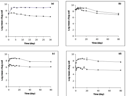
Following inoculation into sterile soil at −2.11 kPa, an initial increase up to 2 order of magnitude was observed in viable cell concentration (Fig. 1a) and remained in that level for up to 50 days. However, in non-sterile soil at −2.11 kPa viable cell concentration decreased by 1 order of magnitude by day 30, approximately 100 fold less than in sterile soil. In sterile soil viable cell concentration increased slightly at early stages after inoculation at matric potential −30 kPa (Fig. 1b) and from day 20 it started to decrease, showing that although viable population were still large, it was still lower than that observed in initial inocula. In non-sterile soil following inoculation, viable cell concentration after a slight increase at early days, decreased by 1 order of magnitude by day 80 and reached the same cell number as observed in sterile soil (Fig. 1b). An initial increase in the early days was also seen in sterile soil at matric potential −750 kPa and −1500 kPa which was greater than −30 kPa and lower than that at −2.11 kPa (Fig. 1c-1d). Viable cell concentrations in both matric potential (−1500 kPa and −750 kPa) decreased after day
20, however still 107 cells were alive after 80 days incubation. In non-sterile soil at −750 kPa viable cell did not change during 80 days incubation (Fig. 1c). In spite of small fluctuations in viable cell concentrations at –1500 kPa, significant change was not observed in non-sterile soil at this matric potential (Fig. 1d).
Luminescence Activity of Ralstonia eutropha H850 Lr
Luminescence initially increased in sterile soil and then was relatively constant for 40 days at a level that was higher than initial values (Fig. 2a). However, in non-sterile soil, luminescence gradually decreased by 2 orders of magnitude at this matric potential (−2.11 kPa). In both sterile and non-sterile soils at −30 kPa, light output increased for the first days of incubation and then decreased rapidly by 2 orders of magnitude up to day 40, after which luminescence remained constant (Fig. 2b). An initial increase was observed at −750 kPa in luminescence values of both sterile and non-sterile soils, in spite of lower light output at −30 kPa (Fig. 2c). At
−750 kPa, in non-sterile soil, luminescence decreased more rapidly than in sterile soil and differences between sterile and non-sterile soil was more apparent. At −1500 kPa despite greater decline in initial inoculum size, light out put in sterile soil changed in similar manner to −30 kPa and −750 kPa (Fig. 2d). In non-sterile soil, luminescence activity changed in very similar manner to viable cell concentration and differences between luminescence values in sterile and non-sterile soil was greater than −30 kPa and −750 kPa.
Statistical Analysis
GLM procedure was used to calculate the significance of the effect on viable cell concentration and luminescence of three factors, autoclaving, time and their interactions (Table 1). The data shows that all factors had a significant effect on both viable cell concentration and luminescence.
0 2 4 6 8 10
0 5 10 15 20 25 30
Time (day)
Log m
ean c
fu/
g soi
l
(a)
0 2 4 6 8 10
0 20 40 60 80
Time (day)
Log
m
e
an cf
u/
g soi
l
(b)
0 2 4 6 8 10
0 20 40 60 80
Time (day)
Lo
g m
ean cf
u/
g
s
o
il
(c)
0 2 4 6 8 10
0 20 40 60 80
Time (day)
L
o
g
m
e
an
cf
u
/g
so
il
(d)
Figure 1. Changes in viable cell concentration of Ralstonia eutropha H850Lr in sterile (▲) and non-sterile (■) microcosms following incubation at (a) −2.11 kPa, (b) −30 kPa, (c) −750 kPa, and (d) −1500 kPa. Bars on graph indicate standard errors of the mean (SEM). Where no bars are seen, SEMs were less than the symbol size.
Discussions
The results obtained in this study suggest that matric potential had a significant effect on the viable cell concentration of R. eutropha H850Lr following
inoculation into soil, which support previous studies on the effects of matric potential by Rattray et al. (1992)
and Meikle et al. (1995). It is found that, because of
higher moisture content at −2.11 kPa, and therefore availability of more nutrients, R. eutropha H850Lr
survived better comparing with lower matric potentials. However, the decrease in viable cell concentrations in sterile soil was more obvious at −2.11 kPa and −30 kPa soil than −750 kPa and −1500 kPa suggesting that matric potentials lower than −750 would probably have the same effect on the survival of this particular microbe. In non-sterile, R. eutropha H850Lr could
survive better at −30 kPa than at −2.11 kPa with a high moisture content, possibly because of better conditions for this GEM at this matric potential provided in the soil microcosms. The significant difference in viable cell
concentration observed between sterile and non-sterile soil and also among the matric potentials studied in non-sterile soil could be explained by the grazing activities of indigenous protozoa as a significant component in the survival and establishment of bacterial inocula [6]. Also the predatory activities of protozoa are considered to be affected by the matric potential of the soil, since protozoa are dependent upon water for their dispersal and movement through soil [17]. It has been observed that the largest protozoan populations are found to exist in water saturated soils, and the lowest concentrations in dry soil [2].
The prokaryotic lux genes encoding luciferase have
been used as markers to examine viable but nonculturable (VBNC) state as well as the survival rate of allochthonous bacteria in the environment [26,27]. Eberl et al. (1997) showed that by employing a
luciferase-marked derivative of P. putida KT2442 in
0 1 2 3 4 5 6 7
0 5 10 15 20 25 30
Time (day) Log m e an l u m ine sc e n ce ( R LU /g soi l) (a) 0 1 2 3 4 5 6 7
0 20 40 60 80
Time (day) Log m e an l u m ine s c e n c e ( R LU /g soi l) (b) 0 1 2 3 4 5 6 7
0 20 40 60 80
Time (day) L o g m e a n lu min e s c e n c e ( R L U /g s o il) (c) 0 1 2 3 4 5 6 7
0 20 40 60 80
Time (day) Log m e an l u m ines c e n ce ( R LU /g s o il ) (d)
Figure 2. Changes in luminescence in sterile (▲) and non-sterile (■) microcosms inoculated with R. eutropha H850 Lr and adjusted to matric potentials (a) −2.11 kPa, (b) −30 kPa, (c) −750 kPa, and (d) −1500 kPa. Bars on graph indicate standard errors of the mean (SEM). Where no bars are seen, SEMs were less than the symbol size.
Table 1. Values of probabilities calculated using GLM procedure to assess the effects of matric potential, time and autoclaving soil on viable cell concentration, and luminescence in soil microcosms inoculated with Ralstonia eutropha H850Lr
Viable Cell Concentration Luminescence
Autoclaving P < 3.54 × 10⁻¹¹ P < 3.54 × 10⁻¹¹
Time P < 3.54 × 10⁻¹¹ P < 3.54 × 10⁻¹¹
Matric Potential P < 3.54 × 10⁻¹¹ P < 3.54 × 10⁻¹¹
Autoclaving × Time P < 3.54 × 10⁻¹¹ 2.7 × 10⁻¹⁰
Autivclaving × Matric Potential P < 3.54 × 10⁻¹¹ P < 3.54 × 10⁻¹¹
Time × Matric Potential 3.54 × 10⁻¹¹ P < 3.54 × 10⁻¹¹
Autoclaving × Time × Matric Potential 0.106 5.01 × 10⁻⁹
Luminescence values were closely related to viable cell concentrations, showing the advantage of bioluminescence-marker systems compared to other methods for measuring of activity such as respirometry
potentials from saturated condition to dry condition in non-sterile soil, R. eutropha H850Lr could survive
better at −30 kPa. Also bioluminescence marker systems used for monitoring the genetically engineered R. eutropha H850 Lr proved to be an appropriate marker
system for assessing the activity of above GEM even in the presence of indigenous micro organisms. However, in spite of their proficiency in detecting culturable bacteria in natural environments, techniques using luminometry to measure in situ activity and potential
activity of introduced micro organisms are limited in their ability to confirm the VBNC state. The reason for this is that in situ detection of lux-encoded luciferase
activity is dependent on energy reserves within cells, but energy reserves of bacteria in the environment may be too low to allow in situ detection of high energy requiring enzyme systems. Therefore, to overcome a green fluorescence protein (GFP) from Aequorea victoria as a marker can be used to verify the VBNC
state of R. eutropha H850 Lr isolated from the
environment. GFP is a very good marker system that can be easily detected by the conventional antibiotic resistance, fluorescence colony counting, and measuring direct fluorescence with spectrofluorometry or epifluorescence microscopy. In addition, the green fluorescent phenotypes are detectable in all growth phases even under starved conditions. Unge et al.
(1999) developed a dual marker system for simultaneous quantification of bacterial cell numbers and their activity with the luxABand gfp genes. Their
dual marker system allowed simultaneous monitoring of the metabolic activity and cell number of a specific bacterial population in situin environmentalsamples.
References
1. Bedard D.L., Haber M.L., May R.J., and Berennan M.J. Evidence for novel mechanisms of polychlorinated biphenyl metabolism in Alcaligenes eutrophus H850. Appl. Environ. Microbiol., 53: 1103-12 (1987).
2. Darbyshire J.F. Effect of water suctions on the growth in soil of the ciliate Colopoda steinii and the bacterium Azotobacter chroococcum. J. Soil Sci., 27: 369-376 (1976).
3. Dighton J., Jones H.E., Robinson C.H., and Beckett J. The role of abiotic factors, cultivation practices and soil fauna in the dispersal of genetically modified microorganisms in soils. Appl. Soil Ecol., 5:109-131 (1997).
4. Dwyer D.F., Rojo F., and Timmis K.N. Fate and behaviour in an activated sludge microcosm of a genetically engineered microorganism designed to degrade substituted aromatic compounds. In: Sussman M., Collins C.H., Skineer F.A., and Stewart-Tull D.E. (Eds), The Release of Genetically Engineered
Microorganisms. London, San Diego: Academic Press Ltd, pp. 77-88 (1988).
5. Guerin W.F. and Boyd S.A. Maintenance and induction of naphthalene degradation activity in Pseudomonas putida and Alcaligenes sp. under different culture conditions. Appl. Environ. Microbiol., 61: 4061-4068 (1995).
6. Habte M. and Alexander M. Protozoan density and the coexistence of protozoan predators and bacterial prey. Ecology, 59: 140-146 (1978).
7. Hickey W.J., Searles D.B., and Focht D.D. Enhanced mineralization of polychlorinated biphenyls in soil inoculated with chlorobenzoate-degrading bacteria. Appl. Environ. Microbiol., 59: 1194-1200 (1993).
8. Marshall T.J., Holmes J.W., and Rose C.W. Soil physics. 3rd Edition. Cambridge University Press, Cambridge (1996).
9. Meikle A., Amin-Hanjani S., Glover L.A., Killham K., and Prosser J.I. Matric potential and the survival and activity of a Pseudomonas fluorescens inoculum in soil. Soil Biol. Biochem., 26: 747-755 (1995).
10. Park K.S., Sims R.C., Dupont R.R., Doucette W.J., and Matthews J.E. Fate of PAH compounds in types: influence of volatilization, abiotic loss and biological activity. Environ. Toxicol. Chem., 9: 187-195 (1990). 11. Pickup R.W. and Saunders J.R. Detection of genetically
engineered traits among bacteria in the environment. Trends in Biotechnol., 8: 329-335 (1990).
12. Postma J., Hok-A-Hin C.H., and Oude Voshaar J.H. Influence of the inoculum density on the growth and survival of Rhizobium leguminosarum biovar trifolii introduced into sterile and non-sterile loamy sand and silt loam. FEMS Microbiol. Ecol., 73: 49-58 (1990).
13. Rattrary E.A.S., Prosser J.I., Killham K., and Glover L.A. Luminescence-Based nonextractive technique for in situ detection of Escherichia coli in Soil. Appl. Environ. Microbiol., 56: 3368-3374 (1990).
14. Rattray E.A.S., Prosser J.I., Glover L.A., and Killham K. Matric potential in relation to survival and activity of a genetically modified microbial inoculum in soil. Soil Biol. Biochem., 24: 421-425 (1992).
15. Ryder M. Key issues in the deliberate release of genetically-manipulated bacteria. FEMS Microbiol. Ecol., 15: 139-146 (1994).
16. Shaw J.J., Dane F., Geiger D., and Kloepper J.W. Use of bioluminescence for detection of genetically engineered microorganism released into environment. Appl. Environ. Microbiol., 58: 267-273 (1992).
17. Sleigh M. The Biology of Protozoa. Edward Arnold, London, pp. 273-274 (1973).
18. Van Dyke M.I., Lee H., and Trevors J.T. Survival of lux-marked Alcaligenes eutrophus H850 in PCB-contaminated soil and sediment. J. Chemical Technol. Biotechnol., 65: 115-122 (1996).
19. Vanderberg P.A. Degradative bacteria. In: Levin M.A., Seidler R.J., and Regul M. (Eds) Microbial Ecology: Principals, Methods and Application. New York: Mc Graw-Hill, pp. 435-453 (1992).
growth kinetics. Appl. Microbiol. Biotechnol., 36: 548-552 (1992)
21. Ritchie J.M., Campbell G.R., Shepherd J., Beaton Y., Jones D., Killham K., and Artz R.R.E. A Stable Bioluminescent Construct of Escherichia coli O157:H7 for Hazard Assessments of Long-Term Survival in the Environment. Appl. Environ. Microbiol., 69: 3359-3367 (2003).
22. Richard J., Whittington D., Marshall J., Nicholls P.J., Marsh I.B., and Reddacliff L.A. Survival and Dormancy of Mycobacterium avium subsp. Paratuberculosis in the Environment. Appl. Environ. Microbiol., 70: 2989-3004 (2004).
23. Waddell, T.E., and Poppe C. Construction of mini-Tn10luxABcam/Ptac-ATS and its use for developing a bacteriophage that transduces bioluminescence to Escherichia coli O157:H7. FEMS Microbiol. Lett., 182: 285-289 (2000).
24. Turnbull, G.A., Morgan J.A., Whipps W.J.M., and Saunders J.R. The role of bacterial motility in the survival and spread of Pseudomonas fluorescens in soil and in the attachment and colonisation of wheat roots. FEMS
Microbiol. Ecol., 36: 21-31 (2001).
25. Campbell G.R., Prosser J.I., Glover L.A., and Killham K. Detection of Escherichia coli O157:H7 in soil and water with multiplex PCR. J. Appl. Microbiol., 91: 1-7 (2001). 26. Duncan S., Glover L.A., Killham K., and Prosser J.I.
Luminescence-based detection of activity of starved and viable but nonculturable bacteria. Appl. Environ. Microbiol., 60: 1308-1316 (1994).
27. Oliver J.D., McDougald D., Barrett T., Glover L.A., and Prosser J.I. Effect of temperature and plasmid carriage on nonculturability in organisms targeted for release. FEMS Microbiol. Ecol., 17: 229-238 (1995).
28. Eberl L., Givskov M., Poulsen L.K., and Molin S. Use of bioluminescence for monitoring the viability of individual Pseudomonas putida KT2442 cells. FEMS Microbiol. Let., 149: pp. 133-140 (1997).