RESEARCH
Stabilization of Foxp3 expression
by CRISPR-dCas9-based epigenome editing
in mouse primary T cells
Masahiro Okada
*, Mitsuhiro Kanamori, Kazue Someya, Hiroko Nakatsukasa and Akihiko Yoshimura
*Abstract
Background: Epigenome editing is expected to manipulate transcription and cell fates and to elucidate the gene
expression mechanisms in various cell types. For functional epigenome editing, assessing the chromatin context-dependent activity of artificial epigenetic modifier is required.
Results: In this study, we applied clustered regularly interspaced short palindromic repeats (CRISPR)-dCas9-based
epigenome editing to mouse primary T cells, focusing on the Forkhead box P3 (Foxp3) gene locus, a master tran-scription factor of regulatory T cells (Tregs). The Foxp3 gene locus is regulated by combinatorial epigenetic modi-fications, which determine the Foxp3 expression. Foxp3 expression is unstable in transforming growth factor beta (TGF-β)-induced Tregs (iTregs), while stable in thymus-derived Tregs (tTregs). To stabilize Foxp3 expression in iTregs, we introduced dCas9-TET1CD (dCas9 fused to the catalytic domain (CD) of ten-eleven translocation dioxygenase 1 (TET1), methylcytosine dioxygenase) and dCas9-p300CD (dCas9 fused to the CD of p300, histone acetyltransferase) with guide RNAs (gRNAs) targeted to the Foxp3 gene locus. Although dCas9-TET1CD induced partial demethylation in enhancer region called conserved non-coding DNA sequences 2 (CNS2), robust Foxp3 stabilization was not observed. In contrast, dCas9-p300CD targeted to the promoter locus partly maintained Foxp3 transcription in cultured and primary T cells even under inflammatory conditions in vitro. Furthermore, dCas9-p300CD promoted expression of Treg signature genes and enhanced suppression activity in vitro.
Conclusions: Our results showed that artificial epigenome editing modified the epigenetic status and gene
expres-sion of the targeted loci, and engineered cellular functions in conjunction with endogenous epigenetic modification, suggesting effective usage of these technologies, which help elucidate the relationship between chromatin states and gene expression.
Keywords: Treg, Foxp3, CRISPR, dCas9, TET1, p300, Epigenome editing
© The Author(s) 2017. This article is distributed under the terms of the Creative Commons Attribution 4.0 International License (http://creativecommons.org/licenses/by/4.0/), which permits unrestricted use, distribution, and reproduction in any medium, provided you give appropriate credit to the original author(s) and the source, provide a link to the Creative Commons license, and indicate if changes were made. The Creative Commons Public Domain Dedication waiver (http://creativecommons.org/ publicdomain/zero/1.0/) applies to the data made available in this article, unless otherwise stated.
Background
Epigenetic marks of histone modification and DNA cyto-sine methylation determine cell identity and function by regulating transcriptional activity at individual loci. Arti-ficial epigenome editing is a novel strategy for manipulat-ing cell fate by altermanipulat-ing the specific epigenomic landscape and can help elucidate the mechanisms between
chro-matin states and gene expression [1]. Epigenome editing
tools are fusion proteins consisting of a DNA-binding
domain fused with epigenetic-modifying enzymes. Previ-ous reports have shown that DNA-binding proteins, such as zinc finger protein (ZFP) and transcription activator-like effector (TALE) protein, can be used for targeted epigenome editing by fusion with
epigenome-modify-ing enzymes [2, 3]. Although their programmability is
verified, there is a disadvantage to designing extensive site-specific constructions. The clustered regularly inter-spaced short palindromic repeats (CRISPR)-associated protein 9 (Cas9) system (CRISPR-Cas9 system) from Streptococcus pyogenes has been used for genome edit-ing by inducedit-ing a guide RNA (gRNA) sequence-specific
Open Access
double-strand DNA break. Due to its simple design and high efficiency, the CRISPR-Cas9 system is expected to be utilized extensively in high-throughput and
multi-targeted genome editing [4]. Catalytic inactive Cas9
(dCas9) is also recruited to the targeted sequence of the DNA locus, and various fusion proteins with dCas9 can be used for target-specific transcriptional activation and
repression [5, 6]. For epigenetic modifications, dCas9
fusion with p300, lysine-specific demethylase 1 (LSD-1), Krüppel-associated box (KRAB), DNA methyltrans-ferase 3a (DNMT3a), and ten-eleven translocation (TET) dioxygenase 1 (TET1) enable gene expression regulation
by modifying epigenetic states [7–11]. These biological
devices were developed by using cultured cell lines and clearly proposed their versatile performance. However, on the basis that gene transcription is complexly regu-lated by epigenetic modifications in our body, it is easy to suppose the effectiveness of epigenome editing differs among target loci and cells. Therefore, applying them to primary tissues or cells and evaluation of their activity
is expected in the next studies [12]. In primary immune
cells, recent research has applied CRISPR-dCas9-based epigenome editing to human primary T lymphocytes,
mainly for silencing gene expression [13]. However, only
a few studies used epigenome editing mainly for activat-ing gene expression in primary immune cells. Further-more, little is known about the relationship between artificial epigenome editing and endogenous epigenetic modifications in immune cells.
Regulatory T cells (Tregs) play a pivotal role in regulat-ing immune responses and maintainregulat-ing immunological tolerance. Treg adoptive transfer therapy is expected to provide a clinical cure for various immunological
disor-ders [14–16]. Tregs are mainly generated via two
differ-ent routes. The first is through direct developmdiffer-ent from Treg progenitor cells in the thymus by thymic antigen presentation with high affinity. These Tregs are called naturally occurring Tregs (nTregs) or thymic Tregs (tTregs). The second is through differentiation from naïve CD4 T cells in the periphery by antigen presentation with transforming growth factor (TGF)-β. These Tregs are called induced Tregs in vitro (iTregs) or peripherally
induced Tregs (pTregs) [17, 18]. Both Tregs have
simi-lar suppression activity and markedly express Forkhead box P3 (Foxp3), a master transcriptional factor for Tregs. Foxp3 expression is required for the differentiation and maintenance of Treg function by expressing Treg signa-ture genes and suppressing effector T cell (Teff) genes
[19–23]. The number of available nTregs is limited. It is
thought that antigen-specific iTregs could be substituted for nTregs, because iTregs are induced and expanded with antigen specificity in vitro. However, Foxp3 expres-sion is unstable in iTregs owing to the lack of active
epigenetic modifications compared with tTregs [24, 25].
Hence, some remaining issues must be resolved prior to the clinical application of ex vivo-expanded iTregs, since iTregs lose Foxp3 expression easily and convert to other
pathogenic T cell subsets in vivo [26–28].
The epigenetic modification of the Foxp3 locus,
pro-moter, and three enhancer regions called conserved non-coding DNA sequences (CNS)1, CNS2, and CNS3, plays pivotal roles in the sustainable expression of Foxp3
[29]. Various transcriptional factors induce active
his-tone modification, such as H3K27 acetylation and H3K4
methylation [30]. Also, the microbial fermentation
prod-uct butyrate enhances histone acetylation of the Foxp3
promoter locus and promotes the induction of pTregs
in the intestine [31, 32]. In addition to histone
modifica-tions, DNA cytosine methylation also effects stable Foxp3 expression. nTregs show a Treg-specific demethylation
pattern. Importantly, the Foxp3 CNS2 locus is also
main-tained under hypomethylation in nTregs; this
hypometh-ylation contributes to the stable expression of Foxp3 [24,
25, 33]. Recent research has shown that TET family
teins are extensively involved in this demethylation
pro-cess and maintain Treg stability [34, 35]. In fact, some
epigenetic-modifying compounds, such as histone
dea-cetylase (HDAC) inhibitors [36], DNMT inhibitors [37],
and TET activators [38], are known for their potential use
in effective iTreg induction. However, their target loci are not limited because of low specificity, and there is a risk of undesirable effects like those observed with many of the epigenetic-modifying compounds used to treat
can-cer [39]. It is essential for functional iTregs to modulate
epigenetic modification at necessary locus and not to modulate unnecessary excess locus.
In this study, we established two epigenome-modifying systems based on CRISPR-dCas9 technology and applied
them to the Foxp3 gene locus. We aimed to investigate
the cross-talk of epigenome editing and endogenous cellular responses in primary immune cells and to lay a foundation for future clinical development. To stabilize Foxp3 expression in artificially epigenome-edited iTregs:
dCas9 fused with TET1CD was targeted to the Foxp3
CNS2 locus, and dCas9 fused with p300CD to the Foxp3
dCas9-p300CD epigenome-edited iTregs also showed high expression of Treg signature genes and enhanced suppression activity. Through various T cell culture con-ditions, we concluded that epigenome editing technology can be used in targeted epigenome research, and effec-tiveness depends on culture conditions. We expect that our study becomes the premise for broad clinical applica-tion in the future.
Methods Mice
Foxp3-hCD2-hCD52-KI mice originated from the labo-ratory of Dr. S Hori (Labolabo-ratory of Microbiology, Gradu-ate School of Pharmaceutical Sciences, the University of Tokyo, Tokyo, Japan).
Antibodies and reagents
For flow cytometry analysis, fluorescein isothiocyanate (FITC), phycoerythrin (PE), peridinin chlorophyll pro-tein-cyanine 5.5 (PerCP-Cy5.5), allophycocyanin (APC), PE-Cy7, and APC-Cy7-conjugated antibodies were pur-chased from BioLegend (San Diego, CA, USA) or eBio-science (San Diego, CA, USA). The following clones were used: anti-CD4 (RM4-5), Foxp3 (FJK16s), hCD2 (RPA2.10), CD25 (PC61.5), CTLA-4 (UC10-4F10-11), and CD45.1 (A20). Fixable Viability Dye eFluor 780 (FVD780) was used to remove dead cells. Cytokines (mouse interleukin-2 (IL-2), IL-12, IL-4, and IL-6) were purchased from Peprotech (Rocky Hill, NJ, USA), and human TGF-β1 was purchased from BioLegend. LY2157299 was purchased from Shanghai Biochempart-ner Co., Ltd (Hubei, China).
Cell culture
Human embryonic kidney cells 293 (HEK293T cells) were obtained from the American Type Culture Collection (ATCC) and maintained in Dulbecco’s modified Eagle medium (DMEM, 4500 mg/l glucose) supplemented with 10% fetal bovine serum (FBS). The 68-41 cells were a gift from Dr. M. Kubo (Division of Molecular Pathology, Research Institute for Biomedical Science, Tokyo
Univer-sity of Science, Tokyo, Japan) [40]. They were maintained
in Roswell Park Memorial Institute (RPMI) 1640 medium supplemented with 10% FBS and 55 µM 2-mercaptoeth-anol (2-ME). Primary T cells were maintained in RPMI 1640 medium supplemented with 10% FBS, 55 µM 2-ME, 1% penicillin/streptomycin, 2 mM l-glutamine, and 100 nM non-essential amino acid solution. All cells were
cultured in a humid, 5% CO2, 37 °C incubator.
Plasmid constructions
LentiCRISPR (Addgene, catalog no. 49535) was mutated at amino acid positions D10A and H840A by mutagenesis
polymerase chain reaction (PCR) to construct Flag-dCas9-P2A-puro. Mouse TET1 catalytic domain (TET1CD) or p300CD was amplified from complementary DNA (cDNA) and subcloned into a MIGR vector. TET1CD or p300CD and internal ribosome entry site green fluorescent protein (IRES-GFP) sequences were amplified with a Gly– Gly–Gly–Gly–Ser linker and recombined into Flag-dCas9-P2A-puro instead of Flag-dCas9-P2A-puro. Flag-dCas9-TET1CD or p300CD and IRES-GFP were recombined into pMXs-GW vectors. Amino acid sequences of each construct are
detailed in the supplementary material (Additional file 1).
For gRNA expression, DsRed was recombined into len-tiCRISPR instead of Cas9-P2A-puro. Next, U6-gRNA-EFS-DsRed was recombined into CSII vector. Each gRNA expression vector was generated by the annealing of the oligonucleotides, followed by ligation into BsmBI-digested gRNA expression vectors based on CSII vector for lenti-viral infection. In some cases, U6-gRNA-EFS-DsRed was recombined into pMXs-GW vectors for retroviral infec-tion. gRNA off-target predictions were performed by
CCTop—CRISPR/Cas9 target online predictor [41]. Max.
total mismatches, core length, and max. core mismatches were set to 4, 12, and 2, respectively. Predicted off-target
loci were listed in Additional file 2: Table S1.
Retroviral or lentiviral production
pMXs and pCL-Eco were co-transfected for retrovi-ral production, or CSII, pMDLg/pRRE, and VSV-G/ Rsv-Rev were co-transfected for lentiviral production into HEK293T cells using polyethylenimine MAX (PEI-MAX), followed by a medium change to remove the transfection reagents. Virus-containing medium was har-vested, filtered (0.45 µm), and then concentrated by cen-trifugation overnight.
Establishment of dCas9‑TET1CD or p300CD stable 68‑41 cells and gRNA transduction
Primary T cell culture
Naïve CD4+ T cells (CD4+CD25-hCD2-CD62L+)
were isolated using magnetic-activated cell sorting (MACS) from the spleen and lymph nodes of 6- to 8-week-old-male Foxp3-hCD2-hCD52-KI mice. Sple-nocytes and lymphocytes were red blood lysed and depleted using AutoMACS (Miltenyi Biotec, Tokyo, Japan) with biotin-conjugated anti-B220 (RA3-6B2), CD8a (53-67), CD49b (DX-5), CD11b (M1/70), CD11c (N418), CD25 (PC61), TER119 (TER-119), Ly6G (RB6-8C5), T cell receptor (TCR)-γδ (GL3), and sometimes hCD2 (RPA2.10) (BioLegend or eBioscience), or CD4 isolation kit (Miltenyi Biotec) with anti-CD25 and sometimes hCD2, and then with anti-Biotin micro-beads or streptavidin micromicro-beads (Miltenyi Biotec).
CD4+ T cells were further incubated with CD62L
microbeads (Miltenyi Biotec), and CD4+CD62L+ T
cells were positively selected by using AutoMACS. The
purity was almost >95%. Isolated naïve CD4+ T cells
(3–4 × 10⁵ cells) were cultured on 24-well plates under
iTreg conditions using anti-CD3e (plate-coated 2C11, 4 µg/ml), anti-CD28 (PV1.17.10, 1.2 µg/ml), anti-IFN-γ (R4-6A2, 5 µg/ml), anti-IL-4 (11B11, 5 µg/ml), recom-binant human TGF-β (2 ng/ml), and IL-2 (20 ng/ml). On day 2, dCas9-TETCD or p300CD and gRNA were co-transduced with 6 µg/ml polybrene (Merck Mil-lipore, Billerica, MA, USA) or 10 µg/ml Protransdu-zin A (Immundiagnostik AG, Bensheim, Germany) (mainly for sorting experiment), with centrifugation at 2500 rpm for 2 h at 35 °C. The next day, co-transduced iTregs were harvested and further cultured for 2 days
under Th1, Th2, and Th0 + IL-6 conditions using
anti-CD3e (plate-coated, 4 µg/ml), anti-CD28 (1.2 µg/ml), and IL-2 (20 ng/ml). Conditions were as follows: Th1, anti-IL-4 (5 µg/ml), IL-12 (20 ng/ml); Th2, anti-IFN-γ
(5 µg/ml), IL-4 (20 ng/ml); Th0 + IL-6, anti-IFN-γ
(5 µg/ml), anti-IL-4 (5 µg/ml), IL-6 (20 ng/ml); Th0, anti-IFN-γ (5 µg/ml), anti-IL-4 (5 µg/ml).
For helper T cell subsets skewing, naive CD4+ T cells
(3–4 × 10⁵ cells) were cultured on 24-well plates under
Th1, Th2, and Th17 conditions. Th1 and Th2 were the same as above: Th17, anti-IFN-γ (5 µg/ml), anti-IL-4 (5 µg/ml), human TGF-β (0.5 ng/ml), IL-6 (20 ng/ml).
For TGF-β signal inhibition experiment, indicated con-centration of TGF-β was used for iTreg condition in the presence of LY2157299 or anti-TGF-β (1D11).
For intracellular cytokine staining, cells were stimu-lated with 50 ng/ml phorbol myristate acetate (PMA), 1 µg/ml ionomycin, and Brefeldin A solution for 4 h. Cells were harvested and stained with FVD780, anti-CD4, hCD2, and CD25. For intracellular staining, anti-IFN-γ, anti-IL-2, and anti-CTLA-4 were used after fixation buffer and permeabilization buffer (eBioscience).
Western blot analysis
The cells were lysed using an immunoprecipitation lysis buffer (50 mM Tris–HCl (pH 7.5), 150 mM NaCl, 10 mM ethylenediaminetetraacetic acid (EDTA, pH 8.0), 1% sodium deoxycholate, 1% Triton X-100, 5 μg/ml leupep-tin, and 1 mM PMSF). The cell lysates were centrifuged, and the supernatants were mixed with 5*sodium dode-cyl sulfate (SDS) sample buffer (10% SDS, 40% glycerol, 0.2 M Tris–HCl (pH 6.8), 0.025% bromophenol blue, and 50 mM dithiothreitol [DTT]). After boiling, the samples were separated through electrophoresis and transferred to polyvinylidene difluoride (PVDF) membranes. The membranes were probed with anti-Flag (M2; Sigma-Aldrich, St. Louis, MO, USA) and anti-α-tubulin (DM1A; Sigma-Aldrich) and detected using the Chemi-Lumi One system (Nacalai Tesque, Kyoto, Japan).
Bisulfite sequencing
Cells were lysed using Wizard SV Lysis Buffer (Promega Corporation, Madison, WI, USA). Genomic DNAs were isolated by phenol–chloroform extraction, isopropanol precipitation, and 70% ethanol purification. Genomic DNAs were digested with BamHI, and the same amount of digested DNA (<1.5 µg) was aligned to 19 µl by adding
H2O. Next, 1.2 µl of 5 M NaOH was added and incubated
at 37 °C for 15 min. Then, 121.2 µl of bisulfite mixture was added and incubated for 1 h at 80 °C. The bisulfite mix-ture (121.2 µl) consisted of 3.6 M sodium bisulfite (1.92 g
in 4.4 ml H2O; 107 µl), 0.57 mM hydroquinone (11 mg in
10 ml H2O; 7 µl), and 0.3 M NaOH (5 M NaOH; 7.2 µl).
After purification and extraction using a 50-µl GP3 solu-tion from a column using a FastGene Gel/PCR Extracsolu-tion Kit (Nippon Genetics, Tokyo, Japan), 3 µl of 5 M NaOH was added and incubated for 5 min at 37 °C to complete the bisulfite reaction. Bisulfite products were precipi-tated using isopropanol with 10 µg glycogen and rinsed using 70% ethanol. The Foxp3 CNS2 locus was amplified by Quick Taq HS DyeMix (Toyobo Life Science, Tokyo, Japan) polymerase with a bisulfite sequence primer (for-ward primer: TTTTGGGTTTTTTTGGTATTTAAGA and reverse primer: AACTAACCAACCAACTTCCTA-CACTAT designed by MethPrimer [online]) and then subcloned into pGEM-T EASY Vector (Promega Corpo-ration). Plasmids were purified and sequenced using SP6 primer. Methylation analysis was performed using the quantification tool for methylation analysis (online).
Real‑time PCR analysis
Corporation) after treatment with RNase inhibitor from human placenta (Nacalai Tesque). cDNA was synthe-sized by reverse transcription by a High-Capacity cDNA Synthesis Kit (Applied Biosystems, Thermo Fisher Scien-tific K.K., Kanagawa, Japan) from RNA. Real-time PCR (RT-PCR) analysis was performed using an iCycler iQ multicolor RT-PCR detection system with SsoFast Eva-Green Supermix (Bio-Rad Laboratories, Hercules, CA, USA). Amplification primers (mFoxp3, forward primer: CCCAGGAAAGACAGCAACCTT, reverse primer: TTCTCACAACCAGGCCACTTG, mHPRT1, for-ward primer: TGAAGAGCTACTGTAATGATCAGTC, reverse primer: AGCAAGCTTGCAACCTTAACCA) were used for quantification.
Chromatin immunoprecipitation assay
Cells were fixed with 1 ml of 1% formaldehyde for 10 min at room temperature and washed two times using phos-phate-buffered saline (PBS). Then, cells were lysed using lysis buffer (1% SDS, 10 mM EDTA (pH 8.0), 50 mM Tris–HCl (pH 8.0), and protease inhibitor cocktail (Nacalai Tesque)). Genomes were sonicated to a mean size of 300 bp using an Acoustic Solubilizer (Covaris, Woburn, MA, USA) with the “300 bp shearing pro” pro-gram. Genomic fragment solutions were diluted tenfold with chromatin immunoprecipitation (ChIP) dilution buffer (0.01% SDS, 1.1% Triton-X, 1.2 mM EDTA (pH 8.0), 16.7 mM Tris–HCl (pH 8.0), 167 mM NaCl, and protease inhibitor cocktail). The input samples were col-lected, and the remainders were immunoprecipitated with 1 µg of Anti-acetyl-Histone H3 Antibody (Merck Millipore) or control rabbit immunoglobulin (Ig) G (Santa Cruz Biotechnology, Inc, Dallas, TX, USA), fol-lowed by incubation with dynabeads protein G (Invit-rogen, Thermo Fisher Scientific K.K.). Precipitants were washed in order, using low salt wash buffer (0.1% SDS, 1% Triton-X, 2 mM EDTA (pH 8.0), 20 mM Tris–HCl (pH 8.0), and 150 mM NaCl), high salt wash buffer (0.1% SDS, 1% Triton-X, 2 mM EDTA (pH 8.0), 20 mM Tris– HCl (pH 8.0), and 500 mM NaCl), LiCl buffer [0.25 M LiCl, 1% NP-40, 1% sodium deoxycholate, 1 mM EDTA (pH 8.0), and 10 mM Tris–HCl (pH 8.0)], and TE buffer (twice). Chromatin samples were eluted twice by incu-bation with 100 µl of elution buffer (1% SDS, 0.1 M
NaHCO3, and 10 mM DTT) for 15 min at room
tempera-ture. Followed by de-cross-linking using incubation at 65 °C overnight, 8 µl of 5 M NaCl was added to elution products. Next, 20.36 µl proteinase K mixture was added and incubated at 45 °C for 6 h. The proteinase K mixture (20.36 µl) consisted of 0.5 M EDTA (pH 8.0): 4 µl, 0.5 M Tris–HCl (pH 6.8): 16 µl, 20 mg/ml proteinase K: 0.36 µl. Purification and extraction were performed using a 50-µl GP3 solution from a column using a FastGene Gel/PCR
Extraction Kit (Nippon Genetics). ChIP products were analyzed by quantitative PCR. Amplification primers (forward primer: CCCTGCAATTATCAGCACACAC, reverse primer: ATCAGCCTGGCTTGTGGGAAAC) were used for quantification.
In vitro Treg suppression assay
Responder effector cells (CD4+CD25-) isolated by
MACS as described above from Ly5.1 cognate mice were labeled with 2 µM carboxyfluorescein diacetate succin-imidyl diester (CFSE) in 37 °C PBS for 10 min and then washed with sufficient RPMI medium, termed as Teff. Splenic CD11c-positive cells isolated by CD11c beads (Miltenyi Biotec) were used as antigen-presenting cells. iTregs co-transduced with dCas9-p300CD and gRNAs,
gated on CD4+GFP+DsRed+hCD2+ cells, were sorted
using a cell sorter SH800. Next, 4 × 10⁴ Teff and 2 × 10⁴
splenic dendritic cells (DCs) with 1 µg/ml soluble
anti-CD3e, with or without 1 × 10⁴ iTreg, were cultured in a
96-well U-bottomed dish for 4 days. CFSE dilution and Foxp3 (hCD2) expression were analyzed.
Statistical analysis
All values are presented as the means ± standard
devia-tions (SDs). Unpaired Student’s t tests were used, and
p < 0.05 was defined as statistically significant.
Results
Constructions and expression
For targeted epigenome editing, we constructed a retro-viral expression system for TET1CD and dCas9-p300CD fusion proteins. Mouse TET1CD H1620Y, D1622A, and mouse p300CD D1398Y were mutated for catalytic inactive mutants. As these retroviral vectors contain the IRES-GFP sequence, the expression of fusion proteins can be monitored by GFP expression. For gRNA expression, we constructed retroviral or lentiviral expres-sion systems that contain DsRed as the fluorescence
marker (Fig. 1a). We confirmed the expression of fusion
proteins in HEK293T cells using western blotting
meth-ods (Fig. 1b).
dCas9‑TET1CD induced demethylation of the Foxp3 CNS2 locus, but weakly sustained Foxp3 expression
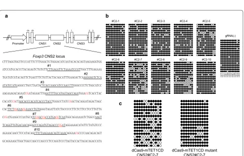
The Foxp3 CNS2 locus contains 12 CpG sites, and its methylation or demethylation status is extensively involved in the unstable or stable Foxp3 expression phe-notype, respectively. To edit the methylation status, we
designed 10 gRNA sequences at the Foxp3 CNS2 locus
(Fig. 2a) and transduced them into a 68-41 T cell line that
stably expressed dCas9-TET1CD. The Foxp3 CNS2 locus
a b
LTR IRES GFP LTR
LTR dCas9
TET1CD
IRES GFP LTR
p300CD GGGGS linker
GGGGS linker
LTR dCas9 TET1CD IRES GFP LTR
GGGGS linker
LTR dCas9 p300CD IRES GFP LTR
GGGGS linker
D1398Y H1620Y, D1622A
LTR U6 gRNA EFS DsRED LTR
Psi RRE cPPT U6 gRNA EFS DsRED WPRE
dCas9
Flag
Flag
Flag
Flag
Flag-dCas9-mTET1CD or mutant
-tubulin
Flag-dCas9-TET1CD
Flag-dCas9-p300CD
Flag-dCas9-mp300CD or mutant
-tubulin
- WT H1620Y, D1622A
- WT D1398Y 196
64
47
196 64 47 kDa
kDa
Fig. 1 CRISPR-dCas9-based epigenome editing for primary T cells. a A retroviral vector for the expression of dCas9-epigenome regulator fusion proteins from Moloney murine leukemia virus promoter long terminal repeats (ΔLTRs) and green fluorescent protein (GFP) from an internal ribo-somal entry site (IRES). Retroviral and lentiviral vector for bicistronic expression of the gRNA from a U6 promoter (U6) and DsRed from a short EF1a promoter (EFS). b Protein expression of dCas9-epigenome regulator fusion proteins in transfected HEK293T cells was detected by western blot against anti-Flag antibody. Anti α-tubulin antibody was used for loading control
#C2-1 #C2-2 #C2-3 #C2-4 #C2-5
#C2-6 #C2-7 #C2-8 #C2-9 #C2-10
gRNA(-)
+427
5
+431
1
+432
7
+436
1
+438
8
+439
3
+444
2
+446
1
+446
6
+447
3
+453
5
+460
5
dCas9-mTET1CD
CNS2#C2-7 dCas9-mTET1CD mutant CNS2#C2-7
a b
c
Foxp3 CNS2 locus
#2 #1
#3
#4
#5
#6
#7 #8
#9
#10
Promoter CNS1 CNS2 CNS3
-2a -2b -1 1 2 3
Fig. 2 dCas9-TET1CD-mediated demethylation of the Foxp3 CNS2 locus. a Sequence at the Foxp3 CNS2 locus is shown. Each gRNA sequence is
underlined and numbered #C2-1 to #C2-10. Specific CpG sites are lettered red. b and c The methylation status of CpG sites at the Foxp3 CNS2 locus in dCas9-TET1CD and each gRNA-expressing 68-41 cells (b) and dCas9-TET1CD or the TET1CD catalytic mutant and gRNA #C2-7 expressing 68-41 cells (c) was determined by bisulfite sequence analysis. The 68-41 cells stably expressing dCas9-TET1CD were transduced with each gRNA expres-sion lentivirus and sorted (b). The 68-41 cells were co-transduced with dCas9-TET1CD or TET1CD mutant and gRNA CNS2#C2-7 and sorted (c). A
the Foxp3 CNS2 locus was analyzed by bisulfite sequenc-ing. The results revealed that several gRNA sequences, such as #C2-7 and #C2-5, could induce demethylation to some extent (approximately 30% by #C2-7), whereas
#C2-1 and #C2-10 had little effect (Fig. 2b). The catalytic
inactive mutant of TET1CD induced less with #C2-7, indicating that demethylation by dCas9-TET1CD was
TET enzyme activity dependent (Fig. 2c). We selected
#C2-1 as a negative control gRNA and #C2-7 as a posi-tive control gRNA. Unlike the reported demethylation
pattern by TALE-TET1 fusion proteins [42],
dCas9-TET1CD fusion proteins could demethylate some CpG sites distant from the designed gRNA sequences. These
findings coincide with previous reports [11, 43].
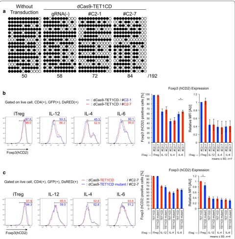
Next, we applied dCas9-TET1CD to primary T cells from male Foxp3-hCD2-hCD52-KI mice under iTreg skewing conditions and confirmed its demethylation activity in iTregs. Unlike 68-41 T cell lines, iTregs showed slight demethylation at the CNS2 locus at the basal level in the absence of gRNAs, and this demethylation was enhanced by co-transduction of dCas9-TET1CD with
gRNAs (Fig. 3a). We then examined the promotive effect
of dCas9-TET1CD on Foxp3 stability in iTregs. Foxp3 stability under inflammatory conditions was
investi-gated using the following method. Naive CD4+ T cells
were cultured under iTreg skewing conditions for 3 days,
resulting in >90% Foxp3(+) cells. iTregs were harvested
and further cultured under the same iTreg conditions (for positive control) or under inflammatory cytokine (in the presence of IL-12, IL-4, or IL-6) conditions for 2 days. This re-stimulation destabilized Foxp3 expres-sion, which was monitored by surface hCD2 staining correlated with intracellular Foxp3 staining as shown
in Additional file 3: Figure S1. The results coincide with
previous reports [44–47]. To retain GFP and DsRed
fluo-rescence, we monitored Foxp3 expression by hCD2 with-out intracellular staining. Using this method, compared
with no gRNA-transduced cells (GFP(+)DsRed(−) cells),
dCas9-TET1CD and gRNA co-transduction yielded
sta-bilized Foxp3 expression (Additional file 3: Figure S2a).
Since demethylation occurred in #C2-1 co-transduced iTregs to some extent, a partial stabilization effect was observed in #C2-7 co-transduced iTregs in comparison
with #C2-1 co-transduced iTregs (Fig. 3b), which were
confirmed by Foxp3 mRNA expression (Additional file 3:
Figure S2b). Comparable to the dCas9-TET1CD mutant, similar stabilization effects were detected to some extent
(Fig. 3c). These data indicated that dCas9-TET1CD for
the Foxp3 CNS2 locus had a certain stabilizing effect for Foxp3 expression, but its effect was weak especially
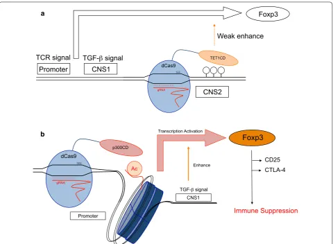
when exposed to inflammatory cytokines (Fig. 7a). A
previous report suggested that inflammatory cytokine signals (IL-4/STAT6, IL-6/STAT3) recruit DNMT1 and
DNMT3a to the CNS2 locus after stimulation, leading
to Foxp3 loss even in nTregs [46]. We speculated that
dCas9-TET1CD targeted to the CNS2 locus competes with methyltransferases under inflammatory condi-tions, resulting in earlier loss of demethylation function than under iTreg conditions. In fact, Foxp3 mean fluo-rescence intensity (MFI) was greater in dCas9-TET1CD than in TET1CD catalytic inactive mutant under iTreg
conditions (Fig. 3c), but was weakened by inflammatory
stimuli.
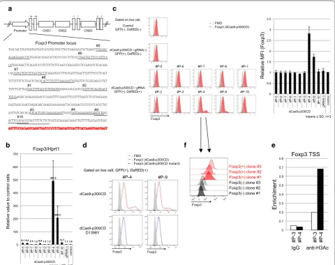
dCas9‑p300CD induced acetylation of the Foxp3 promoter locus and induced stable expression of Foxp3 in a cultured T cell line
Foxp3 expression is induced by histone acetylation of the promoter locus. We designed 10 gRNA sequences at the Foxp3 promoter locus (Fig. 4a) and transduced them into the 68-41 T cell line that stably expressed dCas9-p300CD. The 68-41 cell line showed little Foxp3 expres-sion. We measured the amount of mRNA expression induced by dCas9-p300CD. We observed that #P-4 and #P-9 strongly activated Foxp3 transcription, and that #P-5, #P-6, #P-1, and #P-10 induced moderately
acti-vated Foxp3 transcription (Fig. 4b). Next, we assessed
protein expression. A small but significant fraction of endogenous Foxp3 expression could be detected in #P-4
and #P-9 transduced cells (Fig. 4c). This induction was
dependent on p300CD autoacetylation activity, since the
catalytic inactive mutant could not induce it (Fig. 4d).
We selected #P-3 as a negative control gRNA and #P-4 as a positive control gRNA. In #P-4 transduced cells, the histone acetylation of the Foxp3 promoter locus was pro-moted compared with #P-3, correlating with
transcrip-tional activation (Fig. 4e).
Although similar expression levels of dCas9-p300CD and #P-4 are gated, approximately 10% of the popula-tion significantly expressed Foxp3. By limiting dilupopula-tion, we isolated several clones that stably expressed a high amount of Foxp3, and others that never expressed it
(Fig. 4f). Although we could not explain the mechanism
of this bipolarization phenomenon, the data suggested that it was not due to the oscillation of the cell popula-tion. The data indicated that targeted histone acetyla-tion could strongly maintain epigenetic modificaacetyla-tion and transcriptional activation in certain specific cells.
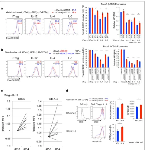
dCas9‑p300CD induces stable Foxp3 expression in primary T cells
We applied this system to primary T cells. Under helper T cell culture skewing conditions, we co-transduced dCas9-p300CD and #P-3 and #P-4 into isolated naïve
CD4+ T cells and investigated Foxp3 expression. Foxp3
and a notably superior enhancing effect was observed under the Th17 condition (IL-6 and TGF-β), as shown
in Fig. 5a. IL-2 was added to all skewing conditions
to improve T cell proliferation and transduction effi-ciency. Thus, the majority of the population expressed Foxp3, even under Th17 conditions. TGF-β induces
a
b
c
50 58 72 84 /192
dCas9-TET1CD
gRNA(-) #C2-1 #C2-7
Without Transduction
iTreg IL-12 IL-4 IL-6
Foxp3(hCD2)
Gated on live cell, CD4(+), GFP(+), DsRED(+)
97.5
98.0 53.3 66.2 45.3 52.9 50.1 60.0
: dCas9-TET1CD / #C2-1 : dCas9-TET1CD / #C2-7
Foxp3 (hCD2) positive cells [%
]
means ± SD, n=7 iTreg iTreg
Relative MFI [AU]
*
0 10 20 30 40 50 60 70 80 90 100
#C2-1 #C2-7 #C2-1 #C2-7 #C2-1 #C2-7 #C2-1 #C2-7
iTreg IL-12 IL-4 IL-6 0 0.2 0.4 0.6 0.8 1 1.2
#C2-1 #C2-7 #C2-1 #C2-7 #C2-1 #C2-7 #C2-1 #C2-7
iTreg IL-12 IL-4 IL-6
Foxp3 (hCD2) Expression
iTreg IL-12 IL-4 IL-6
Foxp3(hCD2)
Gated on live cell, CD4(+), GFP(+), DsRED(+) : dCas9-: dCas9-TET1CDTET1CD mutant / #C2-7 / #C2-7
97.9
98.3 65.5 59.5 52.8 52.8 53.6 51.2
Foxp3 (hCD2) positive cells [%
]
means ± SD, n=4 iTreg iTreg
Relative MFI [AU]
*
0 10 20 30 40 50 60 70 80 90 100
TET1CD
TET1CD mutant
TET1CD
TET1CD mutant
TET1CD
TET1CD mutant
TET1CD
TET1CD mutant
iTreg IL-12 IL-4 IL-6 0 0.2 0.4 0.6 0.8 1 1.2
TET1CD
TET1CD mutant
TET1CD
TET1CD mutant
TET1CD
TET1CD mutant
TET1CD
TET1CD mutant
iTreg IL-12 IL-4 IL-6
Foxp3 (hCD2) Expression
Foxp3 mainly through the Foxp3 CNS1 enhancer locus
by Smad2 and Smad3 signals [29, 48, 49]. This indicates
that the artificial histone acetylation of the promoter locus activated transcription from the promoter locus, and it was augmented by the TGF-β signal, which prin-cipally activates the enhancer locus. In other words, the TGF-β signal additionally activated Foxp3 transcription, even when the promoter locus was artificially opened. This possible enhanced activity of dCas9-p300CD under TGF-β signal condition was confirmed by blocking its signals. Low-dose TGF-β enhanced Foxp3 expression
within #P-4 co-transduced cells than #P-3, and this effect was cancelled by treating LY2157299 (TGF-β recep-tor kinase inhibirecep-tor) or anti-TGF-β antibody, indicating the involvement of TGF-β signal in enhancing
dCas9-p300CD activity (Additional file 3: Figure S3). Then, we
speculated whether co-transduction of dCas9-p300CD and #P-4 facilitated TGF-β signal to engage in transac-tivation by investigating Foxp3 expression in T cell plas-ticity culture (Th1 to iTregs). Since helper T cell subset plasticity is strictly regulated, Foxp3 cannot be induced
by TGF-β in already differentiated Th1 cells [50]. As we
Foxp3 Foxp3
#P-4 #P-9
dCas9-p300CD
dCas9-p300CD D1398Y
: FMO
: Foxp3 (dCas9-p300CD) : Foxp3 (dCas9-p300CD mutant)
Gated on live cell, GFP(+), DsRED(+)
a
b
c
d f e
Foxp3/Hprt1
9.1 9.4 2.5 1.2 6.9 1.5 1.2 491.2
206.5
12.7 1.1 1.0
0 100 200 300 400 500 600 700
#P-5 #P-6 #P-7 #P-8 #P-1 #P-2 #P-3 #P-4 #P-9 #P-10 gRNA(-) control dCas9-p300CD
Relative value to control cells
means ± SD, n=3
0 0.5 1 1.5 2 2.5 3 3.5
#P-5 #P-6 #P-7 #P-8 #P-1 #P-2 #P-3 #P-4 #P-9 #P-10 gRNA(-) control dCas9-p300CD
means ± SD, n=3
Relative MFI (Foxp3
)
0 0.1 0.2 0.3 0.4 0.5 0.6 0.7 0.8
IgG anti-H3Ac
#P-3
#P-4
#P-3
#P-4
Enrichimen
t
Foxp3 TSS
Foxp3(-) clone #1 Foxp3(-) clone #2 Foxp3(-) clone #3
Foxp3(+) clone #1 Foxp3(+) clone #2 Foxp3(+) clone #3
Foxp3 #5
#6
#7
#8
#1 #2
#3 #4 #9
#10
Foxp3 Promoter locus
Promoter CNS1 CNS2 CNS3
-2a -2b -1 1 2 3
Control GFP(-), DsRED(-)
dCas9-p300CD / gRNA(-) GFP(+), DsRED(-)
dCas9-p300CD / gRNA GFP(+), DsRED(+)
Foxp3
#P-5 #P-6 #P-7 #P-8 #P-1
#P-2 #P-3 #P-4 #P-9 #P-10 Gated on live cell,
: FMO
: Foxp3 (dCas9-p300CD)
CD4
Foxp3-hCD2
#P-3 #P-4
Th1
Th2
Th17
0.10 3.13
0.09 2.63
47.5 65.2
** ** *
* *
Foxp3 (hCD2) positive cells [%
]
Promoting Effect [%
]
0 10 20 30 40 50 60 70 80 90 100
#P-3 #P-4 #P-3 #P-4 #P-3 #P-4 Th1 Th2 Th17
0 2 4 6 8 10 12 14 16 18 20
Th1 Th2 Th17
Gated on live cell, CD4(+), GFP(+), DsRED(+)
means ± SD, n=3 Transduction
0 2
day
CD4+CD62L+ Foxp3 expressionAnalysis of
4 Th1 (IL-12)
Th2 (IL-4)
Th17 (IL-6, TGF- ) 3
Half medium change
#P-3 #P-4
Th1 Th1
Th1 iTreg
0 1 2 3 4 5 6 7 8 9 10
Th1 iTreg Th1 iTreg #P-3 #P-4
Th1 without TCR
Foxp3 (hCD2) positive cells [%
]
*
means ± SD, n=3
Th1
iTreg (TGF- ) Th1 (IL-12) Transduction
0 2 3 5
day
CD4+CD62L+ Analysis of
Foxp3 expression
Gated on live cell, CD4(+), GFP(+), DsRED(+)
CD4
Foxp3-hCD2
0.61
0.52
6.45
9.40 a
expected, Foxp3 expression was promoted in dCas9-p300CD and #P-4 co-transduced cells, even when differ-entiated Th1 cells were further treated with TGF-β. This
promotion effect never occurred in #P-3 cells (Fig. 5b).
Epigenome editing could thus be a novel method for con-verting the T cell subset.
Then, we examined the maintenance of Foxp3 by dCas9-p300CD in iTregs. The stability of Foxp3 under inflammatory cytokines was investigated (same as Addi-tional file 3: Figure S1). We observed that iTregs co-transduced with dCas9-p300CD and #P-4 retained a high amount of Foxp3 compared with #P-3 under
inflamma-tory conditions (Fig. 6a). We confirmed this maintenance
is actually dependent on p300CD autoacetylation activity by co-transduction with p300 catalytic inactive mutant
(Fig. 6b). Moreover, the Treg signature genes CD25 and
CTLA-4 were slightly but significantly upregulated under
IL-12 conditions in #P-4 transduced iTregs (Fig. 6c).
Finally, we examined the iTreg suppression activity in vitro. iTregs co-transduced with dCas9-p300CD and
gRNA were sorted (Additional file 3: Figure S4a). Splenic
DCs were used as antigen-presenting cells, and the prolif-eration of effector T cells was further suppressed by #P-4 transduced iTregs, which correlated with Foxp3
stabiliza-tion (Fig. 6d). Similar tendency was observed in
compari-son with catalytic activity (Additional file 3: Figure S4b).
These data showed that applying dCas9-p300CD to pri-mary T cells, especially iTregs, could modify both tran-scription and cell function. These data also clarified one aspect of the Foxp3 transcriptional activation
mecha-nisms (Fig. 7b).
Off‑target analysis of selected gRNAs
CRISPR-Cas9 or CRISPR-dCas9-based technologies
are constantly at risk of off-target activity [51, 52]. For
clinical usage or to validate results, we have to consider
off-target effects. We used the CCTop online tool [41],
and selected gRNA sequences (#C2-1, #C2-7, #P-3, and #P-4) were investigated for potential off-target sites. We observed that the selected gRNA sequences had at least three mismatches on similar sequences, and most off-target candidate sequences were localized in intergenic
regions (Additional file 2: Table S1). In our study, which
mainly focused on mice experiments and revealing the relationships between epigenetics and gene expression, all candidate genes were not strongly involved in direct Foxp3 induction or Treg functions to the best of our knowledge. For future clinical usage, we have to re-select gRNA sequences in the human genome and investigate off-target activity in our next study.
Discussion
Artificial targeted epigenome editing mediated by CRISPR-dCas9 can be utilized to clarify the relationship between chromatin states and gene expression and to develop novel clinical strategies. Previous research pro-posed this biological device and demonstrated its univer-sal performance and efficiency at the targeted locus. In this study, we expanded epigenome editing to mouse
pri-mary T cells, with a focus on the Foxp3 locus to elucidate
epigenetic regulation mechanisms, and to advance future clinical usage in immunotherapy.
In this study, we applied TET1CD and
dCas9-p300CD to the Foxp3 CNS2 and promoter locus,
respectively, and attempted to generate Foxp3 stability-enhanced iTregs. We succeeded in epigenome editing at both loci, and the histone acetylation at the promoter locus strongly activated Foxp3 expression, but the DNA demethylation at the CNS2 locus slightly affected for Foxp3 expression.
dCas9-TET1CD demethylated the CNS2 locus, but did not intensely stabilize Foxp3 expression under inflam-matory conditions. Although a certain level of stabi-lization was achieved by co-transduction with gRNA #C2-1 or #C2-7, we could not observe a similar statisti-cally significant stabilization effect in comparison with dCas9-TET1CD catalytic activity. We speculate that demethylation efficiency is not sufficient for stable Foxp3 expression, as seen in nTregs, because even nTregs lose Foxp3 expression under inflammatory conditions, and
the CNS2 locus was methylated in a parallel way [46,
53, 54]. Endogenous epigenetic modifiers could have
excluded dCas9-TET1CD targeted to the CNS2 locus in iTregs under inflammatory conditions. In addition,
(See figure on previous page.)
0.9 0.95 1 1.05 1.1 1.15 1.2
#P-3 #P-4 0.9 0.95 1 1.05 1.1 1.15 1.2 1.25 1.3 1.35 1.4
#P-3 #P-4
CD25 CTLA-4 Relative MF I Relative MF I iTreg IL-12 a b c
iTreg IL-12 IL-4 IL-6
Foxp3(hCD2)
Gated on live cell, CD4(+), GFP(+), DsRED(+)
99.1
98.7 77.3 89.9 55.9 81.4 66.4 86.4
: dCas9-p300CD / #P-3
: dCas9-p300CD / #P-4 *
* * **
means ± SD, n=4
* **
Foxp3 (hCD2) positive cells [%
]
Relative MFI [AU]
iTreg iTreg 0 10 20 30 40 50 60 70 80 90 100
#P-3 #P-4 #P-3 #P-4 #P-3 #P-4 #P-3 #P-4
iTreg IL-12 IL-4 IL-6
0 0.2 0.4 0.6 0.8 1 1.2
#P-3 #P-4 #P-3 #P-4 #P-3 #P-4 #P-3 #P-4
iTreg IL-12 IL-4 IL-6
Foxp3 (hCD2) Expression
98.0
96.3 77.7 64.7 47.4 27.0 73.8 59.2
iTreg IL-12 IL-4 IL-6
Foxp3(hCD2)
Gated on live cell, CD4(+), GFP(+), DsRED(+) : dCas9-: dCas9-p300CD mutant / #P-4p300CD / #P-4
Foxp3 (hCD2) positive cells [%
]
Relative MFI [AU]
iTreg iTreg
** ** **
** ** **
means ± SD, n=4 0 10 20 30 40 50 60 70 80 90 100 p300CD p300CD mutant p300CD p300CD mutant p300CD p300CD mutant p300CD p300CD mutant
iTreg IL-12 IL-4 IL-6
0 0.1 0.2 0.3 0.4 0.5 0.6 0.7 0.8 0.9 1
p300CD p300CD mutant p300CD p300CD mutant p300CD p300CD mutant p300CD p300CD mutant
iTreg IL-12 IL-4 IL-6
Foxp3 (hCD2) Expression
CFSE
Foxp3(hCD2) CFSE
CD45.1(-) CD45.1(+)
Teff only Teff : Treg = 4 : 1
: dCas9-p300CD / #P-3
: dCas9-p300CD / #P-4 Gated on live cell, CD4(+)
0 5 10 15 20 25 30 35
#P-3 #P-4 0 5000 10000 15000 20000 25000 30000 35000
#P-3 #P-4
0 20 40 60 80 100
#P-3 #P-4
Undivided cells [%
]
hCD2(+) cells [%
]
CFSE MFI
** *
*
means ± SD, n=3
d
dCas9-TET1CD itself impedes interaction of the Foxp3 CNS2 locus with other endogenous transcriptional fac-tors. As dCas9 itself is reported to inhibit transcription,
and it is utilized in CRISPR interference technology [55].
When comparing dCas9-TET1CD with its catalytic inac-tive mutant, the difference in the Foxp3 stabilization effect was smaller than the difference between #C2-1 and #C2-7. We presume that dCas9-TET1CD (or even dCas9-TET1CD catalytic inactive mutant itself) targeted by #C2-7 was not protected from DNMT1 or DNMT3a recruited by inflammatory signals. In fact, in iTregs, the CNS2 locus was not passively regulated, and TET1CD catalytic inactive mutant decreased Foxp3 expression as measured by MFI, indicating that dCas9-TET1CD inac-tive mutant had a negainac-tive effect on Foxp3 transcription.
Recent research reported that the dCas9-TET1CD system in combination with repeating peptide array SunTag technology or engineered gRNA technology, in which bacteriophage MS2 RNA elements are inserted,
succeeded in upregulating gene expression via
consider-able targeted demethylation of some promoter loci [43,
56]. Another study proposed that modified dCas9, with
its degradation controlled by a chemical compound, could prevent dCas9 fusion proteins from remaining at
targeted loci [57]. Additionally, SaCas9, which is smaller
than SpCas9, is reported to overcome the size problem
[58, 59]. In the future study, we plan to use these
modi-fied dCas systems in order to improve demethylation efficiency.
In accordance with a previous report, we confirmed that dCas9-p300CD could induce gene expression
through histone acetylation. Like Hilton et al. [7], we
observed that a single gRNA sequence is sufficient for transcriptional activation, and that this sequence is located approximately 60-bp upstream from the tran-scriptional start site. Additionally, we investigated the potential effects of dCas9-p300CD from two points of view. First, we clarified that artificial histone acetylation
Foxp3
CD25 CTLA-4
Immune Suppression p300CD
dCas9
gRNA
Ac
TGF- signal CNS1 Transcription Activation
Enhance
Promoter Promoter
CNS2
TET1CD
dCas9
gRNA
Weak enhance Foxp3
CNS1
TCR signal TGF- signal
a
b
at the Foxp3 promoter locus activated Foxp3 transcrip-tion, which could be enforced by the TGF-β signal. TGF-β signal is shown to accelerate Foxp3 induction
by modifying the CNS1 enhancer locus [60]. It means
that dCas9-p300CD targeted to the promoter locus did not mask the other enhancer locus function; rather, it could be activated. To dissect the locus specific regula-tion clearly, examining the activity of dCas9-p300CD in CNS1 locus-deficient cells is needed in the next study, since TGF-β signal was also reported to effect promoter
locus [61, 62]. In addition to enhanced effectiveness of
dCas9-p300CD by TGF-β signal, dCas9-p300CD tar-geted to the promoter locus was not interfered by inflam-matory cytokines. The cytokine signals IL-12/STAT4, IL-4/STAT6, and IL-6/STAT3 did not mainly target the Foxp3 promoter locus for downregulating Foxp3
tran-scription. Putative STAT6-binding sites even in Foxp3
promoter locus are located further upstream of
gRNA-targeted regions [63]. We presumed that dCas9-p300CD
was salvaged from inactivation of the Foxp3 gene locus
by remaining at the Foxp3 promoter locus under
inflam-matory conditions. It was expected that applying this sys-tem to various gene loci could identify a novel enhancer element and its regulatory stimuli or factors. Second, we showed that artificial histone acetylation not only sustained Foxp3 expression, but also reinforced Treg function. iTregs transduced with dCas9-p300CD and appropriate gRNA #P-4 highly expressed the Treg sig-nature genes CD25 and CTLA-4, resulting in higher suppression activity. This indicates that dCas9-p300CD induced sufficient protein expression for engineering cel-lular functions.
Considering that dCas9-p300CD-mediated gene activa-tion is observed only in a certain fracactiva-tion, but not all of the transduced cells, effectiveness of dCas9-p300CD depends on each transduced cell. Examination of original chroma-tin states or accessibility of epigenetic modifier to the target locus in individual cells will clarify the more effective usage of epigenome editing. For example, H3K27me3, inactive
epigenetic modification, is marked at the Foxp3 promoter
locus in conventional T cells [64]. Supposing that
dCas9-p300CD has to rewrite this inactive mark with eraser help for transactivation, it is easy to speculate that effectiveness is decreased in such cells than H3K27 unmodified cells. Furthermore, memorization and stabilization of artificially induced epigenetic modification become issue. Our result suggested gene activation is strongly maintained in some cases. Whether this phenomenon was the results of epig-enome editing or stable existence of epigenetic modifier is carefully examined in the next study.
Since Foxp3 locus-targeted epigenome editing
worked well in primary T cells for increasing Treg properties to some extent, further epigenetic modifi-cations to other Treg-characteristic gene loci feasibly convert iTregs to nTregs. In fact, Foxp3 alone is not strictly and not sufficient to determine Treg signature, and multiple co-transcription factors have redundant
functions for Treg physiology [65]. Primarily, we have
to identify these gene loci and modify the epigenetic
status in conjunction with the Foxp3 locus by
multi-ple gRNAs combination. Moreover, for future clinical usage of these epigenome-edited iTregs, optimization for therapeutic effect is required for functional Tregs. Expanding the target genes, suppressive cytokines, or inhibitory molecules seems to be effective for clinical usage. Other transcriptional activation systems can be
applied for this purpose [66, 67].
Finally, we could not verify the function of epigenome-edited iTregs in the in vivo mouse model, because the transduction efficiency was not high enough to obtain a sufficient number of iTregs for disease model study. However, it has been reported that, in contrast to mice T cells, human T cells could be expanded using a rapid
expansion protocol [68]. Moreover, lentivirus-mediated
gene delivery methods have been established for clinical
uses [69]. In our future study, we aim to apply our system
to the human genome and human T cells, and expect its usage in medicine in the future.
Conclusions
We proposed that applying epigenome editing to genes of interest would clarify gene expression regulation mechanisms. Our study firstly investigated the cross-talk of CRISPR-dCas9-based epigenome editing and endogenous cellular signaling in mouse primary T cells,
focusing on Foxp3 gene locus. dCas9-TET1CD and
dCas9-p300CD edited specific CpG sites and chromatin histone, and we showed that subsequent gene activation was occurred by cooperating with the TGF-β signal (in the case of dCas9-p300CD) and was interfered by inflam-matory cytokine signals (in the case of dCas9-TET1CD).
It indicated that different epigenetic states at the Foxp3
Abbreviations
Foxp3: Forkhead box P3; TGF-β: transforming growth factor beta; Tregs: regulatory T cells; TET: Ten-eleven translocation dioxygenase; DNMT: DNA methyltransferase; CNS: conserved non-coding DNA sequences; STAT: signal transducer and activator of transcription; CTLA-4: cytotoxic T-lymphocyte associated protein 4.
Authors’ contributions
MO and AY conceived and designed the study. MO, MK, KS, and HN con-tributed to development of methodology. MK, KS, and HN offered advice on study design, helped with technical or material support. MO performed acquisition and analysis of data. MO and AY wrote the manuscript. MO, MK, KS, HN, and AY helped in manuscript review and revision. AY supervised the study. All authors read and approved the final manuscript.
Acknowledgements
We thank T. Tamiya, N. Shiino, M. Asakawa, Y. Noguchi, H. Yamane, C. Ohkura, and Y. Hirata for their technical assistance and Y. Ushijima for manuscript preparation.
Competing interests
The authors declare that they have no competing interests.
Availability of supporting data
Supplementary material, table, and figures are provided in Additional files 1, 2, and 3, respectively.
Ethical approval and consent to participate
All experiments using mice were approved by and performed in accordance with the guidelines of the Animal Ethics Committee of Keio University, Tokyo, Japan.
Funding
This work was supported by special Grants-in-Aid from the Ministry of Educa-tion, Culture, Sports, Science and Technology of Japan (No. 25221305), Grant-in-Aid for Young Scientists (B) (No. 17K15451), Advanced Research & Develop-ment Programs for Medical Innovation (AMED-CREST), the Takeda Science Foundation, Uehara Memorial Foundation, Mochida Memorial Foundation for Medical and Pharmaceutical Research, Kanae Foundation, and Senshin Medi-cal Research Foundation Keio Gijuku Academic Developmental Funds.
Publisher’s Note
Springer Nature remains neutral with regard to jurisdictional claims in pub-lished maps and institutional affiliations.
Received: 29 December 2016 Accepted: 26 April 2017
References
1. Thakore PI, Black JB, Hilton IB, Gersbach CA. Editing the epigenome: tech-nologies for programmable transcription and epigenetic modulation. Nat Methods. 2016;13(2):127–37.
2. Liu PQ, Rebar EJ, Zhang L, Liu Q, Jamieson AC, Liang Y, Qi H, Li PX, Chen B, Mendel MC, et al. Regulation of an endogenous locus using a panel of designed zinc finger proteins targeted to accessible chromatin regions. Activation of vascular endothelial growth factor A. J Biol Chem. 2001;276(14):11323–34.
3. Konermann S, Brigham MD, Trevino AE, Hsu PD, Heidenreich M, Cong L, Platt RJ, Scott DA, Church GM, Zhang F. Optical control of mam-malian endogenous transcription and epigenetic states. Nature. 2013;500(7463):472–6.
4. Sander JD, Joung JK. CRISPR-Cas systems for editing, regulating and targeting genomes. Nat Biotechnol. 2014;32(4):347–55.
5. Maeder ML, Linder SJ, Cascio VM, Fu Y, Ho QH, Joung JK. CRISPR RNA-guided activation of endogenous human genes. Nat Methods. 2013;10(10):977–9.
6. Perez-Pinera P, Kocak DD, Vockley CM, Adler AF, Kabadi AM, Polstein LR, Thakore PI, Glass KA, Ousterout DG, Leong KW, et al. RNA-guided gene activation by CRISPR-Cas9-based transcription factors. Nat Methods. 2013;10(10):973–6.
7. Hilton IB, D’Ippolito AM, Vockley CM, Thakore PI, Crawford GE, Reddy TE, Gersbach CA. Epigenome editing by a CRISPR-Cas9-based acetyltrans-ferase activates genes from promoters and enhancers. Nat Biotechnol. 2015;33(5):510–7.
8. Kearns NA, Pham H, Tabak B, Genga RM, Silverstein NJ, Garber M, Maehr R. Functional annotation of native enhancers with a Cas9-histone demethy-lase fusion. Nat Methods. 2015;12(5):401–3.
9. Gilbert LA, Larson MH, Morsut L, Liu Z, Brar GA, Torres SE, Stern-Ginossar N, Brandman O, Whitehead EH, Doudna JA, et al. CRISPR-mediated modular RNA-guided regulation of transcription in eukaryotes. Cell. 2013;154(2):442–51.
10. Vojta A, Dobrinic P, Tadic V, Bockor L, Korac P, Julg B, Klasic M, Zoldos V. Repurposing the CRISPR-Cas9 system for targeted DNA methylation. Nucleic Acids Res. 2016;44(12):5615–28.
11. Choudhury SR, Cui Y, Lubecka K, Stefanska B, Irudayaraj J. CRISPR-dCas9 mediated TET1 targeting for selective DNA demethylation at BRCA1 promoter. Oncotarget 2016;7:46545–46556
12. Park M, Keung AJ, Khalil AS: The epigenome: the next substrate for engi-neering. Genome Biol. 2016;17(1):183. doi:10.1186/s13059-016-1046-5 13. Amabile A, Migliara A, Capasso P, Biffi M, Cittaro D, Naldini L, Lombardo
A. Inheritable silencing of endogenous genes by hit-and-run targeted epigenetic editing. Cell. 2016;167(1):219.
14. Sakaguchi S, Ono M, Setoguchi R, Yagi H, Hori S, Fehervari Z, Shimizu J, Takahashi T, Nomura T. Foxp3+ CD25+ CD4+ natural regulatory T cells in dominant self-tolerance and autoimmune disease. Immunol Rev. 2006;212:8–27.
15. Sakaguchi S, Yamaguchi T, Nomura T, Ono M. Regulatory T cells and immune tolerance. Cell. 2008;133(5):775–87.
16. Liston A, Gray DH. Homeostatic control of regulatory T cell diversity. Nat Rev Immunol. 2014;14(3):154–65.
17. Chen W, Jin W, Hardegen N, Lei KJ, Li L, Marinos N, McGrady G, Wahl SM. Conversion of peripheral CD4+CD25- naive T cells to CD4+CD25+ regu-latory T cells by TGF-beta induction of transcription factor Foxp3. J Exp Med. 2003;198(12):1875–86.
18. Josefowicz SZ, Rudensky A. Control of regulatory T cell lineage commit-ment and maintenance. Immunity. 2009;30(5):616–25.
19. Bennett CL, Christie J, Ramsdell F, Brunkow ME, Ferguson PJ, Whitesell L, Kelly TE, Saulsbury FT, Chance PF, Ochs HD. The immune dysregulation,
Additional files
Additional file 1. Amino acid sequences of dCas9-TET1CD and dCas9-p300CD.
polyendocrinopathy, enteropathy, X-linked syndrome (IPEX) is caused by mutations of FOXP3. Nat Genet. 2001;27(1):20–1.
20. Hori S, Nomura T, Sakaguchi S. Control of regulatory T cell development by the transcription factor Foxp3. Science. 2003;299(5609):1057–61. 21. Fontenot JD, Gavin MA, Rudensky AY. Foxp3 programs the develop-ment and function of CD4+CD25+ regulatory T cells. Nat Immunol. 2003;4(4):330–6.
22. Bettelli E, Dastrange M, Oukka M. Foxp3 interacts with nuclear factor of activated T cells and NF-kappa B to repress cytokine gene expres-sion and effector functions of T helper cells. Proc Natl Acad Sci USA. 2005;102(14):5138–43.
23. Ichiyama K, Yoshida H, Wakabayashi Y, Chinen T, Saeki K, Nakaya M, Takaesu G, Hori S, Yoshimura A, Kobayashi T. Foxp3 inhibits RORgammat-mediated IL-17A mRNA transcription through direct interaction with RORgammat. J Biol Chem. 2008;283(25):17003–8.
24. Ohkura N, Hamaguchi M, Morikawa H, Sugimura K, Tanaka A, Ito Y, Osaki M, Tanaka Y, Yamashita R, Nakano N, et al. T cell receptor stimulation-induced epigenetic changes and Foxp3 expression are independent and complementary events required for Treg cell development. Immunity. 2012;37(5):785–99.
25. Miyao T, Floess S, Setoguchi R, Luche H, Fehling HJ, Waldmann H, Huehn J, Hori S. Plasticity of Foxp3(+) T cells reflects promiscuous Foxp3 expres-sion in conventional T cells but not reprogramming of regulatory T cells. Immunity. 2012;36(2):262–75.
26. Xu L, Kitani A, Fuss I, Strober W. Cutting edge: regulatory T cells induce CD4+CD25-Foxp3- T cells or are self-induced to become Th17 cells in the absence of exogenous TGF-beta. Journal of immunology. 2007;178(11):6725–9.
27. Zhou X, Bailey-Bucktrout SL, Jeker LT, Penaranda C, Martinez-Llordella M, Ashby M, Nakayama M, Rosenthal W, Bluestone JA. Instability of the transcription factor Foxp3 leads to the generation of pathogenic memory T cells in vivo. Nat Immunol. 2009;10(9):1000–7.
28. Sekiya T, Kondo T, Shichita T, Morita R, Ichinose H, Yoshimura A. Suppres-sion of Th2 and Tfh immune reactions by Nr4a receptors in mature T reg cells. J Exp Med. 2015;212(10):1623–40.
29. Zheng Y, Josefowicz S, Chaudhry A, Peng XP, Forbush K, Rudensky AY. Role of conserved non-coding DNA elements in the Foxp3 gene in regulatory T-cell fate. Nature. 2010;463(7282):808–12.
30. Sekiya T, Kashiwagi I, Inoue N, Morita R, Hori S, Waldmann H, Rudensky AY, Ichinose H, Metzger D, Chambon P, et al. The nuclear orphan recep-tor Nr4a2 induces Foxp3 and regulates differentiation of CD4+ T cells. Nature communications. 2011;2:269.
31. Furusawa Y, Obata Y, Fukuda S, Endo TA, Nakato G, Takahashi D, Nakanishi Y, Uetake C, Kato K, Kato T, et al. Commensal microbe-derived butyrate induces the differentiation of colonic regulatory T cells. Nature. 2013;504(7480):446–50.
32. Arpaia N, Campbell C, Fan X, Dikiy S, van der Veeken J, deRoos P, Liu H, Cross JR, Pfeffer K, Coffer PJ, et al. Metabolites produced by commen-sal bacteria promote peripheral regulatory T-cell generation. Nature. 2013;504(7480):451–5.
33. Morikawa H, Ohkura N, Vandenbon A, Itoh M, Nagao-Sato S, Kawaji H, Lassmann T, Carninci P, Hayashizaki Y, Forrest AR, et al. Differential roles of epigenetic changes and Foxp3 expression in regulatory T cell-specific transcriptional regulation. Proc Natl Acad Sci USA. 2014;111(14):5289–94. 34. Yang R, Qu C, Zhou Y, Konkel JE, Shi S, Liu Y, Chen C, Liu S, Liu D, Chen
Y, et al. Hydrogen sulfide promotes TET1- and TET2-mediated Foxp3 dem-ethylation to drive regulatory T cell differentiation and maintain immune homeostasis. Immunity. 2015;43(2):251–63.
35. Yue X, Trifari S, Aijo T, Tsagaratou A, Pastor WA, Zepeda-Martinez JA, Lio CJ, Li X, Huang Y, Vijayanand P et al. Control of Foxp3 stability through modulation of TET activity. J Exp Med. 2016;213(3):377–397
36. Tao R, de Zoeten EF, Ozkaynak E, Chen C, Wang L, Porrett PM, Li B, Turka LA, Olson EN, Greene MI, et al. Deacetylase inhibition promotes the gen-eration and function of regulatory T cells. Nat Med. 2007;13(11):1299–307. 37. Lal G, Zhang N, van der Touw W, Ding Y, Ju W, Bottinger EP, Reid SP, Levy
DE, Bromberg JS. Epigenetic regulation of Foxp3 expression in regulatory T cells by DNA methylation. J Immunol. 2009;182(1):259–73.
38. Sasidharan Nair V, Song MH, Oh KI. Vitamin C facilitates demethyla-tion of the Foxp3 enhancer in a TET-dependent manner. J Immunol. 2016;196(5):2119–31.
39. Hibino S, Saito Y, Muramatsu T, Otani A, Kasai Y, Kimura M, Saito H. Inhibi-tors of enhancer of zeste homolog 2 (EZH2) activate tumor-suppressor microRNAs in human cancer cells. Oncogenesis. 2014;3:e104. 40. Kubo M, Kincaid RL, Webb DR, Ransom JT. The Ca2+
calmodulin-activated, phosphoprotein phosphatase calcineurin is sufficient for positive transcriptional regulation of the mouse Il-4 gene. Int Immunol. 1994;6(2):179–88.
41. Stemmer M, Thumberger T, Keyer MD, Wittbrodt J, Mateo JL. CCTop: an intuitive, flexible and reliable CRISPR/Cas9 target prediction tool. PLos One. 2015;10(4): e0124633
42. Maeder ML, Angstman JF, Richardson ME, Linder SJ, Cascio VM, Tsai SQ, Ho QH, Sander JD, Reyon D, Bernstein BE, et al. Targeted DNA demethyla-tion and activademethyla-tion of endogenous genes using programmable TALE-TET1 fusion proteins. Nat Biotechnol. 2013;31(12):1137–42.
43. Xu X, Tao Y, Gao X, Zhang L, Li X, Zou W, Ruan K, Wang F, Xu GL, Hu R. A CRISPR-based approach for targeted DNA demethylation. Cell discovery. 2016;2:16009.
44. Yang XO, Nurieva R, Martinez GJ, Kang HS, Chung Y, Pappu BP, Shah B, Chang SH, Schluns KS, Watowich SS, et al. Molecular antagonism and plasticity of regulatory and inflammatory T cell programs. Immunity. 2008;29(1):44–56.
45. Dominguez-Villar M, Baecher-Allan CM, Hafler DA. Identification of T helper type 1-like, Foxp3+ regulatory T cells in human autoimmune disease. Nat Med. 2011;17(6):673–5.
46. Feng Y, Arvey A, Chinen T, van der Veeken J, Gasteiger G, Rudensky AY. Control of the inheritance of regulatory T cell identity by a cis element in the Foxp3 locus. Cell. 2014;158(4):749–63.
47. Bothur E, Raifer H, Haftmann C, Stittrich AB, Brustle A, Brenner D, Bollig N, Bieringer M, Kang CH, Reinhard K et al. Antigen receptor-mediated depletion of FOXP3 in induced regulatory T-lymphocytes via PTPN2 and FOXO1. Nat Commun. 2015;6:8576
48. Tone Y, Furuuchi K, Kojima Y, Tykocinski ML, Greene MI, Tone M. Smad3 and NFAT cooperate to induce Foxp3 expression through its enhancer. Nat Immunol. 2008;9(2):194–202.
49. Takimoto T, Wakabayashi Y, Sekiya T, Inoue N, Morita R, Ichiyama K, Takahashi R, Asakawa M, Muto G, Mori T, et al. Smad2 and Smad3 are redundantly essential for the TGF-beta-mediated regulation of regulatory T plasticity and Th1 development. J Immunol. 2010;185(2):842–55. 50. Murphy KM, Stockinger B. Effector T cell plasticity: flexibility in the face of
changing circumstances. Nat Immunol. 2010;11(8):674–80.
51. Hsu PD, Scott DA, Weinstein JA, Ran FA, Konermann S, Agarwala V, Li YQ, Fine EJ, Wu XB, Shalem O, et al. DNA targeting specificity of RNA-guided Cas9 nucleases. Nat Biotechnol. 2013;31(9):827.
52. Pattanayak V, Lin S, Guilinger JP, Ma EB, Doudna JA, Liu DR. High-through-put profiling of off-target DNA cleavage reveals RNA-programmed Cas9 nuclease specificity. Nat Biotechnol. 2013;31(9):839.
53. Takahashi R, Nishimoto S, Muto G, Sekiya T, Tamiya T, Kimura A, Morita R, Asakawa M, Chinen T, Yoshimura A. SOCS1 is essential for regulatory T cell functions by preventing loss of Foxp3 expression as well as IFN-gamma and IL-17A production. J Exp Med. 2011;208(10):2055–67.
54. Nair VS, Song MH, Ko M, Oh KI. DNA demethylation of the Foxp3 enhancer is maintained through modulation of ten-eleven-translocation and DNA methyltransferases. Mol Cells. 2016;39(12):888–97.
55. Qi LS, Larson MH, Gilbert LA, Doudna JA, Weissman JS, Arkin AP, Lim WA. Repurposing CRISPR as an RNA-guided platform for sequence-specific control of gene expression. Cell. 2013;152(5):1173–83.
56. Morita S, Noguchi H, Horii T, Nakabayashi K, Kimura M, Okamura K, Sakai A, Nakashima H, Hata K, Nakashima K et al. Targeted DNA demethyla-tion in vivo using dCas9-peptide repeat and scFv-TET1 catalytic domain fusions. Nat Biotechnol. 2016;34:1060–1065.
57. Balboa D, Weltner J, Eurola S, Trokovic R, Wartiovaara K, Otonkoski T. Conditionally stabilized dCas9 activator for controlling gene expres-sion in human cell reprogramming and differentiation. Stem cell Rep. 2015;5(3):448–59.
58. Ran FA, Cong L, Yan WX, Scott DA, Gootenberg JS, Kriz AJ, Zetsche B, Shalem O, Wu X, Makarova KS, et al. In vivo genome editing using Staphy-lococcus aureus Cas9. Nature. 2015;520(7546):186–91.
• We accept pre-submission inquiries
• Our selector tool helps you to find the most relevant journal
• We provide round the clock customer support
• Convenient online submission
• Thorough peer review
• Inclusion in PubMed and all major indexing services
• Maximum visibility for your research
Submit your manuscript at www.biomedcentral.com/submit
Submit your next manuscript to BioMed Central
and we will help you at every step:
60. Josefowicz SZ, Niec RE, Kim HY, Treuting P, Chinen T, Zheng Y, Umetsu DT, Rudensky AY. Extrathymically generated regulatory T cells control mucosal T(H)2 inflammation. Nature. 2012;482(7385):395–U1510. 61. Harada Y, Harada Y, Elly C, Ying G, Paik JH, DePinho RA, Liu YC.
Tran-scription factors Foxo3a and Foxo1 couple the E3 ligase Cbl-b to the induction of Foxp3 expression in induced regulatory T cells. J Exp Med. 2010;207(7):1381–91.
62. Maruyama T, Li J, Vaque JP, Konkel JE, Wang WF, Zhang BJ, Zhang P, Zamarron BF, Yu DY, Wu YT, et al. Control of the differentiation of regula-tory T cells and T(H)17 cells by the DNA-binding inhibitor Id3. Nat Immu-nol. 2011;12(1):86–U114.
63. Takaki H, Ichiyama K, Koga K, Chinen T, Takaesu G, Sugiyama Y, Kato S, Yoshimura A, Kobayashi T. STAT6 inhibits TGF-beta 1-mediated Foxp3 induction through direct binding to the Foxp3 promoter, which is reverted by retinoic acid receptor. J Biol Chem. 2008;283(22):14955–62. 64. Kitagawa Y, Ohkura N, Kidani Y, Vandenbon A, Hirota K, Kawakami R,
Yasuda K, Motooka D, Nakamura S, Kondo M, et al. Guidance of regulatory T cell development by Satb1-dependent super-enhancer establishment. Nat Immunol. 2017;18(2):173–83.
65. Fu WX, Ergun A, Lu T, Hill JA, Haxhinasto S, Fassett MS, Gazit R, Adoro S, Glimcher L, Chan S, et al. A multiply redundant genetic switch ‘locks in’ the transcriptional signature of regulatory T cells. Nat Immunol. 2012;13(10):972–80.
66. Gao X, Tsang JC, Gaba F, Wu D, Lu L, Liu P. Comparison of TALE designer transcription factors and the CRISPR/dCas9 in regulation of gene expres-sion by targeting enhancers. Nucleic Acids Res. 2014;42(20):e155. 67. Chavez A, Tuttle M, Pruitt BW, Ewen-Campen B, Chari R, Ter-Ovanesyan
D, Haque SJ, Cecchi RJ, Kowal EJ, Buchthal J, et al. Comparison of Cas9 activators in multiple species. Nat Methods. 2016;13(7):563–7. 68. Hippen KL, Merkel SC, Schirm DK, Sieben CM, Sumstad D, Kadidlo DM,
McKenna DH, Bromberg JS, Levine BL, Riley JL et al. Massive ex vivo expansion of human natural regulatory T cells (T-regs) with minimal loss of in vivo functional activity. Sci Transl Med. 2011;3(83): 83ra41. doi:10.1126/scitranslmed.3001809