M E T H O D O L O G Y
Open Access
Alu
SINE analyses of 3,000-year-old human
skeletal remains: a pilot study
Maximilian Kothe, Verena Seidenberg, Susanne Hummel and Oliver Piskurek
*Abstract
Background:As Short Interspersed Elements (SINEs), human-specificAluelements can be used for population genetic studies. Very recent inserts are polymorphic within and between human populations. In a sample of 30 elements originating from three differentAlusubfamilies, we investigated whether they are preserved in prehistorical skeletal human remains from the Bronze Age Lichtenstein cave in Lower Saxony, Germany. In the present study, we examined a prehistoric triad of father, mother and daughter.
Results:For 26 of the 30Aluloci investigated, definite results were obtained. We were able to demonstrate that presence/absence analyses ofAluelements can be conducted on individuals who lived 3,000 years ago. The preservation of the ancient DNA (aDNA) is good enough in two out of three ancient individuals to routinely allow the amplification of 500 bp fragments. The third individual revealed less well-preserved DNA, which results in allelic dropout or complete amplification failures. We here present an alternative molecular approach to deal with these degradation phenomena by using internalAlusubfamily specific primers producing short fragments of
approximately 150 bp.
Conclusions:Our data clearly show the possibility of presence/absence analyses ofAluelements in individuals from the Lichtenstein cave. Thus, we demonstrate that our method is reliably applicable for aDNA samples with good or moderate DNA preservation. This method will be very useful for further investigations with moreAluloci and larger datasets. Human population genetic studies and other large-scale investigations would provide insight intoAluSINE-based microevolutionary processes in humans during the last few thousand years and help us comprehend the evolutionary dynamics of our genome.
Keywords:Transposable elements,Alu, SINE, retrotransposon, aDNA,ancientDNA, DNA preservation, Lichtenstein cave, Bronze Age
Background
After the discoveries of Barbara McClintock on Zea
Mays [1, 2], much research has been conducted in the field of transposable elements (TEs). It is now known that TEs, long classified as junk DNA [3], have major ef-fects on the genomes of all organisms. For example, they may affect gene functions or alternate transcription rates [4–9]. In eukaryotes, TEs are mostly inherited vertically from generation to generation and in rare cases horizon-tally, e.g. via a viral vector [10]. In humans, TEs make up a great part of the total genome. Estimates vary from ~45 % [11] to ~69 % [12]. Thousands of new TE loci have re-cently been identified in the human 1,000 Genome Project
[13, 14]. The transposition mechanism of TEs can gener-ally be divided into two classes: class I retrotransposons and class II DNA transposons. While DNA transposons move via a“cut-and-paste” mechanism, the retrotranspo-sons move by a “copy-and-paste” mechanism. As class l elements, the non-autonomous Short Interspersed Ele-ments (SINEs) present the largest group of TEs in eukaryotic genomes in terms of copy numbers [11]. More than 200 SINE families have been identified so far [15]. Their sequence information can be retrieved at SINEBase [15] and RepBase [16]. The retrotransposition mechanism of a SINE requires a LongInterspersed Element (LINE)-encoded protein from a LINE partner with reverse tran-scriptase and endonuclease activity [17].
The absence of an element at a specific locus can be described as the ancestral state, whereas presence is the * Correspondence:[email protected]
Department of Historical Anthropology and Human Ecology, Georg-August-University, 37073 Göttingen, Germany
derived state [18, 19]. Due to the irreversibility of an in-sertion and its homoplasy-free character, SINE inin-sertions are a powerful tool for phylogenetic analyses [20, 21]. The most abundant SINEs in humans are the primate-specific Aluelements, reaching a copy number of about 1.1 million [11, 22]. Their partner LINEs are L1 elements that represent a family of mammalian retrotransposons that have been replicating and evolving for more than 100 Myr [23].Aluelements usually have a length of approxi-mately 300 base pairs. They began to expand with the pri-mate radiation 65 Mya and peaked in activity 40 Mya. It is believed that only a few“Master Genes”are retroposition-ally competent [24]. Due to accumulations of new muta-tions, over evolutionary time, new Alu subfamilies are created. The 7SL RNA-derivedAluelements can be classi-fied in three subfamilies J, S and Y, with AluJ being the oldest, followed by AluS and AluY as the youngest and only active subfamily [22]. Within theAluY elements, the subfamilies AluYa5 and AluYb8 are the groups with the largest numbers of copies. Some of these elements retro-transposed so recently that they are absent in other pri-mate lineages and are even polymorphic between and within human populations [25–28]. These polymorphic elements are perfectly suited for population genetic and phylogenetic studies. In cases of rapid radiation of taxa or simultaneous lineage divergence, some TEs may not show the real phylogenetic state. This phenomenon is called incomplete lineage sorting [29–32]. Nonetheless, poly-morphicAluelements are excellent ancestry markers for resolving relationships within and between human populations [33]. In a genome-wide study of poly-morphic TEs in 2,504 individuals across 26 human populations, Rishishwar et al. [14] recently showed that the genetic diversity represented by TE polymor-phisms, mainly byAlu elements, reflects known patterns of human evolution.Aluelements and TEs in general in-sert nearly randomly into the genome, exist in large copy numbers, and are mostly non-autonomous [34, 35]. Our genome is constantly in evolutionary change [36]. Nor-mally, the long-term effects of gene evolution and function alternation become visible [37, 38]. The effects of short-term or microevolutionary processes can be detected by analyzing the presence/absence situations of human spe-cificAluelements.
For such analyses, human remains with well-preserved DNA are required. Usually, DNA degradation in bones is too advanced for analyses of fragments that exceed 200–300 base pairs [39, 40], but it was proven, for ex-ample, that larger fragments of 397 bp from bone sam-ples of the Lichtenstein cave can be amplified, too [41]. The main causes of DNA loss in remains are autolysis directly after death, hydrolisis, and oxidation [42, 43]. The degree of post-mortem DNA degradation depends on environmental factors such as acid conditions,
microbial activity, and high mean temperatures [42]. On the other hand, constant low temperatures and neutral or slightly alkaline pH-values provide optimal conditions for DNA preservation [40, 42]. These conditions are found in the Lichtenstein cave near Osterode in Lower Saxony, Germany. For thousands of years, the cave has had a constant temperature of 6–8 °C. Additionally, the skeletal remains were coated with a gypsum layer, which causes a slightly basic environment and is thus perfect conditions for preserving bones and DNA. Previous studies on these remains revealed kinship relations be-tween many individuals [44, 45]. These results are based on genetic fingerprinting, mtDNA, and Y-haplotypes [46, 47]. In the present work, a triad of father, mother, and daughter [44, 45] was chosen for the investigations. Besides kinship calculations, STR fingerprints are used for personal identification due to the unique pattern of STRs. In this study, a genetic STR fingerprint multiplex analysis is used to ensure the authenticity of DNA ex-tracts by monitoring for potential contaminations from the laboratory staff.
Considering the rules of Mendelian inheritance, the known kinship relation between the chosen individuals is helpful for revealing potential false negative results. Especially in aDNA analyses, the phenomenon of allelic dropout is common. Large alleles are more frequently af-fected by allelic dropout than short alleles are, depending on the degree of DNA fragmentation of the remains [39].
In the present work, the presence/absence situation of 30Aluloci was investigated for three members of a pre-historic family (father, left femur DO 1911; mother, left femur DO 3756; daughter, left femur DO 3750) and two modern individuals of Caucasian origin as positive con-trols. A presence band is defined as theAlulocus where the element is inserted, resulting in a long amplification fragment, an absence band as the locus where the Alu element is not inserted, which appears as a shorter frag-ment on the gel. We show that it is possible to amplify,
Aluloci, including flanking regions with fragment lengths up to 500 bp, for the 3,000-year-old remains in the Lichtenstein cave. We also demonstrate an alternative ap-proach for cases in which, due to DNA degradation, the classic PCR approach failed to amplify the longer presence fragments. Additionally, we give a brief statement about questions to be raised in further investigations.
Results/Discussion
Presence/Absence analyses
classical PCR approach and internal Alu amplification are presented in Table 1 (for the molecular approach, see methods). In addition to the three prehistoric sam-ples, two modern positive controls were investigated (CAU_1 and CAU_2). CAU_1 originates from a Cauca-sian American person; CAU_2 is a person of Central European origin. Randomly selected loci were chosen and verified by cloning and sequencing (accession num-bers KU323383-KU323387) to ensure the authenticity of the bands (Additional file 2).
The homozygous results for the prehistoric individuals are represented only by“+”or“‐”, to include the possibility of allelic dropout events. In contrast, the homozygous re-sults for the modern samples are indicated by“+/+”or“-/-”, due to the reliability of analyses of modern DNA. In this study, a “definite result” is defined as successful product amplification for all three samples (father, mother and daughter) per Alu locus based on both molecular ap-proaches (FAP and if necessary IAP, see Table 1) upon con-dition that the family situation is congruent. Alu loci that
Table 1Alupresence/absence results for all examined individuals
-; -/-: genomic sequence withoutAluinsert +; +/+: genomic sequence includingAluinsert n.b.: no band
FAP: result after amplification with flankingAluprimers Yellow boxes: amplification with internalAluprimer (IAP) CR: Combined result of both approaches for DO 3750 Bold: complete result
are marked yellow show the incongruence of the family situation in relation to Mendelian inheritance, or the ampli-fication failed completely. This is best explained by the phenomenon of allelic dropout, which is known and com-mon in aDNA analyses. The presence band is periodically not amplified because allelic dropout usually impacts larger alleles. A low number of intact targets is one reason why some alleles may not occur at all or may not reach the detection limits of the devices of electrophoresis [39].
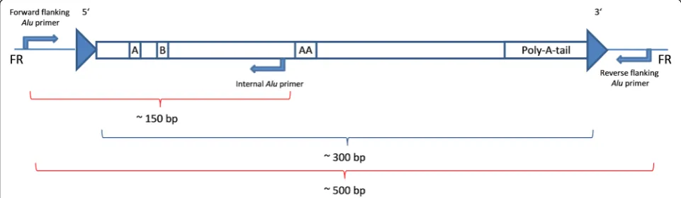
Obviously incomplete and incongruent results were subjected to an alternative molecular approach. Using an internalAluprimer, the predicted fragment length of the amplicon was reduced to ~150 bp (Fig. 1). The internal primers were designed based on an alignment ofAlu se-quences of the respective subfamily and consequently are very specific to each AluY subfamily as described by Nelson et al. [49] or Kass and Batzer [50]. This type of amplification worked in seven cases for the sample DO 3750 (Fig. 2). The heterozygous results for Alu_16,
Alu_26 and Alu_27 for the daughter (‘CR’ in Table 1) represent a combination of both amplification ap-proaches. Further internalAluprimer analyses were not possible, due to a depleted DNA extract (Alu_4,Alu_25; marked red). Loci with exclusively absence bands for the prehistoric individuals, in particular, should be checked by internal Alu amplification. The advantage of this method is that the amplification of short fragments (usu-ally ~150 bp) still proves the presence of an insert. In this study, this approach was applied only in those cases where the Alu amplification results are not in concord-ance with the family situation or where the amplification totally failed for DO 3750. Based on previous analyses on this prehistoric triad, it is known that the DNA is less well-preserved in DO 3750 and best-preserved in DO 1911. Consequently, the chance of allelic dropout events for DO 3750 is more likely than for DO 3756 and DO
1911. Fragments of such short lengths (~150 bp) are usually not affected by allelic dropout. However, the in-ternal primer approach cannot be applied in isolation because it does not indicate heterozygous states.
Proof of kinship in the prehistoric samples and authenticity
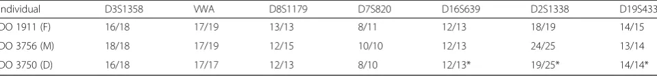
The authenticity of the aDNA was ensured by amplifying STR-based genetic fingerprints. Table 2 presents the consensus results of the Heptaplex STR analyses for the DNA extracts used. Table 3 shows the consensus results for seven additional STR systems. A full list of all results achieved can be found in the supplementary files (Additional file 3). Nearly all amplifications were con-ducted with DNA material taken from the left femur. The genetic fingerprint results of the STR systems D16S539, D2S1338 and D19S433 for the daughter were not achieved with DNA material from her left femur (DO 3750), but from her left humerus (DO 3994). The results of all 13 STR systems for the three prehistoric individuals were used for a kinship calculation resulting in a kinship probability of 99.999 %. All single al-lele frequencies were taken from the online database allstr.de [51]. Given this proven kinship, false homozygous
Alu results for the daughter can be clearly detected by contradiction between the parental alleles and the Men-del’s laws of inheritance.
Conclusion
The study clearly demonstrates the possibility of pres-ence/absence analyses of TEs in 3,000-year-old human remains from the Lichtenstein cave. These and earlier results indicate and prove the high quality of DNA pres-ervation and the applicability of molecular analyses using the remains from this cave [44, 45, 52], but could not yet show the amplification of 500 bp fragments. Of
Fig. 1The amplification via an internalAluprimer results in amplicons of ~150 bp. The reverse flankingAluprimer is replaced by an internalAlu
30 loci, we initially achieved 22 definite results (FAP in Table 1). With additional amplification using internal
Alu primers, we could add four more results (Alu_3,
Alu_14, Alu_19 and Alu_20), thus, 26 definite results (FAP and IAP in Table 1). The following Alu loci were incongruent with the family situation: Alu_2, Alu_7,
Alu_16, Alu_26 andAlu_27. Amplification with internal
Alu primers could place Alu_16, Alu_26 and Alu_27 in congruence with the family situation. The proposed veri-fication technique is to check for possible presence bands by amplification with an internal Alu primer to get short target sequences of ~150 bp. Fragment lengths of more than 200 bp tend to be affected more often by allelic dropout events; therefore, short amplicons should be used. Thus, in further analyses, results that show only absence bands should be subjected to this strategy. Even less well-preserved DNA can be analyzed by this ap-proach. The present study constitutes the basis for further investigations with more Alu loci and larger samples for microevolutionary studies in Central Europe. Such large-scale investigations would provide insight intoAlu SINE-based microevolutionary processes in humans during the last few thousand years and help us comprehend the evo-lutionary dynamics of our genome. Current projects, like the 1,000 Human Genome Project, investigate human
genetic variation and the interrelation of genotypes and phenotypes as well as variants in annotated genes and inherited genetic disorders [13, 53]. Through computa-tional biology, the 1,000 Genome Project recently pro-vided a genome-wide catalog of Alu polymorphisms for human populations [14]. A database with these group-specific insertions of polymorphic Aluelements is useful for future analyses with a larger dataset of Bronze Age Lichtenstein individuals –for instance, to investigate the geographic origin of Lichtenstein family members, who belong to the longest known family tree in the world. Through large-scale Alu element analyses of many in-dividuals from the Bronze Age Lichtenstein cave, we may be able to detect human variability and evolution within one geographic region on a timeline. These data would represent a great supplement to recent human population genetic studies based on TEs.
Methods
Samples and DNA extraction Samples
The skeletal material used for the present thesis originates from the Bronze Age Lichtenstein cave near Osterode in Lower Saxony, Germany. All bone material from the cave is stored at -20 °C in the Department of Historical
Fig. 2The photo shows seven successfully amplified amplicons of an internalAluprimer-based amplification. The expected fragment lengths vary from 118-194 bp. The marks on the base pair ladder are situated at 150 bp and 350 bp. For these sevenAluloci, the presence band for DO 3750 was proved via internalAluamplification. The asterisks indicate reverseAluinserts. In these cases, the primer pairings are an internalAluprimer with the reverseAluflanking primer, whereas the samples without asterisk were amplified with an internalAluprimer and the forwardAlu
flanking primer
Table 2Heptaplex based fingerprint results for all investigated individuals
Individual Amelo D13S317 D21S11 D18S51 TH01 D5S818 FGA
prehistoric DO1911 (F) X/Y 12/12 30.2/32.2 15/17 9.3/9.3 11/12 21/22
DO3756 (M) X/X 8/9 28/29 16/16 9/9.3 12/12 21/23
DO3750 (D) X/X 9/12 29/32.2 16/17 9/9.3 11/12 21/23
modern Cau_1 X/Y 12/13 28/32.2 15/18 9/9.3 11/13 20/21
Anthropology and Human Ecology of the Göttingen University, Lower Saxony, Germany. The DNA of the ancient individuals was extracted from three different members of a prehistoric family: father (left femur DO 1911), mother (left femur DO 3756) and daughter (left femur DO 3750). In all three cases, the DNA was extracted from the middle of the diaphysis. The modern DNA of the person from the United States of America was extracted from lymphocytes (CAU_1), and was provided with full written consent. This sample was ordered from“The Interstate Companies”(Memphis, Tennessee, USA) blood bank. The DNA of the modern positive control CAU_2 was extracted from cells of the buccal mucosa.
aDNA extraction from skeletal material with the QIAvac-24-plus
Fragments about 1 cm2in size are sawed out of the mid-dle of the diaphysis of the left femora. All outer surfaces of the fragments are removed to minimize the risk of a contamination with modern human DNA from, e.g., the excavation personal. The fragments are crushed with a steel mortar before they are powdered in a swing mill for 3 min at 24 swings per second. Afterwards, 0.25 g of the powder is transferred into a 15 ml FalconTube and 3900 μl of EDTA UltraPure™ 0.5 M pH 8 (Invitrogen™) and 100 μl of Proteinase K (600mAnson-U/ml) are added. This mixture is incubated for 18 h at 37 °C in a rotator. Now, an additional 50 μl of Proteinase K are added and the mixture is incubated at 56 °C for 2 h in a rotator. 50 μl of SDS (10 mg/ml) are added, followed by an incubation time of 5 min at 65 °C. The lysate is centri-fuged at 3300rcf for 3 min to sediment surplus organic material. The lysate is transferred into a 50 ml FalconTube that contains 16 ml PB-Buffer (Qiagen) and 100μl sodium acetate buffer (pH 5.2, 3 M, Sigma). After manually mix-ing the lysate, it is centrifuged at 3300rcf for 3 min. The DNA clean-up is conducted with minElute spin columns and funnels for large volumes using the QIAvac-24-plus (Qiagen). Deviating from the protocol, three wash steps with PE-buffer (Qiagen) are performed. The DNA is eluted in 60μl RNase-free water (also cf. [54]).
Modern DNA
Blood sampleThe DNA of the blood sample from CAU_1 is extracted with the Wizard Genomic DNA Purification Kit
(Promega) following the producer’s protocol for extraction from whole blood samples (300μl).
Buccal mucosa swab sample A buccal mucosa swab from CAU_2 is transferred into a 2 ml reaction tube. 400 μl of G2-buffer (Qiagen) and 10μl of Proteinase K are added, followed by incubation for 1 h at 56 °C and 350 rpm on an Eppendorf thermomixer comfort. After-wards, 200 μl of the lysate is transferred into a clean tube and 1 ml of PB-buffer and 100μl of sodium acetate buffer are added. After manually mixing the lysate, it is centrifuged at 3300rcf for 3 min. Now the DNA is cleaned up with minElute spin columns and large volume funnels as described above.
Aluloci and primer design
Alu loci were chosen based on earlier publications with a population-genetics focus [23–26]. The site-specific
Alu sequence was determined by using RepeatMasker [55]. An additional 500 bp flanking sequence on each site was extracted from the human reference genome (hg38) in NCBI [56]. The locus-specific primers were designed with PrimerSelect, version 10.1.2 (DNASTAR). The primer characteristics are a strong 5′and a weak 3′ end by not exceeding a length of 30 bp; further, primer dimerization and hairpin formation were avoided to en-hance the specificity and sensitivity of the reaction [38]. The total length of the target sequence (including the
Aluinsert) should be as short as possible, which usually resulted in amplicons of 450 bp to 500 bp (cf. also Additional file 4 for detailed information).
The internal Alu primers were designed based on a highly conserved region of the Alu sequence that is specific to the respective subfamily. Therefore, randomly selectedAluinserts of the respectiveAluY subfamily were aligned. The amplification always includes theAluhead.
A full list of the primer sequences is shown in the Additional file 5.
PCR
All PCRs are conducted under the same conditions apart from the annealing temperatures. Depending on the en-ergy profiles and melting temperatures of the primer sets and based on preliminary primer tests, different annealing temperatures, varying from 52 to 60 °C, are chosen. The amplification is conducted using the following cycling
Table 3Additional fingerprint results for the prehistorical individuals
Individual D3S1358 VWA D8S1179 D7S820 D16S639 D2S1338 D19S433
DO 1911 (F) 16/18 17/19 13/13 8/11 12/13 18/19 14/15
DO 3756 (M) 18/18 17/19 12/15 10/10 12/13 24/25 13/14
DO 3750 (D) 16/18 17/17 12/13 8/10 12/13* 19/25* 14/14*
program: Initial hot start at 95 °C for 5 min; 40 cycles with denaturation at 94 °C for 1 min, annealing at 52–60 °C for 1 min, elongation at 72 °C for 1 min; a final soak at 10 °C for 10 min. The PCR is composed of 12.5 μl of Multiplex PCR Mastermix (Qiagen), 1 μl each forward and reverse primer, (both 20 μM working solution), 5μl DNA for aDNA samples and 0.5 μl DNA (plus 4.5 μl RNase free water) for modern DNA samples and 5.5μl of RNase-free water to get a final volume of 25 μl per reaction.
The amplification with an internalAluprimer was per-formed with an elongation time of 20 s. All other param-eters are identical to the classical PCR approach.
For proof of authenticity, every DNA extract used in the study here presented was subjected to STR-typing by a multiplex amplification as described previously [57]. Deviating from this work, the sex discriminating amelo-genin gene is arranged in the blue dye panel. The reac-tion mix is composed of 12.5 μl Multiplex PCR Mastermix (Qiagen), 2.85 μl multiplex primer mix, 4.65μl RNase-free water and 5μl DNA extract.
Gel electrophoresis and fragment length estimation
Each amplification result is checked by ethidium bromide stained agarose gel electrophoresis (2.5 %). The fragment length determination is performed with a 50 bp molecular ladder (Invitrogen). For the electrophoresis, usually a volt-age of 120 V and a run time of 30 min are applied.
The STR products are separated in a 50 cm capillary on an ABI 3500 Genetic Analyzer (Applied Biosystems) using POP-7™ Polymer for 3500/3500xL Genetic Ana-lyzers and the 3500 Data collection Software (all Applied Biosystems). Allele determination is performed with GeneMapper Software 5 (Applied Biosystems).
Cloning and sequencing
Cloning of PCR products is conducted with the Blue/ White-Selection based pGEM®-T Easy Vector System (Promega). Deviating from the manufacturer’s protocol, 300μl SOC medium (Invitrogen) is used to suspend the cells. Additionally, 50μl– 100μl of the cell suspension are plated. The Colony-PCR Mastermix is identical to the other PCRs except for the PCR primers. The primers pUC/M13 forward and reverse (Promega) are used in working concentrations of 20 μM. One colony replaces the DNA inset. The Colony PCR is conducted with the following program: Initial denaturation at 94 °C for 3 min; 30 cycles with denaturation at 94 °C for 30 s, an-nealing at 55 °C for 1 min, elongation at 72 °C for 50 s; final elongation at 72 °C for 2 min and final soak at 10 ° C for 10 min. PCR products are purified with an isopro-panol purification protocol: the PCR product is incu-bated with 83 μl HPLC water, 100 μl isopropanol (100 %) and 10μl sodium acetate (3 M) for 10 min, then
centrifuged at 13,200 rpm for 10 min in a conventional tabletop microcentrifuge. The supernatant is discarded and 150 μl of ethanol (70 %) is added. After another 10 min of centrifugation at 13,200 rpm, the supernatant is discarded, the pellet is dried and the desired amount of RNafree water is added for resuspension. The se-quencing reaction is composed of 4μl Sequencing Buffer (5x), 2μl BigDyeTerminator v1.1, 0.3μl primer (20μM), 6.7 μl HPLC water and 7 μl purified PCR product. Se-quencing is performed in forward and reverse direction with the following program: Initial heating step at 94 °C for 3 min; 33 cycles with denaturation at 94 °C for 30 s, annealing at 55 °C for 1 min and elongation at 72 °C for 2.5 min; soak at 10 °C. Sequencing products are purified with NucleoSeq® columns (Macherey-Nagel). The prod-ucts are separated in a 50 cm capillary on an ABI 3500 Genetic Analyzer (Applied Biosystems) using POP-7™ Polymer for 3500/3500xL Genetic Analyzers and the 3500 Data collection Software (all Applied Biosystems). The sequences are edited in BioEdit version 7.2.5 [58] and submitted to a BLAST analysis. Finally, sequence data with the following accession numbers were depos-ited in GenBank: KU323383-KU323387.
Kinship calculation
For kinship calculation (Reverse Parentage Index; RPI), the genotype probabilities are calculated: RPI = X/Y. The numerator (X) is the probability that a woman ran-domly selected from a population is type AB, that a man randomly selected from a population is type CD and that the child is type BC. The child gets one of the two alleles of the father and the mother, respectively. The probabil-ity that one allele of one parent is inherited by the child is 0.5. The denominator (Y) is the probability that a woman randomly selected from a population and unre-lated to the child is type AB, that a man randomly selected from a population and unrelated to the child is type CD, and that a child randomly selected from a population is type BC (also cf. [59, 60]). The reverse parentage index for one STR system is calculated as follows:
RPI¼X
Y¼
2PAPB2PCPD0:50:5
2PAPB2PCPD2PBPC
All single RPIs are multiplied to get a combined RPI.
Additional files
Additional file 1:Genomic locations of all 30Aluloci. This file contains precise locations of all 30Aluloci investigated. (XLS 37 kb)
Additional file 2:Sequences of randomly selected loci. This file contains sequencing results of 3 randomly selected loci with 5 sequences in total to check for authenticity. (PDF 18 kb)
prehistoric individuals investigated in this study. Sheets 1-3 contain the results of all 13 STR systems that were used for kinship calculation. The fourth sheet contains the STR-typing results that were obtained to check the authenticity of the DNA extracts. (XLSX 23 kb)
Additional file 4:Detailed information on theAluloci. The Microsoft Excel file contains detailed information about the exact lengths of the amplicons for every genomicAlulocus investigated in this study. Additionally shown are the lengths of the A-tails and the Target Site Duplications (TSD), as well as the sequences of the TSDs. (XLS 36 kb)
Additional file 5:List of allAluprimers used. The Microsoft Excel file contains all primer sequences that were used in this study. (XLSX 10 kb)
Abbreviations
aDNA:ancientDNA; LINEs: long interspersed elements; SINEs: short interspersed elements; TEs: transposable elements.
Competing interests
The authors declare that they have no competing interests.
Authors’contributions
MK carried out the molecular genetic studies and generated all results that are based on presence/absence analyses and drafted most parts of the manuscript. OP designed the project and participated in molecular studies and data interpretation. Information about the samples from the Lichtenstein cave as well as STR amplifications was generated by VS. In addition VS was involved in data interpretation. SH participated in project design and data interpretation. This manuscript was critically reviewed by OP, VS and SH. All authors read and approved the final manuscript.
Acknowledgements
TheAluSINE-based work was supported by the University of Göttingen with funding provided by the Lower Saxony Ministry for Science and Culture (Ministerium für Wissenschaft und Kultur, MWK)to OP. The research on the skeletal remains from the Lichtenstein cave was funded by theMWKto SH. We want to thank Stefan Flindt from theLandkreis Osterode am Harz (Krei-sarchäologie)Lower Saxony for enabling this research by allowing the use of the invaluable sample material. His long-term cooperation in research on human remains from the Lichtenstein cave is highly appreciated.
Received: 23 December 2015 Accepted: 31 March 2016
References
1. McClintock B. The origin and behavior of mutable loci in maize. Proc Natl Acad Sci U S A. 1950;36:344–55.
2. McClintock B. Induction of Instability at Selected Loci in Maize. Genetics. 1953;38:579–99.
3. Doolittle WF, Sapienza C. Selfish genes, the phenotype paradigm and genome evolution. Nature. 1980;284:601–3.
4. Callinan PA, Batzer MA. Retrotransposable Elements and Human Disease. In: Volff JN, editor. Genome and Disease. Genome Dyn, vol. 1. Basel: Karger; 2006. p. 104–15.
5. Volff JN. Turning junk into gold: domestication of transposable elements and the creation of new genes in eukaryotes. Bioessays. 2006;28:913–22. 6. Piskurek O, Jackson DJ. Transposable Elements: From DNA Parasites to
Architects of Metazoan Evolution. Genes. 2012;3:409–22. 7. Heras SR, López MC, Olivares M, Thomas MC. The L1Tc non-LTR
retrotransposon ofTrypanosoma cruzicontains an internal RNA-pol II-dependent promoter that strongly activates gene transcription and generates unspliced transcripts. Nucleic Acids Res. 2007;35(7):2199–214. 8. Estecio MRH, Gallegos J, Dekmezian M, Lu Y, Liang S, Issa JPJ. SINE
Retrotransposons Cause Epigenetic Reprogramming of Adjacent Gene Promoters. Mol Cancer Res. 2012;10(10):1332–42.
9. Sasaki T, Nishihara H, Hirakawa M, Fujimura K, Tanaka M, Kokubo N, Kimura-Yoshida C, Matsuo I, Sumiyama K, Saitou N, Shimogori T, Okada N. Possible involvement of SINEs in mammalian-specific brain formation. Proc Natl Acad Sci U S A. 2008;105(11):4220–25.
10. Piskurek O, Okada N. Poxviruses as possible vectors for horizontal transfer of retroposons from reptiles to mammals. Proc Natl Acad Sci U S A. 2007;104: 12046–51.
11. Lander ES, Linton LM, Birren B, Nusbaum C, Zody MC, Baldwin J, et al. Initial sequencing and analysis of the human genome. Nature. 2001;409:860–921. 12. De Koning AP, Gu W, Castoe TA, Batzer MA, Pollock DD. Repetitive Elements
May Comprise Over Two-Thirds of the Human Genome. PLoS Genet. 2011;67:183–93.
13. Abecasis GR, Altshuler D, Auton LD, Durbin RM, Gibbs RA, Hurles ME, et al. A map of human genome variation from population-scale sequencing. Nature. 2010;467(7319):1061–73.
14. Rishishwar L, Tellez Villa CE, Jordan IK. Transposable element polymorphisms recapitulate human evolution. Mobile DNA. 2015;6:21.
15. Vassetzky NA, Kramerov DA. SINEBase: a database and tool for SINE analysis. Nucleic Acids Res. 2013;41:D83–9.
16. Jurka J, Kapitonov VV, Pavlicek A, Klonowski P, Kohany O, Walichiewicz J. Repbase Update, a database of eukaryotic repetitive elements. Cytogenet Genome Res. 2005;110:462–7.
17. Okada N, Hamada M, Ogiwara I, Ohshima K. SINEs and LINEs share common 3′sequences: A review. Gene. 1997;205:229–43.
18. Okada N, Shedlock AM, Nikaido M. Retroposon mapping in molecular systematics. Methods Mol Biol. 2004;260:189–226.
19. Shedlock AM, Kazuhiko T, Okada N. SINEs of speciation: tracking lineages with retroposons. TRENDS Ecol Evol. 2004;19(10):545–53.
20. Shedlock AM, Okada N. SINE insertions: powerful tools for molecular systematics. Bioessays. 2000;22(2):148–60.
21. Schmitz J, Ohme M, Zischler H. SINE insertions in cladistic analyses and the phylogenetic affiliations ofTarsius bancanusto other primates. Genetics. 2001;157(2):777–84.
22. Deininger PL.Aluelements: know the SINEs. Genome Biol. 2011;12:236. 23. Furano AV. The biological properties and evolutionary dynamics of
mammalian LINE-1 retrotransposons. Prog Nucleic Acids Res Mol Biol. 2000; 64:255–94.
24. Okada N, Shedlock AM, Nikaido M. Retroposon Mapping in Molecular Systematics. In: Mobile Genetic Elements: Protocols and Genomic Applications, Methods in Molecular Biology. Totowa, New Jersey, U.S.A.: Humana Press; 2004. vol. 260. p. 189–226.
25. Carrol ML, Roy-Engel AM, Nguyen SV, Salem AH, Vogel E, Vincent B, Myers J et al. Large-scale Analysis of theAluYa5 and Yb8 Subfamilies and their Contribution to Human Genomic Diversity. J Mol Biol. 2001;311:17–40. 26. Carter AB, Salem AH, Hedges DJ, Nguyen Keegan C, Kimball B, Walker JA,
et al. Genome wide analysis of the humanAluYb8 lineage. Hum Genomics. 2004;1:167–78.
27. Otieno AC, Carter AB, Hedges DJ, Walker JA, Ray DA, Garber RK, Anders BA et al. Analysis of the humanAluYa-lineage. J Mol Biol. 2004;342:109–18. 28. Garber RK, Hedges DJ, Herke SW, Hazard NW, Batzer MA. TheAluYc1
subfamily: sorting the wheat from the chaff. Cytogenet Genome Res. 2005; 110:537–42.
29. Nikaido M, Piskurek O, Okada N. Toothed whale monophyly reassessed by SINE insertion analysis: The absence of lineage sorting effects suggest a small population of a common ancestral species. Mol Phylogenet Evol. 2007;43:216–24.
30. Nishihara H, Maruyama S, Okada N. Retroposon analysis and recent geological data suggest near-simultaneous divergence of the three superorders of mammals. Proc Natl Acad Sci U S A. 2009;106(13):5235–40. 31. Churakov G, Kriegs JO, Baertsch R, Zemann A, Brosius J, Schmitz J. Mosaic retroposon insertion patterns in placental mammals. Genome Res. 2009; 19(5):868–75.
32. Matzke A, Churakov G, Berkes P, Arms EM, Kelsey D, Brosius J, et al. Retroposon Insertion Patterns of neoavian Birds: Strong Evidence for an Extensive Incomplete Lineage Sorting Era. Mol Biol Evol. 2012;29(6):1497–501. 33. Watkins WS, Rogers AR, Ostler CT, Wooding S, Bamshad MJ, Brassington AM,
et al. Genetic variation among world populations: inferences from 100 Alu insertion polymorphisms. Genome Res. 2003;13(7):1607–18.
34. Kajikawa M, Okada N. LINEs Mobilize SINEs in the Eel through a Shared 3′ Sequence. Cell. 2002;111(3):433–44.
35. Dewannieux M, Esnault C, Heidmann T. LINE-mediated retrotransposition of markedAlusequences. Nat Genet. 2003;35:41–8.
36. Simonti CN, Capra JA. The evolution of the human genome. Curr Opin Genet Dev. 2015;35:9–15.
37. Nekrutenko A, Li WH. Transposable elements are found in a large number of human protein-coding genes. Trends Genet. 2001;17:619–21. 38. Cordaux R, Batzer MA. The impact of retrotransposons on human genome
39. Hummel S. Ancient DNA Typing. Methods, Strategies and Applications. Berlin, Germany: Springer; 2003.
40. Höss M, Jaruga P, Zastawny TH, Dizdaroglu M, Pääbo S. DNA damage and DNA sequence retrieval from ancient tissues. Nucleic Acid Res. 1996;24(7): 1304–7.
41. Haack K, Hummel S, Hummel B. Ancient DNA fragments longer than 300 bp. Anthrop Anz. 2000;58:51–6.
42. Burger J, Hummel S, Herrmann B, Henke W. DNA preservation: A microsatellite-DNA study on ancient skeletal remains. Electrophoresis. 1999; 20:1722–8.
43. Lindahl T. Instability and decay of the primary structure of DNA. Nature. 1993;32(422):709–11.
44. Schultes T. Typisierung alter DNA zur Rekonstruktion von Verwandtschaft in einem bronzezeitlichen Skelettkollektiv. 2000. Dissertation, University of Göttingen.
45. Schilz F. Molekulargenetische Verwandtschaftsanalysen am prähistorischen Skelettkollektiv der Lichtensteinhöhe. 2006. Dissertation, University of Göttingen.
46. Schultes T, Hummel S, Herrmann B. Ancient DNA-typing approaches for the determination of kinship in a disturbed collective burial site. Anthrop Anz. 2000;58(1):37–44.
47. Schilz F, Hummel S, Herrmann B. Design of a multiplex PCR for genotyping 16 short tandem repeats in degraded DNA samples. Anthrop Anz. 2004; 62(4):369–78.
48. ensembl.org. http://www.ensembl.org/index.html.
49. Nelson DL, Ledbetter SA, Corbo L, Victoria MF, Ramirez-Solis R, Webster TD, Ledbetter DH, Caskey CT.Alupolymerase chain reaction: A method for rapid isolation of human-specific sequences from complex DNA sources. Proc Natl Acad Sci U S A. 1989;86:6686–90.
50. Kass DH, Batzer MA. Inter-AluPolymerase Chain Reaction: Advancements and Applications. Anal Biochem. 1995;228:185–93.
51. allstr Autosomal Database for short tandem repeats. http://allstr.de/allstr/ searchMarker.seam. Accessed 3rd September 2015.
52. Hummel S, Schmidt D, Kremeyer B, Herrmann B, Oppermann M. Detection of the CCR5-Δ32 HIV resistance gene in Bronze Age skeletons. Genes Immun. 2005;6:371–4.
53. Konkel MK, Walker JA, Hotard AB, Ranck MC, Fontenot CC, Storer J et al. Sequence Analysis and Characterization of Active HumanAlusubfamilies Based on the 1000 Genomes Pilot Project. Genome Biology and Evolution. 2015; doi:10.1093/gbe/evv167.
54. Frischalowski M, Seidenberg V, Großkopf B, Wulf FW, Hummel S. Molekulargenetische Untersuchung des Verwandtschaftsverhältnisses von möglichen Mutter-Kind-Bestattungen aus dem frühzeitlichen Eldagsen. Nachrichten aus Niedersachsens Urgeschichte. 2015;84:(in press). 55. Smit AFA, Hubley R, Green P. RepeatMasker Open-4.0. 2013-2015. http://
www.repeatmasker.org.
56. National Center for Biotechnology Information. http://www.ncbi.nlm.nih. gov/assembly/GCF_000001405.26.
57. Seidenberg V, Schilz F, Pfister D, Georges L, Fehren-Schmitz L, Hummel S. A new miniSTR heptaplex system for genetic fingerprinting of ancient DNA from archaeological human bone. J Archaeol Sci. 2012;39(10):3224–9. 58. Hall TA. BioEdit: a user-friendly biological sequence alignment editor and
analysis program for Windows 95/97/NT. Nucl Acids Symp. 1999;41:95–8. 59. Brenner CH. Symbolic Kinship Program. Genetics. 1997;145:535–42. 60. Gjertson DW, Brenner CH, Baur MP, Carracedo A, Guidet F, Luque JA, et al.
ISFG: Recommendations on biostatistics in paternity testing. Forensic Sci Int Genet. 2007;1:223–31.
• We accept pre-submission inquiries
• Our selector tool helps you to find the most relevant journal
• We provide round the clock customer support
• Convenient online submission
• Thorough peer review
• Inclusion in PubMed and all major indexing services
• Maximum visibility for your research
Submit your manuscript at www.biomedcentral.com/submit