BMC Physiology 2001,
1 :13
Research article
The dynamics of venous return and response to hypervolemia in the
toad,
Bufo marinus
(L.)
Erin E Killorn and Daniel P Toews*
Address: Department of Biology, Acadia University, Wolfville, Nova Scotia BOP 1X0, Canada
E-mail: Erin E Killorn - [email protected]; Daniel P Toews* - [email protected] *Corresponding author
Abstract
Background: Venous return from the posterior region of amphibians travels by either two renal portal veins to the kidney or a central abdominal vein that drains into the hepatic portal system. The relative proportions of blood flow in these vessels has never been measured nor has a modification of flow been determined when venous return increases by changes in blood volume during hypervolemia or during increased volume input from the posterior lymph hearts.
Results: Venous return from the posterior region of Bufo marinus was measured under resting conditions and in response to a systemic hypervolemia. Doppler flow probes were positioned on the renal portal and ventral abdominal veins, and flow was recorded as injections of artificial plasma equaling 100% of the animal's plasma volume were administered through the sciatic artery. Resting flow was found to be 5.54 ± 2.03 ml min-1 kg-1 in the paired renal portal veins, and 7.31 ± 0.89 ml
min-1 kg-1 in the ventral abdominal vein. While renal portal flow was found to increase by a factor
of 2.4 times during the first 10 min of hypervolemia, ventral abdominal flow only increased by a factor of 1.3.
Conclusions: Our results quantify the contribution to circulation from posterior venous return in the toad Bufo marinus. A preferential movement of excess fluid through the renal portal pathway was also demonstrated, supporting the possibility of water elimination via the renal portal circulation, especially during periods of high water influx into the animals.
Background
The distribution of blood flow through the amphibian body is crucial to many vital homeostatic functions, in-cluding those involving respiration, nutrition and elimi-nation of unwanted materials. Although blood flow has been quantified for the primary arterial components of the circulatory system, the magnitude and partitioning of venous return from the posterior regions through sev-eral major organs is unknown. Blood returning from this area in an anuran amphibian may pass through one of
two major pathways; the first carrying blood through the hepatic portal system, and the second traversing the kid-neys via the renal portal system, a component of circula-tion of relatively unknown funccircula-tion [1].
Through direct measurement of arterial flow in Bufo
marinus, West [2] determined cardiac output to be 57.2
ml min-1 kg-1. Aortic blood flow represented 44.9% of this value, with the pulmocutaneous artery carrying 48.4% and the carotid artery 8.0% of cardiac output [2]. Published: 10 October 2001
BMC Physiology 2001, 1:13
Received: 6 June 2001 Accepted: 10 October 2001
This article is available from: http://www.biomedcentral.com/1472-6793/1/13
Of blood pumped to the posterior of the animal through the aorta, a significant amount passes through vessels surrounding the intestine and through the kidneys via
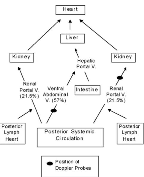
the renal arteries. The remaining blood bathes tissues of the abdomen and hind limb region and returns to the heart through the two main pathways, the renal portal and ventral abdominal veins [1].
The common pelvic and femoral veins join to form the renal portal veins, just distal to the drainage site for the posterior lymph hearts into the circulation (Fig. 1) [1]. The renal portal veins run anteriorly along the kidneys, producing networks of vessels that cover the dorsal sur-face of the organs [3]. As the branched vessels traverse the kidneys, they give rise to peritubular vessels, which surround the kidney nephra. Deeds et al. [4] found that in the perfused bullfrog kidney, the renal portal circula-tion contributed up to 72% of the perfusate, indicating the apparent importance of this portal circulation to re-nal blood flow.
The perfusion of the renal portal vessels through the kid-neys is still uncertain. However, it is generally accepted that the efferent arterioles from the glomerular capillar-ies empty into the peritubular capillarcapillar-ies, serving as a link between arterial and portal circulations [5]. Al-though it has been suggested that this close association may allow for filtration of renal portal blood, Deeds et al.
[4] found that in the perfused bullfrog kidney, only 0.52% of the filtrate was derived from the portal per-fusate, likely due to the high resistance bridge between the two circulations. Efferent renal veins eventually col-lect the bulk of renal blood from the peritubular capillar-ies, and join to form the posterior vena cava [1].
The ventral abdominal vein represents an alternate path-way for venous return from the posterior region (Fig. 1). This vein runs anteriorly from the junction of the com-mon pelvic veins, and joins the hepatic portal system as it enters the liver [1]. The common pelvic and femoral veins have interconnections, providing a link between the two venous return pathways [1]. Ohtani and Naito [3] have suggested that the connection may serve as a means of delivering blood from the ventral abdominal vein to the kidneys.
The delicate balance between water loss and uptake in amphibians makes an understanding of venous return from this region even more significant. Terrestrial am-phibians show a behavior known as the water absorption response, using a portion of their ventral skin called the pelvic patch to take up extraneous water [6]. Although it has long been assumed that this external water moves into the circulatory system, one study shows movement of tritiated water directly into the lymph sacs from the environment [7]. The connection of lymphatic and circu-latory systems through the renal portal veins has elicited suggestions of rapid elimination of excess incoming fluid
via the renal portal vessels [8].
This study will measure flow through both the renal por-tal and ventral abdominal veins in the cane toad, Bufo
marinus, using Doppler flow probes, to determine
rela-tive contributions of the two main pathways to venous return from the posterior regions (Fig. 1). An arterial vol-ume load will also be performed in order to examine pos-sible physiological consequences of volume stress on venous return.
Results
Resting blood flow
Values for resting renal portal and ventral abdominal ve-nous blood flow were determined using 5 min pre-injec-tion levels. Measured flow from a single renal portal vessel was doubled for each individual. Throughout the text, all mention of renal portal blood flow refers to dou-Figure 1
bled values. Renal portal venous flow was 5.5 ± 2.0 ml min-1 kg-1, while flow in the ventral abdominal vessel was 7.3 ± 0.9 ml min-1 kg-1.
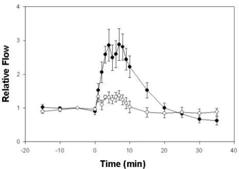
Blood flow during hypervolemia
Following an injected 100% increase in plasma volume there were significant increases in blood flow in ventral abdominal and renal portal veins during the first 10 min period but not in the time following 10 min post infusion (Fig. 2). Flow in the renal portal veins increased by a fac-tor of 2.4 (t14 = 6.6; p < 0.001) and in the ventral abdom-inal vein by a factor of 1.3 (t14 = 3.36; p = 0.0047). Following the 10 min post infusion period blood flow in all veins decreased rapidly and flows did not differ signif-icantly from the pre infusion level (p > 0.29).
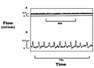
Deviation from resting renal portal blood flow pattern Blood flow in approximately 3/4 of the renal portal ve-nous flow tracings, from 30 to 90 min post injection, showed periods where a deviation from the normal pat-tern was apparent (Fig. 3). This patpat-tern involved sharp pulsatile increases in blood flow, distinguishing these tracings from the normal, slow wave pulsations seen in the simultaneous ventral abdominal tracings and in the resting renal portal and ventral abdominal venous trac-ings. The rate of renal portal pulsations was 48.0 ± 6.3 beats min-1. The rate of slow wave pulsations in the
ven-tral abdominal vein during the same time period was 23.3 ± 4.4 beats min-1. The resting rate of slow wave pul-sation, identical in both vessels, was 19.5 ± 3.0 beats min
-1, calculated using 10 min pre-injection values. The sharp
pulsatile increases above baseline renal portal venous levels generated flows of 6.2 ± 2.9 ml kg-1 h-1, and stroke volumes of 0.0018 ± 0.0006 ml kg-1.
Discussion
The pathway of venous return from the posterior regions of Bufo marinus is via either the renal portal or hepatic portal systems before returning to the heart [1]. Resting blood flow in the two renal portal vessels combined was quite similar to that of the single ventral abdominal vein (which drains into the hepatic portal vein), with a slightly higher value observed for the ventral abdominal vessel. Total flow in the two vessel systems was 12.85 ml min-1 kg-1, which comprises the total blood flow returning from the hind limb region of the toad. Ventral abdominal flow accounted for 57% of this venous return, with renal por-tal flow contributing the remaining 43% (Fig. 1).
West [2], using similar techniques to those used in this study, found cardiac output in Bufo marinus to be 57.2 ml min-1 kg-1, with aortic blood flow being 26.8 ml min-1 kg-1 [2]. Using values obtained in this study, venous re-turn from the hind limbs of the toad would thus repre-sent approximately 1/4 of cardiac output.
When blood flow was examined following an infused doubling in plasma volume, both renal portal and ventral abdominal blood flow increased significantly during the 10 minute post infusion period but flow in the vessels leading to the kidneys was greater. The site of injection, the existence of a vascular connection between the two flow pathways and the large volume of fluid injected would support an equal distribution of excess fluid in both pathways. Our results strongly suggest a preferen-tial movement of fluid through the renal portal system.
A favored movement of fluid through the renal portal veins would support the hypothesis that, in a natural aquatic setting, the rapid elimination of incoming water could be made possible by the renal portal system [8]. Carter [8] postulates that this excess water may move from the peritubular capillaries into the tubular lumen due to osmotic concentration differences, and therefore be excreted as urine. In this case, fluid would move in a preferred direction such that its rapid elimination is maximized. It has also been suggested that the femoral vein may serve as a functional connection between the renal portal and ventral abdominal veins [3]. This would seem to be supported if increased venous flow in the hind limbs is primarily directed through the renal portal veins. This connection could then serve as a means for Figure 2
delivery of increased volumes of blood to the kidneys, al-lowing for the elimination of excess fluid.
Interestingly, the pattern of venous blood flow observed in both of the vessels throughout the experiments was a regular, slow-wave type pulsation. Previous work has shown heart rate in Bufo marinus to be 39.9 beats min-1 and lung ventilation rate to be 16.3 ventilations min-1 in normoxic conditions [9]. The calculated resting rate of 19.5 ± 3.0 pulsations min-1 in the vessels appears closer to values for lung ventilation, however the rhythmic na-ture of the pulsation pattern seems consistent with changes due to heart contraction. Although we did not monitor lung ventilation, it is possible that ventilatory pressure changes within the toad result in regular oscil-lations in venous flow. Pulsations in flow due to negative pressure from the heart, however, seem unlikely, as both of the veins studied branch into portal systems before emptying into the vena cava.
Much of the speculation on pulsation patterns arose di-rectly as a result of the observation of flow "spikes" in the renal portal tracings, which appeared as flow returned to normal after hypervolemia. The sharp extra pulsations observed were less rhythmic than the resting pulsations and contrasted with the flow tracings recorded simulta-neously in the ventral abdominal vein. The pattern of sharp, irregular peaks corresponds remarkably with tracings observed when lymph flow is monitored directly from lymph heart efferent vessels using similar tech-niques [10]. Williams et al. [11] found that 50 min after a
doubling in plasma volume, lymph heart rate was ap-proximately 50 beats min-1, consistent with the "spike" rate of 48.0 ± 6.3 pulsations min-1 calculated in this study during a similar time frame. It would appear that these tracings reflect contributions to renal portal blood flow by lymph, which is ejected from the nearby posteri-or lymph hearts.
Previous measurement of lymph heart function in re-sponse to systemic hypervolemia has shown lymph flow from a single posterior heart to be 20.7 ml kg-1 h-1 and stroke volume to be 0.0074 ml kg-1 at the 50 min post-in-jection mark [11]. Our values, determined through meas-urement of flow peaks above baseline renal portal flow, are considerably lower, with lymph flow being 6.2 ml kg min-1, and stroke volume 0.0018 ml kg-1. These meas-urements of lymph flow may vary due to a more "down-stream" location and the indirect method of lymph flow measurement used in this study. In addition lymph heart flow "spikes" in the renal portal veins were not visible under normal resting conditions. Williams et al. [11] found that, 50 min following a 100% increase in plasma volume, lymph flow was increased, although this change was not significant. It is possible that these sharp pulsa-tile increases in flow are peaks of the lymph heart systolic output, which has been increased such that it becomes visible, superimposed upon the larger flow of the renal portal vein.
The contribution of lymph flow to renal portal flow can also be considered with respect to resting state. Jones et al. [10] found lymph flow in Bufo marinus under normal conditions to be 25.9 ml kg-1 h-1 from a single posterior lymph heart. Using renal portal flow values measured in this study, it is estimated that almost one-sixth of the re-nal portal circulation consists of fluid originating in the posterior lymph hearts.
Conclusions
The shift in distribution of blood flow from the posterior end of the toad during volume stress demonstrates the dynamic nature of the anuran circulatory system. The possibility of the elimination of excess water via the re-nal portal system illustrates the necessity of a greater un-derstanding of the association between lymphatic and circulatory systems. This association was clearly empha-sized by the quantification of lymphatic function within the circulatory system that was demonstrated in this study.
Material and methods
Animals
Cane toads, (Bufo marinus, L.; 245–326 g hydrated mass), were obtained from commercial suppliers (Boreal Laboratories, St. Catherine's, ON, Can.; Charles D. Sulli-Figure 3
Flow tracings from the renal portal vein of Bufo marinus
showing sharp extra pulsations at: A) 50 min post injection, flow values shown for renal portal vein; and B) 55 min post injection with recording expanded, flow values shown for pulsations above resting renal portal flow. The injection was performed at t = 0 min. Chart speeds are: A) 1 mm sec-1; and
van Co. Inc., Nashville, TN, USA). Animals were main-tained in fiberglass aquaria (0.9 m × 0.6 m × 0.6 m)filled with sand to a depth of approximately 5 cm, and allowed free access to water. Toads were force fed raw beef liver once a week, the water was changed weekly and the sand was replaced every second week.
Surgical procedure
Toads of either sex were chosen for experimentation and anaesthetized in a solution of 2 g l-1 of aminobenzoic acid ethylester (MS-222, Sigma Chemical Co.) and 2 g l-1 of NaHCO3 in tapwater until the corneal reflex was absent.
At a point on the median line of the ventral surface of the toad, 2 cm anterior to the juncture of the torso with the hind limb, a transverse incision of about 2 cm was made and the skin was separated from the underlying tissue. A longitudinal incision of about 0.5 cm was made through the muscle at a position 0.5 cm lateral to the ventral ab-dominal vein, and a small length of this vein was separat-ed from the abdominal wall by removal of the connective tissue. The vein was fitted with a Doppler flow probe (si-lastic cuff, i.d. 1.0 mm; Iowa Doppler Products, Iowa City, LA, USA) and the probe was secured to the muscu-lature between the ventral abdominal wall and the skin using 5–0 gauge silk sutures. The leads of the probe were tied down to the skin in 3 locations, and the initial inci-sion was closed using 3–0 gauge silk sutures.
On the dorsal surface an incision was made just above the junction of the renal portal vein and the kidney, just anterior to a posterior lymph heart. As with the ventral abdominal vein, the renal portal vein was dissected free and fitted with a Doppler flow probe (silastic cuff, i.d. 0.8 mm; Iowa Doppler Products, Iowa City, IA, USA), which was placed as close to the point of entry of the vein into the kidney as possible without restriction of flow. The probe was secured to the surrounding musculature, the leads of the probe were tied down to the skin in 3 loca-tions, and the incision was closed. For arterial infusion purposes, the sciatic artery was then cannulated using previously described methods [12].
Following surgery the animals were placed under run-ning water to recover, and then moved to 6 L covered plastic boxes containing 2–3 cm of dechlorinated tap wa-ter and several air ventilation and lead exit holes cut in the top. Animals were allowed to recover for a minimum of 18 h following surgery. During all experiments ani-mals were freely moving and unanaesthetized in the chambers.
Experimental procedure
For measurement of venous flow, the leads of the Dop-pler flow probes were connected to a pulsed DopDop-pler
flowmeter (model 545c-4; Bioengineering, University of Iowa), which was then connected to a two-channel Gould chart recorder.
Resting renal portal and ventral abdominal venous flows were recorded for 15 min, at a chart speed of 1 mm s-1. Following this period, the animals were volume loaded through the sciatic artery cannula with a solution of Bo-vine Albumin (3.60 g 100 ml-1; [13]) in MacKenzie's sa-line [14]. Plasma volume was doubled, assuming 7.4 ml of plasma for 100 g of animal [15]. An infusion pump was used to inject fluid over a period of 4.5 to 5.5 min. Meas-urements were recorded for 90 min from the onset of in-jection, and flow was determined at time 0, then every min for 10 min, every 5 min until 45 min and at 60, 75 and 90 min. For those animals in which the flow probes remained stable for a 24 h period following the initial tri-al, a second injection was performed. This injection was identical to the initial one, and measurements were tak-en at the times outlined for the first injection.
Following experimentation, each Doppler flow probe was calibrated by removal of the vessel from the animal with the probe still in place. The vessel was then cannu-lated with polyethylene tubing (PE-100 for the ventral abdominal vein; PE-50 for the renal portal vein), and whole blood from an exsanguinated toad was perfused through the vessel using an infusion pump. This allowed determination of absolute flow in ml min-1 for each probe and animal.
Statistical analysis
To correct for large differences among toads in the basal flow rates for both arteries, we standardized all observa-tions by dividing by the flow rate at t = -5 min. This helps to correct for differences resulting from animal size and physical condition at the time of experimentation. We then further simplified the analysis by dividing the set of experiments into three groups – prior to the infusion, 0– 10 min post infusion, and >10 min post infusion, and cal-culated the mean flow rate for each artery, for each toad, over each period.
Acknowledgments
Financial support for this work was provided by an NSERC operating grant to D.P.T. and an N.S.E.R.C. Undergraduate Student Research Award to E.E.K. Thanks are expressed to Judy Jones for technical assistance and Philip Taylor for statistical assistance.
References
1. Roth JJ: Vascular supply to the ventral pelvic region of anurans is related to water balance.J. Morphol 1973, 140:443-460 2. West NH: Cardiac output in conscious toads (Bufo marinus).
J. Exp. Biol 1994, 186:315-323
3. Ohtani O, Naito I: Renal microcirculation of the bullfrog, Rana catesbiana. A scanning electron microscope study of vascu-lar casts.Archiva Histologica Japanica 1980, 43:319-330
4. Deeds DG, Sullivan LP, Fenton RA, Tucker JM, Cuppage FE: Func-tion and structure of perfused bullfrog kidney.Am. J. Physiol 1977, 233:481-490
5. Morris JL, Campbell G: Renal vascular anatomy of the toad (Bu-fo marinus).Cell Tissue Res 1978, 189:501-514
6. Hillyard SD, Hoff KvS, Propper C: The water absorption re-sponse: A behavioral assay for physiological processes in ter-restrial amphibians.Physiol.Zool 1998, 71:127-138
7. Wentzell LA, McNeil SA, Toews DP: The role of the lymphatic system in water balance processes in the toad Bufo marinus (L.).Physiol.Zool 1993, 66:307-321
8. Carter D: Structure and function of the subcutaneous lymph sacs in the Anura (Amphibia). J.J. Herp 1979, 13:321-327 9. Boutilier RG, Toews DP: The effect of progressive hypoxia on
respiration in the toad Bufo marinus.J. Exp. Biol 1976, 68:99-107 10. Jones JM, Gamperl AK, Farrell AP, Toews DP: Direct measure-ment from the posterior lymph hearts of hydrated and dehy-drated toads (Bufo marinus).J. Exp. Biol 1997, 200:1695-1702 11. Williams JG, Jones JM, Toews DP: Effects of hypervolemia on
car-diac on posterior lymph heart function in the toad Bufo marinus (L.).Physiol. Zool 1998, 71:458-468
12. Boutilier RG, Randall DJ, Shelton G, Toews DP: Acid-base relation-ships in the blood of the toad Bufo marinus. I. The effects of environmental CO2.J. Exp. Biol 1979, 82:331-344
13. Hillman SS, Zygmunt A, Baustian M: Transcapillary fluid forces during dehydration in two amphibians. Physiol. Zool 1987,
60:339-345
14. de la Lande IS, Tyier MJ, Pridmore BJ: Pharmacology of the heart of Tiligua (Trachysaurus) rugosa (the sleepy lizard).Australian Journal of Experimental Biological Medical Science 1962, 40:129-137 15. Baustian M: The contribution of lymphatic pathways during
recovery from hemorrhage in the toad Bufo marinus.Physiol. Zool 1988, 61:555-563
Publish with BioMed Central and every scientist can read your work free of charge
"BioMedcentral will be the most significant development for disseminating the results of biomedical research in our lifetime."
Paul Nurse, Director-General, Imperial Cancer Research Fund
Publish with BMC and your research papers will be:
available free of charge to the entire biomedical community
peer reviewed and published immediately upon acceptance
cited in PubMed and archived on PubMed Central
yours - you keep the copyright
[email protected] Submit your manuscript here:
http://www.biomedcentral.com/manuscript/