R E V I E W
Open Access
Transposable elements in
Drosophila
Vincent Mérel, Matthieu Boulesteix, Marie Fablet and Cristina Vieira
*Abstract
Drosophilahas been studied as a biological model for many years and many discoveries in biology rely on this species. Research on transposable elements (TEs) is not an exception.Drosophilahas contributed significantly to our knowledge on the mechanisms of transposition and their regulation, but above all, it was one of the first organisms on which genetic and genomic studies of populations were done. In this review article, in a very broad way, we will approach the TEs ofDrosophilawith a historical hindsight as well as recent discoveries in the field.
Keywords:Population genomics,Drosophila, intra and interspecific TE diversity, epigenetics
Background
A few words about Transposable Elements
Transposable elements (TEs) are selfish genetic elements that are able to multiply in a genome by copying them-selves to other locations. This particular property allows them to persist and multiply in populations without the need of providing any advantage to the host [1–3]. Dis-covered in maize in the late 1940’s by Barbara McClin-tock, they were understudied for decades [4, 5]. With the advent of molecular biology, notably their use for genetic engineering, an enormous amount of work has been done on TEs. The first sequencing projects stimu-lated the interest in these sequences, as they under-scored their ubiquitous character. Indeed, TEs are found in virtually all eukaryotic species investigated so far [6– 9]. They may represent up to 80% of a genome, as in Maize [10]. Additionally, one may expect these large ele-ments, up to 20 kb, possessing coding sequences, regula-tory sequences, and a unique epigenetic profile, to produce large-effect mutations [11, 12]. Actually, TEs have been shown to profoundly impact not only ge-nomes, from chromosomal rearrangements to genome size, but also individuals, from deleterious to adaptive ef-fects. Like many other research topics in biology, re-search on TEs owes much toDrosophila.
A few words aboutDrosophila
The Drosophila genus is estimated to include several thousand species [13] sharing their most recent common ancestor ~25-40 My ago [14]. So far, ~1500 drosophilid species have been described. The most extensively stud-ied Drosophila species is, by far, Drosophila melanoga-ster. Originating from Sub-Saharan Africa, it has colonized all continents, except for Antarctica, as a hu-man commensal [15,16]. During the last 15,000-20,000 year, it expanded its range to Europe and Asia and was only recently introduced to Australia and the Americas (~200 years ago) [17]. D. melanogaster is raised in the lab since the beginning of the XXth century [16, 18]. Easy to maintain and having a short generation time, this species has been extensively studied since then. Nowadays, a search for the terms“Drosophila”and “mel-anogaster” onpubmedreturns approximately 55,000 ref-erences, with more than 2000 published in 2018.
A great number of genetic tools, such as genetic transformation vectors using TEs, and the P-element in particular [19], the GAL4/UAS system to study gene expression, or more recently, the CRISPR/Cas9 system for site-specific genome engineering, are avail-able for Drosophila species (see [18] for review). In addition to genetic tools, genome sequencing is rela-tively easy in this genus. Due to their relarela-tively small size, Drosophila genomes can be sequenced at rela-tively low cost [20]. D. melanogaster genome was
© The Author(s). 2020Open AccessThis article is licensed under a Creative Commons Attribution 4.0 International License, which permits use, sharing, adaptation, distribution and reproduction in any medium or format, as long as you give appropriate credit to the original author(s) and the source, provide a link to the Creative Commons licence, and indicate if changes were made. The images or other third party material in this article are included in the article's Creative Commons licence, unless indicated otherwise in a credit line to the material. If material is not included in the article's Creative Commons licence and your intended use is not permitted by statutory regulation or exceeds the permitted use, you will need to obtain permission directly from the copyright holder. To view a copy of this licence, visithttp://creativecommons.org/licenses/by/4.0/. The Creative Commons Public Domain Dedication waiver (http://creativecommons.org/publicdomain/zero/1.0/) applies to the data made available in this article, unless otherwise stated in a credit line to the data.
* Correspondence:[email protected]
among the first eukaryotic genomes sequenced, and is arguably the best annotated genome so far. A lot of sequencing data are available in the Drosophila genus. The genome of at least 46 species were sequenced and assembled [21]. In addition, in D. melanogaster, several studies aimed at sequencing either individuals or populations (PoolSeq) [22–29]. This sequencing ef-fort benefited largely from diverse consortia. One of the first, and probably one of the best-known, the Drosophila melanogaster Genetic Reference Panel (DGRP) consortium made available the genomic se-quence of more than two hundred inbred lines from an American population [22, 24]. At a broader geo-graphical scale, the global diversity lines consortium sequenced a panel of 84 worldwide strains [29]. We also should mention the European Drosophila Popula-tion Genomics Consortium (DrosEU) which recently produced PoolSeq data fom 48 European population samples [28]. Nowadays, more than 1,121 individual Drosophila genomes are available [30], as well as pooled genomes from 30 localities in Europe and 23 in North America. For some individual genomes of the DGRP, data about gene expression and various phenotypic traits are also available [22, 31–34]. DGRP lines and a large variety of mutants and natural strains of D. melanogaster, collected from all over the world at different times, are currently maintained and available for researchers [35]. In addition, more than 250 species are accessible [36]. From an ecological/ genomics perspective, Drosophila species offer a unique opportunity to perform comparative studies. For instance, the pair D. melanogaster/D. simulans, with a short time of divergence (around 1.5 My), share a common geographical range, as both are cosmopolitan species, but have very different ecol-ogies, the former being close to human habitats and the second being found only in forest environments [14, 37]. Other Drosophila species, such as D. suzukii, are classified as invasive species, and represent an op-portunity to study the genomic determinants of the invasive process. A last example that we can cite is the use of Drosophila species as models for speciation studies. This has been done extensively using the spe-cies close to D. melanogaster (D. simulans, D. sechel-lia and D. mauritiana) [38–40] and species from the repleta group (D. mojavensis and D. arizonae [41–44]; D. buzzatii and D. koepferae[45–47].
A few words about Transposable Elements &Drosophila
Drosophila has been used as a model to study TEs for more than forty years now. The activity of the then-called “mobile dispersed genes” was already studied at the beginning of the 80’s [48,49]. Even before, they were studied as the uncharacterized inducers of the hybrid
dysgenesis phenomenon [50,51], in which the transmis-sion of some genetic factor by the male but not the fe-male resulted in a sterile progeny. Since then, research on TE in Drosophila heavily benefited from the advan-tages provided by this model, from genetic engineering to sequencing techniques. Not only the molecular mech-anisms beyond the hybrid dysgenesis are now much bet-ter understood, but the study of this phenomenon also led to major discoveries in TE regulation, such as regula-tion by small RNAs. In this review, we aimed at giving an overview of the accumulated knowledge on Trans-posable Elements from molecular aspects to populations genomics in Drosophila, comparing theD. melanogaster to otherDrosophilaspecies where relevant.
TE diversity
About the classification
distinction also exists between autonomous TEs,i.e.TEs able to move by themselves, and non-autonomous TEs, i.e. TEs relying on other TEs to move, usually because they lack a certain protein.
Class I TEs: retrotransposons
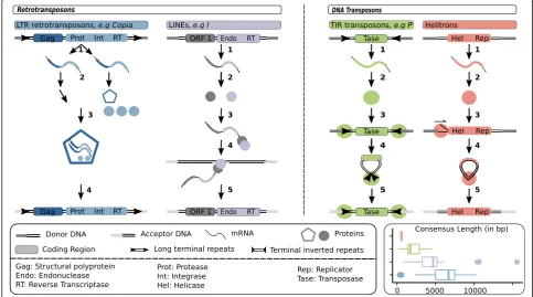
Class I TEs are also called retrotransposons. They trans-pose via an RNA intermediate. The RNA intermediate is transcribed from a genomic copy, then reverse-transcribed into DNA by a TE-encoded reverse tran-scriptase. Each complete replication cycle produces one new copy. Retrotransposons can be divided into five or-ders: long terminal repeat (LTR) retrotransposons, Dic-tyostelium intermediate repeat sequence (DIRS)-like elements, Penelope-like elements (PLEs), long inter-spersed nuclear elements (LINEs) and short interinter-spersed nuclear elements (SINEs). All of them are present in Drosophila,but LTR retrotransposons and LINEs are by far the most abundant [20,55].
In Drosophila, LTR retrotransposons usually range from 5 to 7 kb (Fig. 1) [11, 57–59]. They owe their names to the direct Long Terminal Repeats (~300-400 bp) flanking them. They typically display two genes: gag
and pol.gag encodes the capsid, andpolencodes a pro-tease (Prot), an integrase (Int) and a reverse transcriptase (RT) with an RNase domain. After the transcription step, some transcripts will be translated while the others may end up transposed (Fig.1) (see [60] for more details on transposition mechanisms). The protease of pol cleaves Pol into a protease, an integrase and a reverse transcript-ase [61]. The Gag protein assemble into a capsid that makes a particle around untranslated transcripts, the integrase, reverse transcriptase and a tRNA [62]. Because the formed ribonucleoprotein (RNP) does not comprise the transcript from which proteins were translated, we typically refer to a trans-preference mechanism of RNP assembly. Using the tRNA as a primer for synthesis, the reverse transcriptase initiates the production of double stranded DNA from the TE transcript [63]. After reverse transcription, the particle falls apart, the integrase recog-nizes the two ends of the cDNA and inserts them into the host genome. Upon integration, LTR retrotranspo-sons produce a TSD of 4-6 bp [11]. Note that the LTR order is further divided into five superfamilies: Copia (e.g. Copiaand 1731 families),Gypsy (e.g. HMSBEAGLE and 412 families), Bel-Pao (e.g. BEL, Roo and Max
families),Retrovirusand Endogenous RetroViruses (ERV). According to Wicker and colleagues classification, Retro-virusesand ERVs also have an envelope gene (env). The corresponding protein allowsRetrovirusesto infect other cells. In Drosophila, few families have been shown to possess an env coding ORF, for example Idefix, Gypsy, Tirant and ZAM families [58, 64,65]. Note that the in-sect endogenous retroviruses belong to theGypsy super-family, and that their origin is distinct from that of vertebrate ERVs [66]. Infectious properties have been demonstrated forGypsyandZAMfamilies [67,68].
LINEs are 3 to 5 kb-long, and generally contain two ORFs (Fig. 1) [11, 59, 69–71]. The first ORF encodes a protein with both RNA binding and nucleic acid chaperone properties [72,73]. The second ORF encodes a protein that displays two domains: an endonuclease (Endo) and a Reverse Transcriptase [74,75]. Contrary to LTR retrotransposons, LINEs exhibit a cis-preference mechanism of RNP assembly. After translation, the pro-tein(s) bind to the mRNA molecule from which they ori-ginate, and form an RNP in the cytoplasm [76] (see [77] for more details on transposition mechanisms). The ri-bonucleoprotein particle moves back to the nucleus, and the protein cuts a single strand of the host genome at the point of insertion. The exposed 3’end allows the ini-tiation of reverse transcription (target-primed reverse transcription). Subsequent events remains unclear, how-ever the following has been proposed. During or after re-verse transcription, the second strand of the host genome is cleaved. The newly reverse transcribed single-stranded DNA binds to the generated 3’ extremity, and this extremity acts as a primer for the synthesis of the second strand of DNA. LINEs generate TSDs of various sizes upon insertion. Note that, probably as a conse-quence of early termination of reverse transcription, transposition may result in creation of 5’ - truncated copies [78].
As mentioned above, besides LTR retrotransposons and LINEs that are abundant in Drosophila genomes, Class I comprises three other orders: DIRS, PLEs and SINEs. To our knowledge, DIRS and SINEs have not been found in Drosophila so far [20,79]. PLEs were ini-tially discovered inD.virilisand are involved in the hy-brid dysgenesis phenomenon (Table 1). These TEs are present at least in the virilis group and in D. willistoni [89]. PLEs resemble LINEs, in a sense that they encode an endonuclease and a reverse transcriptase. However, they possess terminal repeats that can be in a direct or an inverse orientation.
Class II TEs: DNA transposons
Class II TEs are DNA transposons. They do not trans-pose via an RNA intermediate but via a DNA intermedi-ate. There are four orders: terminal inverted repeat
(TIR) transposons, Crypton, Helitron and Maverick. TIRs and Helitrons are the most abundant in Drosophila.
TIR Transposable Elements are typically ranging from 1.5 to 3 kb in D. melanogaster, and are characterized by their TIRs of variable lengths (Fig. 1) [11, 59, 90, 91]. TIRs encode one unique protein called transposase (Tase). The transposition mechanism begins with two transposases recognizing and binding to the TIRs [92]. Transposases dimerize and cleave the ends of TIRs forming a free complex containing the TE [93]. The formed entity binds to the target DNA locus, where the transposon is integrated. The TSD size and the se-quences of TIRs are highly variable across the nine known superfamilies [11]. Although the transposition mechanism in itself is not replicative, such TEs can in-crease their copy numbers in two ways. First, by trans-posing during chromosomal replication from a position that has already been replicated to a position ahead of the replication fork [94]. Second, they can exploit gap re-pair following excision to create an extra copy at the donor site [95].
The Helitron order, which is represented by the unique Helitron superfamily, gave rise to rather small TEs inD. melanogaster(< 1 kb, Fig.1) [11,96,97]. Heli-trons encode one unique protein with both a DNA heli-case (Hel) and a replicator (Rep) domain. Because Helitrons were discovered only in 2001, and the lack of active Helitron examples limits experimental work, Heli-tron transposition mechanisms remain murky. However, using an artificially reconstructed active Helitron, Gra-bundzija and colleagues provided new insights and sug-gested the model synthesized hereafter [98]. First, the plus strand, the original donor strand, is nicked at the 5’-extremity of the TE and a replication fork is created. DNA replication results in a reconstituted double stranded donor site and a double stranded TE circle. This step may be repeated several times, producing sev-eral TE circles. Moreover, on the TE circles, a second DNA cleavage may occur on the original donor strand, a new replication fork established, and two double stranded transposon circles obtained from one. Finally, the double stranded TE may be integrated at the ac-ceptor site. Note that the small sizes of Helitrons in D. melanogaster are explained by their non-autonomous character.
TE abundance
The Drosophila melanogaster reference genome
homologous to a library of Drosophila TE consensus sequences available in the RepBase database, and the bioinformatic tool OneCodeToFindThemAll to recon-stitute TE copies [56, 100, 101]. As previously re-ported, D. melanogaster genome contains ~20% of TEs [55, 102]. Note that a significant variation exists regarding these estimates [103–105]. These differences are likely to be at least partly explained by the gen-ome assembly, or the part of the gengen-ome assembly that is analyzed, or both. For example, the Drosophila
12 genomes consortium considered only the best-assembled part of the genome, likely representative of the euchromatic portion of the genome, and found the TE content ranging from 2 % to 8 % (see Popula-tion Genomics secPopula-tion for details about TE density in different genomic regions). On the contrary, even if far from reporting the entire sequence of heterochro-matic regions, the assembly used in Fig. 2 comprises at least 20 Mb of heterochromatic sequences, i.e. ~15% of the 140 Mb assembly [106]. Nevertheless, Table 1Hybrid dysgenesis
InDrosophila, some intraspecific crosses were observed to produce sterile females [50,51,80–85]. This phenomenon is called hybrid dysgenesis (Ovaries pictures from ref 81). It happens when males possessing a particular TE, hereafter referred as the inducer TE, are crossed with females whose genome is devoid of this TE. On the contrary, the reciprocal cross leads to viable and fertile individuals. The explanation is related to piwi-interacting RNAs (piRNAs), small RNAs repressing TEs with sequence complementarity (see the piRNA section). Because piRNAs are maternally transmitted, in dys-genic crosses the inducer TE insertion is transmitted to the progeny without the piRNAs directed against it [86]. In the reciprocal cross, both the in-ducer TE insertion and its piRNAs are transmitted, allowing the control of the TE family in the progeny, and the hybrid to be fertile.
the relative abundance of the different TE orders is globally conserved across studies and similar to what is represented in Fig. 2 [55, 102, 103, 105]. Retrotran-sposons, and essentially LTRs and LINEs (respectively 12% and 5% of the genome in our analysis), contrib-ute substantially to D. melanogaster TE content. DNA transposons correspond to a smaller proportion of the genome: we found that they represent less than 2%, including 0.9% for Helitrons and 0.7% for TIR ele-ments. This ten-fold difference in terms of genomic sequence occupancy between retrotransposons and DNA transposons is mostly due to the larger size of retrotransposons (Fig. 2). Indeed, in terms of insertion numbers we found 11,657 DNA transposons (6,284 Helitrons and 5,373 TIR elements) and 23,148 retro-transposons (14,540 LTR retroretro-transposons and 8,608 LINEs) (see also [103] and [101]). For each of the four major orders, one superfamily is often over-represented: Gypsy for LTR elements, Jockey for LINEs, P for TIR elements, Helitron for Helitrons. According to our analysis, the different TE orders ex-hibit different numbers of families: indeed, we found insertions belonging to 721 LTR families, 331 LINE families, 213 TIR families and 63 Helitron families. The mean copy number per family is 26, but large variations exist. The family having the highest number of insertions is DNAREP1_DM, for which we found 1,746 copies. This sequence is annotated as a non-autonomous Helitron [107] (but see [97, 108] con-cerning classification).
Interspecific variation
When it comes to TE contents across Drosophila spe-cies, a direct comparison of studies may be difficult. In-deed, authors are free to choose among a large number of programs and methods dedicated to identifying TEs,
which leads to widely different results [105,109]. For ex-ample, using the same TE sequence library but two dif-ferent tools to annotate theD. willistonigenome, the 12 genomes consortium estimated TE content to be either 9 % or 16 %. The library used may also greatly affect re-sults. In the same study, using the same tool, but a D. melanogaster TE sequence library or a de novo library, the authors found either 12 or 20 % TEs in theD. ana-nassae genome. Overall, in this study seven combina-tions of library-detection tools were used, leading to a TE content ranging from less than 10 % to up to 30 % in D. ananassae. The direct comparison of studies may thus be risky. A further layer of complexity comes from the sequencing technology, which impacts the quality of genome assemblies. Short paired-end read based assem-blies lead to underestimation of TE contents compared to Sanger and long read based assemblies [110–112]. For all these reasons, to describe variation of TE contents in the Drosophila genus, here we focus on studies directly aiming at comparing TE amounts across species, and we remain cautious when linking them. For illustrative pur-poses, in addition to the annotation of TE contents inD. melanogaster, we estimated TE genomic sequence occu-pancy and copy numbers in two species: D. simulans and D. virilis (Fig. 2). We used the exact same methods as forD. melanogaster,and we do not expect the TE li-brary to strongly bias the results, as it contains se-quences constructed from the three species, which are among the most - studied with regard to TEs [113,114]. Beyond that, we chose these two species because of their different positions relatively to D. melanogaster in the Drosophila phylogeny. On one hand, D. simulans is a close relative toD. melanogaster;they diverged approxi-mately 1.5 Mya. Both species belong to themelanogaster subgroup within the melanogaster group, itself in the sophophora subgenus [14]. On the other hand, D. Fig. 2TE contents inD. melanogaster,D. simulansandD. virilis(from left to right). Barplots represent TE copy numbers for the top 20 TE
melanogaster and D. virilis diverged about 25 Mya and D. virilisbelongs to a different subgenus, the drosophila subgenus.
The first study intending to compare global TE con-tents across a significant number of Drosophila species was performed by the Drosophila 12 genomes consor-tium. This consortium investigated TE genomic se-quence occupancy in eight species from the sophophora subgenus, mostly from the melanogaster subgroup, and four species from the drosophila subgenus. As stated above, the researchers focused on genomic parts likely to be euchromatic, and they used different methods. Using the method giving the lowest estimates, they found a global range of variation going from 1% to 9% of TEs in the genome. The method leading to the highest estimates resulted in genome containing from 3% to 30% of TEs. Invariably,D. ananassaewas the species with the highest proportion of TEs. The authors chose the most unbiased and conservative method to compare the rela-tive abundance of LTR retrotransposons, LINEs, TIR el-ements and so-called OTHERs among species. They found that the pattern LTRs>LINEs>TIRs>OTHERs is globally conserved across the phylogeny, with LTR retro-transposons usually constituting more than 50% of the repeatome. The two exceptions areD. mojavensisandD. pseudoobscura. In D. mojavensis, LTR elements repre-sent only 45% of the repeatome, and in D. pseudoobs-cura, LTR retrotransposons and LINEs each contribute to roughly 33% of the repeatome. Our analysis shows a slightly different pattern, with equivalent genomic se-quence occupancy for LTR elements and LINEs in D. simulans, and more Helitrons than TIR elements in D. virilis (Fig. 2). Recently, Hill and colleagues investigated both the proportion of TEs and their number of inser-tions in the genomes of five species. Four of these spe-cies were already in the set analyzed by the Drosophila 12 genomes consortium, except for D. innubila. The LTRs>LINEs>TIRs>OTHERs pattern for TE genomic proportions was not respected by any of the considered species. The dominant category differed: the most abun-dant elements are LTR retrotransposons in D. ananas-sae,while they are LINEs inD. pseudoobscura, and DNA transposons in D. innubila. D. ananassae was also the species with the highest TE content, with approximately 35% of TEs in the genome. Considering TE copy num-bers, the authors found a total ranging from 2,000 to 14, 000 depending on the species. Once again, the difference with the previous results may probably be explained by data/method differences. Relative abundances of the dif-ferent TE categories were found to differ across ge-nomes. For example, DNA transposons were the most abundant in D. willistoni,whereas inD. ananassae they were as numerous as LINEs or LTR elements. The study with the largest dataset of species compared in terms of
TE content was published by Sessegolo and collabora-tors [20]. These authors investigated the TE contents of 26 Drosophila species. Once again, the LTRs>LINEs>-TIRs>OTHERs pattern did not hold for many species. The genomic content of repeats ranged from 4.65% in D. busckii to 30.80% in D. suzukii. The authors found a significant effect of phylogenetic inertia on TE content, but because of uneven sampling across the phylogeny, it was difficult to extract a pattern for each subgroup, many being represented by only one species. Overall, the data suggest large variations in the abundance of TEs across theDrosophilagenus.
Intraspecific variation
results were obtained when contrasting copy numbers of BilboandOsvaldo between colonizing and original pop-ulations ofD. buzzatii[127]. In both cases, the study of insertion frequencies suggested that genetic drift associ-ated with a founder effect that accompanied the colonization was responsible for the observed variation of copy numbers. Recently, genomic analyses of Euro-pean D. melanogaster populations from DrosEU con-firmed that intraspecific variation of TE contents may be substantial, and reveals TE proportions ranging from 16% to 21% of genomes [28].
TE activity
Spontaneous rate of transposition
A recent study by Adrion and colleagues [128] provided the first genome-wide estimate of TE movement rate in D. melanogaster. These authors used NGS data to com-pare TE contents across laboratory lines before and after ~150 generations of mutation accumulation. They found that the TE movement rate is slightly lower than the point mutation rate: 2.45 × 10(-9) per site per generation against 2.8 × 10(-9) per site per generation, respectively [129]. The rate of insertions is higher than the rate of deletions: 2.11 × 10(-9) per site per generation against 1.37 × 10(-10) per site per generation, respectively. Con-sidering that there are 270 millions sites in the genome assembly, these numbers correspond to approximately 0.57 insertions and 0.037 deletions per generation. Those estimates were obtained across all TE superfam-ilies and are consistent with previous reports using in situ hybridization to determine transposition events for one or a few families [130–132]. Adrion and colleagues found superfamily-specific insertion and deletion rates to range between 0 and 5.13 × 10(-3) per copy per gen-eration, and between 0 and 1.29 × 10(-4) per gengen-eration, respectively. They also found a significant effect of the genetic background, as previously reported [133–135].
Transposition bursts
Beyond the spontaneous rate of transposition, a sig-nificant number of studies have shown that transpos-ition bursts could occur in Drosophila (see [136] for a review). A burst is characterized by movement of large numbers of TE sequences through the genome during a short evolutionary time [137]. Although these bursts can happen without any apparent reason, they are commonly associated with stressful condi-tions such as extreme temperatures, irradiation, chemical exposure, or viral infection [138–142]. For example, Vasil’eva and colleagues showed that gamma radiation could increase the 412 transposition rate up to 5.6 events per genome per generation. Note that the attempts to induce TE mobilization with thermal shocks led to contradictory results in Drosophila,
Interspecific variation
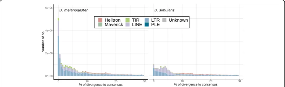
So far, few studies tried to compare TE activity across Drosophila species. In 2011, Lerat and colleagues com-pared the TE contents of four Drosophila species from the melanogaster subgroup: D. melanogaster, D. simu-lans, D. sechellia and D. yakuba[153]. They found that D. simulans, D. sechellia and D. yakuba genomes con-tained a large fraction of degraded copies compared to D. melanogaster. The authors suggested a recent TE ac-tivity in D. melanogaster, compared to the three other species. This can partially be observed when comparing the so-called TE landscapes of D. melanogaster and D. simulans (Fig. 3). These landscapes constitute an easy way to visualize TE activity through time. The X axis corresponds to the divergence of the TE sequences from the consensus, and it can be seen as a proxy of the time passed since the last wave of transposition. In Fig.3, we can see a recent peak of activity of LTR elements, espe-cially inD. melanogaster. InD. simulans, the peak of ac-tivity is also recent but much smaller. Another study was aimed at comparing TE activity betweenD. melano-gaster and D. simulans using NGS population data [155]. Based on TE insertion frequency data, the authors determined that more than 58 families are probably highly active in both species. Half of the TE families show evidence of variation of activity through time, and are not the same depending on the species. Finally, they found that retrotransposons were the most active TEs in D. melanogaster,while DNA transposons were the most active TEs in D. simulans. A recent study compared TE frequencies in five distant points of theDrosophila phyl-ogeny [55]. These species shared a common ancestor around 30 Mya [14]. The authors found evidence that an excess of low frequency insertions is prevailing in the phylogeny and is observed for most TE families. This
suggests that an active repeatome is frequent, at least in theDrosophilagenus.
Impacts of TEs On the genome
TEs play an important role in the structural evolution of genomes through the generation of various types of mutations: chromosomal rearrangements, gene dis-ruption and changes in gene expression. The simplest mechanism by which TEs can cause chromosomal re-arrangements is through participation in an ectopic recombination event [156]. Ectopic recombination corresponds to recombination between more-or-less identical sequences inserted at different locations in the genome, such as TEs [157]. Depending on their relative positions and orientations, their recombin-ation can result in different kinds of chromosomal re-arrangements: duplication, deletion, inversion, or translocation. TEs were associated with chromosomal rearrangements in natura in various species of Dros-ophila, and mainly with inversions [158–161]. In sev-eral cases, ectopic recombination was identified as the cause of these rearrangements [159, 160]. When they insert into genes or their regulatory sequences, TEs can disrupt gene function. A perfect example is the use of the P-element in the Berkeley Drosophila Gen-ome Project [162–164]. The Berkeley Drosophila Gen-ome Project aimed at disrupting each D. melanogaster gene using the P-element in order to decipher gene functions. More than 5,000 genes were disrupted in that way. TEs can affect gene expression in two prin-cipal ways. First, they may bring regulatory sequences (see [165] for a review). For example, Bari-Jheh adds extra antioxidant response elements upstream of the
Jheh1 and Jheh2 genes and is associated with
upregulation of Jheh1 and Jheh2 [166]. Second, the spread of repressive epigenetic marks targeting TEs can reduce the expression of nearby genes (see below, host defenses against TEs), as it was also demon-strated in the Jheh cluster [167]. Lee and Karpen demonstrated recently that the spread of repressive epigenetic marks to nearby DNA occurs for more than half of euchromatic TEs, and can extend up to 20 kb [12]. This effect is TE dependent, copy number dependent, but also species dependent, with stronger epigenetic effect in D. simulans compared to D. melanogaster.
On the individual
While some of the aforementioned genomic changes might remain phenotypicaly silent, others may have dramatic repercussions at the individual level. TEs are responsible for up to 80% of the phenotypic spontaneous mutations observed in D. melanogaster [168] and many observations suggest deleterious ef-fects of TEs in Drosophila. Five to 10 % insertions of active P-elements are estimated to cause recessive le-thal mutations in D. melanogaster [169]. In D. simu-lans, somatic transposition of Mariner decreases lifespan [170]. In 2004, a study used two D. melano-gaster lines with the same genetic background, but different TE copy numbers, to evaluate the impact of TE number on fitness. The authors found differences in fitness and egg hatchability between the two lines, the line with more TEs performing worse than the other. Both homozygous and heterozygous TE inser-tions were shown to have deleterious effects on
fit-ness and its components [134]. Overall, TE
insertions are expected to be generally neutral or deleterious to the host genome [171]. Considering that adaptive mutations are supposed to quickly reach fixation in populations, the low numbers of fixed insertions in D. melanogaster and D. simulans support this theory. In 2006, Burt and Trivers calcu-lated the number of insertions since the divergence between the two species and concluded that, given both genome size and number of fixed insertions, the occurrence and fixation of a beneficial insertion is a really rare event [156]. However, they also underscored the difficulty to detect fixed insertions using in situ hybridization, and suggested it would have been interesting to estimate the rate of fixation from sequencing data. In 2015, using population se-quencing data, Kofler and colleagues estimated the number of fixed insertions in D. melanogaster since its divergence from D. simulans to be approximately 200 [155]. Considering a 1.4 Mya divergence [14], we computed a fixation rate of 1.4 fixed insertions every 10,000 years, i.e. maximum 1.4 beneficial fixed
insertions every 10,000 years. If we update the Burt and Trivers calculation and compare the number of fixed insertions to the total number of insertions over this period: Population size × Insertion rate per genome per generation × Divergence time between D. melanogaster and D. simulans × Number of gen-erations per year = 10 (6) × 0.57 × 1.4 × 10 (6) × 24 = 1.9 × 10 (13) insertions, that is to say 200/(1.9 × 10 (13)) = 1.0 × 10(-11) insertions reaching fix-ation. Finally, we estimated maximum 1.4 beneficial fixed insertions every 10,000 years, or maximum 1 out of 1e11 insertions, being beneficial and fixed. These numbers are upper bounds because all fixed insertions are unlikely to be beneficial. Indeed, most of the fixed insertions are present in regions where the effect of selection is weak, and are essentially old. Therefore, they are more likely to have reached fixation slowly by drift than quickly by positive se-lection [172, 173]. So far, 21 fixed insertions have been identified within or near genomic regions showing low Tajima’s D values, and 12 fixed inser-tions are relatively young. Considering the above, one could expect to find very few putatively adaptive insertions among unfixed insertions. Surprisingly, there are at least 57 of such insertions in the refer-ence genome [173], suggesting a high rate of TE me-diated adaptation recently or even ongoing. The discrepancy between the number of candidates for recent adaptation and the fixation rate was discussed considering the three following points: 1. The migra-tion of D. melanogaster out of Africa may have caused a significant augmentation of the adaptation rate. 2. TE derived adaptations might be ephemeral. 3. Adaptive TE sequences may evolve quicker than neutral insertions, resulting in an underestimation of the number of fixed insertions [174]. One may also add that the TE mutation rate has potentially in-creased recently [175]. It is worth noting that few insertions were clearly associated with an adaptive phenotype so far [166, 176–178]. Interestingly, can-didate adaptive insertions are often close to, or within genes associated with stress response, behav-ior and development. Moreover, two of the historical examples of adaptation associated with TEs corres-pond to two different insertions in the same gene implicated in the response to oxidative stress, cyp6g1, in two different species: D. melanogaster and D. simulans [176, 178, 179].
The case of telomeric elements
chromosome ends is threatening internal regions con-taining essential genes and may contribute to ageing [181]. Organisms have evolved different mechanisms that protect their chromosomes. Usually in eukaryotic genomes a ribonucleoprotein enzyme, the telomerase, mediates the RNA dependent synthesis of tandemly re-peated simple sequences at chromosome ends [182]. In D. melanogaster, the three families, HeT-A, TART and TAHRE,transpose to chromosome extremities, and pro-tect them from shortening [180,183–186]. Many phylo-genetically distinct telomeric retrotransposons have been found in more distant species [187]. All these telomeric elements belong to a single monophyletic clade inside theJockeysuperfamily. The telomeric element phylogeny and species phylogeny are congruent, suggesting vertical transmission from a common ancestor and a conserved host-element relationship [187]. Furthermore, the clade presents evidence of specialization to transpose at chromosome ends [188]. Because of this, the relationship between TEs and their host in this case was referred to as genomic “symbiosis” [188]. However, Saint-Leandre and colleagues investigated more species of the mela-nogaster group [189]. They suggest that these Jockey telomeric elements may have evolved to selfishly over-replicate. In agreement with this hypothesis, they found recurrent gains, losses, and replacements of Jockey telomeric elements. Moreover, in D. biarmipes, the telomere-specialized elements have disappeared completely.
Host defenses
Because of the above-mentioned deleterious effect of TE insertions, several mechanisms of TE control have evolved. Among these, epigenetic modifications play an important role [190]. For example, in mam-mals and plants, TE insertions are usually associated with DNA methylation and histone modifications. Both are related to repressive chromatin states. In Drosophila, DNA methylation has been shown to be almost completely absent, and small RNAs are cen-tral to TE regulation [191, 192]. They may also trig-ger histone tail modifications and chromatin conformation modifications. There are two small RNA pathways controlling TEs in Drosophila: the piRNA and the siRNA pathways. Our purpose here is to give a brief overview of these pathways and their role in shaping TE dynamics. In particular, we refer the reader to [193, 194] for comprehensive re-views on the mechanistic aspects of the piRNA pathway.
The piRNA pathway
The piRNA pathway produces small, single stranded RNAs that were first called rasiRNAs (repeat associated
small interfering RNAs); however, contrary to regular small interfering RNAs, they are 23-30 nt long, and are associated with the Piwi-subfamily Argonaute proteins, which led to their new designation as piRNAs (piwi-interacting RNAs). These piRNAs silence TEs in germ cells, where maintaining the integrity of the genome is of primary importance, as new mutations are passed on to future generations. This pathway is also active in the ovarian somatic follicle cells, which support oogenesis. It prevents endogenous retroviruses, such as Gypsy, from infecting the adjacent oocyte [195]. Research studies in Drosophila were seminal in the piRNA field. Much of what we know today was discovered using this model. In fact, piRNAs were identified for the first time in 2001 in fly testis [196]. They were found to silence Stellate, a gene involved in male sterility. Some of them were even found to be homologous to TEs and assumed to be in-volved in transposon regulation. Moreover, a long-term study of theGypsy family activity led to the discovery of flamenco, a non protein-coding locus producing piRNAs, which was subsequently shown to be involved in the control of other TE families, essentially LTR retrotran-sposons [197,198] (Table2).
piRNAs originate from discrete genomic loci called piRNAs clusters. These loci contain mainly defective TEs and are transcribed into long piRNA precursors (Fig. 4 [202]). Approximately 150 clusters have been identified in the genome of D. melanogaster, represent-ing 3.5% of the assembled genome [208]. The vast ma-jority of them appear to be heterochromatic. The size of piRNA clusters varies substantially, with the largest be-ing 240 kb. Overall, the largest 15 clusters produce a
Table 2theflamencostory
Much of what we know today on the piRNA pathway was discovered using theDrosophilamodel. Especially, a considerable effort started 40 years ago inD. melanogasterled to the early discovery of a gene producing piRNAs and silencingGypsyin a piwi dependent manner (see [199] for a review). This gene namedflamencowas the first piRNA cluster identified.
In the 1980s, theovoDdominant mutation was identified inD. melanogasterand associated with female sterility [200]. Interestingly, crosses betweenovoDmales and females from a particular strain led to the reversion of the phenotype and the recovery of fertility of the daughters, in addition to numerous mutations at other loci. Further work revealed that the particular strain used for the mothers actually displayed high copy numbers of uncontrolledGypsy, whose
transposition intoovoled to a null allele and reversion of sterility [201]. And then, it was demonstrated that the locus controllingGypsyactivity was theflamencolocus, located on the X chromosome and containing a lot of TE sequences [202,203]. In 2004, Sarot and collaborators found out thatGypsytransposition was sensitive to a mutation inpiwi, a gene known to affect RNA-mediated silencing [204]. They also demonstrated that small RNAs homologous toGypsywhere present in silenced tissues. For the first time a gene producing small RNAs was associated with TE silencing, and Piwi was implicated in this process.
large proportion of the total amount of piRNAs: 70% of the piRNAs uniquely mapped to the genome originate from these clusters.
The beginning of piRNA biogenesis is similar in germline and somatic cells (see [193, 194] for detailed reviews). PiRNA cluster transcription is ensured by RNA Pol II and leads to a single stranded long RNA (Fig. 4). Then, piRNA cluster transcripts may enter either the ping-pong pathway or the phased piRNA pathway [208–213]. The ping-pong pathway occurs in germline cells. In this case, guided by a sense piRNA, Argonaute3 (Ago3) binds to a complementary piRNA cluster transcript and cleaves it. Then, Aubergine (Aub) attaches to the newly formed 5’ extremity, slices the transcript and forms an antisense piRNA. Finally, guided by an antisense piRNA, Aub operates a cut in a TE transcript, Ago3 recognizes the result-ing 5’ extremity, cleaves the transcript and forms a sense piRNA. This is the pong pathway or ping-pong loop. The phased piRNA pathway is not specific to germline cells and may also occur in ovarian som-atic follicle cells. Piwi is loaded at the 5’ extremity of the piRNA precursor and Zucchini (Zuc) performs cleavage, generating the piRNA. Piwi is then loaded
again at the 5’ extremity of the precursor piRNA, and the process is repeated in a step-by-step cleavage gen-erating multiple piRNAs. Note that, for clarity, piRNA maturation steps such as trimming are not mentioned here.
After synthesis, piRNAs mediate silencing both at the transcriptional and post-transcriptional levels [214]. The post-transcriptional silencing occurs in the cytoplasm of germline cells only, and corre-sponds to the ping-pong pathway (Fig. 4). At the transcriptional level, a piRNA guides the Piwi protein to a TE insertion, probably due to sequence comple-mentarity with nascent TE transcripts, and mediates local heterochromatin formation by addition of the repressive mark H3K9me3 to histone tails [215–221]. Note that, despite the fact that as early as 2001 piR-NAs were detected in testes, so far most of the work on TE regulation by piRNAs has been done on ovar-ies [196]. Regulation in testes seems to be quite simi-lar to what happens in female germline, with both ping-pong and phased piRNA pathways being active [222–224]. However, contrary to ovaries, the data suggest an Ago-3 independent amplification loop in spermatogenesis.
The siRNA pathway
In addition to piRNAs, sequencing of small RNAs re-vealed the existence of another class of interfering RNAs targeting TEs: endogenous small interfering RNAs, or endo-siRNAs [225–227]. These small RNAs are present in both somatic and germline cells. endo-siRNA sors are double strand RNAs (dsRNAs). These precur-sors may be produced through three distinct mechanisms (Fig.4) [228]. 1. Transcription of the same genomic region in both sense and antisense directions (convergent transcription), then base pairing of the over-lapping region between sense and antisense transcripts. 2. Transcription of complementary sense and antisense transcripts from different genomic regions and base pairing. 3. Base pairing of inverted repetitive elements of one transcript to form a hairpin RNA. The resulting long dsRNA is loaded on Dicer-2 (Dcr-2) and its cofac-tor Loquacious-PD (Loqs-PD) and then processed into 21 nt small double stranded RNAs. They are then loaded on theRNA-induced silencing complex (RISC) including the Ago2 protein. One strand is held and guides the complex to target transcripts that are then cleaved by the RNase domain of Ago2.
Evolution
Several studies demonstrated rapid evolution of anti-TE RNAi genes in Drosophila [47, 229–232]. Indeed, these genes often present signatures of recurrent positive se-lection. By analogy to the signatures of positive selection observed for genes involved in host-parasite interactions, the rapid evolution of anti-TE RNAi genes is often inter-preted as a consequence of an arms race occurring be-tween TEs and TE immunity effectors. Focusing on the piRNA pathway, Blumenstiel and colleagues propose that selection for sensitivity to TE content but also selec-tion for specificity to TE content may drive the rapid evolution of host defense mechanisms [233]. More pre-cisely, concerning the specificity aspect, the authors propose that a too efficient piRNA pathway may induce a too efficient silencing of TE copies that could spread to neighboring genes, which would constitute a cost. They designated this form of off-target gene silencing as “genomic autoimmunity”, an analogous to classic forms of autoimmunity which are caused by an immune re-sponse that incorrectly targets self. Despite the rapid evolution of anti-TE RNAi genes inDrosophila, suggest-ing that host defense mechanisms may vary a lot across the genus, most of the literature on this subject concerns D. melanogaster. A recent study of 20 arthopod species suggests that somatic piRNAs were probably produced in the ancestral arthropod more than 500 Mya and dem-onstrated that, in contrast to D. melanogaster, D. virilis presents somatic piRNAs [234]. This suggests a loss of the piRNA pathway in the soma ofD. melanogaster.
Population genomics
The Drosophila model has been of outstanding import-ance in the field of population genomics of TEs. The ease to get and maintain wild type strains was obviously a key factor, but so was the development of the in situ hybridization method on Drosophila polytene chromo-somes more than 40 years ago [235, 236]. In situ hybridization allows to detect and localize genomic DNA sequences using a labeled sequence (probe) hom-ologous to the targeted sequence. The giant polytene chromosomes are found only in some species and tis-sues, and offer to the researcher a high degree of reso-lution [237, 238]. Using TE probes on salivary gland polytene chromosomes ofDrosophilathird instar larvae, researchers were able to detect and localize TE inser-tions in individuals and thus to accurately estimate TE insertion frequencies in natural populations [239–241].
About the nature of selection acting on TEs
The first in situ hybridization studies evaluating TE in-sertion frequencies in natural populations ofD. melano-gaster demonstrated a predominance of insertions segregating at low frequencies [239–241]. This result ob-tained for specific families was later confirmed at a broader scale. Population sequencing data showed that, in D. melanogaster and D. simulans, more than 80% of TE copies have insertion frequencies lower than 0.2 [155]. This observation is often interpreted as the result of purifying selection acting on TEs. So far, three main hypotheses have been formulated concerning the nature of selection against TEs: 1) the gene-disruption hypoth-esis [3, 242], 2) the ectopic recombination hypothesis [243,244], 3) the deleterious TE-product expression hy-pothesis [245].
The gene disruption hypothesis assumes that inser-tions inside genes or regulatory regions are under strong purifying selection because of their negative ef-fect on the host fitness [242]. A large amount of work supports this hypothesis, demonstrating a depletion of TE insertions in exons and untranslated regions [172, 246–248]. Moreover, Lee and Karpen demonstrated that repressive histone marks affecting euchromatic TEs can spread up to 20 kb both in D. melanogaster and D. simulans, and that this phenomenon is associ-ated with selection against TEs [12]. Therefore, we may extend this hypothesis beyond insertions inside genes or regulatory regions to include insertions close to genes.
meiotic recombination rate, expected to be positively correlated with ectopic recombination rate, should be associated with the strength of purifying selection [137]. First, since long insertions provide longer tar-gets for recombination, one can indeed expect a stronger effect of purifying selection against long TEs in the ectopic recombination hypothesis. The negative correlation between TE size and population frequen-cies suggests that it is actually the case [172, 249]. Second, because ectopic recombination is more likely to occur when TEs are heterozygous, ectopic recom-bination should happen more frequently for TE fam-ilies with a high copy number of polymorphic TEs. Therefore, the negative correlation between TE inser-tion frequencies and copy numbers also supports the ectopic recombination hypothesis [172, 249]. Finally, because ectopic recombination is intrinsically related to the local recombination rate, the fact that low-recombining regions are highly enriched in TEs, and that a negative correlation exists between insertion frequencies and recombination rate [172, 246, 249, 250], constitute one more argument in favor of the ectopic recombination hypothesis. However, this last point may be explained by the Hill-Robertson effect, or the lower density of genes in low-recombining re-gions, or both. The Hill-Robertson effect corresponds to a reduction in the efficiency of selection on a locus due to selection on related loci. If slightly deleterious insertions are close to adaptive mutations, they will be less efficiently removed in low-recombining regions than in high-recombining regions. The lower density of genes in low-recombining regions may explain the higher TE density in these regions because one may expect that TE insertions are strongly counter-selected close to genes (gene disruption hypothesis). However, one paradox exists when considering the ectopic recombination hypothesis. Indeed, considering the higher rate of recombination on the X chromo-some, and the ectopic recombination hypothesis, TE density should be lower on the X chromosome [251]. However, recent studies of D. melanogaster natural populations show different results. TE density was found to be either higher on the X chromosome [246], or similar between the X chromosome and au-tosomes when taking into account differences in the amount of low recombining regions [172]. A higher transposition rate in the X chromosome relatively to autosomes has been proposed as a plausible ex-planation to the observed paradox [137]. Mutation accumulation data recently showed such tendency with a 1.86 fold change for insertion rate on the X chromosome relatively to autosomes [128].
One last hypothesis remains concerning the nature of the purifying selection affecting TEs: the deleterious
TE-product expression hypothesis [245]. Under this model, transcription and translation of TEs may be resource consuming for the host and TE proteins could disrupt cellular processes. According to this hypothesis, and as-suming that full length TEs are more transcribed than nearly complete copies, one may expect complete copies to be under more intense purifying selection than nearly complete copies. However, Petrov and colleagues did not find such effect investigating TE frequencies genome wide [249].
Models of TE dynamics
[197].In this model, after invasion of a host genome, a TE family proliferates until it is trapped, i.e. one inser-tion occurs into a piRNA cluster, then the subsequent production of piRNAs silences the invading family. This model was validated and enriched with populational considerations by Kofler and colleagues [88]. Monitoring the P-Element invasion, in connection with the piRNA pathway, in experimentally evolving populations of D. simulans, they suggested the following three-step model for a TE invasion: 1) TE copies colonize the genome, 2) the first TE insertions in piRNA clusters occur but are not yet sufficient to stop TE proliferation and 3) the TE family is inactivated by the fixation of an insertion within a piRNA cluster. Using simulated data, they were able to demonstrate that this“trap model”accurately de-scribes TE abundance inD. melanogastergermline. They also showed that the suppression of TE activity by segre-gating cluster insertions is reversible. Importantly, they demonstrated that transposition rates and population sizes affected mostly the duration of the invasion steps but not the amounts of accumulating TEs. In fact, the major factor capable of affecting the number of accumu-lating TEs was the piRNA cluster size.
Conclusions
In today's biology research, increasing weight is given to the study of non-model species. This is clearly justified by the diversity of the living world, and even more so for the study of genetic elements as diverse and dynamic as TEs. However, we should not overlook model organisms, because the vast amount of techniques, data collected and knowledge will help us develop and test new hy-potheses. Furthermore, the dissection of conserved path-ways in these organisms, such as the piRNA pathway, should provide results valid for a broad range of species. Despite the fact that Drosophila is an old biological model, it still presents many opportunities for TE re-search. In general, studies of TEs could benefit from uni-fied approaches to identifying and quantifying TEs. As we demonstrated above, the ultimate modelD. melano-gasterappears slightly different from its sister species re-garding TEs—maybe related to the fact that it ended up as the ultimate model species—however, it is clear that the research community greatly benefits from compara-tive genomics in theDrosophilagenus, and a great deal of work remains to be done in Drosophilaand the spe-cies in the group in order to do proper comparative gen-omics. It is clear that the development of long-read technologies will greatly facilitate this work. Another challenge is to understand the activity of TEs and how, in natura, this activity is triggered and controlled. Once again, Drosophilais a model of excellence with the pos-sibility of doing experimental evolution with a follow-up of TE dynamics. At the same time, this will allow a
better understanding of the fine regulation systems of TE activity. Finally, it seems to us that one of the most exciting challenges is to understand the true impact of TEs in adaptive processes, even more so now, with all the gross changes in our environment. Experimental evolution, with different species and different environ-mental factors, are a real opportunity to move forward in this field.
Abbreviations
Ago2:Argonaute2; Ago3: Argonaute3; Aub: Aubergine; Dcr-2: Dicer-2; DIRS: Dictyostelium Intermediate Repeat Sequence; Endo: Endonuclease; Env: Envelope gene; ERV: Endogenous RetroViruses; Int: Integrase; Hel: DNA helicase; LINE: Long Interspersed Nuclear Element; Loqs-PD: Loquacious-PD; LTR: Long Terminal Repeat; piRNA: piwi-interacting RNA; PLE: Penelope-Like Element; Prot: Protease; rasiRNA: repeat associated small interfering RNA; Rep: Replicator; RISC:RNA-induced silencing complex;RNP:RiboNucleoProtein; RT: Reverse Transcriptase; Tase: Transposase; TSD: Target Site Duplication; TE: Transposable Element; TIR: Terminal Inverted Repeat; SINE: Short Interspersed Nuclear Element; siRNA: small interfering RNA; Zuc: Zucchini
Acknowledgments
The authors sincerely thank the anonymous reviewers.
Authors’contributions
VM has drafted the initial version of the review and designed the figures; MB, MF and CV have contributed to the writing of the manuscript. All authors have approved the final version.
Funding
This work was supported by the ANR Exhyb and ANR SWING (grant overseen by the French National Research Agency) and the CNRS.
Availability of data and materials
The datasets analyzed during the current study are available in the following repositories:https://github.com/danrdanny/Drosophila15GenomesProject/ raw/master/assembledGenomes/[99],ftp://ftp.flybase.net/genomes/ Drosophila_melanogaster/dmel_r6.29_FB2019_04/
Ethics approval and consent to participate
Not applicable
Consent for publication
Not applicable
Competing interests
The authors declare that they have no competing interests
Received: 16 February 2020 Accepted: 14 April 2020
References
1. Doolittle WF, Sapienza C. Selfish genes, the phenotype paradigm and genome evolution. Nature. 1980;284:601–3.
2. Orgel LE, Crick FHC. Selfish DNA: the ultimate parasite. Nature. 1980;284: 604–7.
3. Charlesworth B, Charlesworth D. The population dynamics of transposable elements. Genet Res. 1983;42:1–27.
4. Feschotte C, Jiang N, Wessler SR. Plant transposable elements: where genetics meets genomics. Nat Rev Genet. 2002;3:329–41.
5. Ravindran S. Barbara McClintock and the discovery of jumping genes. Proc Natl Acad Sci. 2012;109:20198–9.
6. The Arabidopsis Genome Initiative. Analysis of the genome sequence of the flowering plant Arabidopsis thaliana. Nature. 2000;408:796.
7. Mouse Genome Sequencing Consortium. Initial sequencing and comparative analysis of the mouse genome. Nature. 2002;420:520. 8. C. elegans Sequencing Consortium. Genome sequence of the nematode C.
9. Lander ES, Linton LM, Birren B, Nusbaum C, Zody MC, Baldwin J, et al. Initial sequencing and analysis of the human genome. Nature. 2001;409:860–921. 10. Schnable PS, Ware D, Fulton RS, Stein JC, Wei F, Pasternak S, et al. The B73
maize genome: complexity, diversity, and dynamics. Science. 2009;326:1112–5. 11. Wicker T, Sabot F, Hua-Van A, Bennetzen JL, Capy P, Chalhoub B, et al. A
unified classification system for eukaryotic transposable elements. Nat Rev Genet. 2007;8:973–82.
12. Lee YCG, Karpen GH. Pervasive epigenetic effects of Drosophila euchromatic transposable elements impact their evolution. eLife. 2017;6https://doi.org/ 10.7554/eLife.25762.
13. Singh BN. Species and genetic diversity in the genus Drosophila inhabiting the Indian subcontinent. J Genet. 2015;94:351–61.
14. Obbard DJ, Maclennan J, Kim K-W, Rambaut A, O’Grady PM, Jiggins FM. Estimating divergence dates and substitution rates in the Drosophila phylogeny. Mol Biol Evol. 2012;29:3459–73.
15. Keller A. Drosophila melanogaster’s history as a human commensal. Curr Biol CB. 2007;17:R77–81.
16. Markow TA. The secret lives of Drosophila flies. eLife. 2015;4https://doi.org/ 10.7554/eLife.06793.
17. David JR, Capy P. Genetic variation of Drosophila melanogaster natural populations. Trends Genet TIG. 1988;4:106–11.
18. Hales KG, Korey CA, Larracuente AM, Roberts DM. Genetics on the Fly: A Primer on the Drosophila Model System. Genetics. 2015;201:815–42. 19. Rubin GM, Spradling AC. Genetic transformation of Drosophila with
transposable element vectors. Science. 1982;218:348–53.
20. Sessegolo C, Burlet N, Haudry A. Strong phylogenetic inertia on genome size and transposable element content among 26 species of flies. Biol Lett. 2016;12:20160407.
21. Genome List - Genome - NCBI.https://www.ncbi.nlm.nih.gov/genome/ browse#!/overview/drosophila. Accessed 4 Aug 2019.
22. Mackay TFC, Richards S, Stone EA, Barbadilla A, Ayroles JF, Zhu D, et al. The Drosophila melanogaster Genetic Reference Panel. Nature. 2012;482:173–8. 23. Langley CH, Stevens K, Cardeno C, Lee YCG, Schrider DR, Pool JE, et al.
Genomic Variation in Natural Populations of Drosophila melanogaster. Genetics. 2012;192:533–98.
24. Huang W, Massouras A, Inoue Y, Peiffer J, Ràmia M, Tarone AM, et al. Natural variation in genome architecture among 205 Drosophila melanogaster Genetic Reference Panel lines. Genome Res. 2014;24:1193–208. 25. Pool JE, Corbett-Detig RB, Sugino RP, Stevens KA, Cardeno CM, Crepeau
MW, et al. Population Genomics of Sub-Saharan Drosophila melanogaster: African Diversity and Non-African Admixture. PLoS Genet. 2012;8https://doi. org/10.1371/journal.pgen.1003080.
26. Lack JB, Cardeno CM, Crepeau MW, Taylor W, Corbett-Detig RB, Stevens KA, et al. The Drosophila Genome Nexus: A Population Genomic Resource of 623 Drosophila melanogaster Genomes, Including 197 from a Single Ancestral Range Population. Genetics. 2015;199:1229–41.
27. Machado HE, Bergland AO, Taylor R, Tilk S, Behrman E, Dyer K, et al. Broad geographic sampling reveals predictable, pervasive, and strong seasonal adaptation in Drosophila. bioRxiv. 2019:337543.https://doi.org/10.1101/337543. 28. Kapun M, Barrón MG, Staubach F, Vieira J, Obbard DJ, Goubert C, et al.
Genomic analysis of European Drosophila melanogaster populations on a dense spatial scale reveals longitudinal population structure and continent-wide selection. bioRxiv. 2019:313759.https://doi.org/10.1101/313759. 29. Grenier JK, Arguello JR, Moreira MC, Gottipati S, Mohammed J, Hackett SR,
et al. Global diversity lines - a five-continent reference panel of sequenced Drosophila melanogaster strains. G3 Bethesda Md. 2015;5:593–603. 30. Lack JB, Lange JD, Tang AD, Corbett-Detig RB, Pool JE. A Thousand Fly
Genomes: An Expanded Drosophila Genome Nexus. Mol Biol Evol. 2016;33: 3308–13.
31. Ayroles JF, Carbone MA, Stone EA, Jordan KW, Lyman RF, Magwire MM, et al. Systems genetics of complex traits in Drosophila melanogaster. Nat Genet. 2009;41:299–307.
32. Shorter J, Couch C, Huang W, Carbone MA, Peiffer J, Anholt RRH, et al. Genetic architecture of natural variation in Drosophila melanogaster aggressive behavior. Proc Natl Acad Sci U S A. 2015;112:E3555–63. 33. Durham MF, Magwire MM, Stone EA, Leips J. Genome-wide analysis in
Drosophila reveals age-specific effects of SNPs on fitness traits. Nat Commun. 2014;5:4338.
34. Weber AL, Khan GF, Magwire MM, Tabor CL, Mackay TFC, Anholt RRH. Genome-wide association analysis of oxidative stress resistance in Drosophila melanogaster. PLoS One. 2012;7:e34745.
35. Bloomington Drosophila Stock Center. Bloomingt. Drosoph. Stock Cent.
https://bdsc.indiana.edu/stocks/stockdata.html. Accessed 27 Oct 2019. 36. The National Drosophila Species Stock Center | College of Agriculture and
Life Science.http://blogs.cornell.edu/drosophila/. Accessed 4 Aug 2019. 37. Lachaise D, Cariou M-L, David JR, Lemeunier F, Tsacas L, Ashburner M.
Historical biogeography of the Drosophila melanogaster species subgroup. Hist Biogeogr Drosoph Melanogaster Species Subgr. 1988;22:159–225. 38. McManus CJ, Coolon JD, Duff MO, Eipper-Mains J, Graveley BR, Wittkopp PJ.
Regulatory divergence in Drosophila revealed by mRNA-seq. Genome Res. 2010;20:816–25.
39. Coolon JD, McManus CJ, Stevenson KR, Graveley BR, Wittkopp PJ. Tempo and mode of regulatory evolution in Drosophila. Genome Res. 2014;24:797– 808.
40. Meiklejohn CD, Coolon JD, Hartl DL, Wittkopp PJ. The roles of cis- and trans-regulation in the evolution of regulatory incompatibilities and sexually dimorphic gene expression. Genome Res. 2014;24:84–95.
41. Bono JM, Markow TA. Post-zygotic isolation in cactophilic Drosophila: larval viability and adult life-history traits of D. mojavensis/D. arizonae hybrids. J Evol Biol. 2009;22:1387–95.
42. Lohse K, Clarke M, Ritchie MG, Etges WJ. Genome-wide tests for introgression between cactophilic Drosophila implicate a role of inversions during speciation. Evol Int J Org Evol. 2015;69:1178–90.
43. Sanchez-Flores A, Peñaloza F, Carpinteyro-Ponce J, Nazario-Yepiz N, Abreu-Goodger C, Machado CA, et al. Genome Evolution in Three Species of Cactophilic Drosophila. G3 GenesGenomesGenetics. 2016;6:3097–105. 44. Lopez-Maestre H, Carnelossi EAG, Lacroix V, Burlet N, Mugat B, Chambeyron
S, et al. Identification of misexpressed genetic elements in hybrids between Drosophila-related species. Sci Rep. 2017;7:40618.
45. Vela D, Fontdevila A, Vieira C, García Guerreiro MP. A genome-wide survey of genetic instability by transposition in Drosophila hybrids. PLoS One. 2014; 9:e88992.
46. García Guerreiro MP. Changes of Osvaldo expression patterns in germline of male hybrids between the species Drosophila buzzatii and Drosophila koepferae. Mol Genet Genomics MGG. 2015;290:1471–83.
47. Romero-Soriano V, Modolo L, Lopez-Maestre H, Mugat B, Pessia E, Chambeyron S, et al. Transposable Element Misregulation Is Linked to the Divergence between Parental piRNA Pathways in Drosophila Hybrids. Genome Biol Evol. 2017;9:1450–70.
48. Green MM. Transposable Elements in Drosophila and Other Diptera. Annu Rev Genet. 1980;14:109–20.
49. Ananiev EV, Ilyin YV. A comparative study of the location of mobile dispersed genes in salivary gland and midgut polytene chromosomes of Drosophila melanogaster. Chromosoma. 1981;82:429–35.
50. Picard G. Non-Mendelian Female Sterility in DROSOPHILA MELANOGASTER : Hereditary Transmission of I Factor. Genetics. 1976;83:107–23.
51. Kidwell MG, Kidwell JF, Sved JA. Hybrid Dysgenesis in DROSOPHILA MELANOGASTER: A Syndrome of Aberrant Traits Including Mutation, Sterility and Male Recombination. Genetics. 1977;86:813–33.
52. Kapitonov VV, Jurka J. A universal classification of eukaryotic transposable elements implemented in Repbase. Nat Rev Genet. 2008;9:411–2 author reply 414.
53. Seberg O, Petersen G. A unified classification system for eukaryotic transposable elements should reflect their phylogeny. Nat Rev Genet. 2009; 10:276.
54. Wicker T, Sabot F, Hua-Van A, Bennetzen JL, Capy P, Chalhoub B, et al. Reply: A unified classification system for eukaryotic transposable elements should reflect their phylogeny. Nat Rev Genet. 2009;10:276.
55. Hill T. Transposable element dynamics are consistent across the Drosophila phylogeny, despite drastically differing content. bioRxiv. 2019:651059.
https://doi.org/10.1101/651059.
56. GIRI.https://www.girinst.org/repbase/. Accessed 16 Feb 2020. 57. Mount SM, Rubin GM. Complete nucleotide sequence of the Drosophila
transposable element copia: homology between copia and retroviral proteins. Mol Cell Biol. 1985;5:1630–8.
58. Marlor RL, Parkhurst SM, Corces VG. The Drosophila melanogaster gypsy transposable element encodes putative gene products homologous to retroviral proteins. Mol Cell Biol. 1986;6:1129–34.
59. Lindsley DL, Zimm GG. The Genome of Drosophila Melanogaster. San Diego: Academic Press; 2012.