P R I M A R Y R E S E A R C H
Open Access
Cell-cycle synchronization reverses Taxol
resistance of human ovarian cancer cell lines
Xueqing Wang
1, Lingya Pan
2*, Ning Mao
3, Lifang Sun
1, Xiangjuan Qin
1and Jie Yin
2Abstract
Background:Taxol is a powerful chemotherapy agent leading to mitotic arrest and cell death; however, its clinical efficacy has been hampered due to the development of drug resistance. Taxol specifically targets the cell cycle. Progress through mitosis (M stage) is an absolute requirement for drug-induced death because cell death is markedly reduced in cells blocked at the G1-S transition. The measured doubling time for ovarian cancer cells is about 27 h. As such, during treatment with Taxol most of the cells are not in the M stage of the cell cycle. Thus, the effect of cell-cycle synchronization was investigated in regard to reversing Taxol resistance in ovarian cancer cells. Methods:Giemsa-Wright staining was used for assessing the morphology of the cells. The doubling time of the cells was calculated using formula as follows: Td = In2/slope. The resistant index and cell cycle were measured via MTT assays and flow cytometry. Thymidine was used to induce cell-cycle synchronization, and cell apoptosis rates following exposure to Taxol were measured using a flow cytometer.
Results:The growth doubling time of two Taxol-resistant cell lines were longer than that of Taxol-sensitive cells. Apoptotic rates in Taxol-sensitive and -resistant cell lines after synchronization and exposure to Taxol were all higher compared to unsynchronized controls (p <0.05).
Conclusions:Synchronization of the cell-cycle resulted in an increased effectiveness of Taxol toward ovarian cancer cell lines. We speculated that formation of drug resistance toward Taxol in ovarian cancer could be partly attributed to the longer doubling time of these cells.
Keywords:Thymidine, Cell cycle, Chemoresistance, M stage, Cell-cycle synchronization
Introduction
Ovarian cancer is the third leading cause of death and has the highest mortality rate among the gynecologic malignancies. Because of the effectiveness of Taxol on uncontrolled ovarian cancer, Taxol has quickly become the first-line chemotherapy treatment [1-3]. Taxol has high cytotoxic action on many types of cell lines in vitro, especially ovarian, breast, and lung [4-6]. Although com-bination chemotherapy, such as Taxol and cisplatin, has improved the prognosis for the initial treatment of ovar-ian cancer, the 5-year survival rate of advanced-stage ovarian cancer is still between 15-20%, due to the emer-gence of a broad resistance pattern that is either intrinsic to the tumor or acquired after chemotherapy [7-11].
Acquired resistance to taxol was investigated in the current study.
Taxol was first isolated from the bark of the western yew and has been shown to have cytotoxic activity against a wide range of neoplasms. Taxol is an anti-mitotic agent that binds to microtubules and stabilizes them against depolymerization; therefore, Taxol inhibits cell replication by disrupting normal mitotic spindle formation and arresting cell growth in the M phase of the cell cycle [12-14]. Reversal of drug resistance in can-cer chemotherapy is a complex phenomenon involving diverse molecular mechanisms. Currently, research on drug resistance involving Taxol has been associated with induction of the multidrug resistance (MDR) phenotype, overproduction of p-glycoprotein, mutation of tubulin sites, and abnormal expression of bcl-2 [15-19]. Indeed, such research concerning Taxol resistance emphasizes alterations during the cell cycle.
* Correspondence:lingyapan@hotmail.com
2Department of Obstetrics and Gynecology, Peking Union Medical College
Hospital, Chinese Academy of Medical Science and Peking Union Medical College, Peking, China
Full list of author information is available at the end of the article
Taxol induces apoptosis by blocking cells in the G2/M phase of the cell cycle. Although several studies have suggested a correlation between drug resistance and the cell cycle, the exact mechanisms have not been fully in-vestigated. As such, drug resistance at the molecular level still requires further investigation [20,21]. Normal cells proliferate through the G1, S, G2, M, and G1 stages via serial, strictly monitored mechanisms. Cells with ab-normal cell-cycle progression typically die after under-going apoptosis. The nature of cancer is related to alterations in the mechanisms influencing the cell cycle. The mechanism of action of many kinds of anti-tumor drugs on cancer cells is attributed to the disturbance of cell-cycle control [22-24]. Taxol (also known by its generic name paclitaxel) is known to invoke a mitotic checkpoint; however, the full mechanisms of action re-main incompletely characterized. Cells that are rela-tively resistant to these drugs block mitosis, whereas cells sensitive only transiently block mitosis before undergoing nuclear fragmentation and death. Passage through mitosis is an absolute requirement for Taxol-induced death because death is markedly reduced in cells blocked at G1-S and G2[25,26]. The cell cycle re-flects the station of a group of cells rather than a single cell. While growing in the same medium, all cells are not at the same stage and concordance is absent. This greatly reduces the efficacy of Taxol. The replication time of some ovarian cancer cells is approximately 27 h and resistant cell lines even more longer. During the whole cells cycle, most cells were in G0/G1 or S stage [27]. Thus, a disparity exists between the longer doub-ling time of cancer cells and the shorter window of action in which Taxol functions, as such most cells do not occupy the M stage during the short window of Taxol action. Synchronization of the cell cycle via thy-midine results in arrest of most cells at the S stage be-cause DNA synthesis is not completed and the cells cannot proliferate to the next stage (M phase). After the medium containing thymidine is changed with common medium, most cells gradually enter the M phase at approximately the same time. The effect of cell-cycle synchronization via thymidine on reversing Taxol resist-ance in epithelial ovarian cresist-ancer cell lines was investi-gated in this current study.
Materials and methods Drug and reagents
Taxol was obtained from Bristol-Myers Squibb Co. (Princeton, NJ, USA) and stored at a concentration of 10 nM diluted in DMEM at room temperature. High-glucose medium (HG-MEM) was purchased from Gibco BRL (Grand Island, NY, USA). MTT and DMSO were purchased from Sigma-Aldrich (St. Louis, MO, USA). RNase A was purchased from Beijing NuPu Biological
Company (Beijing, China). A cell apoptosis kit was pur-chased from Beijing BaoSai Technological Corporation (Beijing, China). Thymidine was purchased from Merck & Co (Whitehouse Station, NJ, USA).
Cell lines and cell culture
The SKOV3 cell line was obtained from the Biological Cell Institute of Chinese Peking Union Medical College (Chinese Academy of Medical Science). The Taxol-resistant cell line, SK-TJ2500, was induced from SKOV3 cells by intermittent exposure to 2.5 μM Taxol. While growing to the logarithmic phase, cells were given re-peat stimulation with Taxol for approximately 1 h over a period of 16 months. A2780 and its Taxol-resistant cell line, TA2780, were obtained from the Oncologic Institution of Guang Xi Medical College. All the cells were maintained in Dulbecco’s modified Eagle’s medium (DMEM) con-taining 10% fetal calf serum, 100 U/ml penicillin and streptomycin. Cells were kept at 37°C in a humidified atmosphere of 5% CO2 and 95% air. These cell lines grew in monolayers and were passaged when cultures were 70-80% confluent.
Morphological observations
For light microscopy, exponentially growing cells were transfered to 30-mm dishes containing sterile glass slides and allowed to adhere in 5% CO2 at 37°C for several days. When the cells were 70-80% confluent, the slides were washed, fixed in methanol for 10 min and stained by the Wright-Giemsa method. (Philips, Eindhoven, The Netherlands).
Resistant index (RI) and MTT assays
Doubling time assays
Cells were added to 24-well plates (5 × 10³ cells/well) and cultured at 37°C with 5% CO2. Three duplicate wells were used for each determination. Four cell counts for each replicate from each cell line were made every 24 h for 7 days. The data were subjected to liner regression analysis, in which the doubling time (Td) was calculated from the formula: Td = In2/slope.
Cell-cycle analysis by flow cytometry
Monodispersed cells 1×10⁶ were harvested during the exponential growth phase.
The cells were washed with PBS, fixed in 70% ethanol, and stored at -20°C overnight. The fixed cells were washed twice in PBS, resuspended in PBS containing 200 μg RNase A (Sigma) and incubated at 37°C for 30 min. The samples were stained with 20 μg propidine iodide protected from light for 30 min and the distribution of cells in the cell cycle was analyzed using a flow cytometer. (BD Company, Franklin Lakes, NJ, USA). All experiments were performed at least three times.
Cell-cycle synchronization
When cells growing in 6-well plates had grown to 60-70% confluence, 2 mmol/L thymidine was added to the medium. After 11-16 h of culture, the culture medium was replaced with fresh medium, and the cells were subse-quently treated with thymidine for an additional 16 h (Harper JV 2005 UK). Then, the cells were separated into two groups. One group of cells was collected for cell-cycle analysis and the other group of cells continued culturing and Taxol (100 nmol/l) was added to the medium 8 h later and maintained for 4 h. Finally, the cells were collected 48 h after Taxol was withdrawn, and cell apoptosis rates were determined using a flow cytometer. All experiments were performed at least three times.
Table 1 Resistant index and group doubling time of four kinds of cells
Cell lines Doubling time(hours) Resistang index(RI)
SKOV3 27.49 ± 4.21 1
TJ2500 37.61 ± 3.34** 62.35 ± 11.3
A2780 27.07 ± 8.58 1
TA2780 31.23 ± 6.624* 25 ± 6.5
Legend: **(p<0.01) compared with SKOV3, * (p<0.05) compared with A2780.
Apoptosis assays
After trypsinization and centrifugation, the cells (be-tween 5×10⁵and 1×10⁶) were rinsed twice with PBS and the supernatant was discarded. The cells were then suspended in 0.2 ml of buffer. Annexin-V FITC (10 ul) and PI (5 ml) were added in the dark. The cell suspension was mixed slightly and maintained at room temperature for 15 min and then at 4°C for 30 min. Buffer (0.3 ml) was added to detect the cell apoptotic rate using a flow cytometer over a 1 h period (baosai Technological Corpor-ation Beijing china). The same experiment was repeated at least three times.
Statistical analysis
The experimental data are shown as the mean ± SD. Employing SPSS v.12.0 software (SPSS, Chicago, IL, USA), at-test was used to assess pairwise differences be-tween the treatment and control groups. A p-value of <0.05 was considered statistically significant.
Results
RI and group doubling times
Table 1 shows the different RIs of the sensitive and re-sistant cell lines. The growth doubling time for the sensitive cell lines (SKOV3 and A2780) were about 27 h
and that of resistant cell lines (TJ2500 and TA2780) were statistically longer and difference is significant. Growth curves for the four cell lines are shown in Figure 1.
Analysis of sensitive and resistant cells stained with Giemsa (Figure 1) showed that aberrant nuclei among TJ2500 cells were more prevalent than among SKOV3 cells and that the appearance of the TA2780 cells was mostly fusiform compared to the round shape of A2780 cells. We speculated that these changes in morphology might be related to their sensitivity and resistance to-ward Taxol, as these biological properties likely reflect the growth and fission of cells.
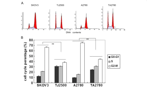
Cell-cycle analysis
Cell cycle analysis was carried out to further investigate the longer doubling times of the resistant cell lines com-pared to the sensitive cell lines. Results in Figure 2 show that the resistant cell lines have a significantly higher proportion of cells existing in the G0-G1 stage of the cell cycle and a significantly reduced number of cells in the S phase compared to the sensitive cell lines. How-ever, the number of cells in the M stage was not changed significantly. The increased number of cells existing in the G0-G1 stage in the resistant cell lines may reduce the potency of Taxol.
Cell-cycle synchronization with thymidine
In order to test the above assumption, we added thymidine to the medium, which prohibited the synthesis of DNA and hampered the progression of the cells into the next stage, resulting in most of the cells arresting in the S phase. The proportion of cells in the G2/M phase was subse-quently reduced. Following the removal of the thymidine, most of the cells gradually entered the M phase several hours later. The time at which most sensitive cells (SKOV3) synchronized with thymidine entered the M phase was approximately 6-8 h; the Taxol-resistant cell line (TJ2500) required 8-10 h. In contrast to Figure 2, the re-sults in Figure 3 show that with the addition of thymidine, more cells of the sensitive cell lines (SKOV3 and A2780) had entered the M phase compared to the resistant cells lines (TJ2500 and TA2780) when assessed at the same time point. This indicated that the speed at which the resistant cells proliferated was slower compared to the sensitive cell lines. We therefore choose 8 h as the time point to add Taxol and to assess apoptosis after synchronization.
Cell apoptotic rates
After synchronization with thymidine, cell lines (both sensitive and resistant) were incubated in common
medium without thymidine for about 8 h and then stim-ulated with Taxol (100 nmol/L) for 4 h. The apoptosis rates of the cells were detected after withdrawed Taxol 48 h later. Independent trials were performed at least in triplicate. Figure 4 shows the significantly different apoptotic rates among the four cell lines compared with unsynchronized controls (p <0.05). Furthermore, apop-totic rates of the resistant cells after synchronization were lower than that of the sensitive cells, especially in regard to the TA2780 cell line.
Discussion
Microtubules represent effective targets in cancer ther-apy. Taxol, a member of the Taxane family of anticancer drugs, stabilizes microtubules. At the molecular level, Taxol binds directly to microtubules, leading to a potent suppression of microtubule dynamics, increased micro-tubule stabilization, and interphase micromicro-tubule bund-ling; consequently, cells undergo robust mitotic arrest and subsequent apoptotic cell death. The mitotic check-point is thought to be invoked by these microtubule-disrupting drugs via mechanisms that monitor correct spindle formation and tension. In normal cells this check-point helps ensure that equal numbers of chromosomes
are distributed to daughter cells and, thus, avoid mis-segregation and the potentially catastrophic consequences of aneuploidy [28,29]. The development of protocols using RNA interference to specifically silence gene expression has provided the ability to test the potential function of specific components of this checkpoint. Our prior research found that these drugs induced nuclear frag-mentation mainly after cells had progressed into mi-tosis; conversely, the lethal effects of these drugs were significantly lessened when cells were prevented from progressing into mitosis [26]. Current models propose that the mitotic checkpoint proteins (BubR1, Bub1, Bub3, Mad1, and Mad2) sense a lack of tension or at-tachment between the kinetochore and microtubules of the mitotic spindle and transmit a “wait signal” to in-hibit the anaphase-promoting complex [30,31]. Certain cancer lines evolved a less robust mitotic checkpoint, and such defects in the mitotic checkpoint may confer a growth advantage by enabling cells to tolerate chromo-somal instability or aneuploidy or by allowing faster proliferation. In support of the latter, Shichiri et al. found few mutations of BubR1 or Bub1 in a series of
could also be reversed by silencing of BubR1 [26,32]. However, others have provided evidence that expression of BubR1 is reduced and the function of the spindle checkpoint is weakened in cell lines with Taxol resistance [33,34]. Drug resistance is often a multifactorial process, and although we cannot exclude the possibility that mech-anisms distinct from the spindle checkpoint contribute to Taxol resistance, there must exist other factors affecting formation of drug resistance during cell cycle. Passage through mitosis is an absolute requirement for drug-induced death. It is tempting to speculate that the clinical response of certain tumors to microtubule-disrupting drugs may be at least partially reflected in the intrinsic sensitivity of cell lines derived from these tumors. For ex-ample, the efficacy of Taxol in treating patients with breast and ovarian cancer is well established, whereas results in treating sarcomas, colorectal, and renal cancer have been disappointing [35-37]. It is likely that these mitotic check-point proteins participate in different pathways to effect orderly mitotic progression; as such, their relationship with Taxol resistance requires further investigation.
The focus of this current study was on the formation of acquired drug resistance. The resistant cells lines in this experiment were obtained by intermittent exposure of sensitive cells to Taxol. The group doubling time of ovarian cancer lines (SKOV3 and A2780) were approxi-mately 27 h, and that of resistant cells (TJ2500 and TA2780) was even longer. Figure 2 show cell cycle of four cells line which unsynchronizated. cells in G0-G1 and S stage occupied 80-90%, M-stage only 10-20%, es-pecially resistant cells (TJ2500, TA2780) in G0-G1 stage much more than sensitive cells lines, difference is signifi-cantly. While growing in the same medium, cells are not all at the same stage of the cell cycle and concordance is absent, which results in a lower efficacy of Taxol, as its effectiveness is limited to a short time period while cells in M stage. Only over a treatment time of approximately 27 h could it be possible to conclude that most cells had passed through the M stage. We speculated that forma-tion of drug resistance toward Taxol in ovarian cancer could be partly attributed to the longer doubling time of these cells.
We used cell-cycle synchronization to test the above as-sumption. After adding a high concentration of thymidine, most of the cells arrested at the S phase and could not pro-gress to the next stage (M phase). After a fresh medium change, most of the cells gradually entered the M phase at nearly the same time, at this point Taxol was added to the medium. Figure 3 show cell cycle of four cells line which synchronizated. cells in G0-G1 and S stage particularly ruduced and M-stage much increased, especially sensitive cells(SKOV3,A2780) in M stage more than resistant cells lines. The apoptotic rates of cells treated with thymidine were significantly increased compared with the cells
without thymidine, and this was not only observed in sen-sitive cells but also in resistant cells. Taxol-resistant cells may require a longer time of action when compared to sensitive cells in order to achieve optimal results; this is be-cause the proliferation period or growth doubling time of the resistant cells was longer and most of these cells were in the non-M phases during the window of Taxol action.
Cell-cycle synchronization resulted in an increase in the number of cells passing through the M stage at a given time and reduced the toxicity of Taxol toward cells in the non-proliferative phase, improving its effect-iveness and decreasing the chance of drug-resistant for-mation. This method may be an efficient approach to strengthen the effectiveness and reverse drug resistance related to Taxol.
However, apoptosis of resistant cells after synchronization was lower than that of sensitive cells, especially in re-gard to TA2780 cells. This observation partly implied that resistance formation towards Taxol is a complex phenomenon involving diverse molecular mechanisms, as opposed to a single factor. The observation that entry into mitosis seems to be a requirement for rapid killing after microtubule disruption in drug-sensitive cells has implications for the sequential application of these drugs and other forms of anticancer treatment. For ex-ample, initial use of Taxol may reduce the lethal effects of microtubule-disrupting drugs. Thus, it may be prefera-ble to administer therapy inducing cell-cycle synchro-nization before paclitaxel treatment to possibly reduce the likelihood of the development of drug resistance.
Abbreviations
SKOV3 and A2780:Taxol sensitive cell lines; TJ2500 and TA2780: Taxol resistant cell lines; RI: Resistant index; IC50: Half-inhibition concentration; HG-DMEM: HIGH-glucose medium; PI: Propidium iodide.
Competing interests
The authors declare that they have no competing interests.
Authors’contributions
XQ Wang participated in the design of the study and drafted the
manuscript. LY Pan conceived of the study and participated in its design and performed the statistical analyses. N Mao participated in the experimental design and implementation. LF Sun and XJ Qin participated in its design and coordination and performed the statistical analyses. J Yin helped to draft the manuscript. All authors read and approved the final manuscript.
Authors’information
XQ Wang worked in the Department of Obstetrics and Gynecology of Beijing Jishuitan Hospital as an attending physician and graduated from Peking Union Medical College Hospital with doctoral degree in 2008. LY Pan worked in the Department of Obstetrics and Gynecology of Peking Union Medical College Hospital, Chinese Academy of Medical Sciences and Peking Union Medical College as a doctoral tutor and medical director. N Mao worked in the Department of Cell Biology, Institute of Basic Medical Sciences, Academy of Military Medical Sciences as a director.
LF Sun worked in the Department of Obstetrics and Gynecology in Beijing Jishuitan Hospital as a medical director.
XJ Qin worked in the Department of Obstetrics and Gynecology in Beijing Jishuitan Hospital as vice-medical director.
Acknowledgements
The authors wish to thank the members of the Department of Cell Biology, Institute of Basic Medical Sciences, and Academy of Military Medical Sciences for their support, advice, and encouragement. The experiments were also supported by the membership of Beijing Jishuitan Hospital, the Fourth Teaching Hospital of Beijing Medical College, and the authors thank them for their discussions and critical review of the paper.
Author details
1
Department of Obstetrics and Gynecology, Beijing Jishuitan Hospital, The Fourth Teaching Hospital of Beijing Medical College, Beijing, China.
2
Department of Obstetrics and Gynecology, Peking Union Medical College Hospital, Chinese Academy of Medical Science and Peking Union Medical College, Peking, China.3Department of Cell Biology, Institute of Basic Medical Sciences, Academy of Military Medical Sciences, Beijing, China.
Received: 16 April 2013 Accepted: 24 July 2013 Published: 30 July 2013
References
1. Slichenmyer WJ, Von Hoff DD:Taxol: a new and effective anti-cancer drug.Anticancer Drugs1991,2:519–530.
2. Vici P, Conti F, Amodio A, Belli F, Della Giulia M, Mariotti S, Gionfra T, Lopez M:Simultaneous infusion of vinorelbin and taxol as first-line
chemotherapy in metastasized breast cancer.Clin Ter1998,149:255–259. 3. Hájek R, Vorlicek J, Slavik M:Paclitaxel (Taxol): a review of its antitumor
activity in clinical studies Minireview.Neoplasma1996,43:141–154. 4. Young RC:Three cycles versus six cycles of adjuvant paclitaxel (Taxol)/
carboplatin in early stage ovarian cancer.Semin Oncol2000,27:8–10. 5. Choi YH, Yoo YH:Taxol-induced growth arrest and apoptosis is
associated with the upregulation of the Cdk inhibitor, p21WAF1/CIP1, in human breast cancer cells.Oncol Rep2012,28:2163–9.
6. Zaniboni A, Ardizzoni A, De Marinis F, Portalone L, Boni C, Meriggi F, Cafferata MA, Ariganello O, Torri V, Neumaier CE, Rosso R:Phase II study of Taxol combi.ned With ifosfamide and carboplatin in the treatment of stage IIIb-IV non-small-cell lung cancer.Am J Clin Oncol2003,26:84–8. 7. Chiou JF, Liang JA, Hsu WH, Wang JJ, Ho ST, Kao A:Comparing the
relationship of Taxol-based chemotherapy response with P-glycoprotein and lung resistance-related protein expression in non-small cell lung cancer.Lung2003,181:267–73.
8. Agarwal R, Kaye SB:Ovarian cancer: strategies for overcoming resistance to chemotherapy.Nat Rev Cancer2003,3:502–16.
9. Lai D, Ho KC, Hao Y, Yang X:Taxol resistance in breast cancer cells is mediated by the hippo pathway component TAZ and its downstream transcriptional targets Cyr61 and CTGF.Cancer Res2011,71:2728–38. Epub 2011 Feb 24.
10. Umezu T, Shibata K, Kajiyama H, Terauchi M, Ino K, Nawa A, Kikkawa F: Taxol resistance among the different histological subtypes of ovarian cancer may be associated with the expression of class III beta-tubulin.
Int J Gynecol Pathol2008,27:207–12.
11. Bai ZG, Qu X, Han W, Ma XM, Zhao XM, Zhang ZT:Expression of taxol resistance gene 1 correlates with gastric cancer patient clinical outcome and induces taxol resistance.Mol Med Report2010,3:1071–8.
12. Wani MC, Taylor HL, Wall ME, Coggon P, McPhail AT:Plant antitumor agents. The isolation and structure of taxol, a novel antileukemic and antitumor agent from taxus brevifolia.J Am Chem Soc1971,93:2325–2327.
13. Radulescu S, Ridgway RA, Appleton P, Kroboth K, Patel S, Woodgett J, Taylor S, Nathke IS, Sansom OJ:Defining the role of APC in the mitotic spindle checkpoint in vivo: APC-deficient cells are resistant to Taxol.
Oncogene2010,29:6418–27.
14. Hornick JE, Bader JR, Tribble EK, Trimble K, Breunig JS, Halpin ES, Vaughan KT, Hinchcliffe EH:Live-cell analysis of mitotic spindle formation in taxol-treated cells.Cell Motil Cytoskeleton2008,65:595–613.
15. Xing H, Wang S, Weng D, Chen G, Yang X, Zhou J, Xu G, Lu Y, Ma D: Knock-down of P-glycoprotein reverses taxol resistance in ovarian cancer multicellular spheroids.Oncol Rep2007,17:117–22.
16. Gazitt Y, Rothenberg ML, Hilsenbeck SG, Fey V, Thomas C, Montegomrey W: Bcl-2 overexpression is associated with resistance to paclitaxel, but not gemcitabine, in multiple myeloma cells.Int J Oncol1998,13:839–48.
17. Panvichian R, Orth K, Day ML, Day KC, Pilat MJ, Pienta KJ:Paclitaxel-associated multimininucleation is permitted by the inhibition of caspase activation: a potential early step in drug resistance.Cancer Res1998,15:4667–72. 18. Li L, Jiang AC, Dong P, Wang H, Xu W, Xu C:MDR1/P-gp and VEGF synergistically enhance the invasion of Hep-2 cells with multidrug resistance induced by taxol.Ann Surg Oncol2009,16:1421–8. 19. Kavallaris M, Burkhart CA, Horwitz SB:Antisense of Ligonucleotides to class
βII- tubulin sensitive drug-resistant cells to Taxol.Br J Cancer1999,80:1020–5. 20. Mao QQ, Bai Y, Lin YW, Zheng XY, Qin J, Yang K, Xie LP:Resveratrol confers
resistance against taxol via induction of cell cycle arrest in human cancer cell lines.Mol Nutr Food Res2010,54:1574–84.
21. Wang TH, Wang HS, Soong YK:Paclitaxel-induced cell death: where the cell cycle and apoptosis come together.Cancer2000,1:2619–28. 22. Edgar BA, Sprenger F, Duronio RJ, Leopold P, O’Farrell PH:Distinct
molecular mechanism regulate cell cycle timing at successive stages of Drosophila embryogenesis.Genes Dev1994,15:440–52.
23. Mohamed FZ, Hussien YM, Albakry MM, Mohamed RH, Said NM:Role of DNA repair and cell cycle control genes in ovarian cancer susceptibility.
Mol Biol Rep2013,40:3757–68.
24. Rastogi N, Mishra DP:Therapeutic targeting of cancer cell cycle using proteasome inhibitors.Cell Div2012,26:7–26.
25. Long BH, Fairchild CR:Paclitaxel inhibits progression of mitotic cells to G1 phase by interference with spindle formation without affecting other microtubule functions during anaphase and telephase.Cancer Res1994, 15(54):4355–61.
26. Lee EA, Keutmann MK, Dowling ML:Inactivation of the mitotic checkpoint as a determinant of the efficacy of microtubule-targeted drugs in killing human cancer cells.Mol Cancer Ther2004,3:661–9.
27. Xue Dong Y, Min L, Ye Y, Ning M, Ling Ya P:Biological comparison of ovarian cancer resistant cell lines to cisplatin and Taxol by two different administrations.Oncol Rep2007,17:1163–1169.
28. Jordan MA, Wilson L:Microtubules and actin filaments: dynamic targets for cancer chemotherapy.Curr Opin Cell Biol1998,10:123–30. 29. Tsukasaki K, Miller CW, Greenspun E, Eshaghian S, Kawabata H, Fujimoto T,
Tomonaga M, Sawyers C, Said JW, Koeffler HP:Mutations in the mitotic check point gene, MAD1L1, in human cancers.Oncogene2001,20:3301–5. 30. Shichiri M, Yoshinaga K, Hisatomi H, Sugihara K, Hirata Y:Genetic and
epigenetic inactivation of mitotic checkpoint genes hBUB1 and hBUBR1 and their relationship to survival.Cancer Res2002,62:13–7.
31. Musio A, Montagna C, Zambroni D, Indino E, Barbieri O, Citti L, Villa A, Ried T, Vezzoni P:Inhibition of BUB1 results in genomic instability and anchorage-independent growth of normal human fibroblasts.Cancer Res
2003,63:2855–63.
32. Hu M, Liu Q, Song P, Zhan X, Luo M, Liu C, Yang D, Cai Y, Zhang F, Jiang F, Zhang Y, Tang M, Zuo G, Zhou L, Luo J, Shi Q, Weng Y:Abnormal expression of the mitotic checkpoint protein BubR1 contributes to the anti-microtubule drug resistance of esophageal squamous cell carcinoma cells.Oncol Rep2013,29:185–92.
33. Masuda A, Maeno K, Nakagawa T:Association between mitotic spindle checkpoint impairment and susceptibility to the induction of apoptosis by anti-microtubule agents in human lung cancers.Am J Pathol2003, 163:1109–1116.
34. Fu Y, Ye D, Chen H, Lu W, Ye F, Xie X:Weakened spindle checkpoint with reduced BubR1 expression in paclitaxel-resistant ovarian carcinoma cell line SKOV3-TR30.Gynecol Oncol2007,105:66–73.
35. Gian VG, Johnson TJ, Marsh RW, Schuhmacher C, Lynch JW:A phase II trial of paclitaxel in the treatment of recurrent or metastatic soft tissue sarcomas or bone sarcomas.J Exp Ther Oncol1996,1:186–90. 36. Patel SR, Papadopoulos NE, Plager C, Linke KA, Moseley SH, Spirindonidis
CH, Benjamin R:Phase II study of paclitaxel in patients with previously treated osteosarcoma and its variants.Cancer1996,78:741–4. 37. Ferlini C, Ojima I, Distefano M, Gallo D, Riva A, Morazzoni P, Bombardelli E,
Mancuso S, Scambia G:Second generation taxanes: from the natural framework to the challenge of drug resistance.Curr Med Chem Anticancer Agents2003,3:133–8.
doi:10.1186/1475-2867-13-77
Cite this article as:Wanget al.:Cell-cycle synchronization reverses Taxol resistance of human ovarian cancer cell lines.Cancer Cell International