The Genomic Standards Consortium
Non-contiguous finished genome sequence and description
of
Cheikh Ibrahima Lo1, Ajay Kumar Mishra1, Roshan Padhmanabhan1,Bissoume Samb Ba2, Amy Gassama Sow2, Catherine Robert1, Carine Couderc1, Ngor Faye3, Didier Raoult1, Pierre-Edouard Fournier1, and Florence Fenollar1*
1Aix-Marseille Université, Unité de Recherche sur les Maladies Infectieuses et Tropicales Emergentes (URMITE), Marseille, France
2Unité de Bactériologie Expérimentale Institut Pasteur de Dakar 36, Dakar, Senegal 3Université Cheikh Anta Diop de Dakar, Laboratoire de Parasitologie générale, Fann
(Senegal)
*Correspondence: Florence Fenollar ([email protected]) Keywords, genome, culturomics, taxono-genomics
strain FF1T, is the type strain o sp. nov., a new
spe-cies within the genus
the fecal flora of a 4-month-old Senegalese child suffering from gastroenteritis. C. dakarense sp. nov. strain FF1T is an obligate anaerobic Gram-positive bacillus. Here we describe the features of
this organism, together with the complete genome sequence and annotation. The 3,735,762 bp long genome (1 chromosome but no plasmid) exhibits a G+C content of 27.98% and contains 3,843 protein-coding and 73 RNA genes, including 8 rRNA genes.
Introduction
strain FF1T (= CSUR P243 =
DSM 27086), is the type strain of
dakarense sp. nov. This bacterium is a
Gram-positive, anaerobic, spore-forming, indole nega-tive bacillus that was isolated from the stool of a 4-month-old Senegalese child suffering from gas-troenteritis as part of a “culturomics” study aim-ing at cultivataim-ing individually all species within human feces.
The elevated cost and lack of intra- and inter-laboratory reproducibility of the “gold standard” of taxonomic tools. i.e. DNA-DNA hybridization and G+C content determination [1], put bacterial taxonomic classification in a precarious state. In addition, the internationally-validated cutoff val-ues of 16S rRNA sequence comparison [2] do not apply to all validly published genera and species. Recently, high throughput genome sequencing and mass spectrometric analyses of bacteria have al-lowed unprecedented access to a wealth of genetic and proteomic information [3]. As a consequence, we proposed to use a polyphasic approach [4] to describe new bacterial taxa, including genome
sequence, MALDI-TOF spectrum and main pheno-typic characteristics [5-11].
The genus
fied among th
[12] and consists of obligate anaerobic rod-shaped bacilli capable of producing endospores [12].
More than 1
scribed to date [13]. Members of the genus
sociated with the commensal digestive flora of mammals, but several are major human
patho-gens, including
and
have gained importance in agricultural and indus-trial applications [16,17].
Here we present a summary classification and a set of features for C. dakarense sp. nov. strain FF1T
(= CSUR P243 = DSM 27086) together with the description of the complete genomic sequencing and annotation. These characteristics support the circumscription of the species C. dakarense sp.
Classification and features
A stool specimen was collected from a 4-month-old Senegalese child suffering from gastroenteritis. In-formed consent was obtained from the child’s par-ents and approval from the ethics committee from the Institut Federatif de Recherche 48 (Faculté de Médecine, Marseille, France). The fecal specimen was preserved at -20°C after collection and sent to Marseille. Strain FF1T (Table 1) was isolated in July
2011 by anaerobic cultivation on 5% sheep blood-enriched Columbia agar (BioMerieux, Marcy
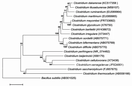
l’Etoile, France). This strain exhibited a 96.90% 16S
rRNA nucleotide sequence similarity with
similarity of the 16S rRNA is not uniform across taxa, this value was lower than the 98.7% threshold recommended by Stackebrandt and Ebers to delin-eate a new species without carrying out DNA-DNA hybridization [30]. In addition, it was consistent with 16S rRNA identity values observed among
val-idated species within the
range from 78.4 to 99.8%.
Table 1. Classification and general features o strain FF1T according to the MIGS recom-mendations [18].
MIGS ID Property Term Evidence codea
Domai TAS [19]
Phylum TAS [20-22]
Cla TAS [23,24]
Current classification Orde TAS [25,26]
Family TAS [25,27]
Genu TAS [12,25,28]
Species IDA
Type strain FF1 IDA
Gram stain Positive IDA
Cell shape Rod-shaped IDA
Motility Motile IDA
Sporulation Sporulating IDA
Temperature range Mesophile IDA
Optimum temperature 37°C IDA
MIGS-6.3 Salinity Growth in BHI medium + 5% NaCl IDA
MIGS-22 Oxygen requirement Anaerobic IDA
Carbon source Unknown NAS
Energy source Unknown NAS
MIGS-6 Habitat Human gut IDA
MIGS-15 Biotic relationship Free living IDA
MIGS-14
Pathogenicity Biosafety level Isolation
Unknown 2
Human feces NAS
MIGS-4 Geographic location Senegal IDA
MIGS-5 Sample collection time June 2011 IDA
MIGS-4.1 Latitude 13.7167 IDA
MIGS-4.1 Longitude - 16.4167 IDA
MIGS-4.3 Depth Surface IDA
MIGS-4.4 Altitude 51 m above sea level IDA
Figure 1. Phylogenetic tree highlighting the position of C. dakarense sp. nov. strain FF1T relative to other type strains
within the
ing CLUSTALW, and phylogenetic inferences obtained using the maximum-likelihood method within the MEGA soft-ware. Numbers at the nodes are bootstrap values obtained by repeating 500 times the analysis to generate a majority consensus tree. gence.
Different growth temperatures (25, 30, 37, 45 and 56°C) were tested. Growth was observed between 25 and 37°C, with optimal growth at 37°C after 24 hours of inoculation in anaerobic conditions. Col-onies were 1.5 mm in diameter and opaque and smooth appearance on blood-enriched Columbia agar. Growth of the strain was tested under an-aerobic and microaerophilic conditions using GENbag anaer and GENbag microaer systems, re-spectively (BioMerieux), and under aerobic condi-tions, with or without 5% CO2. The strain growth
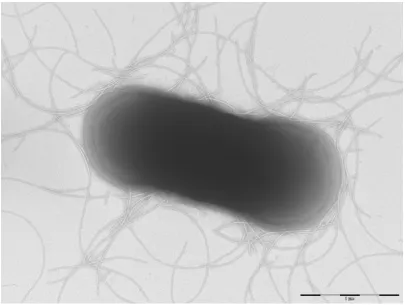
was obtained only in anaerobic conditions. Gram staining showed rod-shaped Gram-positive bacilli able to form spores (Figure 2). The motility test was positive. Cells grown on agar have a mean di-ameter of 1.2 µm (Figure 3).
Strain FF1T exhibited neither catalase nor oxidase
activities. Using API Rapid ID 32A (BioMerieux, Marcy l’Etoile), a positive reaction were observed
for arginine dihydrolase, N-acetyl-β
-glucos-aminidase and pyroglutamic acid arylamidase. Negative reactions were observed for urease, indole and nitrate reduction. Using API 50 CH (BioMerieux, Marcy l’Etoile), positive reactions were observed for galactose, glucose, maltose and saccharose fermentation and negative reaction were observed for ribose, lactose and fructose. C. dakarense is susceptible to amoxicillin,
metroni-dazole, vancomycin, imipenem and rifampicin and resistant to trimethoprim/ sulfamethoxazole. When compared with representative species from
the genusC. dakarense strain FF1T
Figure 2. Gram staining of C. dakarense sp. nov. strain FF1T.
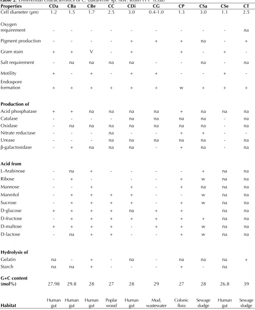
Table 2. Differential characteristics of C. dakarense sp. nov. strain FF1T (Cda)
Properties CDa CBa CBe CC CDi CG CP CSa CSe CT
Cell diameter (µm) 1.2 1.5 1.7 2.5 3.0 0.4-1.0 1.3 3.0 1.1 2.5
Oxygen
requirement - - - na
Pigment production - - - - + + + na - +
Gram stain + + V - + + - + -
Salt requirement - na na na na - - na - na
Motility + - + - + + - - + -
Endospore
formation + + + + + + w + + +
Production of
Acid phosphatase + + na na na na + na na na
Catalase - - - - na na na na - na
Oxidase - na na na na na na na - na
Nitrate reductase - - - na - - + + - -
Urease - - - na na na na na - na
β-galactosidase - + na na na - + na - na
Acid from
L-Arabinose - na + - - - - + na na
Ribose - + - - - + w na na
Mannose - - + + - + na na na
Mannitol - + + + + - - w na na
Sucrose - + + + + - + w na na
D-glucose + + + + na + + na na
D-fructose - + + + + + + + na na
D-maltose + + + + - + + w na na
D-lactose - na + + - - + w na na
Hydrolysis of
Gelatin na - + - na - na na na +
Starch na na + - - - + - na
G+C content
(mol%) 27.98 29.8 28 27 28 29 27 28 26.8 39
Habitat Human gut
Human gut
Human gut
Poplar wood
Human gut
Mud, wastewater
Colonic flora
Sewage sludge
Human gut
Sewage sludge
C. bartlettii (CBa), C. beijerinckii (CBe), C. cellulovorans (CC), C. difficile (CDi), C. glycolicum (CG), C. perfringens (CP),
Matrix-assisted laser-desorption/ionization time-of-flight (MALDI-TOF) MS protein analysis was carried out as previously described [31]. Briefly, a pipette tip was used to pick one isolated bacterial colony from a culture agar plate, and to spread it as a thin film on a MTP 384 MALDI-TOF target plate (Bruker Daltonics, Leipzig, Germany). Eight-een distinct deposits were made for strain FFT
from eighteen isolated colonies. Each smear was overlaid with 2 µL of matrix solution (saturated solution of alpha-cyano-4-hydroxycinnamic acid) in 50% acetonitrile, 2.5% tri-fluoracetic-acid, and allowed to dry for five minutes. Measurements were performed with a Microflex spectrometer (Bruker). Spectra were recorded in the positive linear mode for the mass range of 2,000 to 20,000 Da (parameter settings: ion source 1 (IS1), 20 kV; IS2, 18.5 kV; lens, 7 kV). A spectrum was obtained after 675 shots at a variable laser power. The time of acquisition was between 30 seconds and 1 mi-nute per spot. The eighteen spectra were import-ed into the MALDI BioTyper software (version 2.0,
Bruker) and analyzed by standard pattern match-ing (with default parameter settmatch-ings) against the main spectra of 4,706 bacteria including 216
spec-tra from validly published species of
that are part of the reference data contained in the BioTyper database. The method of identification included the m/z from 2,000 to 20,000 Da. For every spectrum, 100 peaks at most were taken into account and compared with spectra in the database. A score enabled the identification, or not, from the tested species: a score > 2 with a val-idly published species enabled the identification at the species level, and a score < 1.7 did not enable any identification at the genus level. For strain FF1T, the maximal obtained score was lower than
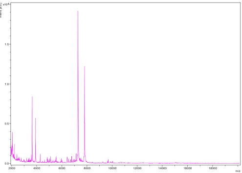
1.9, thus suggesting that our isolate was not a member of a known species. We added the spec-trum from strain FF1T to our database for future
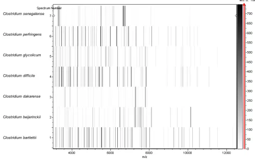
reference (Figure 4). Finally, the gel view allows us to highlight the spectrum differences with
oth-er memboth-ers of the genus
0.0 0.5 1.0 1.5 4 x10
In
te
ns
. [a
.u
.]
2000 4000 6000 8000 10000 12000 14000 16000 18000
m/z
Clostridium senegalense
Clostridium perfringens
Clostridium glycolicum
Clostridium difficile
Clostridium dakarense
Clostridium beijerinckii
Clostridium bartlettii
Figure 5. Gel view comparing C. dakarense sp. nov. strain FF1T spectra with other members of the
raw spectra of all loaded spectrum files arranged in a pseudo-gel like look. The x-axis records the m/z value. The left y-axis displays the running spectrum number originating from subsequent spectra loading. The peak intensity is ex-pressed by a Gray scale scheme code. The color bar and the right y-axis indicate the relation between the color a peak is displayed with and the peak intensity in arbitrary units.
Genome sequencing information
Genome project history
The organism was selected for sequencing on the basis of its phylogenetic position and 16S rRNA
similarity to other members of the genus
human digestive flora aiming at isolating all bacte-rial species within human feces. It was the 94th
genome of a
nome of sp. nov. The
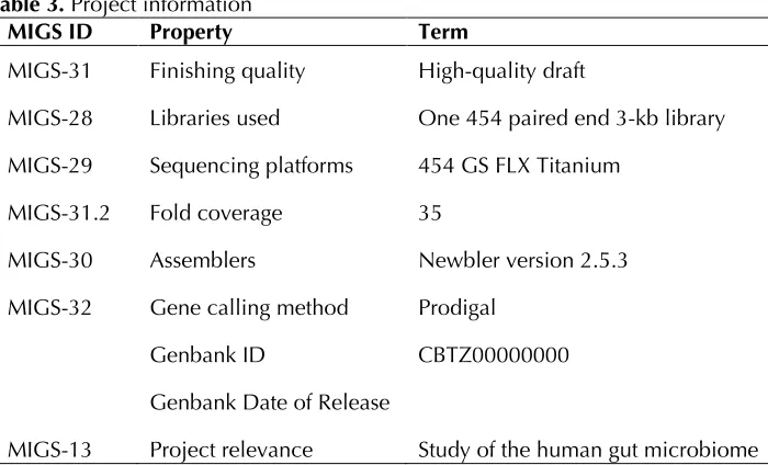
Genbank accession number is CBTZ00000000 and consists of 257 contigs. Table 3 shows the project information and its association with MIGS version 2.0 compliance [32].
Growth conditions and DNA isolation
C. dakarense sp. nov. strain FF1T (= CSUR P243 =
Table 3. Project information
MIGS ID Property Term
MIGS-31 Finishing quality High-quality draft
MIGS-28 Libraries used One 454 paired end 3-kb library MIGS-29 Sequencing platforms 454 GS FLX Titanium
MIGS-31.2 Fold coverage 35
MIGS-30 Assemblers Newbler version 2.5.3 MIGS-32 Gene calling method Prodigal
Genbank ID CBTZ00000000 Genbank Date of Release
MIGS-13 Project relevance Study of the human gut microbiome
Genome sequencing and assembly
This project was loaded twice on a 1/4 region for the paired-end application and once on a 1/8 re-gion for the shotgun on PTP Picotiterplates. The shotgun library was constructed with 500 ng of DNA as described by the manufacturer (Roche) with the GS Rapid library Prep kit. For the paired-end sequencing, 5 µg of DNA was mechanically fragmented on the Hydroshear device (Digilab, Holliston, MA, USA) with an enrichment size of 3-4kb. The DNA fragmentation was visualized using an Agilent 2100 BioAnalyzer on a DNA labchip 7500, which yield an optimal size of 3.6 kb. The library was constructed according to the 454_Titanium paired-end protocol and manufac-turer. Circularization and nebulization were per-formed and generated a pattern with an optimum at 561 bp. After PCR amplification through 15 cy-cles followed by double size selection, the single stranded paired end library was then quantified with Quant-it Ribogreen kit (Invitrogen) on the Genios_Tecan fluorometer at 52 pg/µL. The li-brary concentration equivalence was calculated as 1.7E+08 molecules/µL. The library was stored at -20°C until use.
The shotgun library was clonally amplified with 3cpb in 3 emPCR reactions and the paired end li-brary was amplified with lower cpb (1cpb) in 4 emPCR reactions with the GS Titanium SV emPCR Kit (Lib-L) v2. The yield of the emPCR was 5.37% for the shotgun reads and 19.27% for the paired-end reads, according to the quality expected by the range of 5 to 20% from the Roche procedure. A total of 340,000 beads from the 1/8 region of
the shotgun reads and 790,000 beads from the 1/4 region of the paired-end reads were loaded on the GS Titanium PicoTiterPlates (PTP Kit 70×75) and sequenced with the GS Titanium Sequencing Kit XLR70.
The runs were performed overnight and then ana-lyzed on the cluster through the gsRunBrowser and gsAssembler_Roche. The global 383,079 passed filter sequences generated 96.50 Mb with a length average of 277 bp. These sequences were assembled using the Newbler software from Roche with 90% identity and 40 bp as overlap. Fourteen scaffolds and 257 large contigs (>1500bp) were obtained, for a genome size of 3,735,762 bp.
Genome annotation
Open Reading Frames (ORFs) were predicted us-ing Prodigal [33] with default parameters but the predicted ORFs were excluded if they spanned a sequencing gap region. The predicted bacterial protein sequences were searched against the GenBank database [34] and the Clusters of Orthol-ogous Groups (COG) databases using BLASTP. The tRNAScanSE tool [35] was used to find tRNA genes, whereas ribosomal RNAs were found by using RNAmmer [36] and BLASTn against the GenBank database. Lipoprotein signal peptides and numbers of transmembrane helices were pdicted using SignalP [37] and TMHMM [38] re-spectively. ORFans were identified if their BLASTP
E-value was lower than 1e-03 for alignment length
were smaller than 80 amino acids, we used an E
-value of 1e-05. Such parameter thresholds have already been used in previous works to define ORFans. Artemis [39] was used for data manage-ment and DNA Plotter [40] was used for visualiza-tion of genomic features. Mauve alignment tool was used for multiple genomic sequence align-ment and visualization [41].
To estimate the mean level of nucleotide sequence
similarity at the genome level between C.
dakarense and nine other members of the genus
nomic Identity of gene Sequences (AGIOS) home-made software. Briefly, this software combines the Proteinortho software [42] for detecting ortholo-gous proteins between genomes compared two by two, then retrieves the corresponding genes and determines the mean percentage of nucleotide sequence identity among orthologous ORFs using the Needleman-Wunsch global alignment
algo-rithm. strain FF1T, was
compared t
(GenBank accession number NZ_DS499569),
strain 630 (NC8009089),
ATCC 14880 (ARES01000000),
strain ATCC 13124 (BA000016),
T (CAEV00000000), and
Genome properties
The genome of C. dakarense sp. nov. strain FF1T is
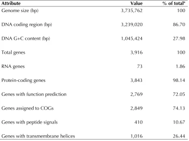
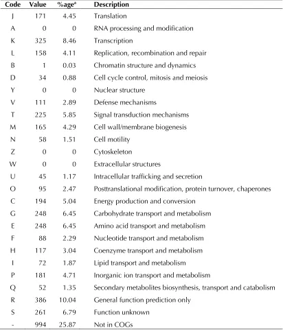
3,735,762 bp long (1 chromosome, but no plas-mid) with a 27,98% G + C content of (Figure 6 and Table 4). Of the 3,916 predicted genes, 3,843 pro-tein-coding genes, and 73 were RNAs. Eight rRNA genes (one 16S rRNA, one 23S rRNA and six 5S rRNA) and 65 predicted tRNA genes were identi-fied in the genome. A total of 2,769 genes (72.05%) were assigned a putative function (by COG or NR blast). Two hundred ninety-eight genes were identified as ORFans (7.75%). The re-maining 515 genes were annotated as hypothet-ical proteins (13, 40%). The distribution of genes into COGs functional categories is presented in Table 4. The properties and the statistics of the genome are summarized in Tables 4 and 5.
Table 4. Nucleotide content and gene count levels of the genome.
Attribute Value % of totala
Genome size (bp) 3,735,762 100
DNA coding region (bp) 3,239,020 86.70
DNA G+C content (bp) 1,045,424 27.98
Total genes 3,916 100
RNA genes 73 1.86
Protein-coding genes 3,843 98.14
Genes with function prediction 2,769 72.05
Genes assigned to COGs 2,849 74.13
Genes with peptide signals 410 10.67
Figure 6. Graphical circular map of the chromosome. From the outside in, the outer two circles show open reading frames oriented in the forward and reverse directions (colored by COG categories), respectively. The third circle marks the rRNA gene operon (red) and tRNA genes (green). The fourth circle shows the G+C% content plot. The in-ner-most circle shows the GC skew, purple and olive indicating negative and positive values, respectively.
Comparison with the genomes from
oth
The genome sequence of
ly available for more than seventy-fiv
species. Here we compared the genome sequence of C. dakarense strain FF1T with than those of
The draft genome sequence of C. dakarense strain
FF1T is smaller than those of
6.0, 3.89, 4.66, 3.84, 4.3 and 3.99 Mb, respectively)
but larger than those ofC.
bartletii (3.26 and 2.97 Mb, respectively). The G+C
content of C. dakarense is lower than those of
29.9, 28.4, 45, 39 and 29.1%, respectively) but
higher than those of
The gene content of C. dakarense is larger than
perfringens,
3,173, 3,761, 2,876, 3,840 and 2,787, respectively)
and smaller than those of
(4,501, 5,243, 4,154 and 4,019, respectively). The ratio of genes per Mb of C. dakarense is larger to
those of
938, 962 and 882, respectively).
The number of orthologous genes shared between
C. dakarense and other compared
cies has been summarized in Table 6. The average percentage of nucleotide sequence identity ranged from 62.05 to 74.5% among previously published
tween C. dakarense and other studied
species, thus confirming its new species status.
Table 5. Number of genes associated with the 25 general COG functional categories. Code Value %agea Description
J 171 4.45 Translation
A 0 0 RNA processing and modification K 325 8.46 Transcription
L 158 4.11 Replication, recombination and repair B 1 0.03 Chromatin structure and dynamics D 34 0.88 Cell cycle control, mitosis and meiosis Y 0 0 Nuclear structure
V 111 2.89 Defense mechanisms
T 225 5.85 Signal transduction mechanisms M 165 4.29 Cell wall/membrane biogenesis N 58 1.51 Cell motility
Z 0 0 Cytoskeleton
W 0 0 Extracellular structures
U 45 1.17 Intracellular trafficking and secretion
O 95 2.47 Posttranslational modification, protein turnover, chaperones C 194 5.04 Energy production and conversion
G 248 6.45 Carbohydrate transport and metabolism E 248 6.45 Amino acid transport and metabolism F 88 2.29 Nucleotide transport and metabolism H 117 3.04 Coenzyme transport and metabolism
I 72 1.87 Lipid transport and metabolism
P 181 4.71 Inorganic ion transport and metabolism
Q 52 1.35 Secondary metabolites biosynthesis, transport and catabolism R 386 10.04 General function prediction only
S 261 6.79 Function unknown - 994 25.87 Not in COGs
Table 6. Numbers of orthologous proteins shared between genomes (upper right)
CDa CC CBe CP CSe CSa CT CBa CG CDi
CDa 3,808 1,045 1,230 1,089 1,131 1,013 806 1,324 1,690 1,203
CC 68.22 4,254 1,490 1,163 1,181 1,057 967 871 1,038 1,021
CBe 68.84 70.36 5,020 1,300 1,289 1,207 968 989 1,204 1,129
CP 70.02 70.43 72.15 2,660 1,168 920 777 845 1,005 1,147
CSe 69.91 70.37 70.82 70.13 3,704 930 821 856 1,134 1,008
CSa 61.94 62.50 62.44 62.22 62.05 4,154 854 833 1,004 998
CT 64.49 64.84 64.56 64.78 64.53 63.83 3,173 713 840 952
CBa 74.98 68.22 68.84 69.46 69.52 62.15 64.73 2,787 1,517 1,303
CG 75.70 68.28 68.83 69.49 69.57 62.26 64.59 76.04 3,840 1,568
CDi 71.34 69.57 68.52 71.52 65.49 66.37 64.32 74.45 74.50 3,798 average percentage similarity of nucleotides corresponding to orthologous protein shared between genomes (lower left) and numbers of proteins per genome (bold). CDa:
C. dakarense; CC:
Conclusion
On the basis of phenotypic, phylogenetic and ge-nomic analyses, we formally propose the creation
of sp. nov. which contains
strain FF1T. This bacterium strain has been
isolat-ed from the fecal flora of a 4-months-old Senega-lese child suffering from gastroenteritis.
Description of
nov.
(da.kar.e′n.se. L. gen. neutr.
n. dakarense, pertaining to, or originating from
Dakar, the capital of Senegal, where the type strain was isolated).
Colonies were 1.5 mm in diameter on blood-enriched Columbia agar and Chocolate agar + PolyViteX. Cells are rod-shaped with a mean
di-ameter of 1.2 μm. Optimal growth is achieved a n-aerobically. No growth is observed in aerobic con-ditions. Growth occurs between 25-37°C, with
optimal growth observed at 37°C, in medium 5% sheep blood-enriched Columbia agar. Cells stain Gram-positive, are endospore-forming, and motile. Catalase, oxidase, urease, indole and nitrate reduc-tion activity are absent. Arginine dihydrolase, N-acetyl-β-glucosanimidase and pyroglutamic acid arylamidase activity are present. Cells are suscep-tible to amoxicillin, metronidazole, vancomycin, imipenem and rifampicin but resistant to trime-thoprim/sulfamethoxazole.
The G+C content of the genome is 27.98%. The 16S rRNA gene sequence and whole-genome shot-gun sequence of C. dakarense strain FF1T (= CSUR
P243 = DSM 27086) are deposited in GenBank under accession numbers KC517358 and CBTZ00000000, respectively. The type strain FF1T
Acknowledgements
The authors thank the Xegen Company (www.xegen.fr)
for automating the genomic annotation process. This study was funded by the Mediterranee-Infection Foun-dation.
References
1. Rossello-Mora R. DNA-DNA Reassociation Methods Applied to Microbial Taxonomy and Their Critical Evaluation. In: Stackebrandt E (ed), Molecular Identification, Systematics, and popu-lation Structure of Prokaryotes. Springer, Berlin, 2006, p. 23-50.
2. Stackebrandt E, Ebers J. Taxonomic parameters revisited: tarnished gold standards. Microbiol To-day 2006; 33:152-155.
3. Welker M, Moore ER. Applications of whole-cell matrix-assisted laser-desorption/ionization time-of-flight mass spectrometry in systematic microbi-ology. Syst Appl Microbiol 2011; 34:2-11
4. Tindall BJ, Rosselló-Móra R, Busse HJ, Ludwig W, Kämpfer P. Notes on the characterization of pro-karyote strains for taxonomic purposes. Int J Syst Evol Microbiol 2010; 60:249-266
5. Kokcha S, Mishra AK, Lagier JC, Million M, Leroy Q, Raoult D, Fournier PE. Non contiguous-finished genome sequence and description of
Stand Genomic Sci
2012; 6:346-355
6. Lagier JC, El Karkouri K, Nguyen TT, Armougom F, Raoult D, Fournier PE. Non-contiguous finished genome sequence and description of
Stand
Ge-nomic Sci 2012; 6:116-125
7. Mishra AK, Gimenez G, Lagier JC, Robert C, Raoult D, Fournier PE. Non-contiguous finished genome sequence and description of
Stand Genomic Sci 2012;
6:304-314
8. Lagier JC, Armougom F, Mishra AK, Ngyuen TT, Raoult D, Fournier PE. Non-contiguous finished genome sequence and description of
Stand Genomic Sci 2012;
6:315-324
9. Mishra AK, Lagier JC, Robert C, Raoult D, Four-nier PE. Non-contiguous finished genome se-quence and description of
Stand Genomic Sci 2012;
6:386-395
10. Mishra AK, Lagier JC, Robert C, Raoult D, Four-nier PE. Non-contiguous finished genome se-quence and description of
Stand Genomic Sci 2012; 7
:1-11
11. Mishra AK, Lagier JC, Rivet R, Raoult D, Fournier PE. Non-contiguous finished genome sequence and description of nov. Stand Genomic Sci 2012; 7:70-81 12. Prazmowski A. "Untersuchung über die
Entwickelungsgeschichte und Fermentwirking einiger Bakterien-Arten." Ph.D. Dissertation, Uni-versity of Leipzig, Germany, 1880, p. 366-371. 13. List of prokaryotic names with standing with
no-menclature.
14. Evaluations and Standards Laboratory.
“Identifica-tion o
15. Wells CL, Wilkins TD. (1996 Spore forming AnaerobicBaron's Medical Mi-crobiology (Baron S et al., eds.) (4th ed.). Univer-sity of Texas Medical Branch.
16. Keis S, Shaheen R, David T. Jones Emended de-scriptions o
Int J Syst
Evol Microbiol 2001; 51:2095-2103
17. Carnahan JE, Castle JE. Nitrogen Fixation. Annu Rev Plant Physiol 1963; 14:125-136.
18. Field D, Garrity G, Gray T, Morrison N, Selengut J, Sterk P, Tatusova T, Thomson N, Allen MJ, Angiuoli SV, et al. The minimum information about a genome sequence(MIGS) specification.
Nat Biotechnol 2008; 26:541-547
19. Woese CR, Kandler O, Wheelis ML. Towards a natural system of organisms: proposal for the
do-mains ArchProc Natl
Acad Sci USA 1990; 87:4576-4579
1978; 28:1-6.
21. Garrity GM, Holt JG. The Road Map to the Man-ual. In: Garrity GM, Boone DR, Castenholz RW (eds), Bergey's Manual of Systematic Bacteriolo-gy, Second Edition, Volume 1, Springer, New York, 2001, p. 119-169.
22. Murray RGE. The Higher Taxa, or, a Place for Everything...? In: Holt JG (ed), Bergey's Manual of Systematic Bacteriology, First Edition, Volume 1, The Williams and Wilkins Co., Baltimore, 1984, p. 31-34.
23. List of new names and new combinations previ-ously effectively, but not validly, published. List no. 132. Int J Syst Evol Microbiol 2010; 60
:469-472
24. Rainey FA. Class Vos P, Garrity G, Jones D, Krieg NR, Ludwig W, Rainey FA, Schleifer KH, Whitman WB (eds), Bergey's Manual of Systematic Bacteriology, Se-cond Edition, Volume 3, Springer-Verlag, New York, 2009, p. 736.
25. Skerman VBD, Sneath PHA. Approved list of bac-terial names. Int J Syst Bacteriol 1980; 30
:225-420
26. Prévot AR. In: Hauderoy P, Ehringer G, Guillot G, Magrou. J., Prévot AR, Rosset D, Urbain A (eds), Dictionnaire des Bactéries Pathogènes, Second Edition, Masson et Cie, Paris, 1953, p. 1-692. 27. Pribram E. Klassification der Schizomyceten.
Klassifikation der Schizomyceten (Bakterien), Franz Deuticke, Leipzig, 1933, p. 1-143. 28. Smith LDS, Hobbs G. Genus III.
Prazmowski 1880, 23. In: Buchanan RE, Gibbons NE (eds), Bergey's Manual of Determinative Bac-teriology, Eighth Edition, The Williams and Wil-kins Co., Baltimore, 1974, p. 551-572.
29. Ashburner M, Ball CA, Blake JA, Botstein D, But-ler H, Cherry JM, Davis AP, Dolinski K, Dwight SS, Eppig JT, et al. Gene ontology: tool for the unification of biology. The Gene Ontology Con-sortium. Nat Genet 2000; 25:25-29
30. Stackebrandt E, Ebers J. Taxonomic parameters revisited: tarnished gold standards. Microbiol To-day 2006; 33:152-155.
31. Seng P, Drancourt M, Gouriet F, La Scola B, Fournier PE, Rolain JM, Raoult D. Ongoing revo-lution in bacteriology: routine identification of bacteria by matrix-assisted laser desorption ioni-zation time-of-flight mass spectrometry. Clin
In-fect Dis 2009; 49:543-551
32. Field D, Garrity G, Gray T, Morrison N, Selengut J, Sterk P, Tatusova T, Thomson N, Allen MJ, Angiuoli SV, et al. The minimum information about a genome sequence (MIGS) specification.
Nat Biotechnol 2008; 26:541-547
33. Prodiga
34. GenBank database.
35. Lowe TM, Eddy SR. tRNAscan-SE: a program for improved detection of transfer RNA genes in ge-nomic sequence. Nucleic Acids Res 1997; 25:955-964
36. Lagesen K, Hallin P, Rodland EA, Staerfeldt HH, Rognes T, Ussery DW. RNAmmer: consistent and rapid annotation of ribosomal RNA genes. Nucle-ic Acids Res 2007; 35:3100-3108
37. Bendtsen JD, Nielsen H, von Heijne G, Brunak S. Improved prediction of signal peptides: SignalP 3.0. J Mol Biol 2004; 340:783-795
38. Krogh A, Larsson B, von Heijne G, Sonnhammer EL. Predicting transmembrane protein topology with a hidden Markov model: application to complete genomes. J Mol Biol 2001; 305
:567-580
39. Rutherford K, Parkhill J, Crook J, Horsnell T, Rice P, Rajandream MA, Barrell B. Artemis: sequence visualization and annotation. Bioinformatics
2000; 16:944-945
40. Carver T, Thomson N, Bleasby A, Berriman M, Parkhill J. DNAPlotter: circular and linear interac-tive genome visualization. Bioinformatics 2009; 25:119-120
41. Darling AC, Mau B, Blattner FR, Perna NT. Mauve: multiple alignment of conserved genomic sequence with rearrangements. Genome Res
2004; 14:1394-1403
42. Lechner M, Findeib S, Steiner L, Marz M, Stadler PF, Prohaska SJ. Proteinortho: Detection of (Co-)orthologs in large-scale analysis. BMC Bioinfor-matics 2011; 12:124
![Table 1. Classification and general features of Clostridium dakarense strain FF1T according to the MIGS recom-mendations [18]](https://thumb-us.123doks.com/thumbv2/123dok_us/697467.2067670/2.612.67.552.246.676/table-classification-general-features-clostridium-dakarense-according-mendations.webp)