Vicente Ibañez Pradas
Received: 15 July 2018 / Revised: 5 September 2018 / Accepted: 18 September 2018 / Published online: 11 October 2018 #The Author(s) 2018
Abstract
Enteric duplication cysts (EDCs) are rare congenital malformations formed during the embryonic development of the digestive
tract. They are usually detected prenatally or in the first years of life. The size, location, type, mucosal pattern and presence of
complications produce a varied clinical presentation and different imaging findings. Ultrasonography (US) is the most used
imaging method for diagnosis. Magnetic resonance (MR) and computed tomography (CT) are less frequently used, but can be
helpful in cases of difficult surgical approach. Conservative surgery is the treatment of choice. Pathology confirms the intestinal
origin of the cyst, showing a layer of smooth muscle in the wall and an epithelial lining inside, resembling some part of the
gastrointestinal tract (GT). We review the different forms of presentation of the EDCs, showing both the typical and atypical
imaging findings with the different imaging techniques. We correlate the imaging findings with the surgical results and the final
pathological features.
Teaching Points
•
EDCs are rare congenital anomalies from the digestive tract with uncertain pathogenesis.
•
More frequently, diagnosis is antenatal, with most EDCs occurring in the distal ileum.
•
Ultrasonography is the method of choice for diagnosis of EDCs.
•
Complicated EDCs can show atypical imaging findings.
•
Surgery is necessary to avoid complications.
Keywords
Cyst . Gastrointestinal tract . Children . Ultrasound . Magnetic resonance
Abbreviations
EDC
Enteric duplication cyst
GT
Gastrointestinal tract
Introduction
Enteric duplication cysts (EDCs) are rare congenital
anoma-lies found anywhere along the gastrointestinal tract (GT) from
the mouth to the rectum; most commonly in the ileum (33%),
followed by the oesophagus (20%), colon (13%), jejunum
(10%), stomach (7%) and duodenum (5%) [1
–
4].
The incidence is 1:4,500 births, found in 0.2% of all
chil-dren, with a slight male predominance [3,
5
–
7].
EDCs are believed to occur between the 4th and 8th weeks
of embryonic development. Their aetiology is still unknown;
several theories have been proposed to explain their
pathophys-iology, but no single hypothesis can justify all duplications,
locations and associated anomalies. Split notochord theory is
often postulated [8]. The luminal recanalisation theory explains
duplications in those portions of the GT that have a solid stage,
* Cinta Sangüesa Nebot1 Radiology Department, Paediatric Imaging Section, Hospital Universitario y Politécnico La Fe, Avenida Fernando Abril Martorell 106, 46026 Valencia, Spain
2
including the oesophagus, small bowel and colon; nevertheless,
it does not explain duplications at other levels. Incomplete or
partial twinning theory could explain the colorectal duplications
that are associated with duplication of genital and urinary
struc-tures. Persistent embryonic diverticula theory suggests that
small diverticula, usually transient along the antimesenteric
bor-der of the intestinal wall, persist and develop intestinal
duplica-tions, although most ECDs are in the mesenteric border. The
intrauterine vascular accident theory suggests that
gastrointesti-nal duplications arise from an intrauterine vascular accident
during early fetal development and may be a valid explanation
for isolated duplication. These different theories lead to think
that the origin of EDCs can be multifactorial [1,
2,
4,
9,
10].
Associated anomalies such as spinal defects, cardiac or
urinary malformations, are reported with an incidence rate of
16
–
26%. Other digestive anomalies are present in about 10%
of cases. Therefore, once an EDC is found, a search for other
anomalies is needed [6,
10
–
12].
EDCs must have three characteristics: an epithelial lining
containing the mucosa of the alimentary tract, an envelope of
smooth muscle, and the cyst must be closely attached to the GT
by sharing a common wall (Fig.
1). The mucosal lining does not
always correlate with the adjacent gastrointestinal tissue, but the
duplications are named according to the part of the GT to which
these are intimately attached. Ectopic gastric mucosa is found in
20
–
30% of these cysts, more frequently in oesophageal and
small bowel duplications [13,
14]. Prominent gastric mucosa
can also be seen as a polypoid mass covering the base of the
cyst, being transmural (Fig.
2) [6]. Ectopic pancreatic mucosa is
most common in gastric duplications [2].
Structurally, EDCs can be either cystic or tubular. Spherical
cysts are the most common duplications (80%) and typically
do not communicate with the adjacent lumen. Tubular
dupli-cation cysts (20%) run parallel to the GT, being communicated
with it (Fig.
3) [4,
12,
15,
16]. Then, when a duplication cyst is
tubular, the connection with GT must be demonstrated for
surgical planning [17].
Multiple duplication cysts are rather uncommon (1
–
7%) [4,
16]. These include multiple EDCs within one segment of the GT
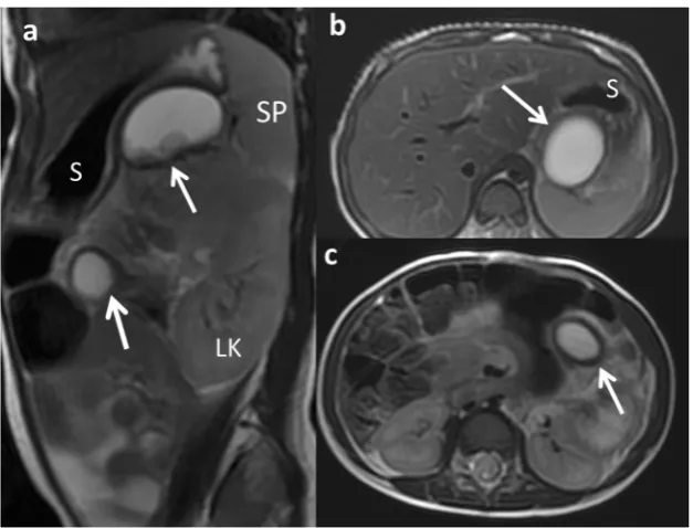
or less frequently in two or more segments (Fig.
4) [11,
16,18,
19].
Atypical EDC is a non-communicating isolated duplication
cyst completely separated from the bowel with no
communi-cation or shared wall. A vascular insult could have led to the
isolation. They are extremely rare [19,
20], especially multiple
isolated EDCs, which are even rarer [19].
The size, location, type, mucosal pattern and presence of
complications produce different clinical presentations and
sev-eral imaging findings of the EDCs. Ultrasonography (US) is
the most used imaging method for diagnosis of abdominal
EDCs. Magnetic resonance (MR) and computed tomography
(CT) are utilised for oesophageal EDCs and for helping in
difficult surgical approaches.
Fig. 1 An 11-month-old boy with abdominal pain is studied.aUS view showing the typical US features of an EDC: an inner hyperechoic epithelial lining containing the mucosa of the alimentary tract (wide arrow) and the outer hypoechoic layer of smooth muscle (white long
arrow), closely attached to the gastrointestinal tract by sharing a common wall.RKright kidney.bSurgical findings: typical ileal EDC.
cDetailed picture of the EDC after resection from the ileal wall
Fig. 2 A 4-year-old boy in a routinary US control of a horseshoe kidney.aAbdominal ultrasound view of an EDC (black arrow) with a peripheral eccentric hypoechoic cap (white arrows).b
We review the different forms of presentation of EDCs,
show-ing both typical and atypical image findshow-ings with the different
imaging techniques. We correlate the imaging findings with the
surgical results and the final pathological features.
Clinical presentations
The intrauterine presentation is increasing, mostly due to the
improvement in prenatal screening US, routine
second-trimester screening and improved imaging resolution.
However, prenatal diagnosis of EDCs is often difficult, and
US identifies only 20
–
30% of them, and sometimes they are
discovered by chance (Fig.
5) [21,
22].
The natural history of EDCs is quite variable. The clinical
presentation or onset symptoms of these malformations range
from infancy and early childhood to adulthood. Almost 70%
of EDCs present symptoms within the first year of life and
85% in the second one [3,
10,
14].
The signs and symptoms depend on the type and location
of the duplication.
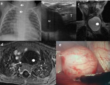
Oral and oesophageal cysts may cause respiratory distress or
dysphagia. Retrosternal pain, haemoptysis and infection can
oc-cur in case of large cysts with rapid growth (Fig.
6) [6,
23].
Gastric and intestinal duplications may produce nausea,
vomiting, abdominal distention or palpable abdominal mass
(Fig.
7). Recurrent abdominal pain is one of the most frequent
forms of presentation and is usually attributed to high pressure
inside the duplication cyst because of the accumulation of
Fig. 4 A 2-year-old girl with a splenic lesion (not shown) is studied with abdominal MR.a
secretions. Intussusception is another complication in which
the cyst serves as a head point and pain, obstruction or
bleed-ing are possible forms of manifestation. Extrinsic compression
of the adjacent bowel is also possible, which causes
obstruc-tion. However, the most serious complications are produced if
gastric mucosa is present within the cyst, leading to
inflam-mation, bleeding, ulceration and even perforation [6,
9,
13,
14,
24,
25].
Nonetheless, EDCs can sometimes be detected
inci-dentally (Fig.
8).
Imaging findings
US is the imaging method of choice in the diagnosis of EDCs;
only limited in the evaluation of oesophageal EDCs.
Fig. 5 A 21-week-old fetus with polyhydramnios and absent normalgastric bubble in the US is studied.a Sagittal FIESTA fetal MRI showing a mediastinal cyst (thick arrow). Oesophageal atresia without fistula is suspected.Bbladder. Trachea (arrows).bCoronal HASTE fetal MRI: detailed view of the mediastinal cyst (thick arrow).c
Postnatal thoraco-abdominal radiograph: the gastric line tip is seen (black arrow) confirming the oesophageal atresia. The absence of air in the abdomen indicates a type-I or -II oesophageal atresia (without fistula). Venous umbilical catheter (white arrow)
Fig. 6 A 10-month-old boy with a congenital cardiopathy presents respiratory distress.aChest X-ray: a left cervicothoracic mass is suspected displacing the trachea to the right (arrows).bChest US: cystic mass (M) with slightly echogenic content inside is seen next to the thymus (T). Its origin is unclear.cCoronal view SSFSE T2 MRI: a well-delineated and hyperintense lesion (star) is seen.
dTransversal view of the lesion in a gadolinium-enhanced VIBE MRI confirms the cystic nature of the mass (star) next to the anterior oesophageal wall (arrow).e
Transoesophageal ultrasound might be useful but is not
rou-tinely used [26,
27].
Classical findings of uncomplicated EDC include:
pres-ence of a cyst in relation to the gut with double-wall or
mus-cular rim sign (gut signature sign), which is caused by inner
hyperechoic mucosa and outer hypoechoic smooth muscle
layer (muscularis propia) (Figs.
1
and
7). However, the
double-wall sign in other cystic lesions (mesenteric cyst,
Meckel
’
s diverticulum or torsioned ovarian cyst) may be seen
[1,
2,
11,
24,
28
–
30].
New US signs are described according to the characteristics
of the EDCs:
–
As an EDC contains the same multi-layered wall
archi-tecture as the normal GT, the sign
Bfive-layered cyst wall
^
is proposed. It corresponds to the innermost hyperechoic
mucosa, hypoechoic muscularis mucosa, hyperechoic
submucosa, hypoechoic muscularis propia and the
outer-most hyperechoic serosa. Identification of all five layers
in a cyst is pathognomonic of EDC. However, this sign is
difficult to demonstrate and needs expertise and
high-resolution US (12
–
18 MHz) [27,
29,
30]. For this reason,
the use of US linear probe is recommendable when the
GT is examined.
–
An EDC shares wall with the adjacent GT. Therefore, the
diagnosis is carried out if it is possible to demonstrate the
BY
^
sonographic configuration of the muscle layer caused
by the splitting of the shared muscularis propria between
the cyst and the adjacent loop. This sign is not described
for other abdominal cysts and reflects one of the
histolog-ical characteristics of the EDCs (Fig.
9) [29,
31].
US is a dynamic study and allows to visualise the
peristal-sis of the cyst wall. It appears as a transient change of the
shape and contour of the cyst because of a concentric
contrac-tion of the cyst wall (Fig.
10) when the transductor stays still
on the cyst for a while [2,
27].
Although EDCs are frequently anechoic or hypoechoic,
mucinous material or septations can be present without being
complicated (Fig.
11) [27].
Complicated EDCs rarely present the classic five layers or
double-wall sign. Ectopic rest of pancreatic tissue can produce
enzymatic destruction of the mucosa with inflammation, as
Fig. 7 An 18-month-old girl with an abdominal mass in the physicalexam is studied.aAbdominal X-ray: round, dense mass is discovered (arrows) in the left upper quadrant.bTransversal US view of the lesion: a cyst with theBdouble-wall^sign: the mucosa is hyperechoic (arrow) and
the muscular layer is hypoechoic (dashed-line arrow). A gastric duplication cyst was suspected.L liver, Ppancreas, AOaorta. c
Laparotomy: antral duplication cyst was found (arrow).Sstomach
Fig. 8 A 12-year-old girl with a gynaecological malformation and haematometrocolpos.aA hypointense lesion is seen (white arrow) next to the rectum in axial T1 weighted pelvic MR.bOn T2-weighted pelvic MR, the cyst next to the left wall of the rectum is seen.cSagittal plane of
well as loss of the wall layers showing a hyperaemic thick
wall. In such cases, the
BY configuration
^
sign helps in
estab-lishing the correct diagnosis of EDC (Fig.
12) [3,
29,
30]. If
ectopic gastric mucosa produces haemorrhage and bleeding,
fluid levels or echogenic debris can be seen. When infection
occurs, ulceration of mucosa can appear, and internal debris
may be seen. The transmural extension can produce important
inflammatory changes in the surrounding mesentery fat
(Figs.
13
and
14) [3,
7,
24]. Ileal EDC, near the ileocecal
valve, can act as intussusception head, showing on US a cystic
mass inside the intussusception requiring emergency
treat-ment (Fig.
15) [2].
In case of atypical or isolated EDC, the pseudokidney sign
is described when there is a complete loss of typical wall
layers because of severe congestion, thus producing a thick
hypoechoic rim with a hyperechoic central layer [32].
US prenatal diagnosis of EDCs includes the same signs
as postnatal cyst: the double
–
wall sign and the presence
of peristalsis. However, on the prenatal US, the
Bdouble
wall
^
is not always seen or can be partial [10,
33,
34], and
Fig. 9 A 3-month-old boy withvomiting is admitted to the emergency room.aUS shows a cystic round-shape lesion with the Bfive-layers sign^(between arrows).bTheBY sign^is seen (long arrow).Starileum,Lliver. Laparoscopic findings: a non-complicated ileal duplication cyst
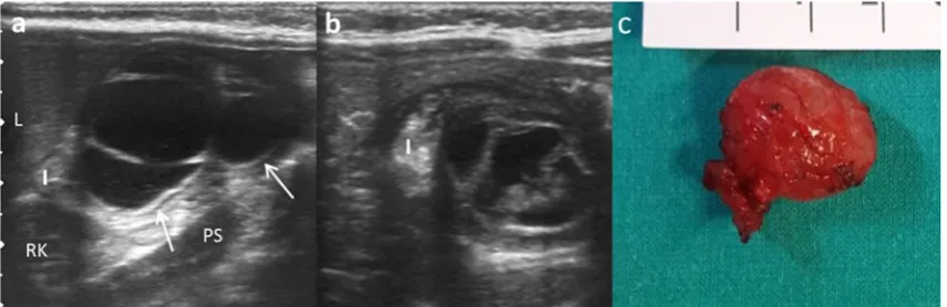
Fig. 10 An 8-month-old boy with abdominal pain.aLongitudinal grey-scale US image showing a cystic lesion with an incomplete septum inside (small white arrows), next to the terminal ileum (big arrow). TheBY^
sign is shown (dashed-line arrow).bUS image obtained a few seconds later: peristalsis of the cyst causes small angulation of the contour and changes shape (black star).cSurgical findings: ileal duplication cyst
Fig. 11 An 8-month-old boy with abdominal pain and abdominal mass on physical exam.aLongitudinal US view of a multiseptated cystic mass in the right flank with theBY^sign (white arrows).Iileum,Lliver,Ps
it may require the differential diagnosis with other cystic
lesions such as mesenteric, omental, ovarian and
choledochal cysts. If it is possible to demonstrate the
pres-ence of peristalsis in the cyst wall, an intestinal origin is
probed. MR imaging is suggested to have a supplemental
value in the assessment of fetal abdominal cysts (Fig.
16)
[10,
35,
36].
CT is not typically performed for evaluation of EDCs
due to radiation. CT may depict the location and
exten-sion of the cyst, as well as complications, the associated
anomalies and anatomical relationship with surrounding
structures. At CT, an EDC manifests as a cystic mass with
a thin and slightly enhancing wall adjacent to the
gastro-intestinal wall. A high attenuation inside the cyst may be
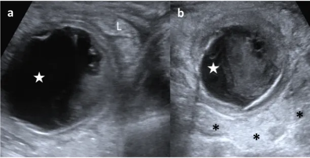
Fig. 12 A 3-week-old term newborn with abdominal distention andgastric intolerance.aLongitudinal US view of the right low quadrant: thickened wall (stars) cystic lesion next to a bowel loop (dashed-line arrow). TheBY^sign between the bowel and the lesion (arrow). b
Power Doppler US demonstrates the significant vascularisation in the cyst wall. cSurgical findings: a cystic tumour next to the ileocecal valve. Pathological findings: ileal duplication cyst with heterotopic pancreatic tissue
Fig. 13 A 3-year-old boy with fever and abdominal pain is studied.aUS shows a cystic mass (star) with internal debris and next to an ileal loop (L).bThe lesion (star) is surrounded by echogenic mesenteric fat (*) as an inflammatory sign. Surgical findings: a 5-cm ileal complicated duplication cyst was found with gastric mucosa with
haemorrhagic and ulcerated walls
Fig. 14 An 8-month-old boy baby with continuous crying is taken to the emergency department. Because of high suspicion of intussusception, a US exam is required.aAbdominal pear-shaped cystic lesion (star) in the left flank was found in a coronal US view. It shows a typical outer
Fig. 15 A 14-month-old girl with acute gastroenteritis and continuous crying.aAbdominal US shows an intestinal intussusception with a cyst (C) as the leading cause.Lliver,RKright kidney.bA detailed US view:
the intussuscipiens (arrows) and intussuscepted bowel (arrowheads) with the cyst inside (C) and the hyperechoic and thickened walls. Surgical findings: ileal duplication cyst as the cause of the intussusception
Fig. 16 A 22-week-old fetus with an abdominal cyst seen on ultrasound is studied with MR.aCoronal fetal FIESTA MR: a cystic lesion is seen next to the stomach (S). Oesophageal lumen is seen (arrows).bFIESTA transversal MR: the hypointense wall of the suspected gastric duplication
cyst (arrow).Lliver,Sstomach.cPostnatal abdominal US view: the lesion (star) imprinting the gastric wall (arrow). The content of the stomach is seen (S).SPspleen. Surgical findings: duplication cyst of the oesophageal-gastric transition
Fig. 17 A 22-month-old boy with fever and abdominal pain. a
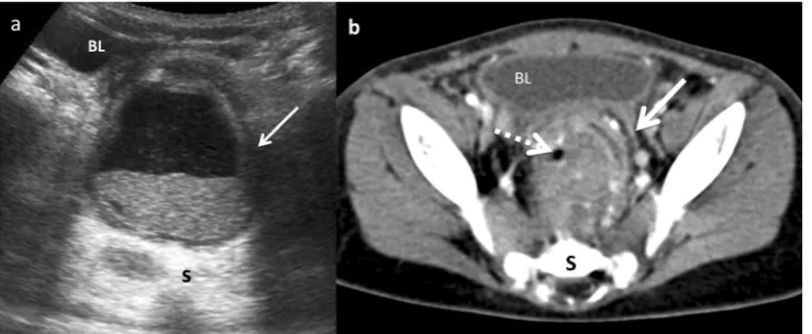
Transversal US view of the pelvis shows a cystic mass between the bladder (BL) and the sacrum (S). The lesion presents anterior and left wall thickening (arrow) and contains a fluid-fluid level suggesting the presence of a complicated rectal mass.b Contrast-enhanced CT
seen due to haemorrhage or proteinaceous material. A
thick enhancing wall, air bubbles inside and
cyst-surrounding inflammation may indicate an EDC
compli-cated by infection (Fig.
17) [1,
11,
12,
15].
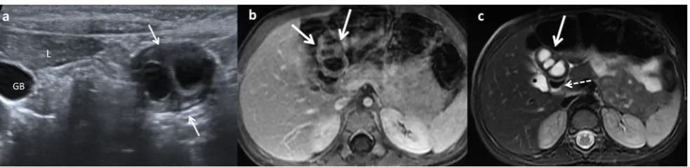
Like CT, MR is not routinely used as a diagnostic
method for EDCs, especially due to sedation requirement.
On MR imaging, most duplications have low signal
inten-sity on T1-weighed images and very high inteninten-sity on
T2-weighted images (Fig.
18). Both CT and MR play a major
role prior to surgery in establishing the relationship
be-tween the cyst and its adjacent structures [12], and in
locations where US presents a limited use, particularly
in oesophageal and rectal duplications [1,
2,
6,
12].
Management
The main considerations in the management of EDCs are: the
condition of the patient, the location of the cyst, whether it
involves one or more anatomic locations, whether its structure
is cystic or tubular, and if it is communicated with the true
intestinal lumen.
With the widespread availability of antenatal diagnosis,
EDCs are often diagnosed prenatally. The optimal time to
perform the resection in children with antenatal diagnosis is
not defined. These patients should undergo early
investiga-tion, followed by early resection even within the first 6 months
of life [3,
37,
38].
Treatment of asymptomatic EDCs remains controversial.
The clinical behaviour of EDCs is unpredictable. EDCs tend
to increase in size gradually and can cause symptoms and
important complications that might be fatal, such as
obstruc-tion, massive bleeding or even a potential risk for malignant
transformation in the adulthood [13,
14,
17,
39].
Early excision is associated with less morbidity and a
shorter length of stay compared to excision in symptomatic
patients. There are significant post-operative morbidities after
resection of complicated EDCs, compared with its elective
surgery. Cyst excision alone could be considered, but if there
is a communication, sometimes a resection of the adjacent
bowel is necessary. It is important to ensure that the cyst is
entirely resected because recurrence or malignant changes
may occur [40].
Currently, minimally invasive surgery is becoming the
elective approach, and most of the cysts can be resected
suc-cessfully, either thoracoscopically or laparoscopically, as long
as an exhaustive imaging diagnosis is available [41].
Conclusions
EDCs are uncommon congenital abnormalities arising anywhere
along the GT. Their clinical presentations vary according to the
site of duplication; ileum appears as the most commonly
in-volved. Nowadays, antenatal diagnosis is becoming more
fre-quent. US is the method of choice to diagnose gastrointestinal
EDCs. Although double-wall US sign in a cyst is the most typical
for diagnosis of EDCs, the findings of the five layers sign or the
BY configuration
^
of the muscular layer are more specific
fea-tures. Complicated cysts present atypical imaging findings. CT
and MR imaging can be required in oesophageal or rectal EDCs
for planning complicated surgical approach. Surgery is necessary
because of the severe complications they can develop. The
diag-nosis is confirmed by histological examination.
Open Access This article is distributed under the terms of the Creative C o m m o n s A t t r i b u t i o n 4 . 0 I n t e r n a t i o n a l L i c e n s e ( h t t p : / / creativecommons.org/licenses/by/4.0/), which permits unrestricted use, distribution, and reproduction in any medium, provided you give appro-priate credit to the original author(s) and the source, provide a link to the Creative Commons license, and indicate if changes were made.
References
1. Hur J, Yoon CS, Kim MJ, Kim OH (2007) Imaging features of gastrointestinal tract duplications in infants and children: from esophagus to rectum. Pediatr Radiol 37:691–699
Fig. 18 A 2-year-old girl with fever and abdominal pain.aAbdominal US view of a complex cystic lesion (white arrows) in the duodenal area, next to the liver (L).GBgallbladder.bAxial gadolinium-enhanced GRE T1 demonstrating the presence of a multilocular cystic mass (white arrows) with wall thickening.cAxial T2 MRI: the mass (white arrow)
2. Berrocal T, Hidalgo P, Gutiérrez J, De Pablo L, Rodríguez-Lemos R (2004) Imagen radiológica de las duplicaciones del tubo digestivo. Radiología 46:282–292
3. Sharma S, Yadav AK, Mandal AK, Zaheer S, Yadav D, Samie A (2015) Enteric duplication cysts in children: a clinicopathological dilemma. J Clin Diagn Res 9:8–11
4. Macpherson RI (1993) Gastrointestinal tract duplications: clinical, pathologic, etiologic, and radiologic considerations. Radiographics 13:1063–1080
5. Tiwari C, Shah H, Waghmare M, Makhija D, Khedkar K (2017) Cysts of gastrointestinal origin in children: varied presentation. Pediatr Gastroenterol Hepatol Nutr 20:94–99
6. Okur MH, Arslan MS, Arslan S et al (2014) Gastrointestinal tract duplications in children. Eur Rev Med Pharmacol Sci 18:1507–1512 7. Van Zitteren LM, Ruppert M, Op de Beeck B, Wojciechowski M (2017) Infected enteric duplication cyst. BMJ Case Rep 21:2017 8. Qi BQ, Beasley SW, Williams AK (2001) Evidence of a common
pathogenesis for foregut duplications and esophageal atresia with tracheo-esophageal fistula. Anat Rec 264:93–100
9. Letelier AM, Barría CM, Beltrán MS, Marcelo A, Moreno CH (2009) Duplicación intestinal: Diagnóstico y tratamiento de una condición inusual. Rev Chil Cir 61:171–175
10. Palacios A, De Vera M, Martínez-Escoriza JC (2013) Prenatal sono-graphic findings of duodenal duplication: case report. J Clin Ultrasound 41(Suppl 1):1-4
11. Udiya AK, Shetty GS, Chauhan U, Singhal S, Prabhu SM (2016) Multiple isolated enteric duplication cysts in an infant—a diagnos-tic dilemma. J Clin Diagn Res 10:TD15–TD16
12. Rasool N, Safdar CA, Ahmad A, Kanwal S (2013) Enteric dupli-cation in children: clinical presentation and outcome. Singapore Med J 54:343–346
13. Erginel B, Soysal FG, Ozbey H et al (2017) Enteric duplication cysts in children: a single-institution series with forty patients in twenty-six years. World J Surg 41:620–624
14. Górecki W, Bogusz B, Zajac A, Soltysiak P (2015) Laparoscopic and laparoscopy assisted resection of enteric duplication cysts in children. J Laparoendosc Adv Surg Tech A 25:838–840
15. Lee NK, Kim S, Jeon TY et al (2010) Complications of congenital and developmental abnormalities of the gastrointestinal tract in ad-olescents and adults: evaluation with multimodality imaging. Radiographics 30:1489–1507
16. Ben-Ishay O, Connlly SA, Buchmiller TL (2013) Multiple duplica-tion cysts diagnosed prenatally: case report and review of the liter-ature. Pediatr Surg Int 29:397–400
17. Olajide AR, Yisau AA, Abdulrasees NA, Kashim IO, Olaniyi AO, Morohunfade AO (2010) Gastrointestinal duplications: experience in seven children and a review of the literature. Saudi J Gastroenterol 16:105–109
18. Chuquisana-Mostacero C, Enríquez de Salamanca-Celada J, Azorín-Cuadrillero D (2017) Dos casos de quiste de intestino an-terior en cavidad oral. Cir Plast Iberolatinoam 43:179–185 19. Mandham P, Ehsan TM, Al-Sibai S, Khan AM, Sankhla D (2014)
Noncommunicating multiple intra-abdominal enteric duplication cysts. Afr J Paediatr Surg 11:276–278
20. Seydafkan S, Shibata D, Sanchez J, Tran ND, Leon M, Coppola D (2016) Presacral noncommunicating enteric duplication cyst. Cancer Control 23:170–174
21. Basany L, Aepala R, Mohan Reddy Bellary M, Chitneni M (2015) Intestinal obstruction due to ileal duplication cyst and malrotation in a preterm neonate. J Neonatal Surg 4:48
22. Nishizawa C, Cajusay-Velasco S, Mashima M et al (2014) HDlive imaging of fetal enteric duplication cyst. J Med Ultrason (2001) 41:511 23. Cuch B, Nachulewicz P, Wieczorek AP, Wozniak M, Pac-Kozuchowska E (2015) Esophageal duplication cyst treated thoracoscopically during the neonatal period. Medicine (Baltimore) 94:e2270
24. Di Serafino M, Mercogliano C, Vallone G (2016) Ultrasound eval-uation of the enteric duplication cyst: the gut signature. J Ultrasound 19:131–133
25. Tong SC, Pitman M, Anupindi SA (2002) Ileocecal enteric dupli-cation cyst: radiologic-pathologic correlation. Radiographics 22: 1217–1222
26. Anupindi SA, Halverson M, Khwaja A, Jeckovic M, Wang X, Bellah RD (2014) Common and uncommon applications of bowel ultrasound with pathologic correlation in children. AJR Am J Roentol 202:946–959
27. Liu R, Adler D (2014) Duplication cysts: diagnosis, management, and the role of endoscopic ultrasound. Endosc Ultrasound 3:152–160 28. Fonseca EKUN, Sameshima YT (2018) Gut signature sign in
en-teric duplication cysts. Abdom Radiol (NY).https://doi.org/10. 1007/s00261-018-1620-2
29. Kumar D, Ramamathan S, Haider E, Khanna M, Otero C (2015) Gastroenterology: revisiting the forgotten sign: five layered gut sig-nature and Y configuration in enteric duplication cysts on high-resolution ultrasound. J Gastroenterol Hepatol 30:1111
30. Cheng G, Soboleski D, Daneman A, Poenaru D, Hurlbut D (2005) Sonographic pitfalls in the diagnosis of enteric duplication cysts. AJR Am J Roentgenol 184:521–525
31. Tritou I, Sfakianaki E, Prassopoulos P (2015) Letter: the sonographic multilaminar appearance is not enough for the diagnosis of enteric duplication cyst in children. AJR Am J Roentgenol 204:222–223 32. Peng HL, Su CT, Chang CY, Lau BH, Lee CC (2012) Unusual
imaging features of completely isolated enteric duplication in a child. Pediatr Radiol 42:1142–1144
33. Gerscovich EO, Sekhon S, Loehfelm TW, Wootton-Gorges SL, Greenspan A (2017) A reminder of peristalsis as a useful tool in the prenatal differential diagnosis of abdominal cystic masses. J Ultrasound 17:129–132
34. Laje P, Flake AW, Adzick NS (2010) Prenatal diagnosis and post-natal resection of intraabdominal enteric duplications. J Pediatr Surg 45:1554
35. Recio Rodríguez M, Martínez de Vega V, Cano Alonso R, Carrascoso Arranz J, Martínez Ten P, Pérez Pedregosa J (2012) MR imaging of thoracic abnormalities in the fetus. Radiographics 32:E305–E321
36. Veyrac C, Couture A, Saguintaah M, Baud C (2004) MRI of fetal gastrointestinal tract abnormalities. Abdom Imaging 29:411–420 37. Laje P, Flake AW, Adzick AS (2010) Prenatal diagnosis and
post-natal resection of intra-abdominal enteric duplications. J Pediatr Surg 45:1554–1558
38. Foley PT, Sithasanan N, McEwing R, Lipsett J, Ford WD, Furness M (2003) Enteric duplications presenting as antenatally detected abdominal cysts: is delayed resection appropriate? J Pediatr Surg 38:1810–1813
39. Ribaux C, Meyer P (1995) Adenocarcinoma in a ileal duplication. Ann Pathol 15:443–445
40. Patiño Mayer J, Bettolli M (2014) Alimentary tract duplications in newborns and children: diagnostic aspects and the role of laparo-scopic treatment. World J Gastroenterol 20:14263–14271 41. Scarpa AA, Ram AD, Soccorso G, Singh M, Parikh D (2017)
Surgical experience and learning points in the management of fore-gut duplication cysts. Eur J Pediatr Surg. https://doi.org/10.1055/s-0037-1607293
Publisher
’
s Note