R E S E A R C H
Open Access
The hepatocyte growth factor-expressing
character is required for mesenchymal
stem cells to protect the lung injured
by lipopolysaccharide in vivo
Shuling Hu, Jinze Li, Xiuping Xu, Airan Liu, Hongli He, Jingyuan Xu, Qihong Chen, Songqiao Liu, Ling Liu,
Haibo Qiu and Yi Yang
*Abstract
Background:Acute respiratory distress syndrome (ARDS) is a life-threatening condition in critically ill patients. Recently, we have found that mesenchymal stem cells (MSC) improved the permeability of human lung
microvascular endothelial cells by secreting hepatocyte growth factor (HGF) in vitro. However, the properties and functions of MSC may change under complex circumstances in vivo. Here, we sought to determine the role of the HGF-expressing character of MSC in the therapeutic effects of MSC on ARDS in vivo.
Methods:MSC with HGF gene knockdown (MSC-ShHGF) were constructed using lentiviral transduction. The HGF mRNA and protein levels in MSC-ShHGF were detected using quantitative real-time polymerase chain reaction and Western blotting analysis, respectively. HGF levels in the MSC culture medium were measured by enzyme-linked immunosorbent assay (ELISA). Rats with ARDS induced by lipopolysaccharide received MSC infusion via the tail vein. After 1, 6, and 24 h, rats were sacrificed. MSC retention in the lung was assessed by immunohistochemical assay. The lung wet weight to body weight ratio (LWW/BW) and Evans blue dye extravasation were obtained to reflect lung permeability. The VE-cadherin was detected with inmmunofluorescence, and the lung endothelial cell apoptosis was assessed by TUNEL assay. The severity of lung injury was evaluated using histopathology. The cytokines and HGF levels in the lung were measured by ELISA.
Results:MSC-ShHGF with markedly lower HGF expression were successfully constructed. Treatment with MSC or MSC carrying green fluorescent protein (MSC-GFP) maintained HGF expression at relatively high levels in the lung at 24 h. MSC or MSC-GFP decreased the LWW/BW and the Evans Blue Dye extravasation, protected adherens junction VE-cadherin, and reduced the lung endothelial cell apoptosis. Furthermore, MSC or MSC-GFP reduced the inflammation and alleviated lung injury based on histopathology. However, HGF gene knockdown significantly decreased the HGF levels without any changes in the MSC retention in the lung, and diminished the protective effects of MSC on the injured lung, indicating the therapeutic effects of MSC on ARDS were partly associated with the HGF-expressing character of MSC.
Conclusions:MSC restores lung permeability and lung injury in part by maintaining HGF levels in the lung and the HGF-expressing character is required for MSC to protect the injured lung.
Keywords:Acute lung injury, Mesenchymal stem cell, Hepatic growth factor, Lung vascular permeability
* Correspondence:yiyiyang2004@163.com
Department of Critical Care Medicine, Zhongda Hospital, Southeast University School of Medicine, No.87 Dingjiaqiao Road, Nanjing 210009, Jiansu, P.R. China
Background
Acute lung injury (ALI) and its severe form, acute re-spiratory distress syndrome (ARDS), are life-threatening conditions in critically ill patients [1]. Although substan-tial progress has been made in the treatment of ALI/ ARDS, especially in the development of lung-protective ventilation and a fluid-conservation strategy, mortality remains high [2, 3]. Novel therapies for ALI/ARDS are therefore needed.
ALI/ARDS are characterized by diffuse injury to the lung endothelial and epithelial cells, which leads to an increase in alveolar-capillary permeability and alveolar pulmonary edema [4]. The lung endothelium is the first barrier to prevent cells and proteins in the blood from infiltrating into the interstitial space and the alveoli of the lung. Therefore, it is essential to restore injured endothelial cells for the treatment of ALI/ARDS.
Bone marrow-derived mesenchymal stem cells (MSC) are one of several types of stem cells that may have therapeutic effects on damaged organs. MSC are capable of differentiating into different types of cells, secreting paracrine soluble factors, releasing microvesicles (MV), homing to injury sites, and so forth. Many studies show that MSC are beneficial in ALI/ARDS [5–7]. They can restore lung protein permeability, reduce inflammation, and improve survival in lipopolysaccharide- (LPS) or Escherichia coli-induced lung injury in mice. However, the underlying mechanism by which MSC restores lung protein permeability in vivo has not yet been fully clarified.
Recently, the strong paracrine property of MSC has been considered to be the principal mechanism for maintaining function in damaged organs [8, 9]. The pulmotrophic factor, hepatocyte growth factor (HGF), which plays an important role in protecting vascular permeability, is a major paracrine soluble factor of MSC. Many studies have detected HGF protein in the cell culture medium of MSC [10, 11]. Furthermore, HGF expression increased when MSC were under hyp-oxic conditions or stimulated with LPS [10]. In our pre-vious in vitro study [12], we found that, by secreting HGF, MSC improved protein permeability across the human lung microvascular endothelial cell (HMVEC) monolayer and repaired intercellular junctions, includ-ing the tight junctions and the adherens junctions of lung endothelial cells. However, in in-vivo conditions, the circumstances for MSC are different due to the un-checked inflammation, severe hypoxemia, and the in-creased pulmonary alveolar and capillary membrane leakage of ARDS. These all may affect the properties and functions of MSC. Hence, we sought to determine whether the HGF-expressing character of MSC would also be critical for MSC to maintain lung permeability and protect the lung from injury in vivo.
Methods
Ethics statement
Male wild-type Sprague-Dawley (SD) rats (Laboratory Animal Center, Shanghai, China) were maintained under specific pathogen-free conditions. The Animal Care and Use Committee of Southeast University approved all ex-periments involving the use of animals.
Cell culture
Rat MSC, isolated from the bone marrow of SD rats, and the MSC culture medium were purchased from Cyagen Bioscience, Inc. (Guangzhou, China). 293 T cells were supplied by Cell Bank of Chinese Academy of Sciences. The supplier identified MSC according to cell surface phe-notypes and multipotency. Fluorescence-activated cell sorting (FACS) analysis characterized surface phenotypes using the following markers: CD90+, CD44+, CD29+, CD34–, CD45–, and CD11b/c–, and the capacities to differentiate into the adipogenic, osteogenic and chondro-genic lineages were determined by staining with oil red-O, alizarin red or alcian blue, respectively, after culturing in adipogenic, osteogenic or chondrogenic differentiation media (Cyagen Bioscience, Inc., Guangzhou, China) for 2–3 weeks (See Additional file 1).
MSC were cultured in the MSC culture medium made from SD rat MSC basal medium containing 10 % SD rat MSC-qualified fetal bovine serum, 1 % penicillin-streptomycin and 1 % glutamine. The culture medium was changed every 2–3 days and the cells were split when they achieved 90 % confluency. The MSC used for in-vitro studies had been passaged 10 times after lentiviral trans-duction. MSC with the total passage number <9 were used in the in-vivo experiments. 293 T cells were cultured in Dulbecco’s modified Eagle’s medium (DMEM) contain-ing 10 % fetal bovine serum (Wisent, Inc., St-Bruno, Quebec, Canada), 1 % L-glutamine, and 1 % penicillin-Streptomycin, and incubated at 37 °C in a humidified atmosphere of 5 % CO2. Additional materials were
pro-vided in Additional file 2.
Lentiviral vector-mediated HGF gene knockdown in MSC
The recombinant plasmid DNAs were extracted from E. coliTop10 and purified using the Plasmid Preparation Kit (GenePharma, Shanghai, China). The purity of the DNA was assessed with a spectrophotometer (Tecan, Switzerland). A260/A280 nm absorbance ratios of 1.8– 2.2 suggested a pure DNA sample. Theses plasmids were then separately co-transfected with three packaging plas-mids (pGag/Pol, pRev, pVSV-G) into 293 T cells using RNAi-mate (Genepharma, Shanghai, China) according to the manufacturer’s instruction. The lentiviral particles were collected and stored at–80 °C for future use. Titer was obtained by GFP expression assay [13].
MSC were seeded and cultured in six-well plates for 24 h. The lentiviral vectors (carrying GFP or LV3-GFP ShRNA HGF) were then added to the wells at a multiplicity of infection (MOI) value of 100:1 and cul-tured with MSC for 24 h. After 24 h, the culture medium was changed, and puromycin was added at the minimal lethal concentration (1.5μg/ml) for trans-fected MSC. The puromycin-resistant cells were then collected.
RNA isolation and quantitative real-time polymerase chain reaction (qRT-PCR)
MSCs treated with LV3-GFP (MSC-GFP) or LV3-GFP-ShRNA HGF (MSC-ShHGF) were collected, respect-ively. Total RNA was isolated from MSC, MSC-GFP or MSC-ShHGF using TRIzol reagent (Takara Bio, Inc., Kyoto, Japan) according to the manufacturer’s protocol. The quality of the RNA was assessed with a spectropho-tometer (Tecan, Switzerland). 260/280 nm absorbance ratios of 1.8–2.2 suggested a pure RNA sample. The RT-PCR primers for rat glyceraldehyde-3-phosphate dehydro-genase (GAPDH) and rat HGF (Table 1) were provided by GenePharma (Shanghai, China). RT-PCR assays were performed following the One-Step RT-PCR protocol described by Funglyn Biotech Inc. (Shanghai, China).
Western blotting analysis
MSC, MSC-GFP, and MSC-ShHGF were collected after transduction with lentiviral vector. Total cellular pro-tein from either MSC, MSC-GFP, or MSC-ShHGF was extracted and separated using SDS-PAGE gels (10 %), as previously described [14]. Protein was then incubated with primary antibodies to HGF (1:600 dilution; Santa
Cruz Biotechnology, Inc., Santa Cruz, CA, USA) or β -actin (1:10,000 dilution; Abcam Ltd., Cambridge, UK). The blots were washed three times and then incubated with goat anti-rabbit IgG conjugated with horseradish peroxidase (HRP; Zhongshan Golden Bridge Biotech-nology Co., Ltd, China). Immunoreactive complexes were visualized using chemiluminescence reagents (Thermo Scientific).
Evaluation of HGF levels by ELISA
MSC, MSC-GFP, and MSC-ShHGF were seeded in a 12-well plate at a density of 1 × 10⁵cells per well. After 12 h the culture medium was changed, and MSC were cultured in an incubator at 37 °C, 5 % CO2for 24 h. The culture
medium was then collected and HGF protein levels in the culture medium were quantified using an enzyme-linked immunosorbent assay (ELISA) kit (ExCellBio, Shanghai, China) according to the manufacturer’s instructions.
LPS-induced ALI in rats
To induce ALI, 6- to 8-week-old wild-type SD rats re-ceived an intratracheal instillation of LPS (2 mg/kg, E. coli 0111:B4; Sigma-Aldrich, St. Louis, MO, USA) dis-solved in 100μl phosphate-buffered saline (PBS; Wisent, Inc., St-Bruno, Quebec, Canada) as described previously [15]. PBS, MSC, MSC-GFP, or MSC-ShHGF (5 × 10⁶ cells resuspended in 100 μl PBS) were injected into the tail vein 5 h after LPS challenge. Rats without LPS chal-lenge were injected with PBS as a control. Rats were sacrificed at 1, 6 and 24 h after MSC injection, and the lung lobes were collected for further analysis.
Measurement of lung edema
Lung wet weight to body weight (LWW/BW) ratios, which reflected the severity of lung vascular permeability and lung edema, were obtained from the control, ALI, MSC, MSC-GFP, and MSC-ShHGF group.
Evans blue dye leakage
For the control, ALI, MSC-GFP, and MSC-ShHGF group (n = 6 per group, see Additional file 3 for sample size calculation), Evans blue dye (20 mg/kg in 1 ml saline; Sigma-Aldrich, St. Louis, MO, USA) was injected into the tail vein of the rats. After 30 min, the right ventricle of the heart was perfused with 100 ml heparinized saline
Table 1The primer sequence of genes
Gene Primer Primer sequence PCR amplified products (bp)
GAPDH Forward primer 5’> GTGCTGAGTATGTCGTGGAGTCT < 3’ 104
Reverse primer 5’> GGAAGGGGCGGAGATGA < 3’
HGF Forward primer 5’> GCACCTCCTCCTGCTTCC < 3’ 288
Reverse primer 5’> CCAAACCCTTTTTTCACTCCA < 3’
to clean up the dye remaining in the lung vascular sys-tem. When all the dye had been cleared from the lung vascular system, the whole lung was collected. Lung tis-sue (100 mg) from the right lobe was then incubated in formamide (Sigma-Aldrich, St. Louis, MO, USA) for 24 h at 60 °C, and the concentration of Evans blue dye was measured using a spectrophotometer at 630 nm (Tecan, Switzerland).
Immunohistochemical staining
Immunohistochemical analysis was performed to deter-mined expression of GFP in the lung. Sections (5 μm thick) were cut from paraffin-embedded tissues. After be-ing routinely deparaffinized and dehydrated, sections were subjected to antigen retrieval by microwave treatment in boiling 0.01 M citrate buffer (pH 6.0) for 20 min. Then the sections were blocked in 1 % bovine serum albumin (BSA) for 1 h at 37 °C and incubated with anti-GFP pri-mary antibody (Abcam Ltd., Cambridge, UK) overnight at 4 °C, followed by HRP conjugated goat anti-rabbit IgG (Santa Cruz Biotechnology, Inc., Santa Cruz, CA, USA). After washing with 0.1 M PBS, the sections were reacted with a staining solution containing 0.03 % 3,3’-diamino-benzidine tetrahydrochloride (Nanjing Lufei Biotechnol-ogy Co., Ltd., Nanjing, China) for 5–10 min at room temperature. Mayer’s hematoxylin was used for counter-staining. The sections were examined with an Olympus IX71 microscope (Olympus Co., Tokyo, Japan). Cells with GFP were counted by a pathologist based on five ran-domly selected high-power fields (×400).
Immunofluorescence
To measure co-localization of VE-cadherin in the lung, rat lungs were collected gently and then fixed in 4 % paraformaldehyde at 4 °C. Twenty four hours later, lung tissue was frozen in optimal cutting temperature medium (OCT; Sakura Finetek USA, Inc., Torrance, CA, USA) and cut into 5-μm-thick sections. Then the sections were stained with anti-VE-cadherin antibody (Santa Cruz Biotechnology, Inc., Santa Cruz, CA, USA), followed by Fluorescence (FITC)-AffiniPure Donkey Anti-Rabbit IgG (H + L; Jackson ImmunoResearch Inc., PA, USA), and mounted with 4,6-diamidino-2-phenylindole (DAPI; Sigma-Aldrich). Fluorescence was monitored with an Olympus IX71 microscope (Olympus Co., Tokyo, Japan). The mean fluorescence intensity of VE-cadherin of five randomly chosen high-power fields per lung lobe section per rat was assessed and calculated.
TUNEL assay
To assess the apoptosis of endothelial cells in the lung, the right lobe was collected and fixed in 4 % paraformalde-hyde at 4 °C for 24 hours, and the lung sections were stained with a TUNEL Apoptosis Assay kit (Nanjing Lufei
Biotechnology Co., Ltd., Nanjing, China). The sections were observed with an Olympus IX71 microscope (Olym-pus Co., Tokyo, Japan). Endothelial cells were quantified by a pathologist based on five randomly selected high-power fields (×400). The number of apoptotic lung endo-thelial cells and total lung endoendo-thelial cells were recorded, and the apoptosis index of lung endothelial cells, which was defined as the number of apoptotic lung endothelial cells divided by the total number of lung endothelial cells, was used to assess the severity of lung endothelial cell apoptosis.
Lung histopathology
To examine the severity of lung injury, the right lobes were collected at 1, 6 and 24 h after MSC infusion and then fixed in 4 % paraformaldehyde. After fixation, the lungs were embedded in paraffin and cut into 5-μm sec-tions. The lung sections were stained using a hematoxylin and eosin staining kit purchased from the Beyotime Insti-tute of Biotechnology. The severity of lung injury was quantified and assessed using a total lung injury score as we previously described [15].
HGF expression in the lung
To detect the HGF levels in the lung, the left lung was collected and stored at–80 °C. The lung tissue was ho-mogenized thoroughly in PBS solution and then centri-fuged at 3000 rpm for 20 min at 4 °C. The levels of HGF in the supernatant were then measured using an ELISA kit (ExCellBio, Shanghai, China), according to the manu-facturer’s instructions.
Measurement of cytokines in the lung
The left lobe was snap-frozen and later processed for lung homogenization. Then the lung homogenate was centri-fuged at 3000 rpm for 20 min at 4 °C and the supernatant was collected. The concentrations of interleukin (IL)-1β and IL-10 in the supernatant were evaluated using ELISA kits (NeoBiosicence, Shenzhen, China), according to the manufacturer’s instructions.
Statistical analysis
Results
The efficiency of lentiviral vector-mediated HGF gene knockdown
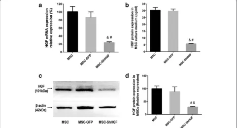
HGF mRNA levels were detected by qRT-PCR. The re-sult showed that HGF mRNA expression was signifi-cantly lower in the MSC-ShHGF group than in the MSC (p< 0.05) and MSC-GFP group (p< 0.05). How-ever, there was no significant difference between the MSC and MSC-GFP group (p> 0.05) (Fig. 1a) in the HGF mRNA expression. The result from Western blot-ting analysis (Fig. 1c and d) showed that the HGF pro-tein expression in the cytoplasm was decreased in the MSC-ShHGF group compared with the MSC (p< 0.05) and MSC-GFP group (p< 0.05). The HGF protein levels in the cell culture medium of the MSC-ShHGF group were also significantly lower those that in the MSC (p< 0.05) and MSC-GFP groups (p< 0.05) (Fig. 1b). However, there was no significant difference in HGF protein levels be-tween the MSC and MSC-GFP group in either the cyto-plasm or the cell culture medium. Taken together, these results suggested that lentiviral-mediated HGF gene knockdown was efficient.
MSC retention in the lung at 24 h
To measure the MSC retention in the lung tissue after cell infusion, immunohistochemical assays were performed for
the GFP carried by MSC. As Fig. 2 shows, no significant differences were found in the retention of MSC with GFP in the lung tissue between the GFP and MSC-ShHGF groups (p> 0.05), demonstrating that HGF gene knockdown in MSC had no significant impact on MSC homing to the lung in respond to lung injury.
HGF levels in the lung after MSC treatment
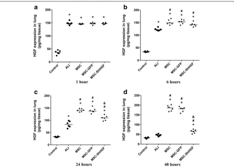
To examine the effect of MSC infusion on HGF levels in ALI rats, we measured the HGF concentration in the lung. As Fig. 3 shows, HGF protein levels in the lung increased at 1 h after LPS challenge and then gradually decreased at 6, 24 and 48 h (p< 0.05). MSC treatment significantly in-creased HGF levels at 6, 24 and 48 h (p< 0.05). When the HGF gene was knocked down in MSC, the HGF level in the lung decreased significantly compared with the MSC or MSC-GFP group at 24 and 48 h (p< 0.05). Since no dif-ferences were found in the retention of MSC with GFP in the lung tissue between the MSC-GFP and MSC-ShHGF groups, these results suggest that MSC regulated HGF level in the injured lung in part through secreting HGF. Additional results were provided in Additional file 2.
The effect of MSC on pulmonary vascular permeability
In this study, LWW/BW and Evans blue dye extravasation were used to evaluate the role of the HGF-expressing
Fig. 1Measurement of hepatocyte growth factor (HGF) expression in mesenchymal stem cells (MSC) after HGF gene knockdown.aEvaluation of HGF mRNA expression in the MSC, MSC-GFP, and MSC-ShHGF groups.bDetection of HGF protein levels in the culture medium of the MSC, MSC-GFP, and MSC-ShHGF groups.cImage for quantification of HGF protein expression in the MSC, MSC-GFP, and MSC-ShHGF groups by Western blotting analysis.
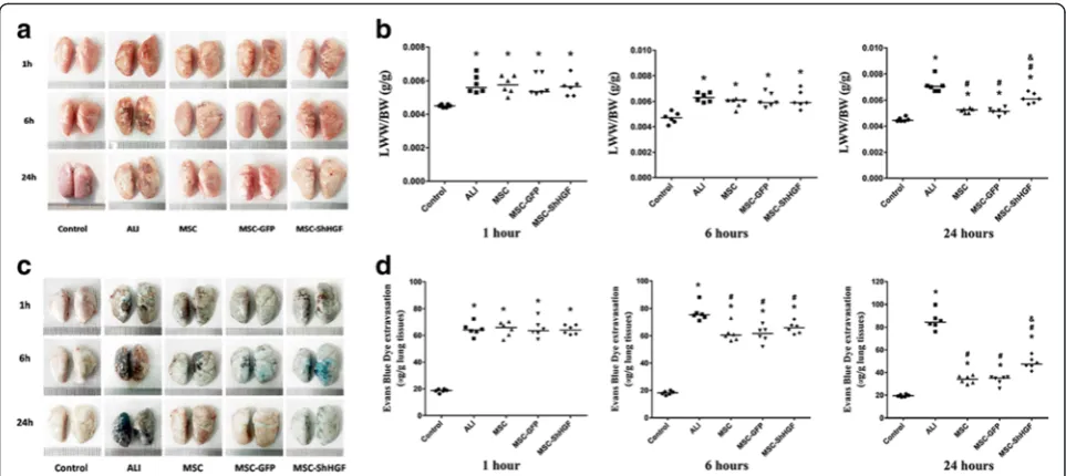
character of MSC in the therapeutic effect of MSC on lung vascular permeability. The results (Fig. 4a and b) showed that LPS stimulation increased LWW/BW mark-edly at 1, 6 and 24 h (p< 0.05). After treatment with MSC, MSC-GFP, or MSC-ShHGF, the LWW/BW was decreased significantly at 24 h in the ALI rats (p< 0.05). More inter-estingly, the LWW/BW in rats treated with MSC with HGF gene knockdown was significantly higher than that in rats treated with MSC or MSC-GFP (p< 0.05). Further-more, as Fig. 4c and d show, LPS stimulation also in-creased Evans blue dye extravasation from the lung vascular to the lung interstitial space and lung alveoli at 1, 6 and 24 h (p< 0.05). With MSC, GFP, or MSC-ShHGF treatment, the Evans blue dye extravasation de-creased markedly at 6 and 24 h (p< 0.05) but, in the MSC-ShHGF group, the Evans blue dye extravasation was sig-nificantly higher compared with the MSC or MSC-GFP group (p< 0.05). These results indicate that MSC improved vascular permeability in lung tissue injured by LPS, and that the HGF-expressing character of MSC was required for MSC to restore this lung vascular permeability.
VE-cadherin expression in the lung following MSC injection
To investigate the effect of MSC with HGF gene knockdown on intercellular junctions in the lungs of rats with ALI, we observed the expression of the adhe-rens junction protein VE-cadherin in the rat lung by
immunofluorescence at 24 h. As Fig. 5 shows, the VE-cadherin expression in lung tissue was decreased markedly after intratracheal instillation of LPS at 24 h (p< 0.05). After treatment with MSC or MSC with HGF gene knockdown, the VE-cadherin expression was increased in the lung compared with the ALI group (p< 0.05). However, the increase in VE-cadherin expression in the MSC-ShHGF group was markedly lower than that in the MSC group (p< 0.05). These re-sults suggest that MSC had a protective effect on the adherens junction of cells in the lung, and that this protective effect required the HGF expressed by MSC.
Assessment of endothelial cell apoptosis in the injured lung after MSC treatment by TUNEL assay
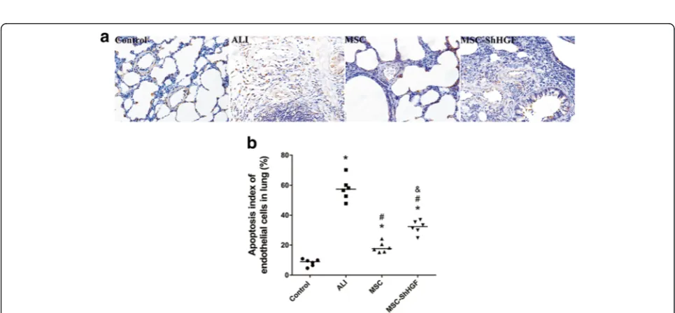
To evaluate the effect of the HGF-expressing character of MSC on endothelial apoptosis in the ALI lung, we carried out a TUNEL assay 24 h after MSC treatment. As Fig. 6 shows, the apoptosis index of the lung endothe-lium was increased after LPS challenge, whereas it de-creased to a large degree after MSC treatment. However, in MSC with gene knockdown, the apoptosis index of the lung endothelium was significantly higher than that in the MSC or the MSC-GFP group. These results indicate that MSC treatment was able to protect lung endothelial cells from apoptosis in the lung of rats with ALI, and that this
protective effect was related to the ability of MSC to re-lease the HGF protein.
Histological evaluation of the therapeutic potential of MSC in ALI rats
As Fig. 7 shows, histopathology indicated extensive in-flammatory infiltrates, marked inter-alveolar septal thickening, and diffuse interstitial and alveolar edema at 1, 6 and 24 h in the ALI group. The lung injury score was 8.23 at 1 h, 10.6 at 6 h and 15.15 at 24 h in the ALI group. The administration of MSC, MSC-GFP, and MSC-ShHGF significantly attenuated lung injury at 24 h (p< 0.05). However, the lung injury score in the MSC-ShHGF group (9.53) was signifi-cantly higher than that in the MSC (5.23) and MSC-GFP group (4.98) (p< 0.05).There was no significant difference in lung injury score between the MSC and MSC-GFP group. These results suggest that MSC had a
protective effect on lung injury, and that this effect was di-minished when the HGF gene was knocked down in MSC.
The effect of MSC on cytokine levels in the injured lung after LPS-induced ALI in rats
In this study, we also investigated the effect of MSC-GFP and MSC with HGF gene knockdown on IL-10 and IL-1β production in the lung. As Fig. 8a shows, the IL-1β level in the lung was markedly increased after LPS challenge at 1, 6 and 24 h (p< 0.05). After treatment with MSC, MSC-GFP, and MSC-ShHGF, IL-1βlevels in the lung were significantly decreased at 6 and 24 h (p< 0.05). There was no significant difference in the lung IL-1β levels between the MSC-ShHGF, MSC, and MSC-GFP group at 6 h. However, there was a significant difference between the MSC-ShHGF and the MSC or MSC-GFP group at 24 h (p< 0.05).
The IL-10 level in the lung was significantly de-creased after LPS stimulation at 1, 6 and 24 h (p< 0.05) (Fig. 8b). However, it was markedly increased after MSC, MSC-GFP, or MSC-ShHGF treatment at 6 and 24 h. In addition, compared with the MSC or MSC-GFP group, the IL-10 level in the lung were signifi-cantly lower in the MSC-ShHGF group. These results indicate that MSC reduced LPS-induced inflammation in the lung, and that the HGF-expressing characteris-tic of MSC played an important part in this effect.
Discussions
Our previous in vitro studies showed that, by secreting the protective soluble factor HGF, MSC are able to re-store the permeability of the HMVEC monolayer follow-ing LPS-induced injury [12]. By producfollow-ing HGF, MSC may maintain the integrity of the injured endothelial monolayer by restoring endothelial intercellular junc-tions, decreasing caveol1 protein expression, and in-ducing proliferation in endothelial cells. Here, we knocked down HGF in MSC to examine the role of the HGF-expressing character of MSC in the therapeutic ef-fect of MSC on lung permeability and lung injury in vivo.
Firstly, we successfully constructed a stable and long-term MSC cell line with low HGF expression by employ-ing a lentiviral vector-mediated HGF gene knockdown technique. Our data showed that MSC-ShHGF at the tenth passage had notably low HGF mRNA and protein expression. Moreover, HGF levels in the cell culture medium of MSC-ShHGF also markedly decreased. The
successful construction of MSC-ShHGF facilitates our further investigations into the role of the HGF-expressing character of MSC in the protective effect of MSC on lung endothelial permeability and lung injury in vivo.
According to our data from the in vivo study, the HGF level in the lung was markedly elevated after LPS challenge. However, it began to decrease at 6 h and was further re-duced to approximately half by 24 h. After MSC treatment, the HGF level increased significantly. Interestingly, when the HGF gene was knocked down in MSC, the HGF pro-tein levels in the injured lung were significantly decreased at 24 h but were still higher than those in the ALI group. However, there was no significant difference in MSC reten-tion in the injured lung between the GFP and MSC-ShHGF groups as shown by the immunohistochemical assay. Taken together, these results suggested that the HGF-expressing character of MSC was required for MSC to increase the HGF levels in the lung, which then exerted a protective effect on the injured lung. There are studies showing that the intravenous or the intra-organ admi-nistration of HGF reduced organ injury [12, 16–18]. Recently, studies [19–21] have also demonstrated that MSC overexpressing HGF elevated the HGF level in in-jured organs and delivered a stronger therapeutic effect compared with normal MSC. Here, our results are consistent with these previous findings.
In this study, data from the LWW/BW and Evans blue dye extravasation assay showed that the HGF-expressing character of MSC played a modest role in the protective effect of MSC on lung endothelial permeability.
Fig. 6The evaluation of vascular endothelial cell apoptosis in the lung.aRepresentative images of lung endothelial cell apoptosis in different groups 24 h after mesenchymal stem cell (MSC) treatment (×400).bThe quantitative result of the apoptosis index of lung endothelial cells in different groups at 24 h after MSC injection.n= 6. *p< 0.05 vs. the control group;#p< 0.05 vs. the ALI group;&p< 0.05 vs. the MSC group.ALI acute lung injury,ShHGFhepatocyte growth factor gene knockdown
expressing MSC have a beneficial effect on maintaining the integrity of the lung endothelium. This may be re-lated to the ability of HGF itself to protect intercellular junctions, as our data demonstrated, preventing endo-thelial cell apoptosis. Previous studies have demon-strated that HGF had a strong angiogenic effect [22–24]. This could facilitate endothelial cell proliferation [25], prevent apoptosis [26, 27], and restore intercellular junc-tions and cytoskeleton structures [28, 29] in vitro. HGF administration could also improve the prognosis of lung in-jury caused by inflammation, oxidative stress, radiation, fi-brosis, and so forth [17, 30, 31], which may be partly related to the effect of HGF on repairing the lung endothelium.
In addition to restoring lung endothelial permeability, the HGF-expressing character also plays a positive role in the beneficial effect of MSC on lung inflammation
induced by LPS. As shown in Fig. 8, IL-1β levels were significantly higher and IL-10 levels were lower in the ShHGF group compared with the MSC or MSC-GFP group. This finding suggested that the ability of MSC to control lung inflammation was diminished after the HGF gene in MSC was knocked down. Possible expla-nations for this result include: 1) HGF has a direct anti-inflammatory effect as demonstrated by previous studies [32, 33]; or 2) the decreased infiltration of immunocytes from the blood to the interstitial space and alveoli of the lung due to the improvement of endothelial permeability related to HGF.
Finally, in the current study, we also assessed lung in-jury using histopathology and lung inin-jury score (Fig. 7). The improvements in histopathology and lung injury score typically occurred 24 h after MSC treatment.
However, in the MSC-ShHGF group this improvement was relatively small compared with the MSC or MSC-GFP group. These results suggested that HGF secretion was required for MSC to exert a better therapeutic ef-fect in ALI. Recently, several studies demonstrated that HGF gene modification (overexpression) in MSC could enhance the therapeutic effect of MSC on injured or-gans such as the heart [34], liver [35], intestine [33], and lung [30]. Here, we inhibited HGF expression in MSC with a method using lentiviral vector and found out that the therapeutic effects of MSC on lung injury were diminished to some degree. This result is consist-ent with the previous studies and indicates a modest role of the HGF-expressing character of MSC in the therapeutic effects of MSC on ALI. In summary, our re-sults showed that the HGF-expressing property of MSC was required for MSC-based therapies in lung injury.
There are some limitations to this study. Firstly, adhe-rens junctions were the only type of intercellular junc-tion we examined in this study. Therefore, further studies are needed to clarify the effect of HGF protein production in MSC on the endothelial tight junctions
and cytoskeleton in rats with ALI. Secondly, we did not investigate the cellular origin of HGF in the lung, which may help us to understand the underlying mechanism of the beneficial effect of the HGF-expressing character of MSC in ALI more clearly. Moreover, we did not explore the effect of MSC with HGF overexpression on ALI. This would also provide some additional evidence for this work. Finally, we only focused on the short-term effect (24 h) of MSC-ShHGF on ALI. The long-term effects remain unknown and merit further investigation.
Conclusion
In conclusion, the results presented here suggest that MSC restores lung vascular permeability, which might be associated with the protective effects of MSC on the adherens junction protein VE-cadherin and lung vascu-lar endothelial cell apoptosis, reducing inflammation, and attenuating lung injury in LPS-induced ALI in rats in part by maintaining the HGF level in the injured lung. Moreover, the HGF-expressing character is re-quired for MSC to protect the injured lung.
Additional files
Additional file 1:Identification of MSC isolated from the bone marrow of SD rats. (DOCX 21.7 mb)
Additional file 2:Additional materials and results. (DOCX 8.71 mb)
Additional file 3:Equation for samlpe size calculation. (DOCX 13.5 kb)
Abbreviations
ALI:acute lung injury; ARDS: acute respiratory distress syndrome;
ELISA: enzyme-linked immunosorbent assay; GFP: green fluorescent protein; HGF: hepatocyte growth factor; HMVEC: human lung microvascular endothelial cell; HRP: horseradish peroxidase; IL: interleukin; LPS: lipopolysaccharide; LV3-GFP: lentivirus carrying GFP; LV3-ShRNA HGF: lentivirus carrying green fluorescent protein and hepatocyte growth factor gene knockdown; LWW/BW: lung wet weight to body weight ratio; MSC: mesenchymal stem cells; MSC-GFP: mesenchymal stem cells carrying green fluorescent protein; MSC-ShHGF: mesenchymal stem cells with hepatocyte growth factor gene knockdown; PBS: phosphate-buffered saline; qRT-PCR: quantitative real-time polymerase chain reaction; SD: Sprague-Dawley; Sh: short-hairpin.
Competing interests
The authors declare that they have no competing interests.
Authors’contributions
SH participated in the study design, performed laboratory work and statistical analysis, prepared the drafts of the manuscript and revised it according to advice from the other authors. JL, XX and HH participated in the laboratory work, performed statistical analysis and drafted the manuscript. AL, JX, QC, SL and LL participated in the study design, and assisted in statistical analysis and drafting the manuscript. HQ and YY were responsible for the study design and revised the manuscript for important intellectual content. All the authors have given final approval of the version to be published and agree to be accountable for all aspects of this work.
Acknowledgements
We all thank Yanli An for assistance with the experimental techniques. We are also thankful to Pablo Gabatto and Fengyun Xu. The authors are grateful for the financial support of the National Natural Science Foundation of China (Grant No. 81170057).
Received: 2 December 2015 Revised: 6 April 2016 Accepted: 11 April 2016
References
1. Rubenfeld GD, Caldwell E, Peabody E, Weaver J, Martin DP, Neff M, et al. Incidence and outcomes of acute lung injury. N Engl J Med. 2005;353(16): 1685–93. doi:10.1056/NEJMoa050333.
2. The Acute Respiratory Distress Syndrome Network. Ventilation with lower tidal volumes as compared with traditional tidal volumes for acute lung injury and the acute respiratory distress syndrome. N Engl J Med. 2000; 342(18):1301–8. doi:10.1056/NEJM200005043421801.
3. Buregeya E, Fowler RA, Talmor DS, Twagirumugabe T, Kiviri W, Riviello ED. Acute respiratory distress syndrome in the global context. Glob Heart. 2014; 9(3):289–95. doi:10.1016/j.gheart.2014.08.003.
4. Matthay MA, Ware LB, Zimmerman GA. The acute respiratory distress syndrome. J Clin Invest. 2012;122(8):2731–40. doi:10.1172/JCI60331. 5. Lee JW, Fang X, Gupta N, Serikov V, Matthay MA. Allogeneic human
mesenchymal stem cells for treatment of E. coli endotoxin-induced acute lung injury in the ex vivo perfused human lung. Proc Natl Acad Sci U S A. 2009;106(38):16357–62. doi:10.1073/pnas.0907996106.
6. Lee JW, Krasnodembskaya A, McKenna DH, Song Y, Abbott J, Matthay MA. Therapeutic effects of human mesenchymal stem cells in ex vivo human lungs injured with live bacteria. Am J Respir Crit Care Med. 2013;187(7):751–60. doi:10.1164/rccm.201206-0990OC.
7. Matthay MA, Goolaerts A, Howard JP, Lee JW. Mesenchymal stem cells for acute lung injury: preclinical evidence. Crit Care Med. 2010;38(10 Suppl): S569–73. doi:10.1097/CCM.0b013e3181f1ff1d.
8. Liang X, Ding Y, Zhang Y, Tse HF, Lian Q. Paracrine mechanisms of mesenchymal stem cell-based therapy: current status and perspectives. Cell Transplant. 2014;23(9):1045–59. doi:10.3727/096368913X667709.
9. Matthay MA. Therapeutic potential of mesenchymal stromal cells for acute respiratory distress syndrome. Ann Am Thorac Soc. 2015;12 Suppl 1:S54–7. doi:10.1513/AnnalsATS.201406-254MG.
10. Crisostomo PR, Wang Y, Markel TA, Wang M, Lahm T, Meldrum DR. Human mesenchymal stem cells stimulated by TNF-alpha, LPS, or hypoxia produce growth factors by an NF kappa B- but not JNK-dependent mechanism. Am J Physiol Cell Physiol. 2008;294(3):C675–82. doi:10.1152/ajpcell.00437.2007. 11. Gnecchi M, He H, Liang OD, Melo LG, Morello F, Mu H, et al. Paracrine action
accounts for marked protection of ischemic heart by Akt-modified mesenchymal stem cells. Nat Med. 2005;11(4):367–8. doi:10.1038/nm0405-367. 12. Chen QH, Liu AR, Qiu HB, Yang Y. Interaction between mesenchymal stem
cells and endothelial cells restores endothelial permeability via paracrine hepatocyte growth factor in vitro. Stem Cell Res Ther. 2015;6(1):44. doi:10. 1186/s13287-015-0025-1.
13. Sastry L, Johnson T, Hobson MJ, Smucker B, Cornetta K. Titering lentiviral vectors: comparison of DNA, RNA and marker expression methods. Gene Ther. 2002;9(17):1155–62. doi:10.1038/sj.gt.3301731.
14. Liu AR, Liu L, Chen S, Yang Y, Zhao HJ, Liu L, et al. Activation of canonical wnt pathway promotes differentiation of mouse bone marrow-derived MSCs into type II alveolar epithelial cells, confers resistance to oxidative stress, and promotes their migration to injured lung tissue in vitro. J Cell Physiol. 2013;228(6):1270–83. doi:10.1002/jcp.24282.
15. He H, Liu L, Chen Q, Liu A, Cai S, Yang Y, et al. Mesenchymal stem cells overexpressing angiotensin-converting enzyme 2 rescue lipopolysaccharide-induced lung injury. Cell Transplant. 2015;24(9):1699-1715. doi:10.3727/ 096368914X685087.
16. Adachi E, Hirose-Sugiura T, Kato Y, Ikebuchi F, Yamashita A, Abe T, et al. Pharmacokinetics and pharmacodynamics following intravenous administration of recombinant human hepatocyte growth factor in rats with renal injury. Pharmacology. 2014;94(3–4):190–7. doi:10.1159/000363412. 17. Meng F, Meliton A, Moldobaeva N, Mutlu G, Kawasaki Y, Akiyama T, et al.
Asef mediates HGF protective effects against LPS-induced lung injury and endothelial barrier dysfunction. Am J Physiol Lung Cell Mol Physiol. 2015; 308(5):L452–63. doi:10.1152/ajplung.00170.2014.
18. Wang X, Li Q, Hu Q, Suntharalingam P, From AH, Zhang J. Intra-myocardial injection of both growth factors and heart derived Sca-1+/CD31–cells attenuates post-MI LV remodeling more than does cell transplantation alone: neither intervention enhances functionally significant cardiomyocyte regeneration. Plos One. 2014;9(2):e95247. doi:10.1371/journal.pone.0095247. 19. Li J, Zheng CQ, Li Y, Yang C, Lin H, Duan HG. Hepatocyte growth factor
gene-modified mesenchymal stem cells augment sinonasal wound healing. Stem Cells Dev. 2015;24(15):1817-1830. doi:10.1089/scd.2014.0521. 20. Seo KW, Sohn SY, Bhang DH, Nam MJ, Lee HW, Youn HY. Therapeutic
effects of hepatocyte growth factor-overexpressing human umbilical cord blood-derived mesenchymal stem cells on liver fibrosis in rats. Cell Biol Int. 2014;38(1):106–16. doi:10.1002/cbin.10186.
21. Kim MD, Kim SS, Cha HY, Jang SH, Chang DY, Kim W, et al. Therapeutic effect of hepatocyte growth factor-secreting mesenchymal stem cells in a rat model of liver fibrosis. Exp Mol Med. 2014;46:e110. doi:10.1038/emm.2014.49. 22. Awada HK, Johnson NR, Wang Y. Dual delivery of vascular endothelial
growth factor and hepatocyte growth factor coacervate displays strong angiogenic effects. Macromol Biosci. 2014;14(5):679–86. doi:10.1002/mabi. 201300486.
23. Yang X, Zhang XF, Lu X, Jia HL, Liang L, Dong QZ, et al. MicroRNA-26a suppresses angiogenesis in human hepatocellular carcinoma by targeting hepatocyte growth factor-cMet pathway. Hepatology. 2014;59(5):1874–85. doi:10.1002/hep.26941.
24. Ding S, Merkulova-Rainon T, Han ZC, Tobelem G. HGF receptor up-regulation contributes to the angiogenic phenotype of human endothelial cells and promotes angiogenesis in vitro. Blood. 2003;101(12):4816–22. doi: 10.1182/blood-2002-06-1731.
25. Yu F, Lin Y, Zhan T, Chen L, Guo S. HGF expression induced by HIF-1alpha promote the proliferation and tube formation of endothelial progenitor cells. Cell Biol Int. 2015;39(3):310–7. doi:10.1002/cbin.10397.
26. Panganiban RA, Day RM. Hepatocyte growth factor in lung repair and pulmonary fibrosis. Acta Pharmacol Sin. 2011;32(1):12–20. doi:10.1038/aps.2010.90. 27. Gazdhar A, Fachinger P, van Leer C, Pierog J, Gugger M, Friis R, et al. Gene
bleomycin-induced lung fibrosis. Am J Physiol Lung Cell Mol Physiol. 2007;292(2):L529–36. doi:10.1152/ajplung.00082.2006.
28. Higginbotham K, Tian Y, Gawlak G, Moldobaeva N, Shah A, Birukova AA. Hepatocyte growth factor triggers distinct mechanisms of Asef and Tiam1 activation to induce endothelial barrier enhancement. Cell Signal. 2014; 26(11):2306–16. doi:10.1016/j.cellsig.2014.07.032.
29. Martin TA, Mansel RE, Jiang WG. Antagonistic effect of NK4 on HGF/SF induced changes in the transendothelial resistance (TER) and paracellular permeability of human vascular endothelial cells. J Cell Physiol. 2002;192(3): 268–75. doi:10.1002/jcp.10133.
30. Gazdhar A, Grad I, Tamo L, Gugger M, Feki A, Geiser T. The secretome of induced pluripotent stem cells reduces lung fibrosis in part by hepatocyte growth factor. Stem Cell Res Ther. 2014;5(6):123. doi:10.1186/scrt513. 31. Chakraborty S, Chopra P, Hak A, Dastidar SG, Ray A. Hepatocyte growth
factor is an attractive target for the treatment of pulmonary fibrosis. Expert Opin Investig Drugs. 2013;22(4):499–515. doi:10.1517/13543784.2013.778972. 32. Molnarfi N, Benkhoucha M, Funakoshi H, Nakamura T, Lalive PH. Hepatocyte growth factor: a regulator of inflammation and autoimmunity. Autoimmun Rev. 2015;14(4):293–303. doi:10.1016/j.autrev.2014.11.013.
33. Wang H, Sun RT, Li Y, Yang YF, Xiao FJ, Zhang YK, et al. HGF gene modification in mesenchymal stem cells reduces radiation-induced intestinal injury by modulating immunity. Plos One. 2015;10(5):e124420. doi: 10.1371/journal.pone.0124420.
34. Loghmanpour NA, Druzdzel MJ, Antaki JF. Cardiac Health Risk Stratification System (CHRiSS): a Bayesian-based decision support system for left ventricular assist device (LVAD) therapy. Plos One. 2014;9(11): e111264. doi:10.1371/journal.pone.0111264.
35. Zhang J, Zhou S, Zhou Y, Feng F, Wang Q, Zhu X, et al. Hepatocyte growth factor gene-modified adipose-derived mesenchymal stem cells ameliorate radiation induced liver damage in a rat model. Plos One. 2014;9(12): e114670. doi:10.1371/journal.pone.0114670.
• We accept pre-submission inquiries
• Our selector tool helps you to find the most relevant journal • We provide round the clock customer support
• Convenient online submission • Thorough peer review
• Inclusion in PubMed and all major indexing services • Maximum visibility for your research
Submit your manuscript at www.biomedcentral.com/submit