R E S E A R C H
Open Access
Impaired endothelial function in pediatric
patients with Turner Syndrome and healthy
controls: a case-control study
Clodagh S O
’
Gorman
1,3,4,5*, Catriona Syme
4, Tim Bradley
2,3, Jill Hamilton
1,3,4and Farid H Mahmud
1,3,4Abstract
Background:Turner Syndrome women are at high risk of vascular disease and the assessment of early risk factors in Turner Syndrome girls is an emerging focus of research. Our objective was to evaluate endothelial function (EF), a preclinical measure of atherosclerosis, in Turner Syndrome girls compared with controls.
Methods:A cross-sectional case-control study of Turner Syndrome girls and healthy controls. Subjects underwent fasting insulin and glucose with calculation of HOMA-IR, fasting lipid profile, anthropometrics, and EF testing using peripheral arterial tonometry (PAT). Subjects, aged 10-18 years, had karyotype-confirmed Turner Syndrome; growth hormone (GH), thyroxine and estrogen use were not exclusion criteria. Controls were age- and BMI-matched healthy girls. Fifteen Turner Syndrome and 15 controls were recruited.
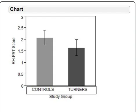
Results:Turner Syndrome girls had lower height, higher HDL and higher waist:height ratio than controls. PAT-hyperemia ratio (RH-PAT) scores were lower in Turner Syndrome (1.64 ± 0.34 vs. 2.08 ± 0.32, p = 0.002) indicating impaired EF. Among Turner Syndrome, RH-PAT did not vary with estrogen therapy or with karyotype 45,XO compared with other karyotypes. However, endothelial function was better in treated compared with GH-untreated Turner Syndrome (1.80 ± 0.36 vs. 1.4 + 0.22, p = 0.02) although there were no differences in HOMA-IR, adiponectin or IGF-1.
Conclusion:Girls with Turner Syndrome exhibit impaired endothelial function compared with controls, which may explain higher risk for vascular disease. GH may protect endothelial function in Turner Syndrome.
Keywords:Turner syndrome, Endothelial function, Adolescents, Pediatrics
Introduction
Turner Syndrome, a common genetic disorder affecting 1 in 2500 live-born females, is caused by complete or partial loss of × chromosome [1]. Despite significant advances in diagnosis and treatment in pediatric settings, Turner Syndrome patients experience high rates of cardi-ovascular disease such that adult females with Turner Syndrome have 3.47 and 2.21 standardized mortality ratios of coronary and cerebrovascular death respectively [2]. The majority of this excess mortality risk encom-passes non-congenital circulatory disease [3]. However, this relates to underlying congenital structural and
functional arterial abnormalities, which predispose these patients to aortic dilatation and aneurysm [4]. Addition-ally, Turner Syndrome patients exhibit a clustering of acquired cardiovascular disease risk factors, including increased rates of hypertension, glucose intolerance, obe-sity and dyslipidemia, which are also impacted by treat-ment regimens including growth hormone (GH) and estrogen [1].
Adolescents and young adults rarely experience cardio-vascular events, and surrogate markers of cardiocardio-vascular disease are needed to evaluate asymptomatic patients. Impaired endothelial function is an initial step in the development of atherosclerosis, and represents an early and reversible step in the vascular disease process [5,6]. The measurement of endothelial function can be used as a surrogate marker to assess cardiovascular risk [7-10].
* Correspondence: clodagh.ogorman@ul.ie
1
Divisions of Endocrinology, The Hospital for Sick Children Research Institute, University of Toronto, Toronto, Canada
Full list of author information is available at the end of the article
In prior clinical studies, peripheral arterial tonometry (PAT) testing has been used to measure endothelial func-tion in both high risk adult [11] and pediatric populafunc-tions [12,13]. Assessment of endothelial function has not pre-viously been reported in pediatric Turner Syndrome populations and it is unclear whether Turner Syndrome is an independent risk factor for endothelial damage or if acquired cardiovascular risk factors impact endothelial function in this high risk patient group [14,15].
The objectives of this study were to evaluate endothelial function, as a marker of early vascular disease, in a group of pediatric Turner Syndrome girls in relation to an age-, sex- and body mass index (BMI)-matched healthy control population, and to assess the impact of disease-related treatment factors and acquired cardiovascular risk factors on endothelial function in the Turner Syndrome study group.
Materials and methods
Institutional ethical approval was obtained for this case-controlled cross-sectional study. Inclusion criteria for Turner Syndrome subjects included: karyotype-confirmed diagnosis of Turner Syndrome; patients followed in our endocrinology clinic; and aged 10-18 years. Individuals receiving human growth hormone (GH), L-thyroxine or estrogen, or with a history of congenital heart disease, were eligible for inclusion. All patients taking L-thyroxine were on stable therapy with thyroid function within the normal range. Subjects took their medications as usual on the day of the study. Controls were healthy girls, age- and BMI-matched to TS subjects and recruited as part of pre-vious studies using the same testing protocol. Exclusion criteria included: inability of the family and/or patient to comply with study protocol; previous diagnosis of Type 1 or Type 2 Diabetes Mellitus; taking medications for the treatment of disorders of glucose, insulin or lipid metabolism.
Each subject attended the Hospital for Sick Children for full evaluation on two separate days. On one day, subjects had fasting bloodwork including cholesterol profile (triglycerides, total cholesterol, high and low den-sity lipoprotein cholesterol [HDL and LDL]), adiponec-tin, insulin and glucose. Homeostatic model assessment of insulin resistance (HOMA-IR) was calculated based on fasting insulin and glucose values: [16].
HOMA - IR =Glucosemmol/L×InsulinmU/L/22.5
Brachial arterial blood pressure (BP) (systolic, diastolic, and mean) and heart rate were recorded in the left arm using an automated Dinamap sphygmomanometer (Cri-tikon Dinamap, Minneapolis, MN, USA). Each subject underwent history, examination and anthropometrics by a single examiner. Height, in metres (m), was measured
using a wall-mounted stadiometer; and weight, in kilo-grams (kg), was assessed using calibrated electronic scales. BMI was calculated as kg/m2. Pubertal status was assessed by a single examiner according to the methods of Tanner [17].
On a different day, subjects returned fasting for endothelial function testing. PAT (Itamar Medical, Cae-sarea, Israel) is a non-invasive test which uses pneumatic probes, similar in shape to a thimble, which cover the fin-gertip and apply a uniform pressure field which allows for the measurement of the pulsatile oscillations of the digital vascular bed microcirculation. The PAT-hyperemia ratio (RH-PAT) shows a strong sensitivity and specificity for the detection of coronary endothelial function, which has been shown to predict cardiovascular events in adult patients [7,10]. PAT has been used previously as a surrogate mar-ker of early endothelial function in paediatric populations [13,18] and, in adolescents it has been validated in a repeated measures study [19]. PAT probes are placed on finger II or III of each hand. After a 5 minute equilibration period, a BP cuff is inflated on the study arm 40 mmHg above systolic BP for 5 minutes. The cuff is then deflated and tonometric recording is completed for an additional 5 minutes. The RH-PAT was determined for each patient and each recording was analyzed individually. Lower RH-PAT index scores indicate worse endothelial function and increased risk for atherosclerosis.
Fifteen Turner Syndrome subjects were recruited, and matched for age and BMI with 15 healthy control girls (Table 1). All girls and their parents consented to partici-pate. All subjects completed the protocol. For the girls with TS, karyotypes were as follows: 45,XO N = 6; 45, XO/46,XX mosaicism N = 1; 45,XO/47,XXX mosaicism N = 1; complex translocations N = 3; isochrome q mate-rial N = 2; 45,XO/46,XY mosaicism N = 2. Two subjects had aortic stenosis, including one with coarctaction repair several years previously. One additional subject had a bicuspid aortic valve. No other TS subjects had any known cardiovascular disease. Fourteen TS subjects were taking medications, including: N = 6 estrogen supple-mentation (N = 1 estrogen only; n = 1 estrogen and pro-gesterone; N = 1 estrogen, progesterone and GH*; N = 3 oral contraceptive pill) and N = 7 GH supplementation (N = 4 GH only; N = 1 GH, estrogen and progesterone*; N = 1 GH and migraine prophylaxis; N = 1 GH with Ritalin and concerta) and N = 1 calcium supplementation (* denotes same patient).
Biochemical analyses
Data analyses
All measures are expressed as mean value ± standard deviation (SD). Baseline characteristics were compared with paired t-tests (due to age- and BMI-matching of patients) and McNemar test. Associations between base-line characteristics and RH-PAT were quantified with Pearson Correlation Coefficients. Linear regression was used to determine whether baseline factors were corre-lated with outcome measures. All analyses were con-ducted with SAS, version 9.1 (SAS Institute Inc., Cary, NC).
Results
There were no statistically significant differences between the Turner Syndrome and control groups for age, BP, weight or BMI. Control subjects were signifi-cantly taller than Turner Syndrome subjects. There was no difference in waist circumference between the groups; but, the waist:height ratio was higher in Turner Syndrome than in controls (Table 1). There were no dif-ferences between the groups for fasting glucose, LDL, total cholesterol, or triglycerides. HDL was higher in TS subjects.
The ages of GH-treated [N = 7] and GH-untreated [N = 8] Turner Syndrome girls were not different (12.7 ± 2.0 years versus 14.3 ± 2.4 years, t = -0.79, p = 0.22) and IGF-1 levels were not different between treated and GH-untreated girls (423.8 ± 122.8 versus 336.7 ± 103.6 mcg/L respectively, p = 0.23). Among the GH-treated Turner Syndrome girls, GH treatment duration was 4.6 ± 1.1 years, with prescribed GH doses 0.35 mg/kg/week. There were no statistical differences in HOMA-IR, adiponectin, lipid measurements or blood glucose between GH-treated and GH-untreated Turner Syndrome girls (see Table 2).
RH-PAT scores were significantly lower in Turner Syn-drome than in control subjects (1.64 ± 0.34 versus 2.08 ± 0.32, p = 0.002). Among girls with Turner Syndrome, RH-PAT scores did not vary with estrogen replacement therapy (1.56 ± 0.30 with estrogen replacement [N = 6] versus 1.69 ± 0.37 without estrogen replacement [N = 9], p = 0.64) or between those with 45,XO karyotype [N = 6] and other karyotypes [N = 9] (1.60 ± 0.46 versus 1.67 ± 0.26 respectively, p = 0.56). However, RH-PAT scores were higher in girls receiving GH therapy than in those not receiving GH therapy (1.86 ± 0.28 versus 1.44 + 0.26 respectively, p = 0.045) and in Turner Syndrome girls receiving GH therapy, PAT scores were similar to control girls (1.86 ± 0.28 versus 2.08 ± 0.32 respectively, p = 0.14).
In Turner Syndrome subjects, RH-PAT score did not correlate with any measures of glucose, adiposity or anthropometrics. In control subjects, RH-PAT corre-lated positively with age and systolic BP, and negatively with LDL and HDL.
Discussion
This study demonstrates that Turner Syndrome girls, compared with healthy age-, sex- and BMI-matched controls, have impaired endothelial function, an early, pre-clinical marker of vascular disease. We did not observe any significant correlations with acquired CVD risk factors present in our TS cohort, including lipid profile, BP or markers of insulin resistance.
The increased risk of cardiovascular complications in Turner Syndrome patients is complex and encompasses both congenital and acquired lesions. Recent data from the UK observed that non-congenital circulatory disease accounted for 41% of absolute excess mortality in
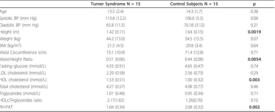
Table 1 Baseline Demographics and results of investigations and PAT compared between girls with Turner Syndrome and Control girls
Turner Syndrome N = 15 Control Subjects N = 15 p
Age 13.5 (2.4) 14.3 (1.7) 0.38
Systolic BP (mm Hg) 113.8 (12.2) 106.6 (5.5) 0.06
Diastolic BP (mm Hg) 65.8 (11.5) 70.18 (3.12) 0.21
Height (m) 1.42 (0.11) 1.64 (0.15) 0.0019
Weight (kg) 44.2 (13.0) 54.5 (15.5) 0.07
BMI (kg/m2) 21.5 (4.5) 20.8 (3.4) 0.64
Waist Circumference (cm) 73.1 (10.9) 71.4 (12.8) 0.71
Waist:Height Ratio 0.51 (0.06) 0.44 (0.08) 0.0054
Fasting glucose (mmol/L) 4.55 (0.91) 4.65 (0.47) 0.74
LDL cholesterol (mmol/L) 2.29 (0.58) 2.56 (0.73) 0.29
HDL cholesterol (mmol/L) 1.53 (0.51) 1.00 (0.32) 0.003
Total cholesterol (mmol/L) 4.27 (0.57) 4.08 (0.77) 0.46
Triglycerides (mmol/L) 1.01 (0.48) 0.95 (0.34) 0.71
HDLc:Triglycerides ratio 2.17(1.82) 1.26(0.70) 0.10
RH-PAT 1.64 (0.34) 2.08 (0.32) 0.002
Turner Syndrome women compared with only 8% for congenital anomalies [3]. Our study describes a young cohort of reasonably healthy Turner Syndrome patients with inherent impaired vascular function, of whom only 3/15 have evidence of some cardiac lesion. The Turner Syndrome patients are compared to BMI-matched healthy controls, because obesity measures do impair endothelial function, as we have previously described in adolescents using the same measure [13]. Previous reports evaluating vascular function in Turner Syn-drome have shown carotid intima media thickening pre-sent in two adult studies, without evidence of abnormal endothelial function (brachial artery reactivity or pulse wave velocity) [20,21]. However, these studies were con-ducted in older Turner Syndrome patients with signifi-cant differences in BP and BMI compared with healthy controls.
We identified higher HDL concentrations in Turner Syndrome subjects compared with controls, and no effect of GH treatment on any measured cholesterol parameter was observed. These results are different with respect to previous studies that have shown higher total cholesterol in TS subjects independent of age, BMI and karyotype [22] and higher total cholesterol but lower HDL in older Turner Syndrome girls compared with controls [23].
Additionally, we identified a tendency towards a decrease in fasting insulin and HOMA-IR with GH ther-apy. Previous reports have been mixed. One study identi-fied increased fasting insulin and glucose in TS girls on GH therapy [24] and another found no difference in
fasting insulin prior to GH therapy in TS girls compared with controls, but found increased fasting insulin following 6-12 months of GH therapy [25]. Compared with controls, in two studies, TS girls on GH therapy had an exaggerated hyperinsulinaemic response to glucose load [24,26]. The suggestion of a protective effect of GH on insulin dynamics and endothelial function in TS girls in this report indicates that confirmatory studies are required, using various modalities to assess endothelial function and glucose-insulin dynamics in TS girls at various ages and stages of puberty.
We did observe that girls with Turner Syndrome who were receiving GH therapy had higher RH-PAT scores than girls with Turner Syndrome who were not receiving GH therapy, suggesting that GH may offer some endothe-lial protection in girls with Turner Syndrome. This was seen in a small number of patients, yet differences in endothelial function were not noted with estrogen replace-ment. Using other measures of endothelial or vascular function, previous studies have identified a protective effect of GH therapy. Flow-mediated dilatation improves in GH deficient adults following 3-18 months of GH ther-apy [27]. GH deficient adolescents had impaired flow-mediated dilatation, and GH induced a larger increase in blood flow in GH treated adolescents than in untreated or healthy controls [28]. Some studies have failed to identify improvements in vascular function following GH therapy, including a recent study of endothelial dysfunction in GH deficient children [29]. Both GH and sex steroids influence vascular function; GH improves cardiac contractility and
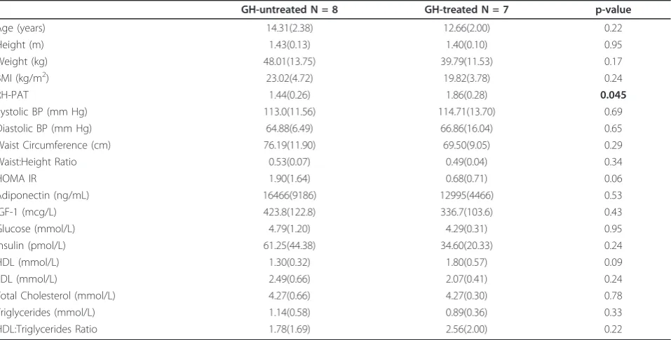
Table 2 Comparison of metabolic parameters between TS subjects GH-treated versus GH-untreated
GH-untreated N = 8 GH-treated N = 7 p-value
Age (years) 14.31(2.38) 12.66(2.00) 0.22
Height (m) 1.43(0.13) 1.40(0.10) 0.95
Weight (kg) 48.01(13.75) 39.79(11.53) 0.17
BMI (kg/m2) 23.02(4.72) 19.82(3.78) 0.24
RH-PAT 1.44(0.26) 1.86(0.28) 0.045
Systolic BP (mm Hg) 113.0(11.56) 114.71(13.70) 0.69
Diastolic BP (mm Hg) 64.88(6.49) 66.86(16.04) 0.65
Waist Circumference (cm) 76.19(11.90) 69.50(9.05) 0.29
Waist:Height Ratio 0.53(0.07) 0.49(0.04) 0.34
HOMA IR 1.90(1.64) 0.68(0.71) 0.06
Adiponectin (ng/mL) 16466(9186) 12995(4466) 0.53
IGF-1 (mcg/L) 423.8(122.8) 336.7(103.6) 0.43
Glucose (mmol/L) 4.79(1.20) 4.29(0.31) 0.95
Insulin (pmol/L) 61.25(44.38) 34.60(20.33) 0.24
HDL (mmol/L) 1.30(0.32) 1.80(0.57) 0.09
LDL (mmol/L) 2.49(0.66) 2.07(0.41) 0.24
Total Cholesterol (mmol/L) 4.27(0.66) 4.27(0.30) 0.78
Triglycerides (mmol/L) 1.14(0.58) 0.89(0.36) 0.33
HDL:Triglycerides Ratio 1.78(1.69) 2.56(2.00) 0.22
vasculature tone [30] while estrogen promotes the release of nitric oxide thus resulting in vasodilation [31]. There are various mechanisms by which GH may improve endothelial function in girls with Turner Syndrome. Firstly, clinical studies suggest that GH may be beneficial in non-GH-deficient (non-Turner Syndrome) individuals with visceral obesity, by reducing abdominal fat and improving insulin sensitivity [32]. Secondly, studies sug-gest that the GH-IGF axis is profoundly altered in Turner Syndrome, specific alterations include a reduction in bioactive IGF-1 [33]. Finally, in studies of animal models of GH deficiency, GH treatment has resulted in increased anti-oxidant capacity and increased resistance to oxidative stressors [34,35].
Physiologically, inherent differences have been described as part of the vasculopathy associated with Turner Syndrome. The precise mechanisms underlying this are likely multi-factorial and related to endothelial injury; alterations in Turner Syndrome patients include abnormi-ties of collagen [36] and impaired release of nitric oxide [37]. Imbalances in matrix metalloproteinases (MMP) activity necessary to maintain normal vessel structure has also been postulated to be present in TS patients [4]. These changes are important in promoting aortic dilation and aneurysm formation; they also likely act in synergy with acquired cardiovascular disease risk factors to pro-mote accelerated atherogenesis in Turner Syndrome.
Several features of this study warrant further comment. This report is limited by the small number of patients stu-died and by the absence of significant correlations between RH-PAT and known traditional cardiovascular risk factors in the Turner Syndrome cohort. We did not observe any significant correlations with acquired CVD risk factors present in our cohort, including abnormal lipid profile, hypertension or insulin resistance. However, our relatively small study size and the lack of wide variation in these variables may account for this finding. We also used his-torical healthy controls who had undergone previous endothelial function testing using the same measure and protocol. While this did allow for matching on the basis of several confounding variables including age, sex and BMI, data relating to pubertal status or markers of insulin resis-tance were not available for comparison in the healthy control group. Nonetheless, given the nature of Turner Syndrome, with requirements for pubertal induction, it is unlikely that groups could be matched simultaneously for age and pubertal status [38]. Finally, we included 1 Turner Syndrome girl with a history of CoArctation repair. But this group of Turner Syndrome girls has not had cardiac MRI performed and, similar to previous studies [39], it is therefore possible that MRI would identify additional girls with structural heart defects. This study confirms that assessment of endothelial function using the PAT techni-que is well-tolerated and suitable for use in high risk,
adolescent pediatric cohorts. However, like other tests of endothelial functionthat serve as surrogate markers of atherosclerosis, additional longitudinal data are required to evaluate any predictive value of these measures for cardiovascular events in adulthood.
Our findings suggest that that Turner Syndrome is an independent risk factor for impaired endothelial func-tion at a young age, but GH therapy may be protective in the Turner Syndrome population. Considering this inherent vasculopathy, it is important to monitor and intervene in these patients to reduce the additional bur-den of acquired cardiovascular disease risk factors (hypertension, glucose intolerance, obesity and dyslipide-mia), which are commonly observed in adolescent and adult Turner Syndrome patients. Regular screening is essential so that sub-clinical treatable conditions can be identified, with the ultimate goal of improving morbidity and mortality.
Conclusions
We identified evidence of impaired endothelial function in pediatric Turner Syndrome subjects, compared with age-and BMI-matched female controls. We suggest that pedia-tricians need to be aware of increased risk of early athero-sclerosis in pediatric age range Turner Syndrome patients, and to intervene with diet and lifestyle modifications and medications as needed. We also suggest that longitudinal studies are required to follow Turner Syndrome girls throughout the age spectrum into adulthood to identify and follow evidence of atherosclerosis.
Abbreviations
PAT: Peripheral Arterial Tonometry; BMI: Body Mass Index; HOMA-IR: Homeostasis Model Assessment - Insulin Resistance; RH-PAT: PAT-hyperemia ratio; HDL: High Density Lipoprotein cholesterol; GH: Growth Hormone; IGF1: Insulin-Like Growth Factor 1; LDL: Low Density Lipoprotein; BP: Blood Pressure; Kg: kilograms; M: metres; CV: coefficient of variation; SD: Standard deviation.
Acknowledgements
This study was supported by a grant from the Adrian and Rita Hudson Foundation. COG was supported by a fellowship from the SickKids Research Institute and by a research award from the Faculty of Medicine, University of Toronto. CS was supported by a fellowship from Heart and Stroke Foundation of Canada. We acknowledge the contribution of our patients and their families in supporting this research.
Disclosures
COG wrote the first draft of this manuscript, and COG was/is not in receipt of payment for writing. This study was supported by a grant from the Adrian and Rita Hudson Foundation. COG was supported by a fellowship from the SickKids Research Institute and by a research award from the Faculty of Medicine, University of Toronto. CS was supported by a fellowship from Heart and Stroke Foundation of Canada.
Author details
1Divisions of Endocrinology, The Hospital for Sick Children Research Institute,
University of Toronto, Toronto, Canada.2Divisions of Cardiology, The Hospital
for Sick Children Research Institute, University of Toronto, Toronto, Canada.
3
Department of Pediatrics, The Hospital for Sick Children Research Institute, University of Toronto, Toronto, Canada.4Physiology and Experimental
Medicine Program, The Hospital for Sick Children Research Institute, University of Toronto, Toronto, Canada.5Department of Paediatrics, Graduate
Entry Medical School, University of Limerick, Limerick, Ireland.
Authors’contributions
CSOG designed the study, collected the data, analysed the data, wrote the manuscript. CS co- collected data and co-analysed data. TB co-designed the study and co-analysed the data. JH participated in study design, data analysis and preparation of the manuscript. FHM conceived the study, designed the study, analysed the data and co-wrote the final manuscript. All authors read and approved the final manuscript.
Competing interests
The authors declare that they have no competing interests.
Received: 29 November 2011 Accepted: 2 April 2012 Published: 2 April 2012
References
1. Hall JG, Gilchrist DM:Turner syndrome and its variants.Pediatr Clin North Am1990,37:1421-1440.
2. Gravholt CH, Juul S, Naeraa RW, Hansen J:Morbidity in turner syndrome.J Clin Epidemiol1998,51:147-158.
3. Schoemaker MJ, Swerdlow AJ, Higgins CD, Wright AF, Jacobs PA:Mortality in women with turner syndrome in Great Britain: a national cohort study.J Clin Endocrinol Metab2008,93:4735-4742.
4. Bondy CA:Aortic dissection in Turner syndrome.Curr Opin Cardiol2008,
23:519-526.
5. Brunner H, Cockcroft JR, Deanfield J, Donald A, Ferrannini E, Halcox J, Kiowski W, Luscher TF, Mancia G, Natali A,et al:Endothelial function and dysfunction. part II: association with cardiovascular risk factors and diseases. a statement by the working group on endothelins and endothelial factors of the european society of hypertension.J Hypertens
2005,23:233-246.
6. Anderson TJ, Uehata A, Gerhard MD, Meredith IT, Knab S, Delagrange D, Lieberman EH, Ganz P, Creager MA, Yeung AC,et al:Close relation of endothelial function in the human coronary and peripheral circulations.
J Am Coll Cardiol1995,26:1235-1241.
7. Bonetti PO, Pumper GM, Higano ST, Holmes DR Jr, Kuvin JT, Lerman A:
Noninvasive identification of patients with early coronary atherosclerosis by assessment of digital reactive hyperemia.J Am Coll Cardiol2004,
44:2137-2141.
8. Bonetti PO, Barsness GW, Keelan PC, Schnell TI, Pumper GM, Kuvin JT, Schnall RP, Holmes DR, Higano ST, Lerman A:Enhanced external counterpulsation improves endothelial function in patients with symptomatic coronary artery disease.J Am Coll Cardiol2003,
41:1761-1768.
9. Kuvin JT, Patel AR, Sliney KA, Pandian NG, Rand WM, Udelson JE, Karas RH:
Peripheral vascular endothelial function testing as a noninvasive indicator of coronary artery disease.J Am Coll Cardiol2001,38:1843-1849. 10. Kuvin JT, Patel AR, Sliney KA, Pandian NG, Sheffy J, Schnall RP, Karas RH,
Udelson JE:Assessment of peripheral vascular endothelial function with finger arterial pulse wave amplitude.Am Heart J2003,146:168-174. 11. Hamburg NM, Keyes MJ, Larson MG, Vasan RS, Schnabel R, Pryde MM,
Mitchell GF, Sheffy J, Vita JA, Benjamin EJ:Cross-sectional relations of digital vascular function to cardiovascular risk factors in the Framingham Heart Study.Circulation2008,117:2467-2474.
12. Mahmud FH, Hill DJ, Cuerden MS, Clarson CL:Impaired vascular function in obese adolescents with insulin resistance.J Pediatr2009,155:678-682. 13. Mahmud FH, Van Uum S, Kanji N, Thiessen-Philbrook H, Clarson CL:
Impaired endothelial function in adolescents with type 1 diabetes mellitus.J Pediatr2008,152:557-562.
14. Jarvisalo MJ, Putto-Laurila A, Jartti L, Lehtimaki T, Solakivi T, Ronnemaa T, Raitakari OT:Carotid artery intima-media thickness in children with type 1 diabetes.Diabetes2002,51:493-498.
15. Aggoun Y, Tounian P, Dabbas-Tyan M, Massih TA, Girardet JP, Ricour C, Sidi D, Bonnet D:Arterial rigidity and endothelial dysfunction in obese children.Arch Mal Coeur Vaiss2002,95:631-635.
16. Matthews DR, Hosker JP, Rudenski AS, Naylor BA, Treacher DF, Turner RC:
Homeostasis model assessment: insulin resistance and beta-cell function from fasting plasma glucose and insulin concentrations in man.
Diabetologia1985,28:412-419.
17. Marshall WA, Tanner JM:Variations in pattern of pubertal changes in girls.Arch Dis Child1969,44:291-303.
18. Haller MJ, Stein J, Shuster J, Theriaque D, Silverstein J, Schatz DA, Earing MG, Lerman A, Mahmud FH:Peripheral artery tonometry demonstrates altered endothelial function in children with type 1 diabetes.Pediatr Diabetes2007,8:193-198.
19. Selamet Tierney ES, Newburger JW, Gauvreau K, Geva J, Coogan E, Colan SD, de Ferranti SD:Endothelial pulse amplitude testing: feasibility and reproducibility in adolescents.J Pediatr2009,154:901-905. 20. Ostberg JE, Donald AE, Halcox JP, Storry C, McCarthy C, Conway GS:
Vasculopathy in Turner syndrome: arterial dilatation and intimal thickening without endothelial dysfunction.J Clin Endocrinol Metab2005,
90:5161-5166.
21. Baguet JP, Douchin S, Pierre H, Rossignol AM, Bost M, Mallion JM:
Structural and functional abnormalities of large arteries in the Turner syndrome.Heart2005,91:1442-1446.
22. Ross JL, Feuillan P, Long LM, Kowal K, Kushner H, Cutler GB Jr:Lipid abnormalities in Turner syndrome.J Pediatr1995,126:242-245. 23. Van PL, Bakalov VK, Bondy CA:Monosomy for the X-chromosome is
associated with an atherogenic lipid profile.J Clin Endocrinol Metab2006,
91:2867-2870.
24. Sas TC, de Muinck Keizer-Schrama SM, Stijnen T, Aanstoot HJ, Drop SL:
Carbohydrate metabolism during long-term growth hormone (GH) treatment and after discontinuation of GH treatment in girls with Turner syndrome participating in a randomized dose-response study. Dutch advisory group on growth hormone.J Clin Endocrinol Metab2000,
85:769-775.
25. Caprio S, Boulware SD, Press M, Sherwin RS, Rubin K, Carpenter TO, Plewe G, Tamborlane WV:Effect of growth hormone treatment on hyperinsulinemia associated with Turner syndrome.J Pediatr1992,
120:238-243.
26. Radetti G, Pasquino B, Gottardi E, Boscolo Contadin I, Aimaretti G, Rigon F:
Insulin sensitivity in Turner’s syndrome: influence of GH treatment.Eur J Endocrinol2004,151:351-354.
27. Pfeifer M, Verhovec R, Zizek B, Prezelj J, Poredos P, Clayton RN:Growth hormone (GH) treatment reverses early atherosclerotic changes in GH-deficient adults.J Clin Endocrinol Metab1999,84:453-457.
adolescents: apparent effects of growth hormone treatment on these parameters.J Clin Endocrinol Metab2005,90:3978-3982.
29. Hoffman RP:Growth hormone (GH) treatment does not restore endothelial function in children with GH deficiency.J Pediatr Endocrinol Metab2008,21:323-328.
30. Capalbo D, Lo Vecchio A, Farina V, Spinelli L, Palladino A, Tiano C, Lettiero T, Lombardi G, Colao A, Salerno M:Subtle alterations of cardiac performance in children with growth hormone deficiency: results of a two-year prospective, case-control study.J Clin Endocrinol Metab2009,
94:3347-3355.
31. Li XP, Zhou Y, Zhao SP, Gao M, Zhou QC, Li YS:Effect of endogenous estrogen on endothelial function in women with coronary heart disease and its mechanism.Clin Chim Acta2004,339:183-188.
32. Attallah H, Friedlander AL, Hoffman AR:Visceral obesity, impaired glucose tolerance, metabolic syndrome, and growth hormone therapy.Growth Horm IGF Res2006,16 Suppl A:S62-67.
33. Gravholt CH, Chen JW, Oxvig C, Overgaard MT, Christiansen JS, Frystyk J, Flyvbjerg A:The GH-IGF-IGFBP axis is changed in Turner syndrome: partial normalization by HRT.Growth Horm IGF Res2006,16:332-339. 34. Ungvari Z, Gautam T, Koncz P, Henthorn JC, Pinto JT, Ballabh P, Yan H,
Mitschelen M, Farley J, Sonntag WE, Csiszar A:Vasoprotective effects of life span-extending peripubertal GH replacement in Lewis dwarf rats.J Gerontol A Biol Sci Med Sci2010,65:1145-1156.
35. Ungvari Z, Sosnowska D, Podlutsky A, Koncz P, Sonntag WE, Csiszar A:Free radical production, antioxidant capacity, and oxidative stress response signatures in fibroblasts from Lewis dwarf rats: effects of life span-extending peripubertal GH treatment.J Gerontol A Biol Sci Med Sci2011,
66:501-510.
36. Gravholt CH, Landin-Wilhelmsen K, Stochholm K, Hjerrild BE, Ledet T, Djurhuus CB, Sylven L, Baandrup U, Kristensen BO, Christiansen JS:Clinical and epidemiological description of aortic dissection in Turner’s syndrome.Cardiol Young2006,16:430-436.
37. Brandenburg H, Steegers EA, Gittenberger-de Groot AC:Potential involvement of vascular endothelial growth factor in pathophysiology of Turner syndrome.Med Hypotheses2005,65:300-304.
38. Caprio S, Boulware S, Diamond M, Sherwin RS, Carpenter TO, Rubin K, Amiel S, Press M, Tamborlane WV:Insulin resistance: an early metabolic defect of Turner’s syndrome.J Clin Endocrinol Metab1991,72:832-836. 39. Ho VB, Bakalov VK, Cooley M, Van PL, Hood MN, Burklow TR, Bondy CA:
Major vascular anomalies in Turner syndrome: prevalence and magnetic resonance angiographic features.Circulation2004,110:1694-1700. doi:10.1186/1687-9856-2012-5
Cite this article as:O’Gormanet al.:Impaired endothelial function in
pediatric patients with Turner Syndrome and healthy controls: a
case-control study.International Journal of Pediatric Endocrinology20122012:5.
Submit your next manuscript to BioMed Central and take full advantage of:
• Convenient online submission
• Thorough peer review
• No space constraints or color figure charges
• Immediate publication on acceptance
• Inclusion in PubMed, CAS, Scopus and Google Scholar
• Research which is freely available for redistribution