BioMedCentral
Cardiovascular Ultrasound
Open Access
Case report
Atypical vessels as an early sign of intracardiac myxoma?
Hans-Peter Dübel
1, Fabian Knebel*
1, Volker Gliech
1, Wolfgang Konertz
2,
Wolfgang Rutsch
1, Gert Baumann
1and Adrian Constantin Borges
1Address: 1Medical Clinic for Cardiology, Angiology, Pulmology, Charité Campus Mitte, University medicine Berlin, Germany and 2Director of the
Clinic of Cardiovascular Surgery, Charité Campus Mitte, University Medicine Berlin, Germany
Email: Hans-Peter Dübel - hans-peter.duebel@charite.de; Fabian Knebel* - fabian.knebel@charite.de; Volker Gliech - volker.gliech@charite.de; Wolfgang Konertz - wolfgang.konertz@charite.de; Wolfgang Rutsch - wolfgang.rutsch@charite.de; Gert Baumann - gert.baumann@charite.de; Adrian Constantin Borges - adrian.borges@charite.de
* Corresponding author
MyxomaCoronary Artery Disease
Abstract
We report on a woman with previously unknown left atrial myxoma, who underwent percutaneous coronary intervention. 45 months after the initial coronary angiography, echocardiography demonstrated a large atrial myxoma, which was not seen echocardiographically before. The retrospective analysis of the pre-intervention coronary angiography revealed atypical vessels in the atrial septum, which are interpreted as early signs of myxoma.
Background
Primary cardiac tumors are rare disorders, with an inci-dence of 0.02% at autopsy. Three quarters of the primary tumors of the heart are benign, half of which are myxomas [1].
As noninvasive cardiac imaging becomes widely available, with increasing resolution provided by echocardiography, computed tomography and magnetic resonance imaging, cardiac tumors are being diagnosed more often. Angiogra-phy, apart from its preoperative role to rule out concomi-tant coronary artery disease, is rarely needed for the diagnostic work-up of cardiac tumors.
This report describes the delayed presentation of a left atrial myxoma which was not depicted in an initial coro-nary angiography performed 51 months earlier in a woman with chest pain.
Case Report
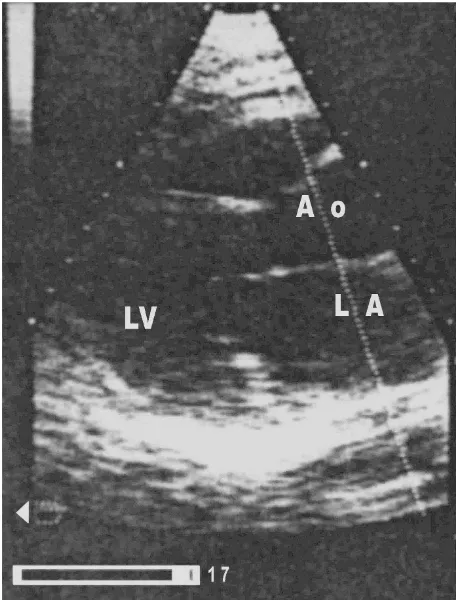
A 62-year-old woman with known metabolic syndrome was referred to our clinic to exclude coronary artery dis-ease invasively. She has been experiencing chest pain for four months which has not increased in frequency or duration since it started. She denied pain at rest, nocturnal pain, difficulty breathing, or palpitations. An echocardio-graphic stress examination revealed significant ischemia with anteroseptal hypokinesia. Cardiac chambers were morphologically normal. The "baseline" echocardiogra-phy and stress echo was unsuspicious of a left atrial myxoma (Fig. 1).
Coronary angiography revealed coronary artery disease with a stenosis of the proximal left anterior descending coronary artery (LAD) near the left main stem. A stent was implanted in the proximal LAD with no residual stenosis. The ventricle was morphologically normal.
Published: 13 August 2004
Cardiovascular Ultrasound 2004, 2:13 doi:10.1186/1476-7120-2-13
Received: 19 July 2004 Accepted: 13 August 2004
This article is available from: http://www.cardiovascularultrasound.com/content/2/1/13
© 2004 Dübel et al; licensee BioMed Central Ltd.
Cardiovascular Ultrasound 2004, 2:13 http://www.cardiovascularultrasound.com/content/2/1/13
Stress echocardiography: long axis view (baseline)
Figure 1
Cardiovascular Ultrasound 2004, 2:13 http://www.cardiovascularultrasound.com/content/2/1/13
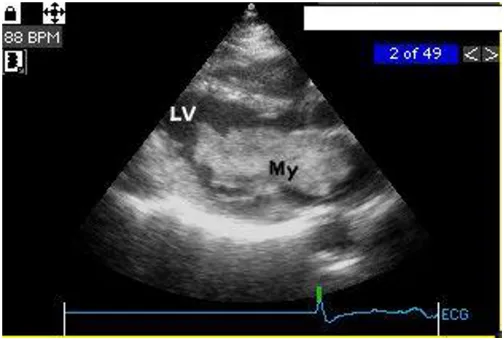
A coronary angiography performed 13 months after stent implantation showed no re-stenosis and a normal left ventricle. No echocardiogram was performed at this time 32 months after the control coronary angiography, the patient was readmitted because of increasing dyspnoea and palpitations. A transthoracic echocardiography dis-closed a big (70 × 30 mm) mass in the left atrium attached to the interatrial septum. The tumor prolapsed into the left ventricle obstructing the mitral valve orifice (Fig 2). The mean pressure gradient across the mitral valve was 8 mm Hg.
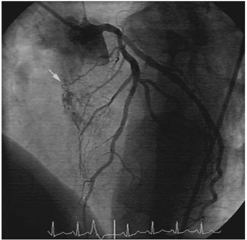
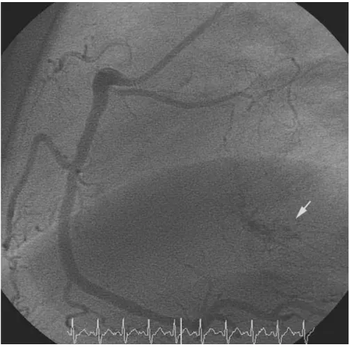
A subsequent coronary angiography and LV and RV cath-eterization detected a mean diastolic pressure gradient of 12 mm Hg between the pulmonary capillary wedge and the left ventricular enddiastolic pressure, and no re-steno-sis of the LAD stent. The angiography was notable for a large area with small atypical, tortuous vessels in the region of the interatrial septum. These vessels were shown
to originate from branches of the right coronary artery (RCA) and the circumflex coronary artery (RCX). (Fig. 5)
Surgery was promptly performed and the tumor was suc-cessfully excised. Histology confirmed the diagnosis of a cardiac myxoma.
A retrospective analysis of the initial coronary angiogra-phies (baseline, 13 and 45 months) disclosed the atypical vessels in a small area of interatrial septum (Fig. 3, 4, 5).
Discussion
Myxomas are benign but potentially dangerous with dis-turbance of rhythm, peripheral embolization or mechan-ical valvular obstruction, of the atrial or ventricular cavity [2,3].
The site, mobility, and size of the myxoma determine the clinical course. Some authors found no correlation
Echocardiography (45 months) long axis view
Figure 2
Cardiovascular Ultrasound 2004, 2:13 http://www.cardiovascularultrasound.com/content/2/1/13
between the size of the tumor and the clinical picture [4], others reported symptoms with left atrial myxomas weighting more than 70 g [5]. The rate of growth of myxo-mas is not exactly known [5,6]. An increase in size of 1.8 – 5.8 cm/year and in weight of up to 14 g/year was
reported [7,8]. The myxoma of our patient reached 70 × 30 mm before being symptomatic.
Myxomas presenting with systemic embolism or intracav-itary obstruction can be easily detected non-invasively. The early depiction of small intracardiac tumors by means
Right coronary artery (baseline) in 90 degree LAO projection
Figure 3
Cardiovascular Ultrasound 2004, 2:13 http://www.cardiovascularultrasound.com/content/2/1/13
of angiography relies on the detection of atypical vessels supplied by branches of the left or right coronary artery. Our case demonstrates that this early angiographic sign is difficult to find. However, vascular malformations are not pathognomonic for myxomas [9] and stromal tumors, such as myxomas, have generally poor blood supply,
hence a coronary angiographic finding with a neo-vascu-larized or highly vascuneo-vascu-larized intracardiac area is more suggestive for other type of cardiac neoplasm. A cluster of small, tortuous and dilated vessels is also seen in old and organised thrombi, haemangiomas and venous
malfoma-Left coronary artery (13 months) in 45 degree LAO and 30 degree CRAN projection
Figure 4
Cardiovascular Ultrasound 2004, 2:13 http://www.cardiovascularultrasound.com/content/2/1/13
tions [10]. The size of malformation has no relation to the size of the tumor [11].
It has been reported in the literature that myxomas can be induced by radiation [12,13]. Between the first
presenta-tion to our clinic and the diagnosis of myxoma, the patient underwent two coronary angiograms including stenting, with a cumulative radiological exposure of about 30 mSv (corresponding to at least 1500 chest X-rays). To
Right coronary artery (RCA) in 60 degree LAO position (45 months, pre-operative coronary angiography)
Figure 5
Publish with BioMed Central and every scientist can read your work free of charge "BioMed Central will be the most significant development for disseminating the results of biomedical researc h in our lifetime."
Sir Paul Nurse, Cancer Research UK
Your research papers will be:
available free of charge to the entire biomedical community
peer reviewed and published immediately upon acceptance
cited in PubMed and archived on PubMed Central
yours — you keep the copyright
Submit your manuscript here:
http://www.biomedcentral.com/info/publishing_adv.asp
BioMedcentral
Cardiovascular Ultrasound 2004, 2:13 http://www.cardiovascularultrasound.com/content/2/1/13
our knowledge, the patient did not undergo any other rel-evant radiation exposure in the past.
In the present case, a retrospective analysis of the patient angiographies disclosed the atypical vessels which were initially overseen. These vessels could have probably been interpreted as an early sign of myxoma.
Author's contributions
HPD and FK have written the manuscript and have equally contributed to this publication. HPD, VG and WR have performed the coronary angiographies. WK has per-formed cardiac surgery. HPD, ACB, FK and GB partici-pated in the design and coordination of the final manuscript. All authors have read and approved the final manuscript.
References
1. Reynen K: Frequency of primary tumors of the heart.Am J Cardiol 1996, 77:107.
2. Colucci WS, Schoen FJ, Braunwald E: Primary tumors of the heart. In: Heart Disease. A Textbook of Cardiovascular Medicine 5th edi-tion. Edited by: Braunwald E. Philadelphia: WB Saunders; 1997:1464-1477.
3. Shapiro LM: Cardiac tumours: diagnosis and management. Heart 2001, 85:218-222.
4. Vassiliadis N, Vassiliadis K, Karkavelas G: Sudden death due to cardiac myxoma.Med Sci Law 1997, 37:76-78.
5. Roberts WC: Primary and secondary neoplasms of the heart. Am J Cardiol 1997, 80:671-682.
6. Reynen K: Benigne Tumoren des Herzens [Benign tumors of the heart].Z Kardiol 1993, 82:749-762.
7. Malekzadeh S, Roberts WC: Growth rate of left atrial myxoma. Am J Cardiol 1989, 64:1075-1076.
8. Pochis WT, Wingo MW, Cinquegrani MP, Sagar KB: Echocardio-graphic demonstration of rapid growth of a left atrial myxoma.Am Heart J 1991, 122:1781-1784.
9. Van Cleemput J, Daenen W, De Geest H: Coronary angiography in cardiac myxoma: findings in 19 consecutive cases and review of literature.Cathet Cardiovasc Diagn 1993, 29:217-220. 10. Fueredi GA, Knechtges TE, Czarnecki DJ: Coronary angiography
in atrial myxoma: Findings in nine cases.AJR Am J Roentgenol 1989, 152:737-738.
11. Chow WH, Chow TC, Tai YT, Yip ASB, Cheung KL: Angiographic visualization of „tumor vascularity" in atrial myxoma. Eur Heart J 1991, 12:79-82.
12. Daoud J, Ben Salah H, Kammoun W, Ghorbel A, Frikha M, Jlidi R, Bes-bes M, Drira MM, Maalej M: Radiation-induced glioblastoma and myxoma after treatment for undifferentiated carcinoma of the naspharynx.Cancer Radiother 2000, 6:469-72.