RESEARCH
Multi-modal liquid biopsy platform
for cancer screening: screening
both cancer-associated rare cells and cancer
cell-derived vesicles on the fabric filters
for a reliable liquid biopsy analysis
Jiyoon Bu
1,2†, Jae‑Eul Shim
1†, Tae Hee Lee
1,3†and Young‑Ho Cho
1*Abstract
Circulating tumor cells (CTCs) are receiving a great amount of scientific interest as a diagnostic biomarker for various types of cancer. Despite the recent progress in the development of highly sensitive CTC isolation devices, post‑ capture analysis of CTCs is still hindered by technical challenges associated with their rarity. Herein, we present a multi‑modal CTC screening platform which is capable to analyze CTCs and CTC‑derived extracellular vesicles (EVs), simultaneously from a single sample. Cytochalasin B (CB) treatment promotes cells to release large number of EVs from their surface, as demonstrated by CB‑treated cells (5 µg/mL for 3 h) secreting 3.5‑fold more EVs, compared to the non‑treated cells. CB further generates 1.7‑fold more EVs from the cells captured on our CTC filtration device (the fabric filter), compared to those from the cell culture flasks, owing to its multiple pore structure design which reduces the non‑specific binding of EVs. Both CB‑treated cancer cells and CB‑induced EVs are found to overexpress tumor‑ associated markers, demonstrating a potential for the development of CTC dual‑screening platform. Collectively, the results presented in this study reveal that our multi‑modal cancer screening platform can synergistically improve the reliability and efficacy of the current CTC analysis systems.
Keywords: Circulating tumor cells, Extracellular vesicles, Cytochalasin B, Polyester fabric, Liquid biopsy, Multi‑modal screening
© The Author(s) 2019. This article is distributed under the terms of the Creative Commons Attribution 4.0 International License (http://creat iveco mmons .org/licen ses/by/4.0/), which permits unrestricted use, distribution, and reproduction in any medium, provided you give appropriate credit to the original author(s) and the source, provide a link to the Creative Commons license, and indicate if changes were made.
1 Introduction
Image-guided tissue biopsy is utilized as the standard diagnostic test for cancer [1]. This traditional biopsy technique has facilitated histological and molecular anal-ysis of tumors, improving the clinical outcomes [2]. How-ever, the results obtained from these biopsy tests often show inconsistent benefits since tumors grow, mutate, and become heterogeneous [2, 3]. Alternatively, liquid
biopsy has been highlighted as an innovative tool for cancer research, which allows non-invasive, repetitive, and routine monitoring for various types of tumor. Liq-uid biopsy refers to all kinds of procedures that detect or quantitatively measure disease-related biomarkers from a human body fluid, mainly from blood [4]. Unlike tra-ditional tissue biopsy techniques, liquid biopsy enables real-time monitoring of the abnormal tissues, providing more detailed information of ongoing tumor progression and therapeutic responses.
Different biomarkers have been employed for the diag-nosis and progdiag-nosis of tumor. Tumor-associated antigens are one of the most well-established biomarkers that are already in clinical use [5]. Carcinoembryonic antigens
Open Access
*Correspondence: [email protected]
†Jiyoon Bu, Jae‑Eul Shim and Tae Hee Lee contributed equally to this work 1 Cell Bench Research Center, Korea Advanced Institute of Science and Technology (KAIST), 291 Daehak‑ro, Yuseong‑gu, Daejeon 34141, Republic of Korea
(CEA), cancer antigen 125 (CA-125), and prostate-spe-cific antigen (PSA) are quantitatively assessed for the detection and screening of colorectal [6], ovarian [7], and prostate tumors [8], respectively. However, these tumor-associated antigens have shown low specificity for differ-entiating the cancer patients from healthy individuals [9]. Hence, the liquid biopsy platforms based on these tumor antigens may result in false negative diagnoses [10].
Circulating tumor cells (CTCs) and exosomes have emerged as potential liquid biopsy biomarkers for deter-mining the histological features, aggressiveness and metastatic potential of the tumor [11, 12]. CTCs are cells that have detached from the primary tumor and circulate through the blood stream [13]. A myriad of technologies have been developed to enrich CTCs from human blood samples, including the method utilizing cancer-targeting capture agents (antibody/peptide-based isolation) and the method employing differences in physical properties between CTCs and other blood components (label-free isolation) [14]. Despite vigorous efforts in developing more sensitive and effective CTC isolation platforms, analyzing tumor markers from CTCs is still challenging due to their low abundance in the blood [15].
In contrast, exosomes, the endosomal-derived vesicles, have obtained great attentions due to their abundance in the body fluid and high stability under varying con-ditions [16, 17]. Exosomes have been known to involve in cell-to-cell communication that their role in can-cer development, progression, and metastasis has been investigated extensively [18, 19]. One of the biggest chal-lenges that needs to be addressed for using exosomes as a tumor biomarker is to minimize their loss during a series of purification processes [17]. Multiple purifica-tion steps are required to enrich exosomes from a large spectrum of cellular debris, which eventually decreases the overall yield of exosomes. Another challenge for the exosome-based cancer screening platforms is that these vesicles are not only secreted from the cancerous cells, but also released from most of the mammalian cells [20]. Thus, separating cancer-associated exosomes from the exosomes of non-cancerous origin requires additional processing beyond their enrichment.
Herein, we propose a novel cancer screening method which could detect the tumor-associated expressions in duplicates, by assessing both CTCs and CTC-derived EVs from a single sample. Our team previously developed a highly-sensitive, viable CTC filtration device made of monofilament polyester, which we named fabric filter [21–23]. Cancer cells captured on the fabric filters are subsequently treated with cytochalasin B (CB), in order to release large number of EVs from the cell surface. Pre-cisely controlling the concentration and treatment time of CB enables cells to secrete large number of vesicles
from their surface, without affecting the expression lev-els of cancerous proteins. Multi-modal analysis of CTCs and CTC-derived EVs could provide a potential to over-come the limitations of both CTC and exosome-based liquid biopsy platforms, which suffer from low sensitivity and specificity, respectively. The results presented in this paper provide an idea for the development of a novel can-cer screening platform which could eventually enhance the detection sensitivity and accuracy of the current liq-uid biopsy systems.
2 Materials and methods
2.1 Cell preparation and cytochalasin B treatment
Human breast cancer cell line, MCF-7, was treated with cytochalasin B (CB, Tocris Biosciences, MO) at varying concentration of 0, 5, 10, 15, and 20 μg/mL and for vary-ing incubation time of 0.5, 3, and 24 h, respectively. Cell viability was measured using two-colored live/dead cell viability assay kit (Life Technology, CA) and AlamarBlue (Thermo Scientific, IL) as described elsewhere [24, 25]. See Additional file 1 for additional details.
2.2 Fabric sheets preparation
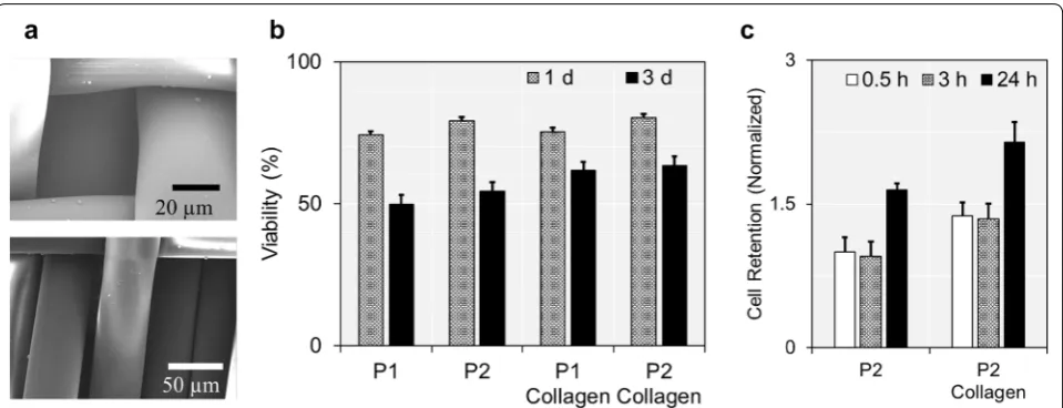
Two types of fabric filters were prepared in this study: the fabric filter prototype 1 (P1) and prototype 2 (P2), which were designed by 2/2 and 3/1 twill patterns, respectively, using 20-denier polyester monofilaments [21, 22]. The details for the filter design and manufacturing process could be found in our previous publications [21, 22]. Briefly, the difference in twill structures affected the size of a slot between the neighboring wefts, as P2 showing larger slot width than P1 (~ 12 µm vs. ~ 8 µm) [21, 22]. Throughout the previous studies, we demonstrated that both fabric filters are capable to capture viable CTCs [21–23]. The structure of each prototype was once more confirmed using SEM. Details could be found in Addi-tional file 1. In this study, the fabric filters were further functionalized with collagen type 1 (3.44 mg/mL), to improve the biocompatibility. Enhancement in biocom-patibility was validated by quantitatively comparing the viability and adhesive properties of the cancer cells after culturing them on either collagen-coated or non-modi-fied fabric filters.
2.3 Imaging and characterization of extracellular vesicles
quantified using BCA assay (Thermo Scientific) and SEM imaging, respectively. See Additional file 1 for additional details.
2.4 Immunofluorescence and immunocytochemistry analysis
We compared the expressions of two cancer-related markers, epidermal growth factor receptor (EGFR) or/ and epithelial cell adhesion molecule (EpCAM), based on immunocytochemistry (ICC) and immunofluores-cence (IF) assays. ICC was performed directly to the CB-treated cells loaded on the fabric filters using the antibody against human EGFR (R&D Systems, MN), as described previously [25]. IF was conducted after retrieving the captured cancer cells from the fabric fil-ters and immobilizing them on the microscope slides. Cells were then stained with both rhodamine-conju-gated EGFR and FITC-conjurhodamine-conju-gated EpCAM (R&D Sys-tems). See Additional file 1 for additional details.
2.5 Quantitative real‑time polymerase chain reaction
EVs were lysed and cancer-associated genes were identi-fied using quantitative real-time polymerase chain reac-tion (qRT-PCR), as described in elsewhere [26]. Primer sequences of EGFR (Hs01076078_m1) and EpCAM (Hs00158980_m1) were purchased from Thermo Fisher Scientifics.
3 Results and discussion
3.1 The effect of cytochalasin B on cell viability and vesicle secretion
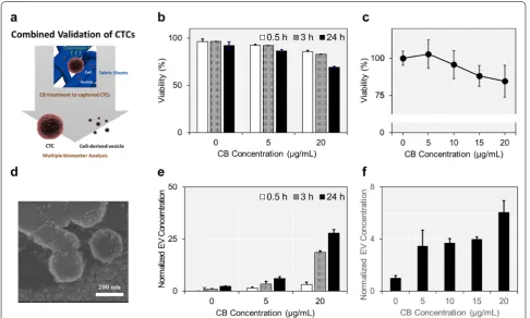
Figure 1a illustrates the overall experimental design of the present study. We first determined the optimal CB concentration and treatment time which could promote cells to release large number of vesicles, while having minimal cytotoxic effect. CB has been well-established as a metabolite which could promote cells to release large number of EVs from their surface by involving in cytoskeleton-membrane interaction [27]. These EVs have been exploited in a wide range of biomedical applications, as they are known to reflect the biological characteristics
Fig. 1 The effect of CB on cell viability and EV secretion: a precisely controlling the CB concentration and treatment time enabled multi‑modal screening of CTCs. CB treatment promoted cancer cells captured on the fabric filters to secrete large number of EVs with minimal cytotoxic effect. b
of the parental cells that they have been originated from [28]. For example, CB-induced EVs have been utilized as a receptor for olfactory sensors and a nano carrier for drug delivery systems [29, 30]. However, when CB con-centration exceeded certain limit, it strongly inhibited actin polymerization and eventually promoted cell death [31]. Rengan et al. treated CB on lymphocytic cells and verified that CB had remarkable cytotoxic effect when its concentration increased from 1 to 10 μg/mL [32]. It is therefore important to determine the optimal CB con-centration and treatment time by balancing its effects on cell viability and vesicle secretion, in order to apply this platform in multi-modal liquid biopsy system based on cell-EV dual screening.
The cells incubated on conventional cell culture flasks were treated with CB at different concentrations and for different time intervals. Cytotoxic effect of CB was inves-tigated using two-colored live/dead staining assay, as demonstrated in Fig. 1b. Cell viability decreased below 90% when the CB concentration exceeded 5 μg/mL or treated over 3 h. Cell viability was assessed more spe-cifically using Alamar blue viability assay after 3 h of CB treatment (Fig. 1c). There were no significant differences in cell viabilities between the cells that have been exposed to CB at the concentration ranging from 0 to 10 μg/mL. However, cell viability reduction was significant when CB concentration exceeded 10 μg/mL, as demonstrated by cells showing 88.1 ± 7.0% (p = 0.031) and 84.6 ± 10.8% (p = 0.057) viabilities at the CB concentration of 15 and 20 μg/mL, respectively. We also found that the cyto-toxicity of CB increased 8% when the treatment time increased from 3 to 6 h (Additional file 1: Figure S1). All together, these results indicated that the cytotoxic effect of CB was not significant at the concentration ≤ 10 μg/ mL and the treatment time ≤ 3 h.
On the other hand, the number of EVs released from the cells increased exponentially as either CB concentra-tion or treatment time increased (Fig. 1d, e). CB-treated cells secreted 2.5-fold (CB concentration of 5 μg/mL; p = 0.001) and 11.5-fold (CB concentration of 20 μg/mL; p = 0.001) more EVs from the cell surface compared to the non-treated cells after 24 h cell incubation. Notably, there was no significant difference between the size of CB-induced EVs and naturally secreted vesicles (Addi-tional file 1: Figure S2). The effect of CB on EV secretion was analyzed in more depth by fixing its treatment time to 3 h (Fig. 1f). CB increased EV secretion by 3.5-fold at the concentration of 5 μg/mL and there was no further significant increment in EV secretion until the concen-tration reached 20 μg/mL. Alternatively, we also quanti-fied the amount of vesicular proteins after CB treatment (Additional file 1: Figure S3). After 3 h-CB treatment, the amount of vesicular proteins increased by 2.0-fold,
compared to that obtained from the first 0.5 h treatment (p = 0.004). The amount of vesicular proteins increased as the treatment time further increased to 24 h, but the difference was not prominent compared to its initial 3 h-treatment. Based on these findings, we decided to treat CB at the concentration of 5 μg/mL for 3 h, which could produce 3.4-fold more EVs compared to naturally secreted vesicles while having non-significant cytotoxic effect.
3.2 Collagen‑coating enhanced biocompatibility of the fabric filters
Prior to applying CB treatment on the captured cancer cells, we determined the best configuration among our CTC capture devices, which could minimize the damage on cells after their capture. Note that the cells should be secured properly for the post-capture CTC analysis, since cell viability could influence expression levels of pro-teins or genes. The viability of cancer cells was measured after isolating them with different types of CTC capture devices, the fabric filter P1 and P2 (Fig. 2a). In our previ-ous studies, we have reported that both fabric filters were capable to isolate viable CTCs with capture efficiency of 70–80% and cell viability of > 80%, at the capture flow rate of 5 mL/h [21–23]. The details are provided in our previ-ous publications. In this study, we found that over 74% of cells were still viable after 24 h-cell culture on the fabric filters (Fig. 2b and Additional file 1: Figure S4). However, this drastically decreased to ~ 50% when the cells were further cultured on the filters for 48 h.
We coated the fabric filters with Collagen type I, a hydrogel which helps cells adhere to the surface and maintain their structure [33]. As expected, the viabil-ity of cancer cells was significantly higher on a colla-gen-coated filter, compared to those on a non-modified filter. The cells on collagen-coated filters showed ~ 1.2-fold higher cell viability than those on non-modified filters (50.1 ± 2.8% vs. 61.5 ± 2.3%; p < 0.001 for P1 and 53.7 ± 2.4% vs. 63.0 ± 4.2%; p = 0.006 for P2; 72 h after cell capture). The collagen-coated filters also achieved higher cell adhesion compared to the bare fabric fil-ters, as demonstrated by collagen-coated filters showing 1.37-fold (p = 0.003), 1.41-fold (p = 0.004), and 1.30-fold (p = 0.005) more number of cells remaining on the sur-face after washing the filters with PBS solution (Fig. 2c). These results were all indicative of collagen treatment facilitating more cancer cells to maintain viability on the fabric filters.
previous study [21]. But conversely, more amount of force could be applied on the cancer cells when captur-ing with P1, which would eventually induce more cell death, compared to P2. Although there was only a slight difference and the results were statistically less relevant, our previous studies demonstrated that the cells cap-tured on P2 were more viable than those capcap-tured on P1, when the cell viability was measured immediately after their capture (90.3 ± 1.4% vs. 85.7 ± 5.7%) [21, 22]. Col-lagen treatment or further cell incubation did not make any changes, as the cells captured on P2 always exhib-ited higher viability than those on P1. Despite both filters demonstrating high performance on keeping cells viable during/after cell capture, we chose collagen-coated P2 prototype as a representative CTC filter model for this study, which had the highest capability in maintaining cell viability.
3.3 Cytochalasin B‑treated cancer cells still express tumor‑specific markers
We quantitatively examined and compared the expres-sion levels of two tumor-associated markers, EpCAM and EGFR, before and after treating CB to the cancer cells on the fabric filters. Human epithelial breast cancer cell line, MCF-7, is well known to exhibit high EpCAM and moderate EGFR expressions [34]. We first assessed the EGFR expression based on ICC analysis, immediately after treating CB to the cells loaded on the fabric filters (Fig. 3a, b). EGFR expression was slightly affected by CB, but the changes were not prominent (6.7% decreased; p = 0.057). This result revealed that EGFR proteins were
still sufficiently presented on the surface of CB-treated MCF-7 cells.
We extended our study by measuring both EpCAM and EGFR expressions on CB-treated cells based on IF staining. CB-treated cells were trypsinized and released from the fabric filters, prior to IF staining. The effect of CB on expression level of tumor-associated surface pro-teins was investigated using IF staining, as described in Fig. 3c, d. CB did not affect expression of both EpCAM and EGFR, as there were no significant differences in expression levels of the proteins between CB-treated and non-treated cells. Particularly, EpCAM and EGFR expressions decreased 11.0% (p =0.132) and 19.9% (p = 0.132), respectively, but were statistically irrelevant. These results confirmed that the CTCs captured on fab-ric filters could be still utilized as a tumor biomarker, even after CB treatment.
3.4 Cytochalasin B‑induced extracellular vesicles express tumor‑associated genes
We treated CB to the cells captured on the fabric fil-ters and compared the number of EVs with the case when CB was treated to the cells on the conventional cell culture flasks (Fig. 4a, b). We hypothesized that the non-specific binding of EVs would decrease on the fabric filters, owing to their multi-pore structures. As expected, the number of EVs produced from the cells on the fabric filters highly exceeded the number of EVs obtained from the cells kept on the conventional cell culture flasks. Cells on the fabric filters produced 1.73-fold (p = 0.011) more EVs and 1.44-fold (p = 0.022) more vesicular proteins, compared to those on the cell
culture flasks. The previous studies showed that the CB-induced EVs could retain membrane proteins of the parental cells [35, 36]. Thus, it could be possible for EVs to bind on the cell culture flasks, which is designed to keep cells attached. However, multi-pore structure of the fabric sheets reduced the non-specific binding of EVs, resulting in higher EV yield compared to the con-ventional cell culture surfaces.
We performed qRT-PCR to verify whether these EVs would reflect the biological status of the parental cells. EpCAM expression of CB-induced EVs did not show any difference with the parental cells (p = 0.969), as described
in Fig. 4c. In case of EGFR mRNA expression, expres-sion in EVs decreased ~ 30% compared to that in their parental cells, but the results were statistically irrelevant (p = 0.165). These findings implied that the CB-induced
EVs could also be employed as the secondary biomarker for CTC-based liquid biopsy platforms.
Until now, we proved that a larger number of cell-derived EVs could be obtained by simply treating CB to the cells captured on the fabric filters. At the opti-mized CB treatment condition (CB concentration of 5 μg/mL and treatment time of 3 h), approximately (9.8 ± 1.3) × 102 EVs were released from a single cell
per an hour. Considering that CTCs were presented in extremely low concentration, analyzing EVs derived from these rare cells might be a challenge. Total amount of tumor-related proteins or nucleic acids in EVs might not reach the detection limit. However, recent advances in nanobiotechnologies could provide solutions to these challenges. Several methods were proposed to capture and detect tumor-related vesicles, without lys-ing them [37–39]. Some of these devices were even capable to analyze the vesicles at a single vesicle level. Combining our CTC validation approach with these
novel vesicle-screening techniques would enable more reliable tumor screening.
4 Conclusion
In this study, we demonstrated a novel CTC-based can-cer screening method which could detect tumor-spe-cific expressions simultaneously from a single sample by measuring: (1) surface protein expressions from the captured cancer cells and (2) tumor-associated gene expressions from EVs secreted from the captured can-cer cells via CB treatment. CB treatment produced large number of EVs from the cell surface, while the cytotoxic effect of CB was not significant at the concentration of 5 µg/mL and treatment time of 3 h. CB was treated on the cells captured on our previously developed CTC cap-ture device, the fabric filter, after functionalizing its sur-face with a type 1 collagen. CB produced approximately 9.8 × 10² EVs from a single cell per an hour on the fab-ric filters, which was 1.73-fold higher than the number of EVs obtained by the cells in conventional T flasks. Most importantly, tumor-associated expressions were detected from both cells and EVs. CB-treated MCF-7 cells exhibited 80–90% expression levels of EpCAM and
EGFR proteins on their surface, while CB-induced EVs also overexpressed these tumor-associated genes. Can-cer screening based on both CTCs and CTC-derived EVs could potentially bring up highly reliable liquid biopsy platform for more accurate tumor diagnosis and effective anticancer therapy.
Supplementary information
Supplementary information accompanies this paper at https ://doi. org/10.1186/s4058 0‑019‑0204‑3.
Additional file 1. Materials and methods, Figure S1. viability of the cancer cells depending on the treatment time at a CB concentration of 5 μg/mL. Figure S2. the size distribuiton of CB‑induced EVs, Figure S3. Amount of vesicular proteins after CB treatment, and Figure S4. two‑ colored live/dead assay for comparing viability of the cancer cells capture on fabric filters.
Abbreviations
CB: cytochalasin B; EVs: extracellular vesicles; CTC : circulating tumor cell; CEA: carcinoembryonic antigens; CA‑125: cancer antigen 125; PSA: prostate‑specific antigen; FE‑SEM: field emission scanning electron microscope; NTA: nano‑ particle tracking analysis; EGFR: epidermal growth factor receptor; EpCAM: epithelial cell adhesion molecule; ICC: immunocytochemistry; IF: immunofluo‑ rescence; qRT‑PCR: quantitative real‑time polymerase chain reaction.
Acknowledgements
This research was supported by the Fundamental R&D Programs for Core Technology of Materials funded by the Ministry of Trade, Industry and Energy (Project No. 10078295).
Authors’ contributions
JB has contributed in the development of experimental design. J‑ES and THL organized and performed the experiment. JB and J‑ES analyzed the results. All authors discussed the results and wrote the manuscript. All authors read and approved the final manuscript.
Funding
This research was supported by the Fundamental R&D Programs for Core Technology of Materials funded by the Ministry of Trade, Industry and Energy (Project No. 10078295).
Availability of data and materials
All data generated or analyzed during this study are included in this published article and its additional file.
Competing interests
The authors declare that there are no competing interests.
Author details
1 Cell Bench Research Center, Korea Advanced Institute of Science and Tech‑ nology (KAIST), 291 Daehak‑ro, Yuseong‑gu, Daejeon 34141, Republic of Korea. 2 Present Address: Pharmaceutical Sciences Division, School of Pharmacy, Uni‑ versity of Wisconsin‑Madison, 777 Highland Ave, Madison, Wisconsin 53705, USA. 3 Present Address: Department of Senior Healthcare, BK21 Plus Program, Graduated School, Eulji University, 77, Gyeryong‑ro 771beon‑gil, Jung‑gu, Daejeon, Republic of Korea.
Received: 3 July 2019 Accepted: 4 October 2019 Fig. 4 EVs obtained from cells captured on the fabric filters: a, b the
References
1. A.L. Tam, H.J. Lim, I.I. Wistuba, A. Tamrazi, M.D. Kuo, E. Ziv, S. Wong, A.J. Shih, R.J. Webster, G.S. Fischer, S. Nagrath, S.E. Davis, S.B. White, K. Ahrar, J. Vasc. Interv. Radiol. 27, 8–19 (2016)
2. D.R. Caswell, C. Swanton, BMC Med. 15, 133 (2017) 3. S. Hong, A.Z. Wang, Adv. Drug Deliv. Rev. 125, 1–2 (2018)
4. W. Zhang, W. Xia, Z. Lv, C. Ni, Y. Xin, L. Yang, Cell Physiol. Biochem. 41, 755–768 (2017)
5. J. Yuan, J. Immunother. Cancer 4, 46 (2016)
6. G. Bonfanti, L. Bombelli, F. Bozzetti, R. Doci, L. Gennari, D. Koukouras, HPB Surg. 3, 29–36 (1990)
7. D.H. Suh, M. Kim, J.Y. Choi, J. Bu, Y.T. Kang, B.S. Kwon, B. Lee, K. Kim, J.H. No, Y.B. Kim, Y.H. Cho, Oncotarget 8, 77195–77206 (2017)
8. R. Mazzucchelli, P. Colanzi, R. Pomante, G. Muzzonigro, R. Montironi, Adv. Clin. Path. 4, 111–120 (2000)
9. X. Han, J. Wang, Y. Sun, Genomics Proteom Bioinform. 15, 59–72 (2017) 10. I.B. Hench, J. Hench, M. Tolnay, Front. Med. 5, 9 (2018)
11. K. Yoonji Kim, B. Jiyoon, C. Young‑Ho, I. Tae Son, Sung‑Bum, J. Micromech. Microeng. 27, 025015 (2017)
12. B. Gold, M. Cankovic, L.V. Furtado, F. Meier, C.D. Gocke, J. Mol. Diagn. 17, 209 (2015)
13. J. den Toonder, Lab Chip 11, 375–377 (2011)
14. V. Murlidhar, L. Rivera‑Báez, S. Nagrath, Small 12, 4450–4463 (2016) 15. J. Bu, Y.T. Kang, Y.J. Kim, Y.H. Cho, H.J. Chang, H. Kim, B.I. Moon, H.G. Kim,
Lab Chip 16, 4759–4769 (2016)
16. B. Février, G. Raposo, Curr. Opin. Cell Biol. 16, 415–421 (2004) 17. Y.T. Kang, Y.J. Kim, J. Bu, Y.H. Cho, S.W. Han, B.I. Moon, Nanoscale. 9,
13495–13505 (2017)
18. D.D. Taylor, C. Gercel‑Taylor, Semin. Immunopathol. 33, 441–454 (2011) 19. A.S. Azmi, B. Bao, F.H. Sarkar, Cancer Metast. Rev. 32, 623–642 (2013) 20. F.J. Verweij, M.P. Bebelman, C.R. Jimenez, J.J. Garcia‑Vallejo, H. Janssen, J.
Neefjes, J.C. Knol, R. de Goeij‑de Haas, S.R. Piersma, S.R. Baglio, M. Verhage, J.M. Middeldorp, A. Zomer, J. van Rheenen, M.G. Coppolino, I. Hurbain, G. Raposo, M.J. Smit, R.F.G. Toonen, G. van Niel, D.M. Pegtel, J. Cell Biol. 217, 1129–1142 (2018)
21. J. Bu, Y.T. Kang, Y.S. Lee, J. Kim, Y.H. Cho, B.I. Moon, Biosens. Bioelectron. 91, 747–755 (2017)
22. J. Bu, Y.J. Kim, Y.T. Kang, T.H. Lee, J. Kim, Y.H. Cho, S.W. Han, Biomaterials 125, 1–11 (2017)
23. J. Bu, Y.‑H. Cho, S.‑W. Han, RSC Adv. 7, 49684–49693 (2017)
24. J. Bu, T.H. Lee, I.S. Kim, Y.‑H. Cho, Sens. Actuat. Chem. 244, 591–598 (2017) 25. T.H. Lee, J. Bu, B.H. Kim, M.J. Poellmann, S. Hong, S.H. Hyun, RSC Adv. 9,
52–57 (2019)
26. J. Gao, X. Liu, F. Yang, T. Liu, Q. Yan, X. Yang, Oncotarget 6, 27187–27198 (2015)
27. M.J. Phillips, M. Oda, I.M. Yousef, K. Funatsu, J. Cell Biol. 91, 524–530 (1981) 28. M.O. Gomzikova, M.N. Zhuravleva, R.R. Miftakhova, S.S. Arkhipova, V.G.
Evtugin, S.F. Khaiboullina, A.P. Kiyasov, J.L. Persson, N.P. Mongan, R.G. Pestell, A.A. Rizvanov, Oncotarget 8, 70496–70507 (2017)
29. H.J. Jin, S.H. Lee, T.H. Kim, J. Park, H.S. Song, T.H. Park, S. Hong, Biosens. Bioelectron. 35, 335–341 (2012)
30. Z. Mao, R. Cartier, A. Hohl, M. Farinacci, A. Dorhoi, T.L. Nguyen, P. Mul‑ vaney, J. Ralston, S.H. Kaufmann, H. Möhwald, D. Wang, Nano Lett. 11, 2152–2156 (2011)
31. M.D. Flanagan, S. Lin, J. Biol. Chem. 255, 835–838 (1980)
32. R. Rengan, H.D. Ochs, L.I. Sweet, M.L. Keil, W.T. Gunning, N.A. Lachant, L.A. Boxer, G.M. Omann, Blood 95, 1283–1292 (2000)
33. H.K. Kleinman, D. Philp, M.P. Hoffman, Curr. Opin. Biotechnol. 14, 526–532 (2003)
34. M. Uchino, H. Kojima, K. Wada, M. Imada, F. Onoda, H. Satofuka, T. Utsugi, Y. Murakami, BMC Cancer 10, 414 (2010)
35. J. Park, J.H. Lim, H.J. Jin, S. Namgung, S.H. Lee, T.H. Park, S. Hong, Analyst 137, 3249–3254 (2012)
36. J.H. Lim, J. Park, E.H. Oh, H.J. Ko, S. Hong, T.H. Park, Adv. Healthcare Mater. 3, 360–366 (2013)
37. K. Lee, K. Fraser, B. Ghaddar, K. Yang, E. Kim, L. Balaj, E.A. Chiocca, X.O. Breakefield, H. Lee, R. Weissleder, ACS Nano 12, 494–503 (2018) 38. J. Park, H.Y. Lin, J.P. Assaker, S. Jeong, C.H. Huang, T. Kurdi, K. Lee, K. Fraser,
C. Min, S. Eskandari, S. Routray, B. Tannous, R. Abdi, L. Riella, A. Chandraker, C.M. Castro, R. Weissleder, H. Lee, J.R. Azzi, ACS Nano 11, 11041–11046 (2017)
39. H. Im, K. Yang, H. Lee, C.M. Castro, Methods Mol. Biol. 1660, 133–141 (2017)
Publisher’s Note