Abstract
Background Multiple different types of mediastinal masses may be encountered on imaging techniques in symptomatic or asymptomatic patients. The location and composition of these lesions are critical to narrowing the differential diagnosis.
Methods Radiological compartmentalisation of the medias-tinum helps in focusing the diagnosis of masses on the basis of their site. Some diseases, however, do not occur exclu-sively in any specific compartment and can spread from one compartment to another.
ResultsTissular components of the mass, the degree of vascularisation and the relationships with mediastinal structures assessed by computed tomography (CT) or magnetic resonance imaging (MRI) are a leading edge of the radiological diagnosis. Special applications at MRI have been developed over the recent years in order to identify accurately tissular components of the medi-astinal masses. The likelihood of malignancy of the mediastinal masses is influenced by the symptomatology
and the age of the patient. This article reviews the most commonly encountered mediastinal masses considering clinical history and manifestations, anatomical position and certain details seen on different imaging modalities that allow correct diagnosis in many cases.
Conclusion Familiarity with the radiological features of mediastinal masses facilitates accurate diagnosis, differenti-ation from other mediastinic processes and, thus, optimal patient treatment.
Teaching Points
•CT and MRI are important for the diagnosis of mediastinal masses.
•The location and tissue characteristics on imaging studies are critical to narrow down the differential diagnosis of mediastinal masses.
• Symptomatology and patient age affect the likelihood of malignancy.
Keywords Mediastinum . Multidetector computed
tomography . Magnetic resonance imaging . Thymoma . Cysts
Introduction
Mediastinal masses span a wide histopathological and radiological spectrum. The most frequent lesions en-countered in the mediastinum are thymoma, neurogenic tumours and benign cysts, altogether representing 60%
of patients with mediastinal masses [1]. Neurogenic
tumours, germ cell neoplasms and foregut cysts repre-sent 80% of childhood lesions, whereas primary thymic neoplasms, thyroid masses and lymphomas are the most
common in adults [1].
S. Juanpere (*)
Department of Diagnostic Radiology,
Hospital Sant Pau i Santa Tecla, 14 Rambla Vella, 43003 Tarragona, Spain
e-mail: sejuanp@hotmail.com
N. Cañete
:
P. Ortuño:
G. Sanchez Department of Diagnostic Radiology,Dr. Josep Trueta University Hospital, Girona, Spain
S. Martínez
Department of Thoracic Surgery, Guy’s Hospital, London, UK L. Bernado
Department of Anatomical Pathology,
The mediastinum is demarcated by the pleural cavi-ties laterally, the thoracic inlet superiorly and the dia-phragm inferiorly. It is further divided into anterior, middle and posterior compartments by many anatomists
[2]. Anterior mediastinal tumours account for 50% of all
mediastinal masses, including thymoma, teratoma,
thy-roid disease and lymphoma [3]. Masses of the middle
mediastinum are typically congenital cysts while those arising in the posterior mediastinum are often
neurogen-ic tumours [4].
Usual symptoms at presentation are cough, chest pain, fever/chills and dyspnea. Localising symptoms are second-ary to tumour invasion (respiratory compromise; paralysis of the limbs, diaphragm and vocal cords; Horner syndrome; superior vena cava syndrome), while systemic symptoms are typically due to the release of excess hormones, anti-bodies or cytokines.
Imaging modalities
Many mediastinal reflections can be appreciated at conven-tional radiography (CR), and their presence or distortion is
the key to the interpretation of mediastinal abnormalities [2].
However, computed tomography (CT) is the most important
tool in the evaluation of a mediastinal mass [5].
Charac-terisation on CT is based on specific attenuation of air,
fat, water and calcium (Fig. 1). High-resolution
multi-planar reformation images display the detailed anatomical relationship of the tumour with the adjacent structures. An excellent soft tissue contrast also designates magnetic resonance imaging (MRI) as an ideal tool to evaluate
tumours of the mediastinum [6]. Assessment of
preoper-ative relationships with the pericardium, heart cavities, spinal cord and vascular involvement is a common indi-cation. Chemical-shift MRI has been shown to be useful
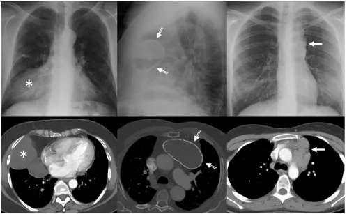
Fig. 1 Conventional radiograph can provide information pertaining to the size, anatomical location and density of a central mass. Right: Frontal chest radiograph shows a sharply defined area of increased opacity with a loss of the cardiac silhouette at the border of the right side of the heart (*). Contrast-enhanced CT scan reveals a thin-walled water-attenuation lesion (*) in the right cardiophrenic angle (pericar-dial cyst).Middle: Lateral chest radiograph and contrast-enhanced CT
scan show a unilocular, well-defined and homogeneously hypodense mass in the anterior mediastinum with peripheral calcification (open
arrows) (thymic cyst).Left: Chest radiograph shows the
or from the digestive tract, and should not be
consid-ered true mediastinal masses (Figs. 2 and 3). Lymph
node enlargement represents a frequent cause of
medi-astinal masses [9].
Fatty masses
Lipomas predominantly occur in the anterior mediastinum and are reported to represent 1.6–2.3% of all primary
mediastinal tumours [10]. At CT, lipomas have
homo-geneous fat attenuation of approximately -100 HU.
Lip-osarcoma frequently occurs in the posterior mediastinum and it is usually symptomatic at the time of
presenta-tion, in contrast to lipoma [11]. Inhomogeneous
appear-ance on CT or MRI differentiates liposarcoma from
lipoma [12].
Thymolipomais a rare, benign, well-encapsulated thymic tumour that accounts for about 2–9% of thymic
neo-plasms [10, 12–14]. Tumours occur most frequently in
the cardiophrenic angle of asymptomatic young adults without sex predilection. The fat content usually con-stitutes 50–85% of the lesion but has been reported to
account for as much as 95% of the tumour [10].
Associations with myasthenia gravis, Graves disease
and haematological disorders have been reported [14].
At CR, thymolipomas may mimic cardiomegaly, exces-sive epicardial fat, diaphragmatic elevation, lobar col-lapse or a pericardial cyst. CT or MRI is required to establish the diagnosis by showing a well-defined en-capsulated mass that has extensive fat content and contains small amounts of solid areas and fibrous septa
[12] (Fig. 4).
Cystic masses
Mediastinal primary cysts represent 15–20% of all
pri-mary mediastinal masses [1, 15]. A smooth or oval
mass with a homogeneous attenuation, with no enhance-ment of cyst contents and no infiltration of adjacent structures are the usual CT features of benign
mediasti-nal cyst (Fig. 5). Any cyst may have a higher
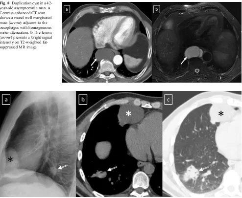
attenua-tion due to its calcic, proteinaceous, mucous or haemorrhagic content. Cysts typically show high signal intensity on T2-weighted MR images. True cystic lesions should be differentiated from the cystic degen-erative changes observed in many solid tumours, in lymphomas before or after treatment, or in abscesses
(Fig. 6).
Bronchogenic cystsresult from abnormal ventral budding or branching of the tracheobronchial tree during
embryo-logic development [15–17]. They are lined by respiratory
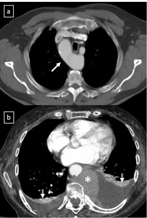
epithelium and their capsule contains cartilage, smooth Fig. 2 a Contrast-enhanced CT scan shows a right-sided aortic arch
(arrow) in an asymptomatic man with an absence of the aortic knuckle
muscle and mucous gland tissue. They are stable in size, except when complicated by infection or haemorrhage. Ap-proximately 40% of bronchogenic cysts are symptomatic,
resulting in cough, dyspnea or chest pain [4]. The
bronchogenic cyst is commonly located in the near carina
(52%) and in the paratracheal region (19%) [5]. The anterior
mediastinum is a rare location of the bronchogenic cyst [17]
(Fig. 7). Air within the cyst is suggestive of secondary
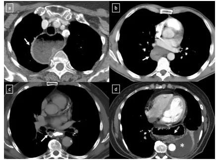
Fig. 3 Different masses arising from the digestive tract.a Oesopha-geal stenosis after Nissen fundoplication (arrow).bPosterior medias-tinal mass (arrow) in relation with squamous-cell carcinoma of the oesophagus.c Oesophageal diverticulum (arrow) in a patient with
oesophageal achalasia. Note the thickened oesophagus.dHiatal hernia is a frequent incidental finding with or without air or air-fluid level
(open arrow). * Pleural effusion
infection and communication with the tracheobronchial tree. Duplication cystsare uncommon lesions lined by gastro-intestinal tract mucosa and generally asymptomatic. How-ever, if they contain gastric or pancreatic mucosa, there is the added risk of haemorrhage or rupture of the cyst from mucosal secretions. The majority of them are detected
adja-cent to or within the oesophageal wall (Fig.8). Duplication
cysts are indistinguishable from bronchogenic cysts on CT and MRI.
Mediastinal neuroenteric cystsare anomalous protrusions of the leptomeninges through intervertebral foramen or defects in the vertebral body. They are associated with
multi-ple vertebral anomalies and with neurofibromatosis [15,16].
Pericardial cystis a benign lesion accounting for 5–10%
of all mediastinal tumours [11]. Most pericardial cysts are
unilocular and commonly located in the right cardiophrenic
space (Fig.9). However, they may occur anywhere in
rela-tion to the pericardium.
Thymic cystrepresents 1% of all mediastinal masses [15]. Congenital cysts derive from remnants of the thymophar-yngeal duct, they are typically unilocular and contain clear
fluid (Fig.10). In contrast, acquired thymic cysts are much
more common, tend to be multilocular (Fig. 11) and may
arise in association with neoplasms such as thymomas, lymphomas or germ cell tumours. Thymic cysts may also be seen in the anterior mediastinum after radiation therapy of Hodgkin’s disease, after inflammatory processes and occasionally in patients with AIDS, particularly in children
[18].
Lymphangioma is a rare benign lesion of lymphatic ori-gin that represents 0.7–4.5% of all mediastinal tumours in
adult population [15]. Lymphangiomas involve the neck or
the axillary region in more than 80% of the cases and the
thorax in 10% of the cases [19] (Fig.12).
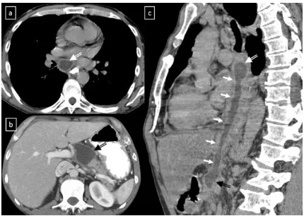
Pancreatic pseudocyst can extend into the mediastinum via the oesophageal or aortic hiatus. CT shows a thin-walled, Fig. 5 A well-marginated mass with a homogeneous attenuation,
usually in the range of water attenuation (0–20 HU) and without an enhancement of the wall or infiltrative appearance are the typical features of benign mediastinal cysts. Probably thymic cystic (a) and pericardial cyst (b)
Fig. 7 Bronchogenic cyst in a 37-year-old man. a Non-contrast-enhanced CT scan shows a homogeneous anterior mediastinal mass with smooth contours and oval shape (arrow). The mass is isodense relative to chest wall muscle.b T2-weighted MR image shows the
same mass (arrow) with markedly high signal intensity.cAlthough its localisation, a bronchogenic cyst was confirmed by histological exam-ination after surgical resection
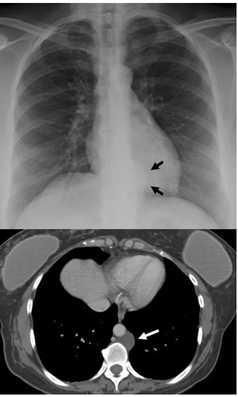
Fig. 9 aLateral chest radiograph of a 58-year-old smoker man allergic to iodine shows a well-defined mass (*) in the cardiophrenic space and a nodular lung opacity (open arrow) in the lower lung parenchyma.b, cNon-contrast-enhanced CT scan confirms the presence of a
fluid-attenuation mass (*) in the right cardiophrenic angle (pericardial cyst) and demonstrates a suspicious lung opacity (open arrows) in the right lower lobe of the lung (squamous-cell carcinoma)
Fig. 8 Duplication cyst in a 42-year-old asymptomatic man.a Contrast-enhanced CT scan shows a round well marginated mass (arrow) adjacent to the oesophagus with homogeneous water-attenuation.bThe lesion
(arrow) presents a bright signal
fluid-containing cyst within the posterior mediastinum which may be in continuity with the intrapancreatic or peripancreatic
fluid collections (Fig.13).
Solid masses
Mediastinal goitre generally represents direct contiguous growth of a goitre into the anterior or superior mediastinum. Typical features of mediastinal goitres are encapsulated and lobulated mass with inhomogeneous appearance with cystic areas, calcifications and marked contrast enhancement.
(Fig. 14). An intrathoracic thyroid mass developing from
heterotopic thyroid tissue without any connection to the
thyroid in the neck is extremely rare (Fig.15a). The
pres-ence of ill-defined margins, invasion of adjacent structures and nearby lymph node enlargement suggests the diagnosis
of thyroid cancer [20] (Fig.15b).
Fig. 10 Congenital thymic cyst in a 47-year-old man. Contrast-enhanced CT scan demonstrates a unilocular unContrast-enhanced lesion in the anterior mediastinum which shows a homogeneous fluid-attenuation
(arrow)
Fig. 11 Acquired thymic cyst in a 43-year-old man.a Contrast-enhanced CT scan shows a well-defined water-attenuation multiloculated mass (*) in the anterior mediastinum. bSagittal T2-weighted MR se-quence demonstrates a multilo-culated mass with typical high signal intensitiy and fine inter-nal septa within (arrow)
Fig. 12 A cystic lymphangioma (also referred to as hygroma) in a 47-year-old woman with retroperitoneal disease. A posterior mediastinal mass with a homogeneous fluid-attenuation is identified on CT (white
arrow) and on posteroanterior chest radiograph as a mass disrupting
Thymic hyperplasia can be divided into two distinct
histological types [13, 14]. True thymic hyperplasia is
defined as enlargement of the thymus, which generally retains its normal shape. This disease entity is observed when a patient is recovering from some recent stress (such as chemotherapy, corticosteroid therapy, irradiation or thermal burns). The phenomenon known as rebound hypeplasia is defined as a greater than 50% increase in
thymic volume over baseline after such stress [14].
Among patients who undergo chemotherapy,
approxi-mately 10–25% may develop rebound hyperplasia [13].
Thymic lymphoid (follicular) hyperplasia of the thymus refers to the presence of an increased number of lym-phoid follicles. It is commonly associated with autoim-mune diseases, being seen in up to 65% of cases with
myasthenia gravis [14], and it has been reported to
occur in the early stages of human immunodeficiency virus infection. At CT it may appear normal (45%),
enlarged (35%) (Fig. 16) or as a focal thymic mass
(20%) [13].
It is important for radiologists to be able to distinguish thymic hyperplasia from neoplasm. Diffuse symmetric
enlargement of the gland, a smooth contour and normal vessels are the key morphological features of hyperplasia, whereas neoplasm tends to manifest as a focal mass with nodular contour and necrotic or calcified foci. Detecting fat in the thymus is particularly relevant in these situations.
According to Takahashi et al. [21], chemical-shift MRI can
be useful in this situation. Thymic hyperplasia reveals a rela-tive signal loss on opposed-phase chemical-shift MRI that it is different from no significant signal change between in-phase and opposed-phase chemical-shift MR images in patients with
malignancy (Fig.17).
Thymomais the most common primary neoplasm of the anterior mediastinum but accounts for less than 1% of all
adult malignancies [22,23]. Thymomas typically occur in
patients older than 40 years of age, being rare in children,
and affecting men and women equally [23]. Between 20%
and 30% of patients with thymoma have pressure-induced
symptoms [13]. Myasthenia gravis associated with
thy-moma occurs most frequently in women. Between 30% and 50% of patients with a thymoma have myasthenia gravis, whereas 10–15% of patients with myasthenia gravis have a thymoma.
Fig. 13 Pancreatic pseudocyst in a 52-year-old man with a recurrent pancreatitis history. Axial (a,b) and oblique-sagittal multiplanar re-construction (c) CT scan show a thin-walled peri-oesophageal
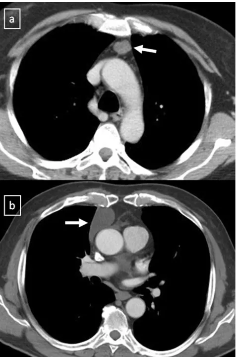
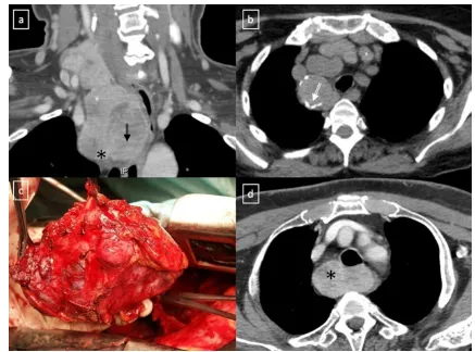
Fig. 14 Mediastinal goitres.aCoronal multiplanar reconstruction CT scan demonstrates an anterior mediastinal mass (*) arising from the thyroid more superiorly. Note the cystic degeneration within goitre (open arrow).b Non-contrast-enhanced CT scan shows a unilateral mediastinal goitre with
peripheral areas of calcification (open arrow).cPhotograph of the resected surgical specimen shows a lobulated and heterogeneous appearance of the mass.dContrast-enhanced CT scan shows a well marginated posterior mediastinal goitre (*) with marked contrast enhancement
Fig. 15 aHeterotopic mediastinal goitre. Sagittal contrast-enhanced CT scan demonstrates a well-defined mass (arrow) located in the retrosternal space which shows an intense and heterogeneous contrast enhancement due to the presence of cystic areas (*). No connection to the thyroid gland from the neck.bAnaplastic thyroid carcinoma in a
The updated histological classification elaborated by WHO in 2004 classified different types of thymomas
(Table1) on the basis of the morphology of the neoplastic
epithelial cells together with the lymphocyte-epithelial cell
ratio [23,24]. In contrast to histological classification, the
stage of thymoma has clinical implications and it is a useful tool for management decisions. The Masaoka-Koga staging system is the most commonly used and describes thymomas
in terms of the local extension of the tumour [14,23–26]
(Table 2). The Masaoka staging system is one of the two
factors, including completeness of surgical resection, that
most strongly correlates with prognosis of thymomas [23].
The role of imaging is to initially diagnose and properly stage thymoma, with emphasis on the detection of local invasion and distant spread of disease. Between 45 and
80% of thymomas are visible by chest radiography [22].
On CT scans, thymomas usually appear as homogeneous solid masses with soft-tissue attenuation and well-demarcated borders, located anywhere from the thoracic inlet to the cardiophrenic angle. Thymomas may be oval, round or lobulated and when they are large, cystic or
ne-crotic degeneration may be shown (Fig.18). Calcification
may be present in the capsule or throughout the mass. Certain findings, such as encasement of mediastinal struc-tures, infiltration of fat planes, irregular interface between the mass and lung parenchyma, and direct signs of vascular
involvement are highly suggestive of invasion (Fig. 19).
Pleural dissemination (“drop metastases”) manifests as
one or more pleural nodules or masses, and they are
almost always ipsilateral to the tumour [23] (Fig. 20).
Thymoma rarely presents with metastatic lymphadenop-athy, metastatic pulmonary nodules or pleural effusion. At MRI, thymomas commonly appear as homogeneous
or heterogeneous masses with low to intermediate signal intensity on T1-weighted images and with high signal
intensity on T2-weighted images (Fig. 21). MRI can
prove useful in identifying the nodular wall thickening detected in cystic thymomas, absent from congenital
cysts [22].
Thymic carcinoma accounts for about 20% of thymic
epithelial tumours with a mean age of 50 years [13]. Typical
appearance is a multilobulated and heterogeneous mass that may contain areas of calcification or haemorrhage. Distant metastasis are present at the initial diagnosis in 50–65%
[13]. Sadohara et al. [27] found that irregular contour,
ne-crotic or cystic components, heterogeneous enhancement, lymphadenopathy and great vessel invasion strongly fav-oured thymic carcinoma.
Thymic carcinoids are rare, well-differentiated
neuroen-docrine tumours, which have a male predilection of 3:1 [13,
14]. They often present with endocrine disorder. Thymic
carcinoid usually manifests as a large anterior mediastinal mass often with metastases.
Lymphoproliferative disorders. Primary mediastinal lym-phoma usually occurs in the anterior mediastinum. Malig-nant lymphoma accounts for nearly 20% of all mediastinum
neoplasms in adults and 50% in children [5]. Lymphomas
are the most common cause of masses in the paediatric
mediastinum [18]. Hodgkin lymphoma (Fig.22) represents
approximately 50–70% of mediastinal lymphomas, while
non-Hodgkin lymphoma comprises 15–25% [4]. Pleural
and pericardial effusions are often common features in all types of lymphoma.
Hodgkin disease (HD) has a bimodal distribution of incidence peaking in young adulthood and again after the
age of 50 years [4]. Most patients experience constitutional
symptoms. Four subtypes of HD are described: nodular sclerosis (by far the most common histological subtype)
(Fig. 23), lymphocyte-rich, mixed cellularity and
lympho-cyte depleted HD [4,5]. CR is abnormal in up to 76% of
patients with HD, often showing enlargement of the
prevas-cular and paratracheal nodes [4]. Characteristic features on
imaging are a homogeneous soft-tissue anterior mediastinal mass with mild to moderate contrast enhancement, irregular contours, surface lobulation, absence of vascular involve-ment, and high prevalence of associated mediastinal
lymph-adenopathy [3]. Cystic and necrotic changes can be
identified.
The two most common forms of mediastinal
non-Hodg-kin disease (NHD) include diffuse large B-cell lymphoma and T-cell lymphoblastic lymphoma. T-cell lymphoblastic lymphoma mainly occurs in children and adolescents. The most common CT appearance is a large mediastinal mass representing thymic and lymph node enlargement, which compresses the airway and cardiovascular structures
(Fig. 24). Low attenuation areas reflecting necrosis are
Table 1 WHO classification for thymoma
2004 WHO classification Description
A (spindle cell thymoma; medullary thymoma) Bland spindle/oval epithelial tumour cells with few or no lymphocytes
AB (mixed thymoma) Mixture of a lymphocyte-poor type A thymoma component and a more lymphocyte-rich type B-like component (smaller and paler than those of B1 or B2 thymomas). Lymphocytes are more numerous than in the type A component, but may be less numerous than in B1 thymomas B1 (lymphocyte-rich thymoma; lymphocytic
thymoma; organoid thymoma; predominantly cortical thymoma)
Epithelial cells with a histological appearance practically indistinguishable from the normal thymus, composed predominantly of areas resembling cortex with epithelial cells scattered in a prominent population of immature lymphocytes
B2 (cortical thymoma) Tumour cells closely resembling the predominant epithelial cells of the normal thymic cortex. A background population of immature T cells is always present and usually outnumbers the neoplastic epithelial cells
B3 (epithelial thymoma; squamoid thymoma) Medium–sized round or polygonal cells with slight atypia. The epithelial cells are mixed with a minor component of intraepithelial lymphocytes
From Travis et al. [24]
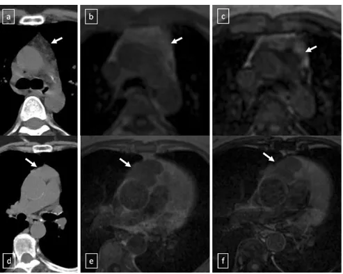
Fig. 17 a–cThymic hyperplasia in a 43-year-old woman (arrows).a Non-contrast-enhanced CT scan reveals a lobulated lesion with smooth margins in anterior mediastinum.bLesion appears slightly hyperin-tense on in-phase gradient-echo T1-weighted MR image.c Opposed-phase gradient-echo T1-weighted MR image shows decreased signal intensity within the lesion, confirming presence of fat.d–f Stage II
commonly seen. Primary mediastinal diffuse large B-cell lymphomas tend to occur in young to middle-aged adults
with a mean age of 30 [5]. It accounts for 7% of all cases of
NHD and about 10% of all cases of high-grade NHD [28].
Table 2 Masaoka-Koga staging system of thymoma
From references [14,23–26]
Stage Description
I Macroscopically and microscopically encapsulated tumour IIa Microscopic invasion through the capsule
IIb Macroscopic invasion into surrounding fatty tissue
III Invasion into a neighbouring organ such as the pericardium, great vessels or lung IVa Pleural or pericardial dissemination
IVb Lymphatic-haematogenous metastases
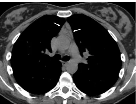
Fig. 18 a,bStage II thymoma (WHO type B1) in a 33-year-old woman who presented with myasthenia gravis. Frontal chest radiograph shows a hilum overlay sign (arrow) of a suggestive anterior mediastinal mass. Contrast-enhanced CT scan confirms the presence of a low-heterogeneous anterior mediastinal mass (arrow). Note the indentation
The tumours appear as a large, smooth or lobulated, anterior mediastinal mass in nearly all patients. On CT the tumours
show low attenuation areas, representing haemorrhage, ne-crosis or cystic degeneration in 50% of the cases and het-tion (arrow). Note the lobulated contour of the mass and the loss of the bronchiolitis in the left lower lobe
Fig. 20 Stage IVa thymoma (WHO type B2) in a 46-year-old man.a Contrast-enhanced CT scan reveals an anterior mediastinal mass
(arrows) with irregular contours, homogeneous enhancement and
pe-ripheral and central calcification as well as a pleural nodule (open
arrow). b On an axial FDG-positron emission tomography (PET)
erogeneous enhancement in about 40% of the cases [5]
(Fig. 25). Primary mediastinal B-cell lymphoma recurs in
the chest. Consequently, chest CT examination alone is
sufficient for routine follow-up of these patients [28].
Germ cell tumours(GCTs) mainly arise in gonads and in the midline of the body as well, the mediastinum being the most common extragonadal site. GCTs account for 10–15% of anterior mediastinal masses in adults and 25% in children
[5]. Only 3% of them arise in the posterior mediastinum [4].
Pathological classifications include teratomas and non-teratomatous germ cell tumours.
Teratoma is the most common mediastinal GCT [4]. Mature teratomas are usually asymptomatic and represent
60–70% of all mediastinal GCTs [5]. They are composed of
well-differentiated benign tissues with predominant ectoder-mal element. If a teratoma contains fetal tissue or neuroen-docrine tissue, it is defined as immature and malignant with a poor prognosis. On CT, teratoma most commonly appears
as a well-defined unilocular or multilocular cystic lesion
containing fluid, soft tissue and fat attenuation (Fig. 26).
Calcifications may be focal, rim-like or, in rare cases, rep-resentative of teeth or bone. Common combinations of internal components of mature teratomas include soft tissue, fluid, fat and calcification in 39%; soft tissue, fluid and fat in 24%; and soft tissue and fluid in 15%. In 15% of the cases, mature teratomas appear as non-specific cystic lesions
with-out fat or calcium [5]. On MRI, teratomas typically
demon-strate heterogeneous signal intensity, representing various internal elements. Fat-fluid levels within the lesion are vir-tually diagnostic of teratoma. Ruptured teratomas show an adjacent consolidation, atelectasis and pleural or pericardial effusion than do unruptured teratomas.
The non-teratomatous germ cell tumours (NTGCT) are rare and malignant tumours which usually occur in young males and most frequently affect the anterosuperior
medias-tinum [29]. These tumours grow rapidly and develop large,
Fig. 21 Axial (a) and coronal multiplanar reconstruction (b) of a non-contrast-enhanced CT scan of a 57-year-old man allergic to iodine with a thymoma. A solid lobulated thymic mass (*) with clumps of calcifi-cations within (arrowhead) is identified. Note the absence of a fat plane between the tumour and the aorta (open arrow).dCoronal T2-weighted MR image shows a typical signal hyperintensity of the
bulky, ill-circumscribed masses with lobulated shape. Pri-mary mediastinal seminomas comprise 25–50% of
malig-nant mediastinal GCTs [4] and occur almost exclusively in
males during the period from the second to fourth decades
of life [5]. At imaging, the tumours typically have
homog-enous appearance and show minimal contrast enhancement. Areas of degeneration due to haemorrhage and coagulation
necrosis may be present (Fig. 27). Metastasis to lymph
nodes and bone does occur. Non-seminomatous germ cell tumours include yolk sac tumours, endodermal sinus tumours, embryonal carcinomas, choriocarcinomas and mixed germ cell tumours, which present as large masses typically with marked heterogeneous attenuation. At
diag-nosis, 85% of patients are symptomatic [4]. Invasion of
Fig. 23 Nodular sclerosis Hodgkin lymphoma in a 44-year-old wom-an. Frontal chest radiograph shows a large, well-defined mediastinal mass with increased density (arrow). Contrast-enhanced CT scan shows a bulky soft tissue mass (arrows) with homogeneous CT-attenuation value occupying prevascular space. Note the left internal
mammarian artery completely surrounded by the lesion. Photomicro-graph reveals numerous neoplastic lacunar cells (arrows) in a back-ground of small lymphocytes, histiocytes and eosinophils, which supports the diagnosis of nodular sclerosis type Hodgkin lymphoma Fig. 22 A 28-year-old man with Hodgkin lymphoma. Frontal chest
radiograph and contrast-enhanced CT scan show a homogeneous soft tissue mass at the level of the subcarina (arrows). An aortopulmonary window lymphadenopathy can be detected on CT scan (open arrow).
adjacent structures and distant metastasis may occur. Pleural and pericardial effusions are common. Measuring AFP and ß-hCG levels is important when making the diagnosis
(Fig.28).
Neurogenic tumoursrepresent approximately 20% of all adults and 35% of all paediatric mediastinal tumours and they are the most common cause of a posterior mediastinal
mass [30]. Seventy to eighty percent of neurogenic tumours
are benign, and nearly half are asymptomatic [4]. These
tumours are generally grouped into:
Peripheral nerve tumours, which are the most common (70%) mediastinal neurogenic tumours and originate from spinal or proximal intercostal nerve; however, they rare-ly arise from the vagus, recurrent laryngeal and phrenic
nerve [30] (Fig. 29). Schwannomas are the most
com-mon (50%) mediastinal neurogenic tumours and
fre-quently affect patients from 20 to 30 years old [30].
They are usually solitary and encapsulated masses, but multiple schwannomas may be associated with neurofi-bromatosis type 2. The tumour may grow through the
adjacent intervertebral foramen and spinal canal to
pro-duce a “dumbbell” or “hourglass” configuration. Cystic
changes and haemorrhage are more common in schwan-nomas than in neurofibromas. Neurofibromas are non-encapsulated soft tissue tumours and account for ap-proximately 20% of mediastinal neurogenic tumours
[30]. A sudden increase in the size of a previously
stable neurofibroma and the presence of neurological symptoms suggests malignant transformation to malig-nant peripheral nerve sheath tumour. These tumours are closely associated with neurofibromatosis and show more heterogeneous signal intensity and contrast en-hancement on MRI.
Sympathetic ganglion tumours, which comprise 25% of mediastinal neurogenic tumours and arise from neuronal
cells rather than from the nerve sheath [30].
Ganglio-neuromas are the most benign and differentiated of the autonomic ganglionic tumours. Radiographically, the tumours are well-marginated, occurring along the ante-rolateral aspect of the spine and spanning three to five
vertebrae. The “whorled appearance” is due to
curvilin-ear bands of low signal intensity that reflects collage-nous fibrous tissue in the mass on T2-weighted images. Most ganglioneuromas show gradual and heterogeneous contrast enhancement. Ganglioneuroblastomas are the least common type of neurogenic tumour and show intermediate features in cellular maturity between neu-roblastoma and ganglioneuroma. Neuneu-roblastomas are highly aggressive and readily metastasising tumours of neuroectodermal origin with a median age at diagnosis
of 22 months [30]. They are heterogeneous and
non-encapsulated lesions, often exhibiting haemorrhage,
ne-crosis, calcification or cystic degeneration (Fig. 30).
Mediastinal paraganglia.Paraganglioma is a rare neuro-endocrine tumour of chromaffin cell origin. One to two percent of extra-adrenal paragangliomas occur in the thorax
[30]. Aortopulmonary paragangliomas are usually
asymp-tomatic, while aortosympathetic paragangliomas (along the Fig. 24 Axial T1-weighted MR image of a 16-year-old man with a
solid, large mass (arrows) in the anterior and superior mediastinum. Supra-aortic trunks are almost completely surrounded by the lesion and trachea (T) is displaced to the contralateral side by the lesion. Patho-logical analysis demonstrated a T-cell lymphoblastic lymphoma
Fig. 25 Primary mediastinal diffuse large B-cell lymphoma in a 19-year-old man. Contrast-enhanced CT scan shows a bulky soft tissue mass in the anterior mediastinum (arrows
sympathetic chain in the posterior mediastinum) occur in symptomatic patients related to the functional activity of the tumour. These masses commonly enhance brightly at
en-hanced CT (Fig.31). A characteristic MRI finding of
para-gangliomas is the presence of multiple curvilinear and punctate signal voids, which reflect high velocity flow in
the intratumoral vessels, described as “salt-and-pepper”
appearance.
Clinical and radiological features of the most common
mediastinal masses are detailed in Table3.
Uncommon mediastinal masses
Parathyroid adenomasmay be seen in ectopic locations, the mediastinum being the most commonly site. High-resolution ultrasonography (US) is recognised as a tool for detecting cervical parathyroid lesions. As it enlarges, an abnormal gland appears as a hypoechoic, and often anecho-ic, lesion, often posterior in location to the thyroid. As the Fig. 26 Chest imaging shows well the highly heterogeneous contents
of mediastinal teratomas.aMature cystic teratoma in a 40-year-old man. Contrast-enhanced CT scan shows a heterogeneous anterior me-diastinal mass with areas of fat (open arrow), calcification (arrow) and fluid attenuation (*). Posterior displacement of mediastinal structures is also be seen. b Photograph of the surgical specimen. c
Contrast-enhanced CT scan of an asymptomatic 24-year-old woman demon-strates a well-defined uniloculated mass located in prevascular space which shows a cystic changes within (*). Non foci of calcification were identified.dThe mass was surgically removed and pathological exam-ination confirmed a benign teratoma
gland enlarges, it can develop lobularity and foci of echoge-nicity. Colour Doppler assessment of parathyroid lesions is a useful integration of grey-scale US and may be helpful in featuring parathyroid lesions. The colour Doppler patterns
termed“parenchymal”(pattern IV, internal flow) and
“vascu-lar pole” (pattern II, focal peripheral flow) are typical of
parathyroid lesions [31]. The different colour Doppler US
patterns seem to be influenced by many factors as the location of the gland and the degree of vascularity. These tumours tend to be small and may contain calcifications at CT. Technetium-99 Sestamibi SPECT scans are more effective for their
diag-nosis (Fig. 32). Fibrosing mediastinitis is a dense fibrosis
which progressively encases and eventually obliterates the
lumen of the mediastinal vessels and airways (Fig.33).
Hae-matoma.High attenuation of haematomas can be observed on
unenhanced CT scans during the first 72 h (Fig.34). When the
hematoma ages its attenuation decreases at CT in a centripetal
fashion.Haemangiomasin the mediastinum are rare and the
may be associated with Rendu-Osler syndrome. Sarcomas
other than vascular or neural origin, including fibrosarcomas, osteosarcomas and chondrosarcomas, are also very uncom-mon. Extramedullary haematopoiesisin posterior mediasti-num is another entity to take into account.
Follow-up
In assessment of mediastinal disease, cross-sectional imag-ing techniques allow excellent visualisation of the medias-tinum. CT is generally the first-choice modality of diagnostic imaging. MRI plays an increasing role in this disease due to the existence of new available MR techniques on mediastinum imaging.
On each of CT and MR scanning, the size of tumour, contour, perimeter of capsule, septum, haemorrhage, Fig. 28 Non-seminomatous malignant germ cell tumour of the
anteri-or mediastinum in a 25-year-old man with chest pain and high serum level ofα-fetoprotein at admission (25.396 ng/ml). Frontal chest radiograph shows a central mass (*). The descending aorta is clearly seen (arrows), indicating that the mass is not within the posterior
mediastinum. Multiple nodules in bilateral pulmonary field are also observed. A contrast-enhanced CT scan confirms a mass of low atten-uation (*) in the anterior mediastinum that compresses the pulmonary artery. Bilateral lung metastasis (arrowheads) and hilar and subcarinal lymphadenopathy is identified (open arrows)
necrotic or cystic component, homogeneity within tumour, presence of mediastinal lymphadenopathy, pleural effusion and great vessel invasion are assessed. In addition, presence of calcification are assessed on CT and signal intensities of the tumour are assessed on MRI.
Imaging plays an essential role in the diagnosis, staging and follow-up of mediastinal disease. Complete resection is the mainstay of treatment in many medias-tinal tumours and the ability to accomplish a complete resection appears to be the most important prognostic factor. Currently, CT is the modality most commonly used for up after treatment. The goal of follow-up is to detect recurrence as early as possible. CT findings may serve as predictors of tumour invasiveness and of postoperative recurrence or metastases.
In Table 4 we summarise some teaching points and
imaging pitfalls for the diagnostic approach to mediastinal masses before and after treatment.
Conclusion
Tumours of the mediastinum represent a wide diversity of disease state. The location and composition of a mass is critical to narrowing the differential diagnosis. The clinical spectrum of mediastinal masses can range from being asymptomatic to producing compressive symptoms. Although many of these masses have similar imaging appearances, clinical history, anatomical position and certain details seen at CT and MRI imaging allow correct diagnosis in many cases.
Fig. 32 Parathyroid adenoma in a 66-year-old man with hypercalcae-mia, hypophosphataemia and elevated PTH values. Tc-99m MIBI scan shows a focus of hyperactivity (black arrow) adjacent to the lower pole
of left thryoid lobule. Enhanced-CT scan shows a superior mediastinal enhanced mass (white arrow)
Acknowledgments The authors gratefully acknowledge the contri-bution of technicians, surgeons and pathologists of the hospitals listed above, without whose efforts this work would not have been possible. The authors thank Isabel Coll for the English correction and assistance.
Open Access This article is distributed under the terms of the Creative Commons Attribution License which permits any use, distribution, and reproduction in any medium, provided the original author(s) and the source are credited.
References
1. Laurent F et al (1998) Mediastinal masses: diagnostic approach. Eur Radiol 8:1148–1159
2. Whiteen CR et al (2007) A diagnostic approach to mediastinal abnormalities. Radiographics 27:657–671
3. Tomiyama N et al (2009) Anterior mediastinal tumors: diagnostic accuracy of CT and MRI. Eur J Radiol 69:280–288
4. Duwe BV et al (2005) Tumors of the mediastinum. Chest 128:2893–2909
with subtle areas of high CT-attenuation values (arrow). Bilateral pleural effusion (*) and a sternum fracture (open arrow) are also
high attenuation value of the lesion compared with muscular tissue
Table 4 Teaching points and imaging pitfalls for the diagnostic approach to mediastinal masses before and after treatment
•CT is accurate in distinguishing mediastinal masses which usually differ in their appearance and the pattern of metastatic spread, both of which are readily detected by chest CT
•Pericardial fat pads and lipomatosis are correctly interpreted as normal findings rather than possible pathological lesions
•When lipoma and liposarcoma are situated in the cardiophrenic space, the imaging findings are very similar to those of Morgagni hernia
•MRI more accurately distinguishes between cystic and solid lesions than CT
•Soft-tissue components associated with cystic lesions can be related to a malignant process (e.g. soft-tissue nodules in a cystic anterior mediastinal lesion suggest that the lesion is a cystic thymoma rather than a congenital cyst)
•Non-neoplastic thymic enlargement must not be confused with thymoma. The normal thymus in young children and the hyperplastic thymus may mimic a mass
•When differentiation between non-neoplastic thymic enlargement and thymoma cannot be achieved at CT or conventional MRI, chemical-shift MRI with in-phase and out-of-phase gradient-echo sequences can be helpful
•Thymoma rarely manifests with lymphadenopathy, pleural effusions, or extrathoracic metastases
•The role of imaging is to initially diagnose and properly stage thymoma, with emphasis on the detection of local invasion and distant spread of disease, to identify candidates for preoperative neoadjuvant therapy
•Late recurrence in thymoma is not uncommon. Imaging of treated patients is directed at identifying resectable recurrent disease, since patients with completely resected recurrent disease have similar outcomes as those without recurrence [23]
•Findings associated with significantly more frequent recurrence and metastases of thymic tumours include lobulated or irregular contour, oval shape, mediastinal fat invasion or great vessel invasion and pleural seeding
•Mediastinal lymphadenopathy, pleural effusions, and pulmonary metastases are characteristic of thymic carcinoma or non-teratomatous germ cell tumour
•Lymphoproliferative disorders typically present pleural effusions, pericardial fluid, and mediastinal lymphadenopathy in many cases
•Heterogeneous appearance (due to necrosis, cystic change, or haemorrhage) is typical of thymic carcinoma, lymphoma, sympathetic ganglion tumour, peripheral nerve tumour and non-teratomatous germ cell tumour. It can be seen in about one-third of thymomas
5. Takahashi K et al (2010) Computed tomography and magnetic resonance imaging of mediastinal tumors. J Magn Reson Imaging 32:1325–1339
6. Puderbach M et al (2007) MR imaging of the chest: a practical approach at 1.5 T. Eur J Radiol 64:345–355
7. Ackman JB et al (2011) MRI of the thymus. AJR Am J Roentgenol 197:W15–W20
8. Gümüştaş S et al (2011) Malignant versus benign mediastinal lesions: quantitative assessment with diffusion weighted MR im-aging. Eur Radiol 21:2255–2260
9. Kim Y et al (2000) Middle mediastinal lesions: imaging findings and pathologic correlation. Eur J Radiol 35:30–38
10. Gaerte SC et al (2002) Fat-containing lesions of the chest. Radio-graphics 22:S61–S78
11. Pineda V et al (2007) Lesions of the cardiophrenic space: findings at cross-sectional imaging. Radiographics 27:19–32
12. Molinari F et al (2011) Fat-containing lesions in adult thoracic imaging. AJR Am J Roentgenol 197:W795–W813
13. Nasseri F et al (2010) Clinical and radiologic review of the normal and abnormal thymus: pearls and pitfalls. Radiographics 30:413– 428
14. Nishino M et al (2006) The thymus: a comprehensive review. Radiographics 26:335–348
15. Jeung M-Y et al (2002) Imaging of cystic masses of the mediasti-num. Radiographics 22:S79–S93
16. Takeda S-I et al (2003) Clinical spectrum of medistinal cysts. Chest 124:125–132
17. Kim JH et al (2003) Cystic tumors in the anterior mediastinum. Radiologic-pathological correlation. J Comput Assist Tomogr 27:714–723
18. Franco A et al (2005) Imaging evaluation of pediatric mediastinal masses. Radiol Clin N Am 43:325–353
19. Charruau L et al (2000) Mediastinal lymphangioma in adults: CT and MRI imaging features. Eur Radiol 10:1310–1314
20. Quint LE (2007) Imaging of anterior mediastinal masses. Cancer Imaging 7 Spec No A: S56–S62
21. Takahashi K et al (2003) Characterization of the normal and hyperplastic thymus on chemical-chift MR imaging. AJR Am J Roentgenol 180:1265–1269
22. Marom EM (2010) Imaging thymoma. J Thorac Oncol 5:S296–S303 23. Benveniste MFK et al (2011) Role of imaging in the diagnosis, staging, and treatment of thymoma. Radiographics 31:1847–1861 24. Travis WD, Brambilla E, Müller-Hermelink HK, Harris CC (eds) (2004) Pathology & genetics: tumours of the lung, pleura, thymus and heart. World Health Organization Classification of Tumours, vol 10. IARC, Lyon
25. Falkson CB et al (2009) The management of thymoma; a system-atic review and practice guideline. J Thorac Oncol 4:911–919 26. Masaoka A (2010) Staging system of thymoma. J Thorac Oncol 5:
S304–S312
27. Sadohara J et al (2006) Thymic epithelial tumors: comparison of CT and MR imaging findings of low-risk thymomas, high-risk thymomas, and thymic carcinomas. Eur J Radiol 60:70–79 28. Boger-Megiddo I et al (2002) Is chest CT sufficient for follow-up
of primary mediastinal B-cell lymphoma in remission? AJR Am J Roentgenol 178:165–167
29. Li T et al (2011) CT findings of primary non-teratomatous germ cell tumors of the mediastinum—a report of 15 cases. Eur J Radiol 81:1057–1061
30. Nakazono T et al (2011) MRI findings of mediastinal neurogenic tumors. AJR Am J Roentgenol 197:W643–W652