R E S E A R C H
Open Access
Reduced culture temperature attenuates
oxidative stress and inflammatory response
facilitating expansion and differentiation of
adipose-derived stem cells
Gal Tirza
1†, Inna Solodeev
1†, Meirav Sela
1, Ilanit Greenberg
1, Metsada Pasmanik-Chor
2, Eyal Gur
3and Nir Shani
3*Abstract
Background:Adipose-derived stem cell (ASC) expansion under atmospheric oxygen levels (21%) was previously shown to cause increased reactive oxygen species (ROS) accumulation and genetic instability compared to cells cultured under physiological oxygen levels (2–8%). However, since culture under physiological oxygen levels is costly and complicated, a simpler method to reduce ROS accumulation is desirable. The current study aimed to determine whether lower culture temperature can reduce ROS production in ASCs without impairing their culture expansion.
Methods:Proliferation, differentiation, ROS accumulation, and gene expression were compared between ASC cultures at 35 °C and 37 °C. ASCs isolated either from rat fat depots or from human lipoaspirates were examined in the study.
Results:Rat visceral ASCs (vASCs) cultured at 35 °C demonstrated reduced ROS production and apoptosis and enhanced expansion and adipogenic differentiation compared to vASCs cultured at 37 °C. Similarly, the culture of human ASCs (hASCs) at 35 °C led to reduced ROS accumulation and apoptosis, with no effect on the proliferation rate, compared to hASCs cultured at 37 °C. Comparison of gene expression profiles of 35 °C versus 37 °C vASCs uncovered the development of a pro-inflammatory phenotype in 37 °C vASCs in correlation with culture
temperature and ROS overproduction. This correlation was reaffirmed in both hASCs and subcutaneous rat ASCs. Conclusions:This is the first evidence of the effect of culture temperature on ASC growth and differentiation properties. Reduced temperatures may result in superior ASC cultures with enhanced expansion capacities in vitro and effectiveness in vivo.
Introduction
Mesenchymal stem cells (MSCs) are multipotent, can be derived from most adult tissues, and have been demon-strated to bear regenerative and immunosuppressive cap-acities in preclinical models [1]. Although first isolated from the bone marrow, MSCs were also later isolated from adipose tissue and termed adipose-derived stem cells (ASCs) [2, 3]. Clinical utilization of MSCs often requires
1 × 10⁶–5 × 10⁶ cells/kg [4] necessitating significant in vitro expansion of cells prior to their application, in-creasing the risk of DNA mutation and genetic instability.
Reactive oxygen species (ROS) are a byproduct of mito-chondrial oxidative phosphorylation but are also generated as cellular signaling molecules by enzymes such as the fam-ily of NOX NADPH oxidases [5]. ROS overproduction leads to various destructive cellular processes, such as aging, DNA damage, and apoptosis [6, 7]. Physiological oxygen levels within the MSC niche were reported to be between 2 and 8% [8]. Elevated oxygen concentrations stimulate increased mitochondrial ROS production by pro-moting higher ROS leakage from the respiratory chain [9]. Consequently, MSC culture at drastically higher
© The Author(s). 2020Open AccessThis article is distributed under the terms of the Creative Commons Attribution 4.0 International License (http://creativecommons.org/licenses/by/4.0/), which permits unrestricted use, distribution, and reproduction in any medium, provided you give appropriate credit to the original author(s) and the source, provide a link to the Creative Commons license, and indicate if changes were made. The Creative Commons Public Domain Dedication waiver (http://creativecommons.org/publicdomain/zero/1.0/) applies to the data made available in this article, unless otherwise stated.
* Correspondence:[email protected]
†Gal Tirza and Inna Solodeev contributed equally to this work.
3The Department of Plastic and Reconstructive Surgery, Tel Aviv Sourasky
Medical Center, affiliated with the Sackler Faculty of Medicine, Tel Aviv University, Weizmann 6, Tel Aviv, Israel
atmospheric oxygen levels (21%), most commonly employed in culture protocols, leads to ROS overproduc-tion, DNA damage, and genetic instability compared to cul-ture under physiological oxygen levels (2–8%) [10–14]. Culture under physiological oxygen conditions also leads to increased proliferation and stem cell potency of both pluri-potent [15–17] and adult stem cells [18–24]. Although adaptation of culture conditions to physiological oxygen levels to prevent excess ROS is appealing, reducing oxygen levels from atmospheric levels is demanding and pricy and requires specialized equipment. Thus, simpler and most cost-effective approaches are desirable [25].
Reduced ROS production can theoretically be achieved by decreasing cellular temperature and consequently reducing cellular metabolism and mitochondrial oxygen consump-tion. Indeed, reduction of body temperature to mild hypothermia was demonstrated to protect against ischemia-induced cardiac damage and stroke [26–28] and to reduce ROS production and NOX activation following stroke [26,
29]. Decreased body temperature was also found to prevent ischemia-induced damage in hibernating animals [30]. This protection results, most probably, from metabolism reduc-tion during hibernareduc-tion, which leads to reduced mitochon-drial activity and reduced ROS production [31,32].
The effect of low temperature on cellular metabolism is also evident in cultured cells, with various cell types dem-onstrating lower cellular metabolism in correlation with temperature reduction [33–36]. However, decreased cul-ture temperacul-ture also leads to a temperacul-ture-dependent reduction in cell proliferation in different cell types [35– 39]. For example, the culture of bone marrow-derived MSCs at 32 °C was demonstrated to attenuate ROS accu-mulation and apoptosis but also cell proliferation [38]. In contrast, long-term bone marrow cultures, first achieved when mesenchymal cells were used to form a niche for hematopoietic stem cells, demonstrated improved culture longevity and hematopoietic cell yields at 33 °C, as com-pared to the conventional culture conditions of 37 °C [40]. The aim of the current study was to examine whether lowering culture temperature can inhibit the development of oxidative stress and its accompanying phenotype in ASC cultures without hindering their proliferation capacity. We hypothesized that only mildly reducing culture temperature to 35 °C may reduce ROS production in ASCs without hindering their proliferation capacity. Proliferation, differentiation, ROS accumulation, and gene expression were therefore compared between ASC cultures at 35 °C and 37 °C.
Methods
Human primary stromal vascular fraction (SVF) cell isolation
Subcutaneous adipose tissue samples were obtained from patients undergoing plastic surgery. All procedures
were performed in accordance with the Declaration of Helsinki and approved by the ethics committee of Tel Aviv Sourasky Medical Center. Written, informed con-sent was obtained from all patients in advance. All sam-ples were waste materials collected as a byproduct of surgery.
Cell culture of human ASCs
Human primary SVF cells from adipose tissue were maintained in their undifferentiated state in high-glucose Dulbecco’s modified Eagle’s medium (DMEM) (Gibco, Paisley, Scotland, UK), supplemented with 10% fetal calf serum (FCS) (Thermo Scientific HyClone, New Zealand), 60μg/ml penicillin, 100μg/ml streptomycin, 50μg/ml kanamycin, 1 mM sodium pyruvate, 2 mM L -glutamine, and non-essential amino acids, under 10% CO2 and atmospheric oxygen conditions at 37 °C. The
medium was changed twice a week, and cells were pas-saged once they reached confluence.
Mild hypothermia
Cells were cultured in the medium described above, under atmospheric oxygen, at 35 °C.
Cell culture of rat ASCs
ASCs were derived from the adipose tissues of Lewis rats (purchased from the Harlan Laboratories Jerusalem, Israel). Cells were isolated from the surgically removed visceral or subcutaneous fat tissue, using 0.1% collage-nase (Sigma), and separated from the fat by centrifuga-tion. The cells were cultured in high-glucose DMEM (Gibco), supplemented with 10% FCS (Thermo Scientific HyClone), 60μg/mL penicillin, 100μg/mL streptomycin, 50μg/mL kanamycin, 1 mM sodium pyruvate, 2 mM L -glutamine, and non-essential amino acids, under atmos-pheric oxygen, at 37 °C. The medium was changed twice a week, and cells were passaged upon reaching confluence.
Mild hypothermia
Cells were cultured in the medium described above, under atmospheric oxygen, at 35 °C.
The Tel Aviv Sourasky Medical Center Institutional Animal Care and Use Committee approved all animal experiments, and all experiments were performed in ac-cordance with institutional guidelines.
Adipogenic differentiation
Confluent cells were cultured in adipogenic medium, composed of DMEM, supplemented with 10% FCS (Thermo Scientific HyClone), 10μg/mL insulin, 1 × 10−
6
M dexamethasone, 0.5 mM 3-isobutyl-1-methyl xanthine (IBMX), and 50μM indomethacin (all pur-chased from Sigma). After 21 days, the cells were fixed
with 4% formalin (20 min, room temperature [RT]) and stained with 0.5% Oil Red O (10 min, RT) (Sigma). Cells were photographed with an Olympus IX71 microscope (Olympus, Tokyo, Japan) equipped with a DP73 camera. Oil Red O was then extracted with 4% IGEPAL (Sigma) in isopropanol and quantified using a TECAN Infinite M200 plate reader (TECAN, Männedorf, Switzerland, emission 520 nm).
Osteogenic differentiation
Confluent cells were cultured in StemPro osteogenesis differentiation medium (Gibco). The induction medium was replaced every 3–4 days. After 21 days, the cells were fixed with 4% formalin (20 min at RT) and stained with 2% Alizarin red (Sigma), pH 4.2 (10 min, at RT). Photo-graphs were taken using an Olympus IX71 microscope with a DP73 camera. Alizarin red was extracted with ex-traction solution (0.5 N HCL and 5% sodium dodecyl sulfate) and quantified at 415 nm using a TECAN
Infin-ite M200 plate reader (TECAN, Männedorf,
Switzerland). Reads were normalized according to cell counts.
Flow cytometry
Apoptosis assessment by annexin/propidium iodide (PI) staining: Cells were stained using an annexin-APC/PI detection kit (BioLegend).
Labeled cells were analyzed using a BD FACS Canto II flow cytometer (Becton Dickinson). Data analysis was performed using the FlowJo software (Tree star, Ash-land, OR, USA).
Reactive oxygen species measurements
Cells were trypsinized and incubated (30 min, 37 °C, in the dark) in PBS containing 10μM 2′,7-dichlorodihydro-fluorescein diacetate (H2DCFDA) (Molecular Probes, Carlsbad, CA, USA). H2DCFDA was detected by flow cytometry in a BD FACS Canto II flow cytometer (BD Biosciences), and data were analyzed using the FlowJo software.
Real-time PCR
RNA was collected from cultured ASCs, during passages 2–5, using a Total RNA Mini Kit (Sigma). cDNA was prepared using M-MLV Reverse Transcriptase (Quanta Biosciences, Gaithersburg, MD, USA), according to the manufacturer’s protocols. Real-time PCR was carried out using the perfeCTa SYBR mix (Quanta Biosciences) and processed using Step One Plus (Applied Biosystems, Foster City, CA, USA), with normalization to Rn18 s. All the primers used in the study are listed in Add-itional file1: Table S1.
CHIP array analysis
Visceral ASCs were grown at 37 °C (triplicates). In addition, visceral ASCs were grown at 35 °C (duplicates). GenEltue Mammalian Total RNA Miniprep kit (Sigma-Aldrich) was used for RNA extraction. Samples were analyzed for gene expression using Affymetrix RaGene-2.1-st-v1 microarray at the Functional Genomics Laboratory, Sackler Faculty of Medicine, Tel Aviv University. Bioinformatics analysis was performed using Partek Genomics Suite ( http://www.par-tek.com/partek-genomics-suite/). In short, cell files were normalized and log2 expression values were obtained, followed by ANOVA statistical evaluation. Differentially expressed genes were extracted using cutoffspvalue < 0.05 and fold-change difference = 2. A heatmap of differentially expressed genes was generated by Partek Genomics Suite.
All genes that were significantly different between vis-ceral ASCs grown under 35 °C and visvis-ceral ASCs grown under 37 °C were analyzed by STRING database [41] protein-protein interaction tool (https://string-db.org/). Enriched processes are shown in Fig.3.
Statistical analysis
The statistical significance of the results was determined using a two-tailed Student’s ttest. pvalues < 0.05 were considered significant.
Results
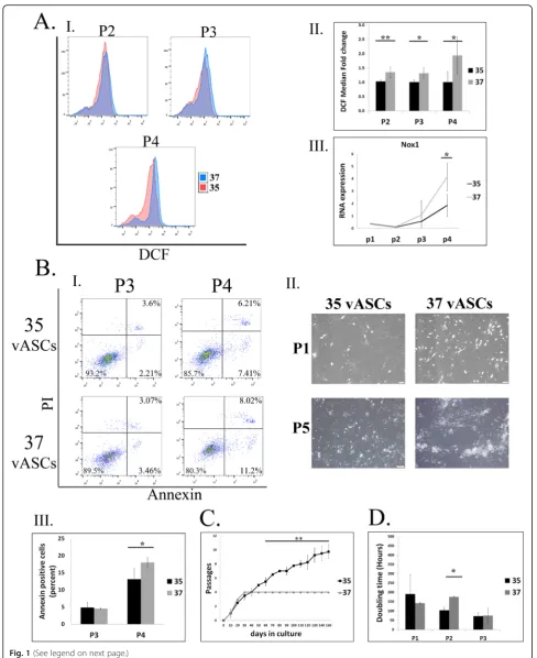
Culture of rat visceral ASCs (vASCs) under 35 °C
attenuates oxidative stress, leading to reduced apoptosis and long-term expansion
Fig. 1(See legend on next page.)
Culture under 35 °C enhances vASC fat differentiation potential
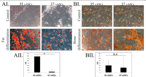
We previously described the low fat differentiation po-tential of vASCs compared to ASCs derived from sub-cutaneous fat, which correlated with measured levels of NOX1-induced oxidative stress [14]. As can be seen in Fig. 2, expansion of vASCs under 35 °C culture condi-tions promoted a drastic increase in fat differentiation as compared to vASCs cultured at 37 °C (Fig. 2A) but not of bone differentiation (Fig.2B).
Gene array analysis identifies 110 genes significantly up-or downregulated in 37 °C compared to 35 °C vASC cultures
In pursuit of the molecular mechanisms responsible for the phenotypic differences between 37 °C vASC and 35 °C vASC cultures, we performed a compara-tive gene array analysis of vASCs grown under both culture conditions. The gene array analysis was per-formed on passage 3 cells, which preceded the stage at which 37 °C vASC cultures underwent (See figure on previous page.)
Fig. 1Culture of vASCs at 35 °C attenuates NOX1-dependent oxidative stress and apoptosis, enabling long-term expansion.vASCs were propagated at 35 °C or 37 °C.AROS accumulation was detected by DCFDA staining and analyzed by flow cytometry at the indicated passages (P). (AI) Representative FACS analyses. (AII) Summary results of three independent repeats at P2–4. Error bars represent SD. (AIII) qRT-PCR analysis of NOX1 RNA levels in cells grown at 35 °C (black line) versus 37 °C (gray line) at the indicated passages (n= two independent repeats).BThe percentage of apoptotic cells at each passage of cells grown at 37 °C and 35 °C was determined by PI/ Annexin staining, followed by FACS analysis. (BI) Representative FACS analyses. (BII) Images of representative 37 °C and 35 °C vASCs at P1 and P5. (BIII) Mean of FACs analyses by-passage rate of apoptosis in 37 °C and 35 °C vASC cultures (n= two independent repeats).CA growth curve comparing the ability of visceral ASCs to undergo long-term expansion at 37 °C versus 35 °C as evident by the number of passages reached against the time in culture in days (n= four independent repeats). Cells were passaged only when reaching confluence. Error bars represent standard deviation (SD).DThe doubling time of 37OC and 35OC vASC was compared (n=two independent repeats). The doubling time of 37 °C and 35 °C vASC was compared (n= two independent repeats). Student’s two-tailedt test for equal variance: **p< 0.01; *p< 0.05
significant apoptosis and expansion arrest. The ana-lysis categorized the 37 °C vASCs and 35 °C vASCs as two separate populations (Fig. 3a). The gene array analysis identified 110 genes that were signifi-cantly (p< 0.05) up- or downregulated (> 2-fold change) in 35 °C vASCs as compared to 37 °C vASCs (Fig. 3a).
Function analysis of differentially expressed genes demonstrates a reduced inflammatory phenotype in 35 °C vASCs
In order to further characterize the changes in gene ex-pression observed between 35 °C vASCs and 37 °C vASCs, the 110 up/downregulated genes were analyzed using STRING database defining protein-protein Fig. 3Gene array analysis, protein-protein interactions, and function analysis of vASC differentially expressed genes (35 °C, 37 °C). RNA was produced from passage 3 35 °C and 37 °C vASCs. Samples were analyzed for gene expression using Affymetrix RaGene-2.1-st-v1 microarray.aCluster analysis of the gene array experiment samples showed a clear separation of each treatment (35 °C and 37 °C). Heatmap of 110 differentially expressed genes (p< 0.05 and fold-change difference = 2) (a). bPresentation of the 110 differentially expressed genes (p< 0.05 and fold-change difference = 2) responsible for the molecular changes elicited by temperature
change (STRING)
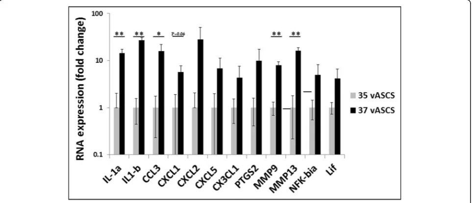
Fig. 4qRT-PCR-based validation of genes highlighted in the functional cluster analysis. RNA was extracted from passage 3 37 °C and 35 °C vASCs, and the gene expression of the indicated genes was compared by qRT-PCR (n= 2–3 independent repeats). Gene expression was validated according to the differential expression in the gene array analysis
interactions and functional enrichment of gene clusters (Fig. 3b). A drastic reduction in the expression level of a range of immune-related factors was noted in 35 °C vASC cultures as compared to 37 °C vASC cultures (Additional file 2: Table S2). An additional gene cluster termed “response to oxygen-containing compounds” containing, amongst other genes, important immune modulators showed reduced gene expression levels in 35 °C vASCs as compared to 37 °C vASCs (Add-itional file2: Table S2). Interestingly, temperature reduc-tion was also found to affect cell proliferation, differentiation, and wound healing potential (Add-itional file 2: Table S2), suggesting a possible influence of culture conditions on the regeneration potential of ASCs. The gene array results (Additional file 3: Table S3) were validated by performing qRT-PCR on 12 repre-sentative genes from all four gene groups; the differential gene expression between groups was preserved (Fig.4).
ROS accumulation in rat subcutaneous ASCs (scASCs) correlates with increased expression of pro-inflammatory cytokines
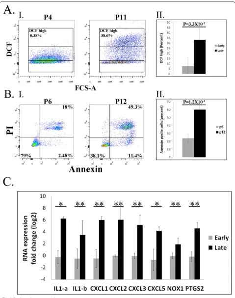
The results presented thus far demonstrated the ten-dency of 37 °C vASC cultures to develop ROS over-accumulation/oxidative stress that was correlated with a shift toward a pro-inflammatory phenotype. We previously reported a reduced tendency of scASCs to accumulate ROS at passages 1–6 compared to vASCs [14]. However, As can be seen in Fig. 5A, rat scASCs cultured at normal culture conditions (37 °C) demon-strated increased ROS accumulation (Fig. 5A) and apoptosis (Fig. 5B) at late passages (> 10) compared to their early counterparts (passages 4–6). Thus, to verify the correlation between ROS accumulation and the development of a pro-inflammatory phenotype, we compared the pro-inflammatory phenotype of early versus late scASCs. As seen with vASCs, increased ROS accumulation at late scASC passages was associ-ated with an increased pro-inflammatory phenotype (Fig. 5C).
Culture of human subcutaneous ASCs at 35 °C attenuates oxidative stress development, leading to reduced apoptosis without impairing expansion
Clinically relevant human ASCs are generally produced from subcutaneous fat extracted by different liposuction techniques. Therefore, we decided to examine whether our findings in rat vASCs and scASCs also apply to hu-man subcutaneous ASCs. To this end, we examined the effect of reduced culture temperature on the phenotype of subcutaneous ASCs by comparing their long-term ex-pansion at 37 °C and 35 °C. Increased ROS accumulation in 37 °C compared to 35 °C ASCs was already evident at passage 3 and was further enhanced with increasing
passages (Fig. 6A). Importantly, however, the culture of ASCs at 35 °C did not affect their expansion rate, as evi-dent from cell quantities comparable to those of 37 °C ASCs at passages 0–3 (Fig.6B). At passage 5, ROS over-accumulation in 37 °C ASCs led also to their increased apoptosis as compared to 35 °C ASCs (Fig.6C). The cor-relation between ROS accumulation and the develop-ment of a pro-inflammatory phenotype was also demonstrated, with human ASCs cultured at 37 °C exhi-biting increased expression of pro-inflammatory cyto-kines (IL-1b, IL-1a, and IL-6) as compared to ASCs cultured at 35 °C (Fig.6D–F).
Discussion
We and others have previously described the tendency of ASCs to accumulate ROS during long-term culturing, leading to DNA damage, apoptosis, expansion arrest, and reduced fat differentiation potential [12,14]. In the current study, we showed, for the first time, that a 2 °C reduction in the culture temperature inhibits ROS over-accumulation in both human subcutaneous ASCs and rat vASCs, resulting in reduced apoptosis and improved long-term expansion. In addition, the culture of rat vASCs at 35 °C led also to their enhanced fat differenti-ation compared to 37 °C vASCs. Gene array analysis demonstrated reduced expression of many immune modulators in 35 °C versus 37 °C ASC cultures, indicat-ing a reduced inflammatory phenotype under 35 °C cul-ture conditions. Functional analysis further identified reduced expression of a cluster of“response to oxygen-containing compounds” genes in 35 °C vASCs compared to 37 °C vASCs, correlating the increased inflammatory phenotype in 37 °C vASCs with their increased ROS pro-duction and oxidative stress. The correlation between ROS overproduction and the pro-inflammatory pheno-type was further corroborated by the observed increased expression of pro-inflammatory genes in both human and rat subcutaneous ASCs in response to ROS accumu-lation. Thus, reducing culture temperature can enhance ASC quality and quantity and prevent culture-induced oxidative stress and cell damage, which may all impact their clinical effectiveness.
Fig. 5(See legend on next page.)
was reported for various cell types [33–36]. Reduced ROS accumulation in 32 °C bone marrow-derived MSC cultures was previously reported [38]. Yet, the culture of both bone marrow-derived MSCs and add-itional cell types at temperatures below 34 °C was also reported to inhibit cell proliferation, [35, 38] hindering cell expansion. In contrast, culture at tem-peratures between 34 and 35.5 °C was reported to promote similar cell proliferation rates as cultures at 37 °C, despite a significant reduction in cellular oxy-gen and glucose consumption [37, 39]. We therefore chose to assess long-term expansion of ASCs under mild hypothermia (35 °C). These culture conditions were able to salvage both rat and human ASCs from ROS overpro-duction and consequential apoptosis. In rat vASCs, redu-cing culture temperature from 37 to 35 °C also prevented apoptosis-induced expansion arrest that was previously observed in 37 °C vASCs, enabling long-term expansion, without affecting proliferation rates; the 35 °C vASC cul-tures proliferated just as effectively as passage 0–3 37 °C vASC cultures prior to expansion arrest. Similar to vASCs, no reduction in the proliferation of 35 °C human ASCs was observed compared to 37 °C human ASCs.
We previously reported that ROS overproduction in vASCs was accompanied by increased expression of NOX1 but not of NOX2 or NOX4. NOX family mem-bers are an important source of intracellular ROS point-ing to NOX1 involvement in ROS accumulation [14]. This was verified when vASCs that were grown under 3% oxygen conditions and expanded long term displayed reduced NOX1 expression and decreased ROS accumu-lation [14]. NOX1 involvement in vASC cytostasis was reaffirmed when cells that were expanded under normal oxygen conditions (21%) in the presence of a specific NOX1 inhibitor, ML171, demonstrated reduced ROS ac-cumulation, reduced apoptosis, and long-term expansion [14]. In the current study, we demonstrated that vASC culturing at 35 °C was associated with an attenuated ROS accumulation in correlation with NOX1 expression, a reduction in apoptosis rates, and long-term expansion of the cell population as compared to vASCs cultured at 37 °C. This indicates that ROS decreased accumulation in 35 °C vASCs compared to 37 °C vASCs resulted, at least in part, from the reduction of NOX1 expression under 35 °C culture conditions.
We previously showed a direct correlation between the reduction of ROS levels and fat differentiation po-tential in 37 °C vASC cultures upon administration of a NOX1-specific inhibitor [14]. Enhanced fat differenti-ation potential in correldifferenti-ation with reduced ROS accumu-lation was similarly observed in scASCs [14]. In the current study, reduced culture temperature increased vASC fat differentiation capabilities in correlation with reduced ROS accumulation. It is thus reasonable to as-sume that the markedly improved fat differentiation po-tential of 35 °C vASC cultures was a direct result of reduced ROS production. It has been generally agreed that ROS accumulation positively regulates fat differenti-ation [42], although the exact source of ROS during adi-pogenesis is still controversial. Thus, unlike the temporal physiological increase in ROS levels, previously reported during fat differentiation, the increased chronic and toxic accumulation of ROS in 37 °C vASC cultures seems to disrupt many physiological processes, including fat differentiation processes.
Comparison of the gene expression pattern of 37 °C versus 35 °C ASCs revealed an increased expression of various positive modulators of immune processes, in-cluding the pivotal IL-1a and IL-1b cytokines and vari-ous chemokines in 37 °C ASCs. Thus, culturing ASCs under slightly hypothermic conditions reduces their in-flammatory phenotype as compared to 37 °C ASCs. In recent years, MSCs have gained much attention due to their potential application as immunomodulators for the treatment of immune-mediated diseases such as auto-immune diseases, graft-versus-host disease (GvHD), and inflammatory diseases [43]. In such cases, it would seem that the reduced inflammatory phenotype of 35 °C vASCs would provide a clinical advantage.
In an earlier study [14], we showed that early-passage scASCs cultured at 37 °C generated less ROS as com-pared to 37 °C vASCs and consequently demonstrated reduced apoptosis and enhanced expansion and fat dif-ferentiation than the former [14]. In the current study, similar to scASCs, vASCs cultured at 35 °C demon-strated reduced ROS accumulation that correlated with their reduced apoptosis and enhanced expansion and fat differentiation compared to 37 °C vASCs. Thus, we speculate that most of the changes in gene expression levels measured in 35 °C vASCs occurred in response to (See figure on previous page.)
Fig. 6(See legend on next page.)
reduced ROS production in these cells. Accordingly, analysis of the gene array results also uncovered en-hanced expression of genes associated with response to oxygen, the majority of which were immune modulators or genes involved in immune regulation, in 37 °C vASCs. Therefore, since 35 °C culture conditions also led to a significant reduction in ROS accumulation in vASCs, we wanted to determine whether ROS accumulation played a direct role in the development of the pro-inflammatory phenotype of 37 °C vASCs. To this end, we compared the expression of pro-inflammatory im-mune modulators between early- and late-passage (> 10) rat scASCs cultured at 37 °C, which display increased oxidative stress at later passages. Indeed, a significant in-crease in the pro-inflammatory phenotype was found in late- compared to early-passage scASCs, in correlation with oxidative stress enhancement, indicating that ROS accumulation and not culture temperature was respon-sible for the phenotypic shift. In accordance, 35 °C hu-man ASCs cultures demonstrated significantly reduced ROS production in correlation with the reduced expres-sion of the pro-inflammatory cytokines IL-1a, IL-1b, and IL-6 compared to 37 °C human ASCs. In conclusion, we propose that the development of intracellular oxidative stress in ASC cultures elicits increased expression of various immune modulators, with a tendency toward a pro-inflammatory phenotype.
Conclusions
Thus, a mild temperature shift in ASC cultures can in-duce profound effects on their in vitro phenotype with-out affecting their proliferation rate, possibly affecting the cells’in vivo clinical efficacy. Given their highly plas-tic nature, the culture conditions of MSCs, in general, and of ASCs, in particular, must be strictly controlled to avoid inter-batch variability. In addition, given the undis-puted harmful effect of oxidative stress on cells, we speculate that a mildly reduced culture temperature may serve as a simple means of preventing ROS overproduc-tion in ASCs.
Supplementary information
Supplementary informationaccompanies this paper athttps://doi.org/10. 1186/s13287-019-1542-0.
Additional file 1: Table S1.Primer list.
Additional file 2: Table S2.Function analysis of the differentially expressed genes between 37 °C and 35 °C ASCs.
Additional file 3: Table S3.Gene array results of the main pro-inflammatory genes appearing in the function analysis of the differentially expressed genes between 37 °C and 35 °C ASCs.
Abbreviations
ASCs:Adipose-derived stem cells; DMEM: Dulbecco’s modified Eagle’s medium; FCS: Fetal calf serum; GvHD: Graft-versus-host disease; H2DCFDA: 2′,7-Dichlorodihydrofluorescein diacetate; MSCs: Mesenchymal stem cells; PI: Propidium iodide; ROS: Reactive oxygen species; RT: Room temperature; scASCs: Subcutaneous ASCs; SVF: Stromal vascular fraction; vASCs: Rat visceral ASCs
Acknowledgements Not applicable
Authors’contributions
GT performed most of the experimental work presented on rat ASCs. IS performed most of the experimental presented on human ASCs and was incremental in the analysis of FACS data. MS was involved in rat ASC long-term culture and in sample preparation for the gene array analysis. MPC per-formed the analysis of the gene array data. IG was involved in the experi-ments presented on rat scASCs and performed the qRT-PCR analysis. NS and EG supervised all the experimental work performed and in getting the fund-ing for the research. NS and GT prepared the manuscript. All authors read and approved the final manuscript.
Funding
We have no funding resources to declare.
Availability of data and materials
The datasets used and/or analyzed during the current study are available from the corresponding author on reasonable request.
Ethics approval and consent to participate
The Tel Aviv Sourasky Medical Center Institutional Animal Care and Use Committee approved all animal experiments.
All procedures were performed in accordance with the Declaration of Helsinki and approved by the ethics committee of Tel Aviv Sourasky Medical Center. Written, informed consent was obtained from all patients in advance.
Consent for publication Not applicable
Competing interests
The authors declare that they have no competing interests. (See figure on previous page.)
Author details
1The Department of Plastic and Reconstructive Surgery, Tel Aviv Sourasky
Medical Center, Tel Aviv, Israel.2The Bioinformatics Unit George S. Wise
Faculty of Life Sciences, Tel Aviv University, Tel Aviv, Israel.3The Department of Plastic and Reconstructive Surgery, Tel Aviv Sourasky Medical Center, affiliated with the Sackler Faculty of Medicine, Tel Aviv University, Weizmann 6, Tel Aviv, Israel.
Received: 23 July 2019 Revised: 10 December 2019 Accepted: 26 December 2019
References
1. Uccelli A, Moretta L, Pistoia V. Mesenchymal stem cells in health and disease. Nat Rev Immunol. 2008;8(9):726–36.
2. Bourin P, Bunnell BA, Casteilla L, Dominici M, Katz AJ, March KL, et al. Stromal cells from the adipose tissue-derived stromal vascular fraction and culture expanded adipose tissue-derived stromal/stem cells: a joint statement of the International Federation for Adipose Therapeutics and Science (IFATS) and the International Society for Cellular Therapy (ISCT). Cytotherapy. 2013;15(6):641–8.
3. Zuk PA, Zhu M, Mizuno H, Huang J, Futrell JW, Katz AJ, et al. Multilineage cells from human adipose tissue: implications for cell-based therapies. Tissue Eng. 2001;7(2):211–28.
4. Oliveira PH, Silva CL, Cabral J. Concise review: genomic instability in human stem cells: current status and future challenges. Stem Cells. 2014;32(11): 2824–32.
5. Brown DI, Griendling KK. Nox proteins in signal transduction. Free Radic Biol Med. 2009;47(9):1239–53.
6. Circu ML, Aw TY. Reactive oxygen species, cellular redox systems, and apoptosis. Free Radic Biol Med. 2010;48(6):749–62.
7. Giorgio M, Trinei M, Migliaccio E, Pelicci PG. Hydrogen peroxide: a metabolic by-product or a common mediator of ageing signals? Nat Rev Mol Cell Biol. 2007;8(9):722–8.
8. Mohyeldin A, Garzón-Muvdi T, Quiñones-Hinojosa A. Oxygen in stem cell biology: a critical component of the stem cell niche. Cell Stem Cell. 2010; 7(2):150–61.
9. Baez A, Shiloach J. Effect of elevated oxygen concentration on bacteria, yeasts, and cells propagated for production of biological compounds. Microb Cell Factories. 2014;13(1):181.
10. Bétous R, Renoud ML, Hoede C, Gonzalez I, Jones N, Longy M, et al. Human adipose-derived stem cells expanded under ambient oxygen concentration accumulate oxidative DNA lesions and experience procarcinogenic DNA replication stress. Stem Cells Transl Med. 2017;6(1):68–76.
11. Boregowda SV, Krishnappa V, Chambers JW, Lograsso PV, Lai WT, Ortiz LA, et al. Atmospheric oxygen inhibits growth and differentiation of marrow-derived mouse mesenchymal stem cells via a p53-dependent mechanism: implications for long-term culture expansion. Stem Cells. 2012;30(5):975–87. 12. Estrada J, Albo C, Benguria A, Dopazo A, Lopez-Romero P, Carrera-Quintanar
L, et al. Culture of human mesenchymal stem cells at low oxygen tension improves growth and genetic stability by activating glycolysis. Cell Death Differ. 2012;19(5):743–55.
13. Kim J-W, Tchernyshyov I, Semenza GL, Dang CV. HIF-1-mediated expression of pyruvate dehydrogenase kinase: a metabolic switch required for cellular adaptation to hypoxia. Cell Metab. 2006;3(3):177–85.
14. Sela M, Tirza G, Ravid O, Volovitz I, Solodeev I, Friedman O, et al. NOX1-induced accumulation of reactive oxygen species in abdominal fat-derived mesenchymal stromal cells impinges on long-term proliferation. Cell Death Dis. 2015;6(4):e1728.
15. Lengner CJ, Gimelbrant AA, Erwin JA, Cheng AW, Guenther MG, Welstead GG, et al. Derivation of pre-X inactivation human embryonic stem cells under physiological oxygen concentrations. Cell. 2010;141(5):872–83. 16. Mathieu J, Zhou W, Xing Y, Sperber H, Ferreccio A, Agoston Z, et al.
Hypoxia-inducible factors have distinct and stage-specific roles during reprogramming of human cells to pluripotency. Cell Stem Cell. 2014;14(5): 592–605.
17. Yoshida Y, Takahashi K, Okita K, Ichisaka T, Yamanaka S. Hypoxia enhances the generation of induced pluripotent stem cells. Cell Stem Cell. 2009;5(3): 237–41.
18. Basciano L, Nemos C, Foliguet B, de Isla N, de Carvalho M, Tran N, et al. Long term culture of mesenchymal stem cells in hypoxia promotes a
genetic program maintaining their undifferentiated and multipotent status. BMC Cell Biol. 2011;12(1):12.
19. Choi JR, Pingguan-Murphy B, Abas WABW, Azmi MAN, Omar SZ, Chua KH, et al. Impact of low oxygen tension on stemness, proliferation and differentiation potential of human adipose-derived stem cells. Biochem Biophys Res Commun. 2014;448(2):218–24.
20. Choi JR, Yong KW, Safwani WKZW. Effect of hypoxia on human adipose-derived mesenchymal stem cells and its potential clinical applications. Cell Mol Life Sci. 2017;74(14):2587–600.
21. Fehrer C, Brunauer R, Laschober G, Unterluggauer H, Reitinger S, Kloss F, et al. Reduced oxygen tension attenuates differentiation capacity of human mesenchymal stem cells and prolongs their lifespan. Aging Cell. 2007;6(6): 745–57.
22. Grayson WL, Zhao F, Bunnell B, Ma T. Hypoxia enhances proliferation and tissue formation of human mesenchymal stem cells. Biochem Biophys Res Commun. 2007;358(3):948–53.
23. Saller MM, Prall WC, Docheva D, Schönitzer V, Popov T, Anz D, et al. Increased stemness and migration of human mesenchymal stem cells in hypoxia is associated with altered integrin expression. Biochem Biophys Res Commun. 2012;423(2):379–85.
24. Valorani M, Montelatici E, Germani A, Biddle A, D'alessandro D, Strollo R, et al. Pre-culturing human adipose tissue mesenchymal stem cells under hypoxia increases their adipogenic and osteogenic differentiation potentials. Cell Prolif. 2012;45(3):225–38.
25. Denu RA, Hematti P. Effects of oxidative stress on mesenchymal stem cell biology. Oxidative Med Cell Longev. 2016; Article ID 2989076:9. 26. Cai L, Stevenson J, Geng X, Peng C, Ji X, Xin R, et al. Combining normobaric
oxygen with ethanol or hypothermia prevents brain damage from
thromboembolic stroke via PKC-Akt-NOX modulation. Mol Neurobiol. 2016. p. 1– 15.
27. Ning X-H, Xu C-S, Song YC, Xiao Y, Hu Y-J, Lupinetti FM, et al. Hypothermia preserves function and signaling for mitochondrial biogenesis during subsequent ischemia. Am J Phys Heart Circ Phys. 1998;274(3):H786–H93. 28. Tissier R, Chenoune M, Pons S, Zini R, Darbera L, Lidouren F, et al. Mild
hypothermia reduces per-ischemic reactive oxygen species production and preserves mitochondrial respiratory complexes. Resuscitation. 2013;84(2):249–55. 29. Zhao H, Steinberg G. Limited therapeutic time windows of
mild-to-moderate hypothermia in a focal ischemia model in rat. Stroke Res Treat. 2011;2011.
30. Dave KR, Christian SL, Perez-Pinzon MA, Drew KL. Neuroprotection: lessons from hibernators. Comp Biochem Physiol B: Biochem Mol Biol. 2012;162(1):1–9. 31. Brown JC, Chung DJ, Belgrave KR, Staples JF. Mitochondrial metabolic
suppression and reactive oxygen species production in liver and skeletal muscle of hibernating thirteen-lined ground squirrels. Am J Phys Regul Integr Comp Phys. 2012;302(1):R15–28.
32. Brown JC, Staples JF. Mitochondrial metabolic suppression in fasting and daily torpor: consequences for reactive oxygen species production. Physiol Biochem Zool. 2011;84(5):467–80.
33. Chuppa S, Tsai YS, Yoon S, Shackleford S, Rozales C, Bhat R, et al. Fermentor temperature as a tool for control of high-density perfusion cultures of mammalian cells. Biotechnol Bioeng. 1997;55(2):328–38.
34. Jorjani P, Ozturk SS. Effects of cell density and temperature on oxygen consumption rate for different mammalian cell lines. Biotechnol Bioeng. 1999;64(3):349–56.
35. Moore A, Mercer J, Dutina G, Donahue CJ, Bauer KD, Mather JP, et al. Effects of temperature shift on cell cycle, apoptosis and nucleotide pools in CHO cell batch cultues. Cytotechnology. 1997;23(1–3):47–54.
36. Weidemann R, Ludwig A, Kretzmer G. Low temperature cultivation—a step towards process optimisation. Cytotechnology. 1994;15(1–3):111–6. 37. Bloemkolk JW, Gray MR, Merchant F, Mosmann TR. Effect of temperature on
hybridoma cell cycle and MAb production. Biotechnol Bioeng. 1992;40(3): 427–31.
38. Stolzing A, Scutt A. Effect of reduced culture temperature on antioxidant defences of mesenchymal stem cells. Free Radic Biol Med. 2006;41(2):326–38. 39. Sureshkumar G, Mutharasan R. The influence of temperature on a mouse–
mouse hybridoma growth and monoclonal antibody production. Biotechnol Bioeng. 1991;37(3):292–5.
40. Dexter TM, Allen TD, Lajtha L. Conditions controlling the proliferation of haemopoietic stem cells in vitro. J Cell Physiol. 1977;91(3):335–44. 41. Szklarczyk D, Morris JH, Cook H, Kuhn M, Wyder S, Simonovic M, et al. The
STRING database in 2017: quality-controlled protein–protein association
networks, made broadly accessible. Nucleic Acids Res. 2016;45(D1):D362– D368.
42. Liu G-S, Chan EC, Higuchi M, Dusting GJ, Jiang F. Redox mechanisms in regulation of adipocyte differentiation: beyond a general stress response. Cells. 2012;1(4):976–93.
43. Zhao Q, Ren H, Han Z. Mesenchymal stem cells: immunomodulatory capability and clinical potential in immune diseases. J Cell Immunother. 2016;2(1):3–20.
Publisher’s Note