Original Article
Shear Bond Strength of a Multi-Mode Adhesive to Bur-Cut and Er,Cr:YSGG
Lased Dentin in Different Output Powers
Niloofar Shadman 1, Shahram Farzin Ebrahimi 1, Sara Amanpour 2, Siavash Mehdizadeh 3
1 Dept. of Operative Dentistry, Oral and Dental Diseases Research Center, School of Dentistry, Kerman University of Medical Sciences,
Kerman, Iran. 2
Dept. of Oral Pathology, Oral and Dental Diseases Research Center, School of Dentistry, Kerman University of Medical Sciences, Kerman, Iran.
3
Postgraduate Student, Dept. of Oral and Maxillofacial Surgery, School of Dentistry, Kerman University of Medical Sciences, Kerman, Iran.
KEY WORDS
Adhesive;
Bond Strength, Laser;
Dentin;
Received January 2018;
Received in Revised Form May 2018; Accepted June 2018;
ABSTRACT
Statement of the Problem: Universal or multi-mode adhesives are new adhesive systems that can be used in both etch-and-rinse (ER) and self-etch (SE) modes. Lesser
technical sensitivity and dual use of these adhesives have made them popular among
dentists. Studies are being conducted to analyze the advantages and disadvantages
these adhesives in different conditions
Purpose: The aim of this study was to compare shear bond strength (SBS) of a multi-mode adhesive in different etching multi-modes to Er,Cr:YSGG laser ablated and bur-cut
dentin.
Materials and Method: Buccal and lingual surfaces of 30 sound human molars, ran-domly divided to three groups, were prepared by bur and Er,Cr:YSGG (4 Watt and 5
Watt, 20 Hz, 96% water, 60% air, and 600-µm spot size) to reach a flat surface in
superficial dentin. Each group was randomly divided into 2 subgroups (ER and SE),
and then Scotchbond Universal adhesive was applied. Composite cylinders were
at-tached to the surfaces and cured. Specimens were stored in 37 °C water for 24 hours
and thermocycled (500 cycles) and were tested for SBS and failure modes were
deter-mined by stereomicroscope. Data was analyzed using SPSS19 and one- way ANOVA and Tukey’s post hoc tests and p< 0.05 was considered as significance level.
Results: Bur-cut dentin with ER method had the highest mean SBS value (33.80 MPa). SBS in bur-cut and 4Watt laser in ER mode were significantly higher than SE
mode (p= 0.002 and p= 0.000 respectively). Highest mean SBS value in lased dentin was achieve in 4 Watt ER mode.
Conclusion: SBS of Scotchbond universal adhesive to dentine is highest in bur-cut and ER mode and in 4-Watt lased-dentin is higher than 5-Watt lased-dentin. Moreover,
in 4-Watt lased-dentin, SBS of ER mode is more than SE mode.
Corresponding Author: Mehdizadeh S., Dept. of Oral and Maxillofacial Surgery, School of Dentistry, Kerman University of Medical Sciences, Kerman, Iran. Tel: +983432119028 Fax: +983432119021 Email: [email protected]
Cite this article as: Shadman N., Farzin Ebrahimi Sh., Amanpour S., Mehdizadeh S. Shear Bond Strength of a Multi-Mode Adhesive to Bur-Cut and Er,Cr:YSGG Lased Dentin in
Different Output Powers. J Dent Shiraz Univ Med Sci., June 2019; 20(2): 118-123.
Introduction
In 1917, Einstein presented laser theory and in 1960, the
first working laser device was manufactured [1]. Er:
YAG and Er,Cr:YSGG lasers are used in dentistry for
hard tissues removal. Er,Cr:YSGG laser with 2.78 µm
wavelength can remove both hard and soft tissues
with-out the vibration and pulpal thermal stress induced by
rotary instruments and less need for local anaesthetics.
Therefore, it has been gaining popularity in treating
tissue water, causing micro explosions which removes
hard tissue. This process is called laser ablation.
Be-cause of micro explosions in laser-ablated surfaces, we
cannot expect a flat surface, but a rough one. With this
property and the lack of smear layer in lased surfaces,
better and stronger adhesion properties are expected but
in studies, we see rather contradictory results [2].
Katuami et al. [3] observed microcracks under hybrid layer in Er:YAG laser irradiated dentin.
Moreo-ver, Dela Rosa et al. [4] described areas of dehydration and loss of protein with malformed hydroxyapatite
crys-tals in Er:YAG laser irradiated dentin. This layer was
3-4 µm in thickness [3-4]. It is suggested that laser ablated
surfaces have fused collagen fibrils and loss of
interfi-brillar space which prevents diffusion of resin to
inter-tubular dentin [2]. Others described various patterns of
micro irregularities, which cause lower bond strengths
[5]. All these superficial changes are related to intensity
of laser radiation [6]. Laser thermomechanical effects
will affect subsurface layer, leading to loss of integrity
and weakening of superficial layer, which is often
ex-plained as the reason for prevalence of cohesive failure
modes in dentin and enamel [4]. For enhancement of
bond strength, this altered surface should be either
changed or removed by chemical or mechanical means
[7].
Universal or multi-mode adhesives are new
adhe-sive systems that can be used in both etch-and-rinse
(ER) and self-etch (SE) modes. Lesser technical
sensi-tivity and dual use of these adhesives have made them
popular amongst dentist. Studies are being conducted to
analyse them in different conditions [8]. Lately, a new
adhesive from this category has been introduced to
mar-ket under the trade name of Scotchbond Universal (3M,
ESPE, USA). Manufacturers claim that this adhesive
has strong and stable bonding properties in both ER and
SE modes that result from its special formulation.
Re-cent studies report various results [8-12], but none of
them studied this adhesive in laser-ablated teeth.
Dunn et al. [5] reported lower shear bond strengths (SBS) in lased dentin and enamel from other
adhesives and considered loss of uniformity of hybrid
layer as the reason. Lee et al. [13] reported that after acid conditioning, lased dentin surfaces reached bond
strengths equal to bur-cut dentin, but without acid
con-ditioning, lower bond strengths were observed.
Marginal integrity and bond strength are the major
factors in assessing success of a restoration and both of
these properties are related to adhesive system
perfor-mance and the condition it is used [14].
In this laboratory study, we aimed to assess and
compare SBS of Scotchbond Universal to Er,Cr:YSGG
lased dentin with different output powers and bur-cut
dentin in two conditioning modes, ER and SE. The null
hypothesis is that mean SBS values do not differ
be-tween different methods of removing dentin and ER or
SE modes.
Materials and Method
In this in vitro experimental study, 30 sound human extracted third molars were used that were either
im-pacted or extracted for orthodontic reasons. Teeth were
cleaned of debris and remaining soft tissues, disinfected
in 0.5% sodium hypochlorite for 10 minutes, and stored
in tap water the whole time.
Plastic cylinders (2.5×2.5cm) were used for
mounting teeth in self-cured acrylic resin (Acropars,
Marlic, Tehran, Iran), 1mm beneath the CEJ line.
Buc-cal and lingual surfaces of each tooth were cut
perpen-dicular to the horizontal line to remove enamel using a
water-cooled air turbine and a diamond bur (long and
flat-end cylindrical 837L, TeezKavan, Tehran, Iran)
attached to a custom surveyor so that the cut surfaces
were completely flat. After exposing superficial dentin
(3*3 mm dentin surface), samples were randomly
divid-ed to three groups including bur-cut (Bur), 4Watt Laser
(4W), and 5Watt Laser (5W). Bur-cut samples received
no further preparation other than using a 600-grit silicon
carbide paper to produce a uniform smear layer. 4W
laser samples were lased with Er,Cr:YSGG laser
(Wa-terlase: Biolase, Irvine, CA, USA) with following
pa-rameters: 4W output power, water pressure 95% and air
pressure 60%. The MZ6 tip with the spot size of 600 µm
was used; holding it 1-2 mm above dentin surface with
a constant sweeping motion until at least 1mm of dentin
was ablated uniformly. 5W laser samples were prepared
just as above, only differing in output power. Each
group was randomly divided to two subgroups of SE
and ER and then Scotchbond Universal adhesive (3M,
ESPE, USA) was applied (Table 1).
Materials used in this study are described in Table
Table 1: Materials used in this study and application techniques of adhesive
Material Type Manufacturer Composition Application technique
Scotchbond Universal
2-step ER or 1-step
SE adhesive 3M, ESPE, USA
10-MDP, HEMA, Vitre-bond copolymer, filler, ethanol, water, initiators,
silane
Etch-and-rinse: acid etching (15s), rinsing (15s), blot-drying, two-coats of adhesive applied (20s) and gently air-drying (5s), light cured for 20s. Self-Etch: two-coats of adhesive applied (20s) and gently air-drying (5s), light cured for 20s. Filtek Z-250 XT
Light-cure nano Hybrid resin
com-posite
3M, ESPE, USA
GMA, UDMA, BIS-EMA, PEGDMA, TEGDMA, zirconium,
silica Ultra Etch Etching agent Ultradent, USA 35% phosphoric acid
diameter of 2mm was placed and fixed by sticky wax on
the bonded surface before composite placement and
curing. Light curing for adhesive and composite was 20
and 40 seconds respectively (Optilux 501, Kerr, USA)
at a light irradiance level of 600 mW/cm2. After curing,
composite plastic mold was removed. Then specimens
were stored in tap water in 37 °C for 24 hours
(Incuba-tor 6520, Behdad, Iran) before receiving 500 cycles of
thermocycling between 5 and 55 °c (with a 60 s dwell
time and a 15 s transfer time). The specimens were
loaded to failure at 0.5mm/min using the Testometric
machine (Testometric M350-10 CT, England) with a
metal rod with a chisel-shaped end adjacent to the flat
ground dentin surface. The SBS values (MPa) were
calculated by dividing peak load at failure at bonded
surface area. One-way ANOVA and Tukey’s post hoc
tests were used with the p< 0.05 considered as the level of significance. Tested specimens were observed under
a stereomicroscope (SEM) (Olympus, DP12, Germany)
at 16x magnification by two calibrated observers
(spe-cialist in operative dentistry) separately for determining
failure modes. Four types of failure modes were
consid-ered in this study including adhesive failure, cohesive
failure in dentin, cohesive failure in composite, and
fi-nally, mixed failure- partially adhesive and partially
cohesive [15].
Results
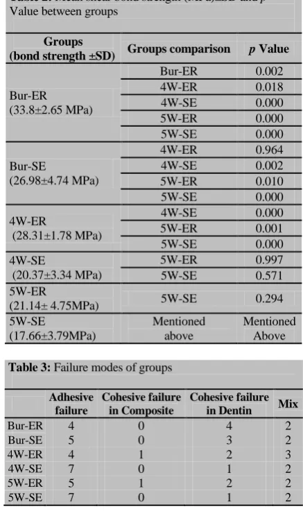
The results of mean SBS values for each subgroup are
shown in Table 2.
Bur-cut dentin with ER mode had the highest
mean SBS value (33.80 MPa). In similar etching
modes, Bur-cut groups showed significantly higher
SBS values than laser ablated groups. In ER mode, 4W
laser showed significantly higher SBS values than 5W
laser, however; it was not statistically significant in SE
mode (p= 0.571). Generally, etching mode affected
SBS values and produced significantly higher bond
strength in ER mode in Bur-cut (p= 0.00) and 4W laser (p= 0.00) but was not statistically significant in 5W laser (p= 0.294). The highest mean SBS value (28.31 MPa) in lased dentin was achieved in 4W ER mode.
Failure patterns are shown in table 3. The Bur-ER
sub-group showed more non-cohesive failure pattern while
other subgroups showed mainly adhesive failure
pat-tern.
Table 2: Mean shear bond strength (MPa)±SD and p Value between groups
Groups
(bond strength ±SD) Groups comparison p Value
Bur-ER (33.8±2.65 MPa)
Bur-ER 0.002
4W-ER 0.018
4W-SE 0.000
5W-ER 0.000
5W-SE 0.000
Bur-SE
(26.98±4.74 MPa)
4W-ER 0.964
4W-SE 0.002
5W-ER 0.010
5W-SE 0.000
4W-ER
(28.31±1.78 MPa)
4W-SE 0.000
5W-ER 0.001
5W-SE 0.000
4W-SE
(20.37±3.34 MPa)
5W-ER 0.997
5W-SE 0.571
5W-ER
(21.14± 4.75MPa) 5W-SE 0.294
5W-SE
(17.66±3.79MPa)
Mentioned above
Mentioned Above
Table 3: Failure modes of groups
Adhesive failure
Cohesive failure in Composite
Cohesive failure in Dentin Mix
Bur-ER 4 0 4 2
Bur-SE 5 0 3 2
4W-ER 4 1 2 3
4W-SE 7 0 1 2
5W-ER 5 1 2 2
5W-SE 7 0 1 2
Discussion
fracture and stress caused by rotary instruments [16].
Thermal injury to pulp, vibration, and annoying noise
are all disadvantages related to rotary instruments,
which are not present in use of laser ablation. Such
mat-ters would help in treatment of anxious patients and
reducing the dose of anesthesia needed [17].
In similar modes of application of Scotchbond
Universal, SBS mean values in bur-cut dentin were
sig-nificantly higher than both 4W and 5W laser ablated
groups. In Esteves-Oliviera et al. study [18], SBS values in both laser-ablated enamel and dentin were lower than
bur-cut surfaces. They concluded that thermal effects of
Er,Cr:YSGG laser causes changes in hydroxyapatite,
leading to more resistance of the surfaces to acid, the
issue, which does not happen in bur-cut dentine [18].
On the other hand, denatured matrix proteins prevent
proper permeation of adhesive in collagen matrix
caus-ing lower bond strengths [4]. In a review of the
litera-ture by Lopes et al., [19] it was mentioned a reduction in bond strength in laser irradiated dentin compared
with conventional methods. Lee et al. [13] reported bet-ter tensile bond strengths in laser-ablated dentin afbet-ter
acid etching of the surface, equal to bur-cut dentin. In
SEM images, acid etched lased dentin was almost
simi-lar to acid etched bur-cut dentin, but without acid
condi-tioning, lased dentin showed an uneven scaly surface.
The reduced bond strengths of lased dentin without acid
etch is due to improper formation of hybrid layer, as
collagen matrix is not fully exposed or permeable for
resin penetration. [13] Shahabi et al. [20] reported simi-lar tensile bond strengths in lased and bur-cut dentin
after acid conditioning.
In this study, in ER mode of 4W subgroup, the
mean SBS value was significantly higher than 5W
subg-roup. Few studies have assessed different settings of Er,
Cr:YSGG laser on tooth surface and resulting bond
strengths [19-21]. Most studies have employed manu-facturer’s recommended settings (4W, 20 Hz, 65% Air, 55% Water) [18-20, 22].
It is asserted that greater power outputs of device
produces more surface heat, causing melting in
hydrox-yapatite, which subsequently leads to more acid
re-sistance [4]. Consequently, more microcracks are
ob-served in SEM evaluations [5], which might be the
rea-son for lower mean SBS value in 5W than 4W
sub-groups. In SE mode, because of lower acidity of
adhe-sive and less penetration of it to dentin, a thinner hybrid
layer will be produced; which might explain why bond
strength was lower in 4W subgroup, though the
differ-ence between 4W and 5W subgroups was not
statistical-ly significant. Higher mean SBS value of Scotchbond
Universal for bur-cut dentin than lased dentin in SE
mode might be due to the mentioned superficial thermal
effects induced by laser as Scotchbond Universal has
mild acidic monomers (pH=2.6) and acts less efficient
in removing mineral content. [23] Sun et al. [6] studied different output powers of Er,Cr:YSGG laser from 1 to
6 Watt on sclerotic dentin with the rest of the settings
unchanged (20 Hz, 65% Air, 55% Water). They
ob-served that surface roughness increased with higher
outputs but decreased in 5W and 6W. Furthermore,
mean open tubular area increased from 1W to 6W but
the difference between 4W, 5W, and 6W groups was
not statistically significant. As a result, 4W group
pro-duced highest mean microSBS value. They considered
surface cracks, observed by SEM, responsible for these
lower bond strengths [6] Likewise, these findings can be
relevant in explaining our results. In studying
Er,Cr:YSGG lased tooth surfaces under SEM, Lee et al.
[13] reported that in output powers higher than 3.5W
microcracks have appeared, air spray at 80% produced
the roughest surface and finally, maximum water output
produced the least carbonized surface. In the present
study, device settings were adopted regarding the
previ-ous studies [6, 13]and considering manufacturer’s
rec-ommendations. Maximum water pressure was selected
as in pressures under 90%, carbonization and charring
of dentin surface was observed, and dry bur-cut dentin
odor was smelled in pilot study.
In the present study, in bur-cut and 4W lased
den-tin, Scotchbond Universal mean SBS value was higher
in ER mode than SE mode. Various bond strength of
Scotchbond Universal has been reported in ER and SE
mode in different studies [8, 10, 12]. Munoz et al. [10] reported no statistically significant difference in
micro-tensile bond strength of different etching modes and in
Takamizawa et al. study, [12] SBS was reported signifi-cantly higher in SE mode. Wagner et al. [8] reported no statistically significant difference in microtensile bond
strength between the two modes. They observed that
although the length of resin tags and hybrid layer
the bond strength did not differ significantly. In
addi-tion, in Ayar et al. study [24], similar bond strength was concluded between two methods. Competitive property
of MDP monomer and Vitrebond copolymer for
bond-ing to hydroxyapatite calcium has been used as an
ex-planation of these various results [8]. On the other hand,
Shadman et al. [9] reported significantly higher SBS values for ER mode than SE mode in sound and caries
affected dentin. They considered better surface
mor-phology and more penetration of resin tags in ER mode
and weak acidity of Scotchbond Universal self-etching
monomers as the reason for higher SBS values of ER
mode [9]. Similar results were revealed in some studies
[25-26]. Difference between the results of these studies
can be due to technical sensitivity of ER mode, dry/wet
bonding [27], samples morphology in microtensile tests
(matchstick or dumbbell shape), type of bond strength
tested (shear or tensile) and the condition samples are
stored in after preparation [28].
Shallow and superficial etching and reduced
mi-cromechanical retention is the main worry of mild SE
adhesives [29]. Conditioning with phosphoric acid in
dentin before applying SE adhesives can produce better
interfacial morphology by creating a thicker hybrid
lay-er and longlay-er resin tags. Elimination of smear laylay-er and
smear plugs by acid conditioning can facilitate
permea-tion of mild SE adhesives [8].
Failure modes in shear tests can differ because of
mechanics of the test and distribution of stress in the
in-terface, thus it does not necessarily imply bond efficacy
[30]. After reviewing failure modes in this study,
non-adhesive failure modes were seen in bur-cut and 4W laser
groups, probably because of higher bond strengths. In
Takamizawa et al. study [12], most frequent failure pat-tern of Scotchbond Universal adhesive in ER and SE
modes was cohesive failure in dentin. In Marchesi et al.
study [31] on microtensile bond strength of Scotchbond
Universal, most frequent failure pattern in ER mode was
cohesive type. The SEM evaluation of the effect of other
laser outputs is suggested for future studies.
Conclusion
Bur-cut dentin had significantly higher SBS values than
lased dentin. In ER mode, 4W lased-dentin had
signifi-cantly higher SBS values than 5W lased-dentin. In
bur-cut and 4W lased-dentin, Scotchbond Universal had
significantly higher SBS values in ER mode. Dominant
failure mode in low SBS value subgroups, such as
5W-ER and 5W-SE, was adhesive failure.
Acknowledgements
This study was funded by Kerman University of
Medi-cal Sciences. The authors express their gratitude toOral
and Dental Diseases Research Center for helping in
laboratory procedures.
Conflict of Interest
The authors declare that they have no conflict of
inter-est.
References
[1] Martens LC. Laser physics and a review of laser
applica-tions in dentistry for children. Eur Arch Paediatr Dent.
2011; 12: 61-67.
[2] Hibst R. Lasers for caries removal and cavity preparation:
State of the art and future directions. J Oral Laser
Applica-tions. 2002; 2: 202-212.
[3] Kataumi M, Nakajima M, Yamada T, Tagami J. Tensile
bond strength and SEM evaluation of Er:YAG laser
irradi-ated dentin using dentin adhesive. Dent Mater J. 1998; 17:
125–138.
[4] Dela Rosa A, Sarma AV, Le CQ, Jones RS, Fried D.
Pe-ripheral thermal and mechanical damage to dentin with
mi-crosecond and sub-mimi-crosecond 9.6 microm, 2.79 microm,
and 0.355 microm laser pulses. Lasers Surg Med. 2004; 35:
214-228.
[5] Dunn WJ, Davis JT, Bush AC. Shear bond strength and
SEM evaluation of composite bonded to Er:YAG
laser-prepareddentin and enamel. Dent Mater. 2005; 21:
616-624.
[6] Sun X, Ban J, Sha X, Wang W, Jiao Y, Wang W, et al.
Effect of Er,Cr:YSGG Laser at Different Output Powers on
the Micromorphology and the Bond Property of
Non-Carious Sclerotic Dentin to Resin Composites. PLoS One.
2015; 10: e0142311.
[7] Obeidi A, McCracken MS, Liu PR, Litaker MS, Beck P,
Rahemtulla F. Enhancement of bonding to enamel and
den-tin prepared by Er,Cr:YSGG laser. Lasers Surg Med. 2009;
41: 454-462.
[8] Wagner A, Wendler M, Petschelt A, Belli R, Lohbauer U.
Bonding performance of universal adhesives in different
etching modes. J Dent. 2014; 42: 800-807.
Ghaderi A. Shear bond strength of different adhesive
sys-tems to normal and caries-affected dentin. J Oral Health
Oral Epidemiol. 2015; 4: 87-93.
[10]Muñoz MA, Luque I, Hass V, Reis A, Loguercio AD,
Bombarda NH. Immediate bonding properties of universal
adhesives to dentine. J Dent. 2013; 41: 404-411.
[11]Perdigão J, Kose C, Mena-Serrano AP, De Paula EA, Tay
LY, Reis A, et al. A new universal simplified adhesive:
18-month clinical evaluation. Oper Dent. 2014; 39: 113-127.
[12]Takamizawa T, Barkmeier WW, Tsujimoto A, Berry TP,
Watanabe H, Erickson RL, et al. Influence of different
etching modes on bond strength and fatigue strength to
dentinusing universal adhesive systems. Dent Mater. 2016;
32: e9-e21.
[13]Lee BS, Lin PY, Chen MH, Hsieh TT, Lin CP, Lai JY, et
al. Tensile bond strength of Er,Cr:YSGG laser-irradiated
human dentin and analysis of dentin-resin interface. Dent
Mater. 2007; 23: 570-578.
[14]Van Landuyt KL, Snauwaert J, De Munck J, Peumans M,
Yoshida Y, Poitevin A, et al. Systematic review of the
chemical composition of contemporary dental adhesives.
Biomaterials. 2007; 28: 3757-3785.
[15]Unlu N, Gunal S, Ulker M, Ozer F, Blatz MB. Influence of
operator. Experience on in vitro bond strength of dentin
ad-hesives. J Adhes Dent. 2012; 14: 223-227.
[16]Baldissara P, Catapano S, Scotti R. Clinical and
histologi-cal evaluation of thermal injury thresholds in human teeth:
a preliminary study. J Oral Rehabil. 1997; 24: 791-801.
[17]Keller U, Hibst R, Geurtsen W, Schilke R, Heidemann D,
Klaiber B, et al. Erbium:YAG laser application in caries
therapy. Evaluation of patient perception and acceptance. J
Dent. 1998; 26: 649-656.
[18]Esteves-Oliveira M, Zezell DM, Apel C, Turbino ML,
Aranha AC, Eduardo Cde P, et al. Bond strength of
self-etching primer to bur cut, Er,Cr:YSGG, and Er:YAG lased
dentalsurfaces. Photomed Laser Surg. 2007; 25: 373-380.
[19]Obeidi A, Liu PR, Ramp LC, Beck P, Gutknecht N. Acid-
etch interval and shear bond strength of Er,Cr:YSGG
laser-prepared enamel and dentin. Lasers Med Sci. 2010; 25:
363-369.
[20]Shahabi S, Chiniforush N, Bahramian H, Monzavi A,
Baghalian A, Kharazifard MJ. The effect of erbium family
laser on tensile bond strength of composite to dentin in
comparison with conventional method. Lasers Med Sci.
2013; 28: 139-142.
[21]Paghdiwala AF, Vaidyanathan TK, Paghdiwala MF.
Eval-uation of erbium:YAG laser radiation of hard dental
tis-sues: analysis of temperaturechanges, depth of cuts and
structural effects. Scanning Microsc. 1993; 7: 989-997.
[22]Jaberi Ansari Z, Fekrazad R, Feizi S, Younessian F,
Kal-hori KA, Gutknecht N. The effect of an Er,Cr:YSGG laser
on the micro-shear bond strength of composite to the
enamel and dentin of human permanent teeth. Lasers Med
Sci. 2012; 27: 761-765.
[23]Isolan CP, Valente LL, Münchow EA, Basso GR, Pimentel
AH, Schwantz JK, da Silva AV, Moraes RR. Bond strength
of a universal bonding agent and other contemporary dental
adhesives applied on enamel, dentin, composite, and
porce-lain. Appl Adhes Sci. 2014; 2: 1–10.
[24]Ayar MK, Erdermir F. Bonding strength of universal
adhe-sives to Er,Cr:YSGG Laser-Irradiated Dentin. Niger J Clin
Pract. 2018; 21: 93-98.
[25]Buyukhatipoglu I, Ozsevik AS, Secilmis A, Usumez A.
Effect of dentin laser irradiation at different pulse settings
on microtensile bondstrength of flowable resin. Dent Mater
J. 2016; 35: 82-88.
[26]Yazici AR, Karaman E, Tuncer D, Berk G, Ertan A. Effect
of an Er, Cr: YSGG laser preparation on dentin bond
strength of a universal adhesive. J Adhes Sci Technol.
2016; 30: 2477-2484.
[27]Moszner N, Salz U, Zimmermann J. Chemical aspects of
self-etching enamel-dentin adhesives: a systematic review.
Dent Mater. 2005; 21: 895-910.
[28]Placido E, Meira JB, Lima RG, Muench A, de Souza RM,
Ballester RY. Shear versus micro-shear bond strength test:
a finite element stress analysis. Dent Mater. 2007; 23:
1086-1092.
[29]Sabatini C. Effect of phosphoric acid etching on the shear
bond strength of two self-etch adhesives. J Appl Oral Sci.
2013; 21: 56-62.
[30]Wei S, Sadr A, Shimada Y, Tagami J. Effect of
caries-affected dentin hardness on the shear bond strength of
cur-rentadhesives. J Adhes Dent. 2008; 10: 431-440.
[31]Marchesi G, Frassetto A, Mazzoni A, Apolonio F, Diolosà
M, Cadenaro M, et al. Adhesive performance of a
multi-mode adhesive system: 1-year in vitro study. J Dent. 2014;