idiopathic inflammatory myopathies
Nickie Seto, … , Lisa G. Rider, Mariana J. Kaplan
JCI Insight.
2020;
5(3)
:e134189.
https://doi.org/10.1172/jci.insight.134189
.
Idiopathic inflammatory myopathies (IIM) are characterized by muscle inflammation and
weakness, myositis-specific autoantibodies (MSAs), and extramuscular organ damage. The
role of neutrophil dysregulation and neutrophil extracellular traps (NETs) in IIM is unclear.
We assessed whether pathogenic neutrophil subsets (low-density granulocytes [LDGs]) and
NETs were elevated in IIM, associated with clinical presentation and MSAs, and their effect
on skeletal myoblasts and myotubes. Circulating NETs and LDGs were quantified and
correlated with clinical measures. Specific MSAs were tested for their ability to induce
NETs. NETs and neutrophil gene expression were measured in IIM biopsies. Whether
NETs damage skeletal myoblasts and myotubes was tested. Circulating LDGs and NETs
were increased in IIM. IIM LDGs had an enhanced ability to form NETs. LDGs and NETs
correlated with IIM disease activity and muscle damage. The serum MSA anti-MDA5
correlated with circulating and tissue NETs and directly enhanced NET formation. An
enhanced neutrophil gene signature was present in IIM muscle and associated with muscle
injury and tissue IFN gene signatures. IIM NETs decreased the viability of myotubes in a
citrullinated histone-dependent manner. Dysregulated neutrophil pathways may play
pathogenic roles in IIM through their ability to directly injure muscle cells and other affected
tissues.
Research Article
Muscle biology
Find the latest version:
Authorship note: AG is deceased. AM and LGR are co–second-to-last authors.
Conflict of interest: The authors have declared that no conflict of interest exists.
Copyright: © 2020, American Society for Clinical Investigation.
Submitted: October 25, 2019 Accepted: January 9, 2020 Published: January 16, 2020. Reference information:JCI Insight. 2020;5(3):e134189.
https://doi.org/10.1172/jci. insight.134189.
Neutrophil dysregulation is pathogenic in
idiopathic inflammatory myopathies
Nickie Seto,1 Jose Jiram Torres-Ruiz,1,2 Carmelo Carmona-Rivera,1 Iago Pinal-Fernandez,3,4,5 Katherine Pak,3 Monica M. Purmalek,1 Yuji Hosono,3 Catia Fernandes-Cerqueira,6 Prateek Gowda,7 Nathan Arnett,7 Alexander Gorbach,7 Olivier Benveniste,8 Diana Gómez-Martín,2
Albert Selva-O’Callaghan,9 José C. Milisenda,10 Josep M. Grau-Junyent,10 Lisa Christopher-Stine,4 Frederick W. Miller,11 Ingrid E. Lundberg,6 J. Michelle Kahlenberg,12 Adam I. Schiffenbauer,13 Andrew Mammen,3,4 Lisa G. Rider,13 and Mariana J. Kaplan1
1Systemic Autoimmunity Branch, Intramural Research Program, National Institute of Arthritis and Musculoskeletal and
Skin Diseases (NIAMS), NIH, Bethesda, Maryland, USA. 2Instituto Nacional de Ciencias Medicas y de la Nutrición Salvador
Zubiran, Mexico City, México. 3Muscle Disease Unit, NIAMS, NIH, Bethesda, Maryland, USA. 4Johns Hopkins University
School of Medicine, Baltimore, Maryland, USA. 5Faculty of Health Sciences and Faculty of Computer Science, Universitat
Oberta de Catalunya, Barcelona, Spain. 6Division of Rheumatology, Department of Medicine, Solna, Karolinska Institutet,
and Rheumatology Clinic, Karolinska University Hospital, Stockholm, Sweden. 7Infrared Imaging and Thermometry Unit,
National Institute of Biomedical Imaging and Bioengineering, NIH, Bethesda, Maryland, USA. 8Department of Internal
Medicine and Clinical Immunology, CHU Paris-GH La Pitié-Salpêtrière-Charles Foix-Hôpital Pitié Salpêtrière, Paris, France.
9Hospital General Universitario Vall d’Hebron, Universidad Autónoma de Barcelona, Barcelona, Spain. 10Hospital Clinic
de Barcelona and Universidad de Barcelona, Barcelona, Spain. 11Environmental Autoimmunity Group, Clinical Research
Branch, National Institute of Environmental Health Sciences (NIEHS), NIH, Research Triangle Park, North Carolina and Bethesda, Maryland, USA. 12Division of Rheumatology, University of Michigan Medical School, Ann Arbor, Michigan, USA. 13Environmental Autoimmunity Group, Clinical Research Branch, National Institute of Environmental Health Sciences
(NIEHS), NIH, Bethesda, Maryland, USA.
Introduction
Idiopathic inflammatory myopathies (IIM) are heterogeneous autoimmune conditions that affect adults and children and are characterized by muscle inflammation and frequent development of extra-muscular manifestations in lung, skin, and joints (1). IIM include several clinicopathologic subtypes: dermatomyositis (DM), polymyositis (PM), immune-mediated necrotizing myopathy, sporadic inclu-sion body myositis, and myositis in association with other autoimmune conditions. The discovery of autoantibodies associated with specific clinical phenotypes has been instrumental in the under-standing of IIM. Myositis-specific autoantibodies (MSAs) target cytoplasmic or nuclear antigens. Abs
against histidyl tRNA synthetase (Jo-1) are the most common in adult IIM and are associated with the anti-synthetase syndrome, a distinct clinical entity (2, 3). Autoantibodies against the IFN-inducible antigen melanoma differentiation-associated protein 5 (MDA5) have been described in DM, in associ-ation with skin ulcerassoci-ation, arthritis, and rapidly progressive interstitial lung disease (ILD) (4).
Although significant work has focused on the role of the adaptive immune system and lymphocyte subsets in IIM, the role of cellular components of the innate immune system is less well understood. In par-ticular, how neutrophils may contribute to this group of diseases has only started to be investigated. In other systemic autoimmune diseases, including systemic lupus erythematosus (SLE), rheumatoid arthritis (RA), and antineutrophil cytoplasmic antibody-associated vasculitis (AAV), neutrophils have recently been pro-posed to play important pathogenic roles, at least in part, through their enhanced ability to form neutrophil extracellular traps (NETs) (5–7). NETs are extracellular lattices that are extruded by neutrophils in response to microbial and sterile danger signals, and they contain nucleic acids, histones, and neutrophil granule proteins. Studies from our group and others have identified NETs as important sources of modified autoanti-gens and immunostimulatory molecules as well as drivers of enhanced type I IFN response (5–8). NETs also induce tissue damage through deleterious effects on endothelial cells, fibroblasts, and macrophages, among various cell types (8–10). In particular, a subset of neutrophils called low-density granulocytes (LDGs), previously identified in increased levels in SLE, AAV, psoriasis, and others inflammatory diseases, has an enhanced capacity to form immunogenic NETs that can cause more tissue damage and are more immuno-stimulatory and interferogenic than NETs synthesized by other neutrophil subsets (5, 7, 11–13).
A previous study in the Chinese population reported that NETs are associated with ILD in IIM, par-ticularly in subjects who are positive for anti-MDA5 autoantibodies (14), indicating that neutrophils poten-tially may be involved in the pathogenesis of this group of conditions. Furthermore, a recent association among circulating NETs, neutrophil activation, and calcinosis in juvenile DM (JDM) has been described, implicating calcium deposits characteristic of this disease with the induction of tissue and peripheral NET formation (15).However, whether aberrant neutrophil responses and NET formation play an important role in IIM and associated muscle damage remains to be systematically studied. Here, we investigated whether neutrophil dysregulation is prevalent in IIM and examined its associations with clinical manifesta-tions and immune perturbances as well as its potential role in muscle damage.
Results
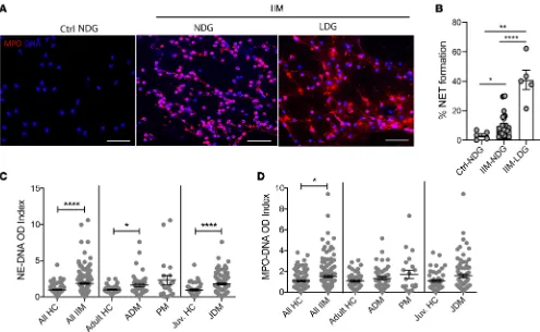
LDGs and NET complexes are present in IIM and associate with clinical manifestations. The demographic and
clinical characteristics of the different IIM cohorts studied are included in the Methods and in Supple-mental Tables 1–4 (suppleSupple-mental material available online with this article; https://doi.org/10.1172/jci. insight.134189DS1). LDGs were detected in various forms of IIM at levels comparable to those observed in SLE, except for in adult DM, in which levels were found to be significantly higher than in SLE (Supple-mental Figure 1). Similar to what has been described in SLE, IIM LDGs displayed enhanced spontaneous ability to form NETs in vitro when compared with healthy control (HC) neutrophils or normal-dense IIM neutrophils (Figure 1, A and B). Circulating NET remnants were measured with 2 assays: human neutro-phil elastase–DNA (DNA) complexes and myeloperoxidase-DNA (MPO-DNA) complexes. HNE-DNA NET complexes were significantly elevated in IIM as a whole, as well as in adult DM and JDM subgroups when compared with age-matched controls, but not in the PM group (Figure 1C). MPO-DNA complexes were significantly elevated in IIM group as a whole when compared with HCs (Figure 1D).
damage (16). LDGs negatively correlated with periungual capillary density in JDM (r = –0.58, P < 0.05), supporting previous observations that lupus LDGs damage endothelial cells (9). No associations between LDG or NETs were observed with calcinosis in the DM or JDM group. Overall, neutrophil subsets and NETs correlated with disease activity in IIM and with the microvascular abnormalities char-acteristic of these conditions. In general, LDGs showed correlations with clinical disease parameters in JDM but not as strongly in adult DM, while correlations of these parameters with NETs were present in both adult and pediatric forms of the disease.
There were no associations between use of specific immunosuppressive therapies and LDG or NET complexes levels, except for a correlation between circulating HNE-DNA NET complexes in the circula-tion with steroid dose in the JDM group (r = 0.29, P < 0.05) but not with other IIM. No associacircula-tions were observed with levels of circulating LDGs and steroid use in any form of IIM.
MSAs are associated with NET levels and directly induce NET formation. When assessing associations
with specific MSA profiles, circulating NET levels (both HNE-DNA and MPO-DNA complexes) were significantly higher in IIM subjects that tested positive for anti-MDA5 MSAs. In addition, NET levels were higher in those subjects that had anti-transcriptional intermediary factor 1 (TIF1, also known as p155/140) autoantibodies, a MSA associated with DM and JDM (17). In contrast, other MSAs (includ-ing anti-Jo1) were not associated with elevated circulat(includ-ing levels of NETs, while LDG numbers did not correlate with any specific MSA (Figure 2, A and B; P > 0.05).
neutrophils when compared with control IgG (Figure 2, C and D), while purified anti-Jo1 MSA (also iso-lated from an adult IIM subject) did not (Figure 2C). Overall, anti-MDA5 MSAs enhanced NET formation and were associated with in vivo neutrophil dysregulation in IIM.
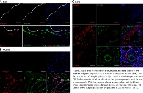
NETs and infiltrating neutrophils are detected in IIM tissues. To further assess whether enhanced NET
nary parenchyma (Figure 3C). Overall, these results indicate that netting neutrophils infiltrate clinically affected tissues in MSA-positive IIM, particularly in association with anti-MDA5. Given the association between anti-MDA5 and the presence of NETs in tissues, and previous reports of high MDA5 mRNA expression during muscle regeneration (18), we assessed whether the MDA5 protein could be detected in these biopsies. Indeed, MDA5 antigen was present in DM muscle and skin tissue biopsies (Supplemental Figure 2A). Purified NETs did not express MDA5, indicating that they were not the source of this anti-gen in skin (Supplemental Figure 2B).
Neutrophil-associated gene expression in skeletal muscle correlate with markers of IIM disease activity and the type I and II IFN gene signatures. RNA-sequencing was performed in muscle biopsy specimens from 39 subjects
with adult DM (11 anti-Mi2-, 12 anti-NXP2-, 11 anti-TIF1-, and 5 anti-MDA5–positive MSAs; Supple-mental Table 4). In addition, 20 normal muscle biopsy specimens were used as comparators. A neutrophil gene signature was evaluated using 5 neutrophil genes: ELANE, MPO, CTSG, S100A8, and S100A9, as previously described (19). Relative to HC muscle, neutrophil-associated gene expression was significantly upregulated in the DM group (Table 1). S100A8, S100A9, and MPO showed a positive correlation with type I IFN-regulated genes (ISG15, IFI6, and MX1) and type II IFN-regulated genes (PSMB8, GPB1, and GBP2; all q values < 0.001), while CSTG showed a positive correlation only with type II IFN-regulated genes (all q values < 0.006; Figure 4A). Muscle strength measurements and creatine kinase (CK) levels obtained near the time of the muscle biopsy were available in 17 DM subjects. S100A8, S100A9, and MPO positively correlated with CK levels and negatively correlated with muscle strength (Figure 4B). Overall, these results indicated that a skeletal muscle neutrophil gene signature was present in DM, in association with muscle damage and with enhanced muscle type I and II IFN responses, pathways considered pathogenic in DM.
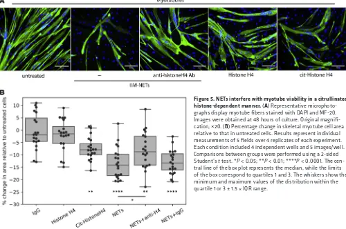
NETs hamper myotube viability. Given that NETs were detected in IIM muscle tissue, we determined
whether they could play a local pathogenic role. Skeletal muscle fibers form as a result of the fusion of the precursors myoblasts into multinucleated fibers called myotubes. To assess whether NETs could directly harm muscle cells, we grew skeletal muscle myoblasts in the presence or absence of spontaneously formed NETs
isolated from IIM subjects. We assessed both the ability of myoblasts to proliferate as well as the viability of skeletal myotubes. NETs did not disrupt the ability of myoblasts to proliferate after 72 hours in culture (Sup-plemental Figure 3A). In contrast, already formed myotubes exposed to NETs showed enhanced cell death that was evident as early as 1 hour after exposure. Significantly fewer myotubes were present by 24–48 hours when cells were exposed to NETs compared with untreated cells (Figure 5 and Supplemental Figure 3, B and C). To assess potential mechanisms of muscle cell damage by NETs, we blocked HNE, a component of NETs previously associated with cell injury. However, adding the HNE inhibitor Sivelestat did not abrogate the damaging effect of NETs on myotube viability (Supplemental Figure 3C). Histones externalized in NETs have recently been implicated in vascular smooth muscle cell damage in the context of atherosclerosis by forming pores in the target cells and inducing cell death (20). During NET formation, histones can undergo citrullination due to the activation of the enzyme peptidylarginine deiminase-4 (PAD4), and these citrulli-nated histones can be extruded as part of the NETs (21). Thus, we assessed whether native or citrullicitrulli-nated histones could cause skeletal myotube cell death. Indeed, while recombinant histone H4 was not toxic to these myotubes, citrullinated histone H4 significantly decreased myotube viability, similar to the effect observed with NETs. Furthermore, a blocking antibody targeting histone H4 partially and significantly reversed the tox-ic effect of the NETs on the skeletal myotubes, while control IgG did not (Figure 5B). Overall, these observa-tions indicated that NETs have the ability to directly disrupt skeletal myotubes, at least in part, through a toxic effect mediated by citrullinated histones present in NETs. These results indicated that NETs in IIM muscle may contribute to the direct tissue damage that represents the hallmark of these diseases.
Discussion
The role of neutrophils in inflammatory myopathies remains poorly characterized, although recent data suggest that neutrophil numbers are associated with decreased patient survival (22). We now report that IIM is characterized by the presence of a pathogenic subset of neutrophils, LDGs, previously described in other autoimmune diseases. As is the case in SLE and AAV (5, 7), LDGs present in IIM subjects are characterized by their enhanced ability to form NETs. The levels of circulating LDGs and/or NETs signifi-cantly associate with disease activity in the muscle and skin and with the presence of specific MSAs (prev-alently with anti-MDA5). Indeed, anti-MDA5 MSAs directly enhance NET formation. Further supporting that specific MSAs induce NET formation, netting neutrophils infiltrate organs affected in DM, including muscle, lung, and skin, in those IIM subjects who are positive for MSAs but not in MSA-negative IIM. Furthermore, the presence of an upregulated neutrophil gene signature in DM skeletal muscle biopsies associates with clinical features of the disease and with the type I–II IFN signatures. All these observations appear to have important pathogenic implications, as NETs purified from IIM neutrophils directly cause damage to myotubes, at least in part, via a citrullinated histone-dependent manner.
It is now generally accepted that inflammatory responses, if tightly regulated, are integral to muscle repair and regeneration (23). However, when inflammation becomes chronic and dysregulated, it can have profound effects on muscle injury and regeneration. Skeletal muscle has a remarkable capacity for repair, even following severe damage. The regenerative capacity of skeletal muscle relies on the presence of satellite cells/myoblasts that retain their ability to proliferate and differentiate to either fuse with existing muscle fibers or with other myogenic cells to generate new fibers (24). Furthermore, chronic inflammatory damage to already formed muscle fibers can significantly affect the function of muscles Table 1. Gene expression (fold change) and q values of 5 neutrophil genes in DM skeletal muscle
compared with healthy skeletal muscle
Gene FC q values
S100A8 2.5 0.01
S100A9 4.4 6.3 × 10⁻⁹
MPO 2.4 2.4 × 10⁻⁶
CTSG 1.3 0.4
ELANE 1.7 0.2
and their ability to recover from injury. The response of the immune system to muscle injury and disease is a complex process. Although myeloid cells, including neutrophils, participate in chronic striated mus-cle injury and regeneration, the role of NETs in this process had not been reported.
It is known that neutrophils can cause muscle fiber damage in vitro and in vivo (25) and that oxi-dants released by these cells can cause severe cellular damage. Recent studies also implicate a role for HNE in muscular dystrophies by impairing myoblast cell survival, proliferation, and differentiation (26). We found that, while NETs do not impair myoblast proliferation, they significantly decrease skel-etal myotube viability. This is relevant, as biopsies from IIM subjects, but not healthy muscle, displayed evidence of infiltrating netting neutrophils and NET structures. Given the high levels of HNE present in NETs, it is possible that it and other proteases play prominent roles in the induction of myotube cell death (26). However, blocking the effects of HNE did not improve myotube viability, while citrullinat-ed histones recapitulatcitrullinat-ed the toxic effect of NETs on myotube viability. This is consistent with a recent report that histone H4 can bind and lyse vascular smooth muscle cells and destabilize atherosclerotic plaques (20). Although that study did not address the role of specific posttranslational modifications of histones in their ability to damage cells, we observed that citrullinated histone H4 has an enhanced ability to damage skeletal myotubes compared with native histone H4. Future studies should address the mechanisms by which specific protein modifications may dictate cell injury in various disease states. Of note, a recent study reported evidence of tissue citrullination in IIM-affected muscles, while was absent in control muscle tissue (27). This supports our observations implicating PAD activation and histone citrullination in skeletal muscle damage.
In other diseases, specific autoantibodies were found to induce enhanced NET formation, including ribonucleoprotein autoantibodies in SLE (13), citrullinated protein antibodies in RA (6), and anti-neutrophil cytoplasmic antibodies in AAV (28). We now report that anti-MDA5 MSAs can have similar effects in IIM and that their presence is associated with enhanced NET formation in circulation and in tissues. The mechanisms of this distinct association need to be further investigated and may have thera-peutic implications. Future studies should also test how other MSAs, besides anti-MDA5 and anti-Jo, may
modulate NET formation and the role of the NET-autoantibody interactions in the degree of induced skeletal myotube damage. It will also be important to enhance understanding of why some disease-specific autoantibodies, but not others, induce NET formation in this and other autoimmune conditions.
a link between calcium deposition in tissues, the calcinosis characteristic of JDM, and the development of neutrophil activation and NETs in muscle tissue (15). This may be an additional mechanism that induces NET formation in these individuals. However, we also observed enhanced NET formation and tissue infil-tration by NETs in the absence of clinical calcinosis and in subsets of IIM not characterized by calcium deposits. Indeed, we found no correlations between circulating NET or LDG levels and calcinosis in DM and JDM. This suggests that other mechanisms in addition to crystals are implicated in enhanced neutro-phil activation and NET formation in IIM and may include specific MSAs. In previous studies, numbers of LDGs and levels of NET formation were not linked to specific medications used to treat autoimmune diseases. In this study, however, some associations were present with the use of corticosteroids. It remains to be determined whether this represents the true effects of the drugs on neutrophil dysregulation in IIM or merely that individuals who require these medications tend to have more severe disease.
Overall, our results indicate that neutrophil dysregulation in IIM may play important pathogenic roles in autoimmune responses and tissue damage and that investigating strategies that modulate aberrant neu-trophil responses in IIM may lead to changes in the outcome of these potentially devastating conditions.
Methods
Patient recruitment for peripheral blood studies. Patients fulfilled probable or definite criteria for IIM from Bohan,
Peter, and colleagues (32) or revised criteria for SLE from the American College of Rheumatology (33) and were enrolled at the NIH Clinical Center in the NIAMS/National Institute of Diabetes and Digestive and Kid-ney Diseases IRB-approved myositis (94-E0165) or SLE (94-AR-0066) natural history studies, respectively. HCs were enrolled from the NIH Blood Bank or the NIH healthy volunteer cohort. MSAs were detected by standard-ized immunoprecipitation and immunoprecipitation-immunoblot methods (34) at the NIH Clinical Center Cen-tral Laboratory. Clinical characterization and measurements of IIM disease activity and damage were obtained as previously described (35, 36). Videocapillaroscopy was quantified as described previously (37).
Quantification of circulating NET complexes. HNE-DNA and MPO-DNA complexes were measured in
plasma as previously described (19). In brief, high-binding 96-well microplates were incubated overnight at 4°C with either rabbit anti-HNE (Calbiochem, 481001) or mouse anti-human MPO (Bio-Rad, clone 4A4, 0400-0002). At 4°C, the plates were washed and blocked with 1% BSA overnight, and then plasma samples diluted in blocking buffer were added and incubated overnight. Plates were washed; they were then incu-bated for 1 hour with anti-ds-DNA (MilliporeSigma, clone BV16-13, MAB030) for the HNE-DNA plate or HRP-conjugated anti-DNA POD (Roche, 11774425001) for the MPO-DNA plate. The HNE-DNA plate was washed and incubated for 1 hour with anti–mouse IgG-HRP conjugate (Bio-Rad, 1721012). ELISAs were visualized with tetramethylbenzidine and stop solution.
Quantification of circulating LDGs. Quantification of circulating LDGs was performed as
previous-ly described (19). In brief, human PBMCs were isolated by Ficoll-Paque Plus density gradient (GE Healthcare, GE17-1440-03), RBCs were lysed with hypertonic solution, and cells were resuspended in FACs buffer. Cells were blocked for 15 minutes with Human TruStain Fc Receptor Blocking Solution (BioLegend, 422301); stained with Abs recognizing human CD10 (BioLegend, clone H10A, 312209), CD15 (BioLegend, clone HI98, 301906), and CD14 (BioLegend, clone HCD14, catalog 325610) or isotype control for 15 minutes at 4°C in the dark; and fixed with 2% paraformaldehyde overnight. Data were collected using a BD FACSCanto RUO and analyzed using FlowJo Software version 10. Cutoff values for positive staining were determined using compensation controls for each fluorophore. LDGs were classified as CD10+CD15+CD14lo, as previously described (11).
Isolation of MSAs. MSAs were isolated as previously reported (38). Plasma from 1 anti-Jo1-positive
prepacked sepharose columns (NHS columns, GE Healthcare) were coupled with 5 or 1 mg recombinant MDA5 and Jo1, respectively, and according to the manufacturer’s instructions. Anti-MDA5 and anti-Jo1 IgG were isolated as previously reported (38). Briefly, plasma was centrifuged at 3000 g for 5 minutes, diluted 1:5 in PBS, and filtered using a 0.45-μM filter, before loading in the HiTrap Protein G HP columns (GE Healthcare) for IgG enrichment. Total IgGs eluted from the column using 0.1 M glycine-HCl, pH 2.7, were immediately neutralized to approximately 7.4 with 1 M Tris, pH 9, and buffer exchanged to PBS by dialysis. Anti-MDA5 IgG and anti-Jo1 IgG were enriched by applying total IgGs to MDA5 or Jo1 affinity columns, as described for the protein G column. Non-MDA5 and non-Jo1 reactive IgGs were collected and used as control Abs (flow through [FT]). No endotoxin contamination was detected in the Ab prepara-tions after buffer exchange to PBS and the preparaprepara-tions were sterile filtered (<0.05 EU/mL). HC IgG was isolated from serum using the Melon Gel IgG Spin Purification Kit (Thermo Fisher Scientific, 45206) and quantified with a BCA assay. The efficiency of anti-MDA5 and anti-Jo1 IgG enrichment from the protein G–derived total IgG was confirmed by ELISA. Streptavidin-coated 96-well plates (50 ng, overnight at 4°C) were first blocked for 1 hour at room temperature with PBST 0.1% BSA, followed by a 1-hour incubation with either recombinant biotinylated MDA5 or Jo1 (1 μg/well). Anti-MDA5 IgG, anti-Jo1 IgG and corre-sponding FT fractions were added to the plates in serial dilutions (ranging from 0.05 ng/mL to 1000 ng/ mL per well) and incubated for 90 minutes. Reactivity was recorded at 450 nm absorbance after adding Peroxidase F(ab′)2 Fragment Goat Anti-Human IgG, Fcγ fragment specific antibody (dilution 1:10,000, ref. 109-036-008, Jackson ImmunoResearch) for 1 hour at room temperature, followed by a 15-minute incuba-tion with 3,3′,5,5′-tetramethylbenzidine substrate (reacincuba-tion stopped with 1 M H2SO4).
Assessment of NET formation by immunofluorescence microscopy. HC neutrophils and IIM neutrophils and
LDGs were isolated as previously described (13). Cells were incubated on poly-L-lysine–coated coverslips for 15 minutes, blocked with Fc-blocking reagent (Miltenyi, 130-059-901), and then treated for 1 hour with anti-Jo-1 IgG, anti-MDA5 IgG, FT of anti-Jo-1 IgG, FT of anti-MDA5 IgG, or HC IgG. Coverslips were gently fixed with 4% paraformaldehyde overnight at 4°C, then stained with rabbit anti-MPO (Agilent Tech-nologies, A039829-2) for 1 hour, followed with Alexa Fluor 555–conjugated donkey anti-rabbit IgG (Life Technologies, A-31572) and Hoechst (Life Technologies, H3570). After mounting with Prolong Gold (Life Technologies, P36930), slides were visualized by microscopy. Three images were taken per coverslip at ×40, and NET and total cell counts were quantified using ImageJ (NIH).
NET collection. NETs were collected as previously described (11). Neutrophils or LDGs were seeded at
5 million cells per well and incubated in 5% CO2 at 37°C. After 4 hours, NETs were harvested using micro-coccal nuclease and centrifuged, and the supernatant was collected and stored at –20°C. Protein content was quantified by BCA assay before use.
Preparation of skin, muscle, and lung biopsies. Only cases that met clinical and histologic criteria
tem-perature with 1:1000 Hoechst 33342 (Thermo Fisher Scientific) and mounted with ProLong Gold Antifade (Thermo Fisher Scientific). For the frozen sections, similar steps were followed starting with the blocking step. The samples were acquired in an Eclipse Ti-E Nikon confocal microscope (Minato).
RNA-sequencing analysis of skeletal muscle. Muscle biopsies used for the RNA-sequencing analysis were
obtained from DM subjects enrolled in IRB-approved longitudinal cohorts from the NIH (IRB 91-AR-0196), the Johns Hopkins Myositis Center (IRB NA_00007454), the Clinic Hospital (Barcelona; IRB HCB/2015/0479), and the Vall d’Hebron Hospital (Barcelona; IRB PR[AG] 68/2008). CK levels and strength assessments obtained closest to the time of muscle biopsy were used to measure the clinical activ-ity. Muscle strength was evaluated by the examining physician using the Medical Research Council scale. This scale was transformed to Kendall’s 0–10 scale; the right- and left-side measurements for arm abduction and hip flexion strength were combined and the average was used for calculations (possible range 0–10), as previously described (36). Normal muscle biopsies were obtained from the Johns Hopkins Neuromuscular Pathology Laboratory (n = 10) and the Skeletal Muscle Biobank of the University of Kentucky (n = 10).
RNA-sequencing was performed as previously described (41). Briefly, RNA was prepared using TRIzol (Invitrogen). Libraries were prepared using the NeoPrep system according to the TruSeq Stranded mRNA Library Prep protocol (Illumina) and sequenced using the Illumina HiSeq 2500 or 3000. Reads were aligned using STAR v.2. (42), the abundance of each gene was quantified using StringTie v.1.3.3. (43), and differential gene expression analysis was performed using DESeq2 v.1.20.0. (44) The Benjamini-Hochberg correction was used to adjust for multiple comparisons, and a corrected P value (q value) of 0.05 or less was considered statistically significant. A neutrophil gene signature was calculated based on the expression of ELANE, MPO,
CTSG, S100A8, and S100A9, as mentioned above. IFN pathway genes were collected from the Reactome
biorepository (https://reactome.org/). General IFN-related genes and genes from the type I and II IFN path-ways were merged in a single list. The 13 genes included in the previously proposed IFN scoring system in myositis were also added to the list (45). The expression of the genes of on list was analyzed in the different MSA and clinical myositis subsets. Gene expression (fragments per kilobase of transcript per million mapped reads [FPKM]) values were log transformed (logFPKM: log2[FPKM+1]) for visualization purposes using the Python programming language and the packages Numpy, Pandas, and Seaborn. Correlation among contin-uous variables was measured using Spearman’s rho. Skin microarray and muscle RNA-sequencing data were deposited in GEO (accession GSE142807 and GSE143323, respectively).
Skeletal myoblast cell cultures. Healthy human skeletal muscle myoblasts (Lonza, CC2580) were grown
following the manufacturer’s recommendations. Myoblasts were seeded at 3500 cells/cm2 and maintained in growth medium (Lonza, CC-3245) at 37°C, 5% CO2, until 50% or 60% confluence, changing growth medium every other day. Myoblasts were then treated with HC NETs, IIM NETs, ionophore, or media in duplicates. Within 10 minutes of treatment, plates were imaged on the IncuCyte ZOOM platform as pre-viously described (46), using phase contrast. Nine areas per well were imaged every 30 minutes for the first 6 hours and every 2 hours for the next 18 hours for 3 days, using a ×20 dry objective. Videos were created using the IncuCyte Basic Software and ImageJ (NIH).
Preparation of citrullinated histone H4. Recombinant human histone H4 (New England Biolabs) and
recombinant PAD4 (Signalchem) were suspended in RPMI containing 2 mM CaCl2 and 1 mM DTT. The reaction was performed at 37°C overnight and stored at –20°C until further use.
Skeletal myotube experiments. Healthy human skeletal muscle myoblasts at passage 4 were cultured in
stained with DAPI (Vector Laboratories; Vectashield Antifade Mounting Medium DAPI, catalog H-1200). Five pictures were taken per well (×10 magnification), using a Revolve microscope (Echo Laboratories Inc). ImageJ (NIH) was used to count DAPI positivity (nuclei) and to calculate the muscle fiber area.
Statistics. All graphs depict the mean ± SEM or median and IQR range unless otherwise stated.
Associ-ations of measurements with clinical characteristics and disease activity indices were assessed with Spear-man’s correlation using IBM SPSS software (IBM Analytics). Student’s 2-tailed t test, Kruskal-Wallis, Mann-Whitney were used as appropriate. Multiple comparisons were analyzed by 1-way ANOVA using GraphPad Prism. P values of less than 0.05 were considered significant.
Study approval. All human studies were reviewed and approved by appropriate institutional review boards
(NIAMS/National Institute of Diabetes and Digestive and Kidney Diseases 94-AR-0066 and 91-AR-0196; NIE-HS 94E0165; University of Michigan HUM00072843; Instituto Nacional de Ciencias Medicas y de la Nutrición Salvador Zubiran 2152; Johns Hopkins University NA_00007454; Vall d’Hebron Hospital PR(AG)68/2008; Hospital Clinic de Barcelona HCB/2015/0479 (Barcelona, Spain). All subjects provided informed consent before their participation in the study according to the standards of the Declaration of Helsinki.
Author contributions
NS, JJTR, CCR, IPF, KP, MMP, YH, CFC, PG, and NA performed experiments and analyzed data. AG, OB, DGM, FWM, IEL, AIS, AM, and LGR supervised data and performed critical analysis of data. ASO, JCM, JMGJ, LCS, and JMK obtained and provided tissues and clinical analyses. AIS, AM, and LGR eval-uated clinical subjects and provided clinical scores and statistical analyses. All authors reviewed the paper. MJK conceived the study, analyzed data, and wrote the manuscript.
Acknowledgments
This work was funded by the Intramural Research Program of the NIH, NIAMS (ZIAAR041199) and the NIEHS (Z01ES101074). This work was also funded in part by the Consejo Nacional de Ciencia y Tecnología (CONACYT SEP-CB2017-2018, A1-S23262). We thank Ira Targoff for MSA testing, Stephen Hewitt for technical support, and Edvard Wigren and Susanne Graslund for recombinant protein preparation.
Address correspondence to: Mariana J. Kaplan, Systemic Autoimmunity Branch, NIAMS/NIH, 10 Center Drive, 12N248C, Bethesda, Maryland 20892, USA. Phone: 301.496.0517; Email: mariana.kaplan@nih.gov.
DGM’s present address is: Tecnológico de Monterrey, Escuela de Medicina y Ciencias de la Salud, Monterrey, Mexico.
OB’s present address is: Department of Internal Medicine and Clinical Immunology, Pitié Salpêtrière Hospital, AP-HP, Sorbonne Université, Paris, France.
1. Selva-O’Callaghan A, Pinal-Fernandez I, Trallero-Araguás E, Milisenda JC, Grau-Junyent JM, Mammen AL. Classification and management of adult inflammatory myopathies. Lancet Neurol. 2018;17(9):816–828.
2. Tournadre A, Miossec P. A critical role for immature muscle precursors in myositis. Nat Rev Rheumatol. 2013;9(7):438–442. 3. Yoshida S, Akizuki M, Mimori T, Yamagata H, Inada S, Homma M. The precipitating antibody to an acidic nuclear protein
antigen, the Jo-1, in connective tissue diseases. A marker for a subset of polymyositis with interstitial pulmonary fibrosis.
Arthri-tis Rheum. 1983;26(5):604–611.
4. Moghadam-Kia S, Oddis CV, Sato S, Kuwana M, Aggarwal R. Antimelanoma differentiation-associated gene 5 antibody: expanding the clinical spectrum in North American patients with dermatomyositis. J Rheumatol. 2017;44(3):319–325. 5. Villanueva E, et al. Netting neutrophils induce endothelial damage, infiltrate tissues, and expose immunostimulatory molecules
in systemic lupus erythematosus. J Immunol. 2011;187(1):538–552.
6. Khandpur R, et al. NETs are a source of citrullinated autoantigens and stimulate inflammatory responses in rheumatoid arthri-tis. Sci Transl Med. 2013;5(178):178ra40.
7. Grayson PC, et al. Neutrophil-related gene expression and low-density granulocytes associated with disease activity and response to treatment in antineutrophil cytoplasmic antibody-associated vasculitis. Arthritis Rheumatol. 2015;67(7):1922–1932. 8. Carmona-Rivera C, et al. Synovial fibroblast-neutrophil interactions promote pathogenic adaptive immunity in rheumatoid
arthritis. Sci Immunol. 2017;2(10):eaag3358.
9. Carmona-Rivera C, Zhao W, Yalavarthi S, Kaplan MJ. Neutrophil extracellular traps induce endothelial dysfunction in systemic lupus erythematosus through the activation of matrix metalloproteinase-2. Ann Rheum Dis. 2015;74(7):1417–1424.
10. Kahlenberg JM, Carmona-Rivera C, Smith CK, Kaplan MJ. Neutrophil extracellular trap-associated protein activation of the NLRP3 inflammasome is enhanced in lupus macrophages. J Immunol. 2013;190(3):1217–1226.
12. Lin AM, et al. Mast cells and neutrophils release IL-17 through extracellular trap formation in psoriasis. J Immunol. 2011;187(1):490–500.
13. Lood C, et al. Neutrophil extracellular traps enriched in oxidized mitochondrial DNA are interferogenic and contribute to lupus-like disease. Nat Med. 2016;22(2):146–153.
14. Peng Y, Zhang S, Zhao Y, Liu Y, Yan B. Neutrophil extracellular traps may contribute to interstitial lung disease associated with anti-MDA5 autoantibody positive dermatomyositis. Clin Rheumatol. 2018;37(1):107–115.
15. Duvvuri B, et al. Neutrophil extracellular traps in tissue periphery in juvenile dermatomyositis [published online ahead of print August 12, 2019]. Arthritis Rheumatol. https://doi.org/10.1002/art.41078.
16. Bertolazzi C, Cutolo M, Smith V, Gutierrez M. State of the art on nailfold capillaroscopy in dermatomyositis and polymyositis.
Semin Arthritis Rheum. 2017;47(3):432–444.
17. Fiorentino D, Casciola-Rosen L. Autoantibodies to transcription intermediary factor 1 in dermatomyositis shed insight into the cancer-myositis connection. Arthritis Rheum. 2012;64(2):346–349.
18. Pinal-Fernandez I, et al. Myositis autoantigen expression correlates with muscle regeneration but not autoantibody specificity.
Arthritis Rheumatol. 2019;71(8):1371–1376.
19. Carlucci PM, et al. Neutrophil subsets and their gene signature associate with vascular inflammation and coronary atherosclero-sis in lupus. JCI Insight. 2018;3(8):99276.
20. Silvestre-Roig C, et al. Externalized histone H4 orchestrates chronic inflammation by inducing lytic cell death. Nature. 2019;569(7755):236–240.
21. Grayson PC, Kaplan MJ. At the Bench: Neutrophil extracellular traps (NETs) highlight novel aspects of innate immune system involvement in autoimmune diseases. J Leukoc Biol. 2016;99(2):253–264.
22. Ha YJ, et al. Baseline peripheral blood neutrophil-to-lymphocyte ratio could predict survival in patients with adult polymyositis and dermatomyositis: A retrospective observational study. PLoS One. 2018;13(1):e0190411.
23. Peake JM, Neubauer O, Della Gatta PA, Nosaka K. Muscle damage and inflammation during recovery from exercise. J Appl
Physiol. 2017;122(3):559–570.
24. Robinson DCL, Dilworth FJ. Epigenetic regulation of adult myogenesis. Curr Top Dev Biol. 2018;126:235–284.
25. Tidball JG, Villalta SA. Regulatory interactions between muscle and the immune system during muscle regeneration. Am J
Physiol Regul Integr Comp Physiol. 2010;298(5):R1173–R1187.
26. Arecco N, et al. Elastase levels and activity are increased in dystrophic muscle and impair myoblast cell survival, proliferation and differentiation. Sci Rep. 2016;6:24708.
27. Makrygiannakis D, et al. Citrullination is an inflammation-dependent process. Ann Rheum Dis. 2006;65(9):1219–1222. 28. Kessenbrock K, et al. Netting neutrophils in autoimmune small-vessel vasculitis. Nat Med. 2009;15(6):623–625.
29. Lu X, et al. The host defense peptide LL-37 a possible inducer of the type I interferon system in patients with polymyositis and dermatomyositis. J Autoimmun. 2017;78:46–56.
30. Sakamoto N, et al. Elevated α-defensin levels in plasma and bronchoalveolar lavage fluid from patients with myositis-associated interstitial lung disease. BMC Pulm Med. 2018;18(1):44.
31. Zhang S, Shu X, Tian X, Chen F, Lu X, Wang G. Enhanced formation and impaired degradation of neutrophil extracellular traps in dermatomyositis and polymyositis: a potential contributor to interstitial lung disease complications. Clin Exp Immunol. 2014;177(1):134–141.
32. Bohan A, Peter JB, Bowman RL, Pearson CM. Computer-assisted analysis of 153 patients with polymyositis and dermatomyo-sitis. Medicine (Baltimore). 1977;56(4):255–286.
33. Hochberg MC. Updating the American College of Rheumatology revised criteria for the classification of systemic lupus erythe-matosus. Arthritis Rheum. 1997;40(9):1725.
34. Targoff IN, et al. A novel autoantibody to a 155-kd protein is associated with dermatomyositis. Arthritis Rheum. 2006;54(11):3682–3689.
35. Rider LG, et al. Update on outcome assessment in myositis. Nat Rev Rheumatol. 2018;14(5):303–318.
36. Rider LG, et al. Measures of adult and juvenile dermatomyositis, polymyositis, and inclusion body myositis: Physician and Patient/Parent Global Activity, Manual Muscle Testing (MMT), Health Assessment Questionnaire (HAQ)/Childhood Health Assessment Questionnaire (C-HAQ), Childhood Myositis Assessment Scale (CMAS), Myositis Disease Activity Assessment Tool (MDAAT), Disease Activity Score (DAS), Short Form 36 (SF-36), Child Health Questionnaire (CHQ), physician global damage, Myositis Damage Index (MDI), Quantitative Muscle Testing (QMT), Myositis Functional Index-2 (FI-2), Myositis Activities Profile (MAP), Inclusion Body Myositis Functional Rating Scale (IBMFRS), Cutaneous Dermatomyositis Disease Area and Severity Index (CDASI), Cutaneous Assessment Tool (CAT), Dermatomyositis Skin Severity Index (DSSI), Skindex, and Dermatology Life Quality Index (DLQI). Arthritis Care Res (Hoboken). 2011;63 Suppl 11:S118–S157.
37. Minniti CP, et al. Vasculopathy, inflammation, and blood flow in leg ulcers of patients with sickle cell anemia. Am J Hematol. 2014;89(1):1–6.
38. Fernandes-Cerqueira C, et al. Patients with anti-Jo1 antibodies display a characteristic IgG Fc-glycan profile which is further enhanced in anti-Jo1 autoantibodies. Sci Rep. 2018;8(1):17958.
39. Keates T, et al. Expressing the human proteome for affinity proteomics: optimising expression of soluble protein domains and in vivo biotinylation. N Biotechnol. 2012;29(5):515–525.
40. Zhong N, et al. Optimizing production of antigens and Fabs in the context of generating recombinant antibodies to human pro-teins. PLoS One. 2015;10(10):e0139695.
41. Amici DR, et al. Calcium dysregulation, functional calpainopathy, and endoplasmic reticulum stress in sporadic inclusion body myositis. Acta Neuropathol Commun. 2017;5(1):24.
42. Dobin A, et al. STAR: ultrafast universal RNA-seq aligner. Bioinformatics. 2013;29(1):15–21.
43. Pertea M, Pertea GM, Antonescu CM, Chang TC, Mendell JT, Salzberg SL. StringTie enables improved reconstruction of a transcriptome from RNA-seq reads. Nat Biotechnol. 2015;33(3):290–295.
44. Love MI, Huber W, Anders S. Moderated estimation of fold change and dispersion for RNA-seq data with DESeq2. Genome
45. Greenberg SA, et al. Interferon-alpha/beta-mediated innate immune mechanisms in dermatomyositis. Ann Neurol. 2005;57(5):664–678.