INVESTIGATION
HIGHLIGHTED ARTICLE
The Basis for Evolution of DNA-Binding Speci
fi
city
of the Aft1 Transcription Factor in Yeasts
Isabelle R. Gonçalves,*,1Natalia Conde e Silva,†,1Cesar La Torre Garay,‡Emmanuel Lesuisse,‡ Jean Michel Camadro,‡and Pierre Louis Blaiseau‡,§,2 *Microbiologie, Adaptation et Pathogénie, Unité Mixte de Recherche 5240, Centre National de la Recherche Scientifique, Université Lyon 1, Bayer CropScience, F-69622 Villeurbanne, France,†Institut de Biologie des Plantes, Centre National de la Recherche Scientifique Unité Mixte de Recherche 8618, Université Paris-Sud, Saclay Plant Sciences, F-91405 Orsay Cedex, France, ‡Mitochondries, Métaux et Stress Oxydatif, Institut Jacques Monod Unité Mixte de Recherche 7592, Centre National de la Recherche Scientifique, Université Paris Diderot, Sorbonne Paris Cité, F-75205 Paris Cedex 13, France, and§Université Pierre et Marie Curie, Paris 06, Unité de Formation et de Recherche 927 Sciences De la Vie, F-75005 Paris Cedex 05, France
ABSTRACT The Saccharomyces cerevisiaeAft1 and Kluyveromyces lactis KlAft are orthologous yeast transcription activators that regulate the expression of the same group of iron-uptake genes but bind to the different DNA sites: TGCACCC forAft1and PuCACCC for KlAft. To establish whether the DNA-binding mechanisms of Aft1and KlAft have diverged during the evolution of the Aft-type transcription factor, we examined the function of a nonconserved region in their DNA-binding domains. A large part of this region is composed of a sequence predicted to be disordered in structure and potentially phosphorylated. We show with deletion mutant analyses that this sequence is essential for the binding ofAft1to its DNA site and for the iron uptake and growth ofS. cerevisiaeunder iron-limited conditions. We constructed hybrid proteins by exchanging the nonconserved regions ofAft1and KlAft. We show that the Aft1region is necessary and sufficient for KlAft to bind efficiently to theAft1DNA site inS. cerevisiaeand to complement the iron-dependent phenotype of theaft1Daft2Dmutant. This demonstrates that the changes in the nonconserved region of the Aft-type DNA-binding domain have led to changes in the DNA-binding specificity and have major consequences for the regulation of iron homeostasis. The combination of bioinformatic and experimental analyses indicates that the sequence TGCACCC is the most probable ancestral Aft-type element. Our findings suggest that the changes in the nonconserved region of the DNA-binding domain are responsible for the evolution of the TGCACCC sequence toward PuCACCC in theK. lactisspecies.
I
RON is an essential element but its accumulation can be toxic for the cell (Halliwell and Gutteridge 1984). There-fore, organisms have developed various regulated systems to adjust the iron uptakeflux to the cell requirements (Blaiseau et al.2010). In fungi, the expression of iron uptake genes is under the control of either of two opposite modes of tran-scriptional regulation. In most fungi, the iron-regulatory pathway is mediated by a Zn-finger GATA-type factor that represses the transcription of the iron-uptake genes underhigh-iron conditions (Haas et al. 2008). In contrast, in the hemiascomycete yeasts Saccharomyces cerevisiae and Kluyveromyces lactis, the transcription of the same group of genes is activated by an Aft-type transcription factor under conditions of low iron availability (Yamaguchi-Iwai
et al. 1995; Conde e Silva et al. 2009). An AFTgene has been identified in the genome of all hemiascomycetes stud-ied except Yarrowia lipolityca. Some hemiascomycete ge-nomes, including that of S. cerevisiae, have two copies of theAFTgene resulting from the whole genomic duplication (WGD) event (Wolfe and Shields 1997). We previously showed that the S. cerevisiaeAft1 and Aft2 paralogs have specialized iron-regulatory functions:Aft1specifically acti-vates the transcription of the genes involved in the iron uptake at the cell surface, whereasAft2activates the tran-scription of genes involved in vacuolar and mitochondrial transport (Courelet al.2005; Dhaouiet al.2011). Exami-nation of the promoter sequences ofAft1- andAft2-regulated
Copyright © 2014 by the Genetics Society of America doi: 10.1534/genetics.113.157693
Manuscript received September 19, 2013; accepted for publication October 21, 2013; published Early Online October 23, 2013.
Supporting information is available online athttp://www.genetics.org/lookup/suppl/ doi:10.1534/genetics.113.157693/-/DC1.
1These authors contributed equally to this work.
genes and DNA-binding analyses show that Aft1 and Aft2
recognize similar but distinct DNA sequences: TGCACCC and PuCACCC, respectively (Yamaguchi-Iwai et al. 1996; Rutherfordet al.2003; Courelet al.2005). The investigation of iron regulation in the pre-WGD speciesK. lactis indicates that KlAft, the ortholog ofAft1andAft2, activates the tran-scription of homologs ofAft1-regulated genes, but not ofAft2 -regulated genes (Conde e Silvaet al.2009). This suggests that
Aft1and KlAft have kept their ancestral iron-regulatory func-tion, which is to control genes encoding the cell surface iron uptake components, whereasAft2evolved to have a new Aft-type regulatory function emerging after the WGD involving the control of intracellular iron transport.
Although Aft1 and KlAft regulate the same group of genes, there is evidence that their iron-regulated mecha-nisms of transcription are different. Heterologous comple-mentation experiments indicate that KlAft does not totally suppress theaft1Daft2D mutant phenotype (Conde e Silva et al. 2009). DNA-binding analyses with Aft1 translated in vitro showed that it binds better to the TGCACCC se-quence than to TACACCC and that a DNA duplex containing the GGCACCC sequence competes poorly with TGCACCC (Yamaguchi-Iwai et al. 1996). In contrast, KlAft binds to promoters containing various ACACCC sequences such as GACACCC, CACACCC, and TACACCC (Conde e Silvaet al. 2009). Consistent with this, bioinformatic analyses identify PuCACCC and TGCACCC as the Aft-type consensus sequen-ces in the promoters of KlAft-regulated andAft1-regulated genes, respectively.
Several examples of the divergence of DNA-binding specificity between orthologous yeast transcription regula-tors with conserved function have been reported (Gasch et al. 2004; Kuoet al. 2010; Baker et al. 2011). However, only one study correlated the change of DNA-binding spec-ificity with a particular change in the amino acid sequence of the DNA-binding domain (Kuoet al.2010). Here, we inves-tigated whether particular differences in the DNA-binding domains ofAft1and KlAft are associated with the differen-ces in their DNA-binding sites. The DNA-binding domain of the Aft proteins has been identified as a WRKY-GCM1
zinc-finger domain (Babuet al.2006). It belongs to the subgroup of the WRKY-GCM1 domains with insert because it contains an insert of 20–90 residues between two conserved cysteine residues that have been suggested to be involved in a
zinc-finger domain (Babuet al.2006; Conde e Silvaet al.2009). We examined the amino acid sequence of the insert and identified a variable sequence (VS) within the insert that is predicted to be disordered and highly phosphorylated. We show that Aft1VS (residues 152–203) is necessary for binding ofAft1to the DNA sequence TGCACCC. Deletion of
Aft1(152–203) led to severe deficiency in both the transport of iron and growth in iron-depleted conditions. By ectopic expression of hybrid proteins, we show that the replacement of the KlAft insert by that ofAft1is necessary and sufficient for KlAft to bind the TGCACCC sequence and complement the iron-dependent aft1Daft2D mutant phenotype. This
demonstrates that the divergence of the DNA-binding spec-ificity between the orthologs Aft1 and KlAft is strictly de-pendent on the nonconserved region inserted into their DNA-binding domains.
Materials and Methods
S. cerevisiae strains
The S. cerevisiae strains used in this study were BY4742 (MATa; his3D1; leu2D0; lys2D0; ura3D0) and SCMC01 (MATa; his3D1; leu2D0; lys2D0; ura3D0; aft1::kanMX4;
aft2::kanMX4).
Plasmid constructions
The plasmidpEG202-AFT1 containing the sequence encod-ing the DNA-bindencod-ing domain of LexA has been described in Blaiseauet al.(2001). Plasmids pCXJ22-AFT1and pCXJ22-KLAFT have been described in Conde e Silvaet al.(2009). The plasmids containing the AFT1D deletion mutants were constructed by using pEG202-AFT1as the PCR tem-plate with the QuikChange Mutagenesis Kit (Stratagene, La Jolla, CA) according to the manufacturer’s instructions. For pEG202-AFT1D(152–203), pEG202-AFT1D(152–181), pEG202-AFT1D(182–203), and pEG202-AFT1D(172–203), the primers used were, respectively, 59-GCTGCTAAAAGG
GGAAGGAACAAGAAAAAAAGATGTGTATCGAGG-39, 59
-GCTGCTAAAAGGGGAAGGAACCCTTCTAATGCCACA
GTAACCAATG-39, 59-GATGACGAATTAGAATATGCGAGTA
AGAAAAAAAGATGTGTATCGAGG-39, 59- CACGAAGACGA
GAAATCCAAGAAGAAAAAAAGATGTGTATCGAGG-39, and
their complements. Plasmids pEG202-KLAFT and pEG202-KLAFTAFT1(143–215) were generated by gap repair in yeast. For pEG202-KLAFT, the cells were cotransformed with BamHI-linearized pEG202 and a DNA fragment produced by PCR from pCXJ22-KLAFT as the template, with the
fol-lowing primers: 59- GGCGGTTGGGGTTATTCGCAACGGCG
ACTGGCTGGAATTCATGAAGCACGAGCTACAGACAC-39 and
59-CCGGAATTAGCTTGGCTGCAGGTCGACTCGAGCGGC
CGC TCATTGAAAAAAGCCGTGAGGTTC-39. For
pEG202-KLAFTAFT1(143–215), cells were cotransformed with RsrII-linearized pEG202-KLAFT and a DNA fragment produced by PCR from pCXJ18-AFT1(Conde e Silva et al.2009) as the template, with the following primers: 59-CATTGAAC GGTCGGATAGCATGAAGATCGTGTTCAAATGTAAAGCTGC
TAAAAGGGGAAGGAAC-39and 59-TTCTCTTCAAAGAGAA
CGTGGCTCGCACTCTGAAGGGACAGTTATTAAACCTCGA TACACATC-39. To construct pEG202-AFT1KLAFT(134–229), we used a two-step PCR protocol (Wach 1996) involv-ing the followinvolv-ing primers: 59-GGCGGTTGGGGTTATTCGC AACGGCGACTGGCTGGAATTCATGGAAGGCTTCAATCCGG
C-39, 59-CCGGAATTAGCTTGGCTGCAGGTCGACTCGAGCG
GCCGCCTAATCTTCTGGCTTCACATACTTC-39, 59-GTATTT
CTATTCTTTTTAACAGCTTTACACTTGAAGACAACTTTAA
ATGCG-39, and 59-ACGTGCCATCGGACCGTACAATTCAT
The sequences underlined are homologous to sequences in pEG202, the sequences in italics are homologous to sequences in KLAFT, and the other sequences are homolo-gous to sequences inAFT1. For thefirst and the second PCR steps, the DNA templates were pCXJ22-AFT1 and pCXJ22-KLAFT, respectively. Thefinal PCR product wasfirst inserted into the vector pCR-2.1-TOPO (Invitrogen) and then trans-ferred to between the BamHI-XhoI sites of pEG202. To ob-tain pCXJ22-KLAFTAFT1(143–215), the AgeI-XhoI fragment of pEG202-KLAFTAFT1(143–215) was inserted between the AgeI-XhoI sites of pCXJ22-KLAFT. Plasmid pEG202-LTAFT express-ing LexA-LTAFT was constructed by integrating the open reading frame of LTAFT in frame with the DNA binding domain of LexA. The genomic DNA from theLachancea ther-motoleransstrain CBS 6340 was used as template for PCR to amplify the open reading frame ofLTAFTwith the following
oligonucleotides: 59-GGCGGTTGGGGTTATTCGCAACGG
CGACTGGCTGGAATTCATGGATCAGTCAGCATTAGACG-39
and 59- CCGGAATTAGCTTGGCTGCAGGTCGACTCGAGC
GGCCGCCTAGAAGTCATTACTATATCCATGAGG-39. The
amplified fragments were digested by EcoRI and NotI and ligated to the DNA-binding domain of LexA derived from the plasmid pEG202 digested with the appropriate restriction enzymes. Plasmid pSH18–34 contains theGAL1 -lexop-lacZ reporter gene from which the upstream activation sequence GAL1 had been deleted and replaced by four lexA operators (Hanes and Brent 1989). Plasmid pFC-W contains the2263/2234 upstream activation sequence of theFET3gene inserted into theCYC1promoter and fused to the LacZgene (Yamaguchi-Iwai et al. 1996). All PCR-generated sequences were confirmed by DNA sequencing. All yeast transformations were performed by the lithium acetate method.
Media, growth conditions, and plate assays
Yeast strains were grown in rich medium (1% yeast extract, 2% peptone) containing either 2% glucose (YPD) or 2% glycerol and in iron- and copper-limiting yeast nitrogen base medium (Bio101 minus iron and copper) containing 0.17% yeast nitrogen base without iron or copper, 0.5% ammonium sulfate, 2% glucose, and the required amino acids and bases. For all experiments, the cells were pregrown at 30° in Bio101 minus iron and copper, supplemented with 1 mM ferric ammonium sulfate. The cultures were then diluted to OD600 = 0.3 and grown in Bio101 minus iron and copper except for ferrichrome uptake experiments for which the di-luted cells were shifted for 6 hr to YPD medium with 200mM bathophenantroline disulfonic acid (BPS). For plate assays, cells were suspended in water at 23106cells/ml, and serial 10-fold dilutions were plated onto YPD without BPS (+Fe) or with 200mM BPS (2Fe) or onto rich medium containing 2% glycerol and incubated at 30°for 3–4 days prior to imaging.
Ferrichrome uptake
Desferri-ferrichrome [purchased from Sigma (St. Louis)] was loaded with 55Fe (30 mCimg21). Iron uptake was
assayed in microtitration plates (Lesuisseet al.2001). Yeast cultures were harvested by filtration and incubated in 50 mM citrate buffer (pH 6.5) containing 5% glucose for 30 min at 30°with 1mM55Fe-ferrichrome (final concentra-tion), collected with a cell harvester (Brandel), and washed on thefilter with water.
b-Galactosidase assay
b-Galactosidase was assayed usingo-nitrophenyl-D
-galacto-pyranoside as described previously (Guarente 1983).
Electrophoretic mobility shift assays
Cultures were prepared as described above and the cell extracts were prepared as described previously (Conde e Silvaet al. 2009). For mobility shift assays, the binding re-action mixtures (20ml) contained 25 mM HEPES, pH 7.6, 60 mM potassium chloride, 7.5% glycerol, 0.1 mM EDTA, 1 mM dithiothreitol, and 5 mM MgCl2. Aliquots of 20–40mg of cell extract and 1mg of poly(dIdC)2were used. The DNA probe consisted of a 155-pb DNA fragment containing the 2251 TGCACCC sequence of theFET3promoter. It was produced by PCR from pFC-W (Yamaguchi-Iwai et al. 1996) as the template, with the following primers: 59-CAATGGAATAG GAAACTTTG-39and 59-GTGTCAGCACTAAAGTTGC-39Probes were end labeled with [g-32P]dATP. Approximately 10,000 cpm of probe (1.7 ng) was used in each binding mixture. Samples were incubated for 30 min at room temperature. Competition assays were performed by adding double-stranded DNA fragments made with unlabeled oligonucleotides to the reaction mixtures and incubating for a further 30 min. To test the presence of the LexA fusion proteins in the formed complexes, we used anti-LexA monoclonal antibodies (Santa Cruz Biotechnology). To test the specificity of the complex formation with respect to the TGCACCC element, competition experiments were performed with 20 pmol of double-stranded oligonucleotides centered on the wild-type TGCACCC [wild type (WT), 59-CTTCAAAAGTGCACCCATTT GCAGGTGCTC-39] or mutated TGCAGGG (M1, 59-CTTCAA AAGTGCAGGGATTTGCAGGTGCTC-39). Samples were loaded onto a 5% polyacrylamide gel in 0.253TBE (22 mM Tris, pH 8.3, 22 mM boric acid, 0.6 mM EDTA) that had been preelectrophoresed for 1 hr at 7.5 V/cm at 7°. Gels were run at 15 V/cm at 7°for 4 hr, dried, and autoradiographed for 16 hr with an intensifying screen.
Genome and protein sequences and detection of orthology and homology
ebi.ac.uk/integr8; data for Candida albicans SC5314, Assembly 21 and C. glabrata CBS138 were from http:// www.candidagenome.org; and data for Pichia stipitis were fromhttp://www.ncbi.nlm.nih.gov.
Comparison of the gene expression data forS. cerevisiae andK. lactisrevealed an ancestral core of genes the expres-sion of which is clearly iron regulated and dependent on either Aft1 or KlAft (Conde e Silva et al. 2009). We used the corresponding genes inS. cerevisiaeto query the databases and combined the results with the following approaches and databases tofind their orthologs when possible or construct the whole gene family in the various hemiascomycetes: (i) the Yeast Gene Order Browser (YGOB) (Byrne and Wolfe 2005), which considers both sequence similarity and geno-mic context (synteny); (ii) comparisons of proteome pairs with InParanoid v4.1 (O’Brien et al.2005), which clusters best-matching pairs and inparalogs; (iii) the database of ortholog groups, orthoDB (Waterhouse et al. 2011); and (iv) for orthologous relations that were too difficult to infer, for the siderophore transporter- and metalloreductase-encoding gene families, homologs were selected using
BLASTP with an arbitrary E-value cutoff of 1e-30, using theS. cerevisiaeproteins as queries.
Identification of iron regulatory elements
The gene sets obtained (see Supporting Information, File S1), composed of genes in each species homologous to
Aft1/KlAft iron-regulated genes, have a strong probability of being regulated both by iron and by an Aft-type protein (if one exists). Hence, they were considered to be iron reg-ulons. For each species, we analyzed the sequences between coordinates 21 and 2800, upstream from the start of the coding sequence (CDS) of the genes in the sets. The MEME program (Bailey and Elkan 1994) was used tofind elements highly conserved in these sets of sequences. This program was used for words of between 6 and 20 nucleotides, with theanrmodel, allowing zero or multiple occurrences of an element per sequence. The statistical significance of an ele-ment identified by this method is based on its log-likelihood ratio, its width and number of occurrences, the background nucleotide frequencies, and the size of the training set. Here, the background model used (fourth-order Markov model)
Figure 1 Variable sequences in the inserted region of the Aft-type DNA-binding domain. Numbers above the multiple-sequence alignment correspond to the position of the residues in Aft1 (i.e., Scer_Aft1). Charged amino acids are highlighted in red (positive) or blue (negative) and predicted phosphorylated sites are shown in green. The phylogenetic tree of theAFTsequences was adapted from the Yeast Genome Order Browser (YGOB) website information (species phylogeny and synteny) and Wanget al.(2009). On this tree, the whole-genome duplication (WGD) and the CTG clade are indicated. Branches leading to the generaVanderwaltozymaandTetrapisisporaare shown in gray because they have lost oneAFTcopy; they represent the lineage among post-WGD species most distant fromS. cerevisiaesuch that it is difficult to infer the orthologs of the remaining copy. Hence, it was arbitrarily decided to consider these copies asAFT1-like genes because the variable sequence is present in the inserted region of their Aft-type DNA-binding domain. Species abbreviations: Scer,Saccharomyces cerevisiae; Sbay,S. bayanus; Cgla,Candida glabrata; Kafr,Kazachstania Africana; Knag,
K. naganishii; Ncas,Naumovozyma castellii; Ndai,N. dairenensis; Tbla,Tetrapisispora blattae; Tpha,T. phaffii; Vpol,Vanderwaltozyma polyspora; Zrou,
was the letter frequencies in all upstream gene sequences from the same species as the set analyzed.
Protein sequence analysis
The MAFFT E-INS-i algorithm was used for multiple Aft-type protein sequence alignment with default settings (Katoh et al. 2005; Katoh and Toh 2008) and the alignment was refined by hand by using the graphical multiple-sequence alignment editor SeaView (Galtier et al. 1996). For each ungapped position onAft1in the alignment, the percentage of sequences sharing theAft1residue was taken as a measure of sequence conservation. Disopred2 (Wardet al.2004) was used to search for intrinsically disordered regions within the protein sequences. Three different software tools were used to identify putative phosphorylation sites: KinasePhos (Huang et al. 2005), NetPhos 2.0 (Blomet al. 1999), and DISPHOS 1.3 (Iakouchevaet al.2004). A site was considered to be a potential phosphorylation site if predicted by two or three of the independent predictors, as in Prevorovskyet al. (2011). All prediction algorithms were run with complete proteins and default settings.
Results
Conservation and composition of the Aft DNA-binding domain
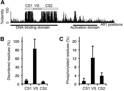
The DNA-binding domain of the Aft-type proteins contains a region of variable length between two cysteine residues that have been suggested to participate in a zinc-finger domain; this region had been named the insert by Babuet al. (2006). The insert sequences (corresponding to residues 143–215 in Aft1) of all known Afts proteins were aligned (Figure 1): most include a variable central regionflanked by two regions rich in basic amino acids. However, the Aft2
orthologs and the Afts of the yeasts from the CTG clade (e.g.,C. albicans) lacked the central region such that their inserts are smaller. The VS (corresponding to residues 152– 203 inAft1) was compared with conservedflanking sequen-ces of the DNA-binding domains, named CS1 and CS2 (Figure 2). VS was predicted to be more disordered than CS1 and CS2 (Figure 2B; paired Mann–Whitney tests, P-value ,0.001). On average, .82% of the residues in all VS but only 8% of those in CS1 and 5% in CS2 were predicted to be disordered. InAft1, 56% of the VS region residues and 11% and 5% for CS1 and CS2, respectively, were predicted to be disordered. Consistent with observations that phosphoryla-tion sites are frequent in disordered regions in proteins (Iakoucheva et al.2004), VS overall were also predicted to be more phosphorylated (on average 12% of the residues) than CS1 (1%) and CS2 (4%) (Figure 2C; paired Mann– Whitney tests, P-value ,0.001). Fifteen percent of the res-idues in Aft1(VS) were predicted to be phosphorylated whereas only 6% of those in CS2 and none of those in CS1 were predicted to be phosphorylated inAft1. Searches in data-bases of experimental mass spectra indicated that the Aft1
(173–203) tryptic peptide was found to be phosphorylated
on the Ser181 and Ser183 residues (PhosphoPep Database). Thus, although VS showed little protein sequence conserva-tion, they appeared to share at least two common proper-ties: being highly disordered and containing a high density of phosphorylation sites.
Aft1(152–203) is required for the binding to the TGCACCC sequence
Conserved disordered regions with rapidly evolving amino acid sequences, such as VS, were recently defined asflexible regions strongly associated with signaling and regulation (Bellay et al.2011). To assess the involvement ofAft1(VS) in the regulatory function of Aft1, we performed deletion mutation analyses (Figure 3). We constructed a mutant de-leted for the entire 152–203 region and mutants deleted for the 152–181, 182–203, and 172–203 subregions (namely D152–203, D152–181, D182–203, and D172–203, respec-tively, Figure 3A) and evaluated their trans-activation and DNA-binding functions. To test the trans-activation func-tion, transcriptional fusions of the proteins to the bacterial LexA DNA-binding domain were constructed and expressed in wild-type cells carrying a LacZ reporter gene driven by four LexA operators (Figure 3B). Equivalent levels of b-galactosidase activity were detected in cells expressing the LexA-Aft1 fusion construct and those expressing the
LexA- deleted Aft1 constructs. This demonstrates that all the deleted mutants retained full trans-activation activity and suggests that the protein stability ofAft1is not affected by the deletions. To evaluate the effect of the deletions on the DNA-binding function of Aft1, the LexA-deleted Aft1
constructs were expressed in aft1Δaft2Δ cells carrying a LacZ reporter gene downstream from theAft1-binding site, TGCACCC. Thus, theb-galactosidase activity gave a measure of the activation of transcription at theAft1-binding site. As previously described (Yamaguchi-Iwai et al. 1996; Courel
et al.2005),Aft1exhibited a strong level ofb-galactosidase activity (Figure 3C). b-Galactosidase activity was 5.5-fold lower with the 152–203 deletion and 2.2- and 1.5-fold lower with the 152–181 and 182–203 deletions, respectively. The construct with the deletion of the 172–203 region resulted in slightly higherb-galactosidase activity (1.2-fold higher). The deletion of the region 152–203 did not affect the level of
Aft1trans-activation per se(Figure 3B), but substantially decreased the activation of transcription at theAft1-binding
site (Figure 3C); this indicates that the 152–203 region is required for the recognition of the TGCACCC sequence.
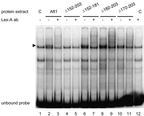
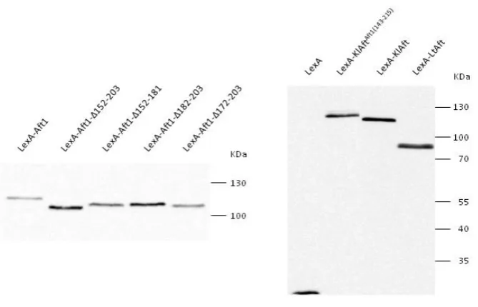
To confirm this result and investigate more directly the role of the 152–203 region for the binding to the TGCACCC sequence, we performed electrophoretic mobility shift assays with extracts from the aft1Δaft2Δ cells expressing LexA, LexA-Aft1, or LexA-deletedAft1constructs and a probe containing the TGCACCCAft1-binding site of theFET3 pro-moter. Western blotting with extract proteins used for mo-bility shift assays and anti-LexA antibodies confirm that the protein stability of the Aft1 protein is not affected by the deletions (seeFigure S1). Extracts containing LexA-Aft1 fu-sion protein gave a high-mobility complex (Figure 4, lane 2) that is absent in extracts containing the sole LexA protein (Figure 4, lanes 1 and 12). Addition of anti-LexA antibod-ies overshifted this complex, but had no effect on other mobility complexes (Figure 4, lane 3). This demonstrated that LexA-Aft1is a component of the higher-mobility complex formed with theFET3promoter sequence. Extracts containing
Figure 3 Deletion of the variable 152–203 Aft1(VS) sequence affects the recognition of the TGCACCC sequence, ferrichrome uptake, and cell growth in iron-depleted conditions. (A) Structure of the Aft1-deleted regions. Shown is a plot of the percentages of identity along the Aft1(143–215) DNA-binding domain insert as described in the legend to Figure 2. The vari-able 152–203 Aft1(VS) sequence is shown as a shaded box. The structures and names of the four deleted regions are depicted below. (B and C)b-Galactosidase activity. LexA-Aft1 deleted proteins have a full capacity fortrans-activation (B) but are affected for the recognition of the
AFT1 DNA-binding site (C). LexA, LexA-Aft1, and LexA-Aft1 deleted derivatives were expressed in wild-type (B) andaft1Daft2D mu-tant strains (C) transformed with either pSH18-34 containing four lexA operators fused to the
LacZgene (B) or pFC-W containing the activa-tion sequence of theFET3gene fused to the
LexA-Aft1(Δ152–181), LexA-Aft1(Δ182–203), or LexA-Aft1
(Δ172–203) exhibit also a complex of high mobility that is overshifted with addition of anti-LexA antibodies (Figure 4, lanes 6–11). In contrast, extracts containing LexA-Aft1
(Δ152–203) did not exhibit the higher-mobility complex (Figure 4, lanes 4 and 5). This showed that the formation of a LexA-Aft1-containing complex is abolished by the 152– 203 deletion but not by the 152–181, 182–203, or 172–203 deletions. In accord with the results presented in Figure 3C, this demonstrated that theAft1(152–203) region is required for the binding to the TGCACCC sequence of theFET3 pro-moter sequence.
Next, we address the functional importance of the Aft1
(152–203) region for iron homeostasis. Aft1 is the major transcriptional regulator of iron transport systems, so we
first conducted iron uptake experiments with the sidero-phore ferrichrome (FCH). FCH transport is mediated by theAft1-regulated genesARN1andSIT1and both their pro-moter regions contain theAft1-binding site TGCACCC (Yun et al.2000a,b). We compared the rate of FCH-55Fe uptake by aft1Δaft2Δcells expressing LexA-Aft1with that of cells expressing the LexA-deleted Aft1 constructs (Figure 3D). FCH accumulation was substantially lower (4.1-fold) in cells expressing LexA-Aft1(Δ152–203) than in cells expressing LexA-Aft1, consistent with a decrease of transcription activ-ity at the Aft1-binding site (Figure 3C). FCH accumulation by cells expressing the other deletion constructs was strictly correlated with the corresponding levels of transcription
activity at theAft1-binding site. The tight correlation between the recognition of the Aft1-binding site and FCH uptake activity (Figure 3, C and D) confirms the importance of
Aft1binding to the TGCACCC promoter sequence for the regulation of FCH uptake.
We compared the growth on agar plates of theaft1Δaft2Δ
mutant expressing LexA-Aft1or the LexA-deletedAft1 con-structs. The aft1Δaft2Δmutant is unable to grow in iron-depleted conditions or on carbon sources like raffinose or glycerol that require functional respiratory activity for their catabolism (Blaiseau et al. 2001; Rutherford et al. 2001). Consistent with previous results (Conde e Silvaet al.2009), the aft1Δaft2Δ mutant phenotypes were complemented by the expression of LexA-Aft1 (Figure 3E). However, the
aft1Δaft2Δcells expressing LexA-Aft1(Δ152–203) grew poorly in iron-depleted conditions and were unable to grow with glycerol as the only carbon source. The aft1Δaft2Δ cells expressing either LexA-Aft1(Δ152–181) or LexA-Aft1(Δ182– 203) showed slight but reproducible growth deficiency on glycerol, whereas the growth of those expressing lexA-Aft1
(Δ172–203) was similar to that of those expressing
LexA-Aft1. These results demonstrate that deletion of the 152– 203 region ofAft1reduced the complementation both of the FCH uptake and of the growth deficiency of aft1Δaft2Δ
mutants in iron-depleted conditions.
The insert of Aft1 (residues 143–215) is necessary and sufficient for the binding of KlAft to the TGCACCC sequence
The DNA-binding mechanisms of Aft1 and KlAft may be different (Conde e Silva et al. 2009). The inserts in Aft1
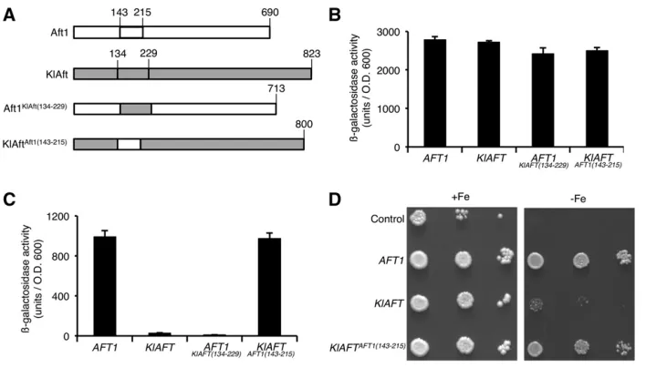
and KlAft are not well conserved (Figure 1), and this diver-gence may be responsible for differences in DNA binding. We used swapping experiments to test this possibility. As the limits of the regions not conserved between Aft1and KlAft are uncertain (Figure 1), we constructed hybrid proteins by exchanging the entireAft1(143–215) and KlAft (134–229) inserts (Figure 5A). The trans-activation and DNA-binding functions ofAft1, KlAft,Aft1KlAft(134–229), and KlAftAft1(143–215) proteins were studied as described in Figure 3, B and C. The native and hybrid proteins exhibited similartrans-activation abilities (Figure 5B). In contrast, the levels of transcription activity at the TGCACCC sequence differed substantially be-tween Aft1 and KlAft and also between the native and the hybrid proteins (Figure 5C). The level ofb-galactosidase ac-tivity in cells expressing KlAft was 35-fold lower than that in those expressing Aft1, indicating that KlAft recognized the TGCACCC sequence very much more poorly than Aft1. The replacement ofAft1(143–215) by KlAft(134–229) resulted in reciprocal opposite effects. The b-galactosidase activity in cells expressing theAft1KlAft(134–229)hybrid was strongly de-creased compared to that in those expressing Aft1. In con-trast, the b-galactosidase activity in cells expressing the KlAftAft1(143–215)hybrid was strongly increased. These fi nd-ings indicate that Aft1(143–215) is necessary and sufficient for KlAft binding to the TGCACCC sequence.
To confirm this important point, we performed electro-phoretic mobility shift assays with extracts from the
aft1Δaft2Δ cells expressing LexA, LexA-Aft1, lexA-KlAft, or LexA-KlAftAft1(143–215) and the probe containing the TGCACCC Aft1-binding site of the FET3 promoter. Western blotting checked that the LexA-KlAft and LexA-KlAftAft1(143–215) proteins were well expressed, at the expected size, in S. cerevisiae (seeFigure S1). As shown in Figure 6, pro-tein extracts containing either LexA-Aft1(lane 2) or LexA-KlAftAft1(143–215) (lane 8) showed a high-mobility complex that is overshifted with addition of anti-LexA antibodies (lanes 3 and 9). In contrast, extracts containing lexA-KlAft (Figure 6, lanes 6 and 7) showed no significant difference from those containing LexA (Figure 6, lanes 1 and 12). To test the specificity of the complex formed with LexA-Aft1
and LexA-KlAftAft1(143–215), we performed competition assays with addition of unlabeled oligomers centered on wild-type TGCACCC (WT) or mutated TGCAGGG (M) sequences. The high-mobility complex formed with both LexA-Aft1 and LexA-KlAftAft1(143–215)was specifically displaced by the ad-dition of excess wild-type but not mutated oligomers (Figure 6, lanes 4 and 5 and 10 and 11). Therefore, the binding of LexA-Aft1 and LexA-KlAftAft1(143–215) was specific for the TGCACCC sequence. Thus, the Aft1(143–215) region con-fers on KlAft the capacity to bind specifically to the TGCACCC sequence. We investigated iron homeostasis in cells expressing KlAftAft1(143–215). We compared the growth of aft1Δaft2Δ cells transformed with monocopy plasmids carrying the AFT1, the KLAFT, or the KLAFTAFT1(143–215) genes controlled by their own promoter (Figure 5D). As pre-viously reported (Conde e Silva et al. 2009), the growth defect of theaft1Δaft2Δmutant was not complemented by the plasmid carryingKLAFT. However, the plasmid carrying KLAFTAFT1(143–215)restored growth to theaft1Δaft2Δmutant,
and therefore KlAft was able to complement theaft1Δaft2Δ
iron-dependent phenotype only if the insert of KlAft was replaced with that ofAft1. This demonstrates the functional importance of the Aft1insert in the regulation of iron ho-meostasis inS. cerevisiae.
Figure 5 The Aft1(143–215) region is nec-essary and sufficient for KlAft to recognize the TGCACCC sequence and complement the growth deficiency ofaft1Daft2Dcells in iron-depleted conditions. (A) Schematic rep-resentation of the native Aft1 and KlAft and hybrid KlAftAft1(143–215) and Aft1KlAft(134–229) proteins in which the Aft1(143–215) and KlAft(134–229) DNA-binding domain inserted regions have been exchanged. (B and C)
b-Galactosidase activity. Aft1, LexA-KlAft, KlAftAft1(143–215), and Aft1KlAft(134–229) proteins have a similar capacity for trans -activation (B) and Aft1(143–215) is necessary and sufficient for KlAft to recognize the TGCACCC sequence (C). Native and hybrid proteins were expressed in wild-type (B) and
aft1Daft2D mutant strains (C) transformed with either pSH18-34 containing four lexA operators fused to the LacZ gene (B) or pFC-W containing the activation sequence of theFET3gene fused to theLacZgene (C). Strains, LacZ repertory systems, and culture conditions were as described in the legend to Figure 3. Results are means6SE from experiments with three independent transformants. (D) Growth ofaft1Daft2Dcells expressing pCXJ22 (Control), Aft1, pCXJ22-KlAft, and pCXJ22-KlAft-Aft1(143–215). Cells were pregrown as in B and C and plated onto YPD without BPS (+Fe) or with 200mM BPS (2Fe).
The TGCACCC sequence is widespread among promoter regions of genes of the iron regulon
in hemiascomycetes
Previous computer analyses identified TGCACCC and PuCACCC Aft-type sequences as consensus sequences in the promoters of iron-regulated genes in S. cerevisiae and K. lactis, respectively (Rutherfordet al.2003; Courelet al. 2005; Conde e Silva et al. 2009). To study Aft-type cis-regulatory sequences and their evolution, we extended our analyses to diverse species belonging to hemiascomycete clades and for which the genome sequences are available. Comparison of gene expression inS. cerevisiaeandK. lactis allowed us to identify a group of Aft-target genes, the ex-pression of which is both iron regulated and dependent on either Aft1 or KlAft. Homologs of this group of genes, re-ferred to here as the iron regulon, were identified in several hemiascomycete species. We analyzed their promoter regions (see Materials and Methods), and the Aft-type ele-ment was found in all hemiascomycetes (Figure 7), except in Y. lipolytica and in species of the CTG clade (C. albicans,
D. hansenii, andP. stipitis). Unlike the other species analyzed, Y. lipolyticaand the CTG clade species possess an ortholog of the iron-responsive GATA-type repressor (Conde e Silva et al.2009) that most probably controls their iron-regulatory pathway, as in most fungi. This has been demonstrated in C. albicans and P. pastoris (Lan et al.2004; Miele et al. 2007). The Aft-type sequence identified in other hemias-comycetes was clearly TGCACCC except in the phyloge-netic branch leading toE. gossypiiandK. lactis.E. gossypii had a small predicted iron regulon (nine genes) and pos-sessed the genome with the highest GC content in the analysis. This led to the detection of an Aft-type sequence with a low confidence level forE. gossypii. As previously reported, (Conde e Silvaet al.2009), we found the shorter and less restrictive Aft-type PuCACCC sequence inK. lactis (Figure 7). The results of this study revealed mainly that the TGCACCC sequence is widespread in the promoter regions of genes of the iron regulons in hemiascomycetes. This suggests that the TGCACCC sequence corresponds to the ancestral Aft-type element and that it has diverged
Figure 7 Divergence of thecis-regulatory sequence between the iron regulons of different hemiascomycete yeasts. Motif logos were constructed using WebLogo (Crookset al.2004) from motif sequences obtained, ase-values, using MEME with gene upstream sequences from inferred iron regulons (seeMaterials and Methods
and File S1). Upstream sequences in
toward the PuCACCC sequence in a recent ancestor of K. lactis.
LtAft, the L. thermotolerans ortholog of Aft1 and KlAft, binds to the TGCACCC sequence and complements the aft1Δaft2Δiron-dependent phenotype
To confirm the results of the bioinformatic analysis described above, we tested the ability of LtAft, the L. thermotolerans ortholog ofAft1 and Klaft, to bind the TGCACCC sequence. We chooseL. thermotoleransbecause theLachanceaspecies have a common ancestor with K. lactismore recent than the common ancestor of K. lactisand S. cerevisiaeand the e-values associated with the predicted TGCACCC sequence identified in the upstream sequences of theL. thermotolerans iron regulon were of high confidence (Figure 7). A LexA-LtAft fusion protein was constructed and we performed electrophoretic mobility shift assays with extracts from theaft1Δaft2Δcells expressing LexA-LtAft and the probe containing the TGCACCC
Aft1-binding site of theFET3promoter. Extracts containing LexA-LtAft gave a mobility complex of high intensity with theFET3promoter (Figure 8A, lane 1). The mobility com-plex formed with LexA-LtAft was overshifted with addition of anti-LexA antibodies (Figure 8A, lane 2) and specifically displaced by the addition of excess unlabeled oligomers cen-tered on the wild-type TGCACCC (WT) sequence but not by those centered on the TGCAGGG (M) mutated sequences (Figure 8A, lanes 3 and 4). These results demonstrated that, in contrast to KlAft, LtAft was able to bind the TGCACCC
Aft1-binding site inS. cerevisiae. We next compared the abil-ity of KlAft and LtAft to complement the aft1Δaft2Δ iron-dependent phenotype. The deficiency of aft1Δaft2Δcells to grow in iron-depleted conditions is not suppressed with a plasmid expressing LexA-KlAft whereas it is totally re-stored with a plasmid expressing LexA-LtAft (Figure 8B). Thus, LtAft was able to bind the TGCACCC sequence and to complement the iron-dependent phenotype of theS. cerevisiae
aft1Δaft2Δ cells. In accord with the bioinformatic data, these experimental results reinforce the hypothesis that the TGCACCC sequence is ancestral and suggest that it diverged toward the PuCACCC sequence after the divergence of the Lachancea yeasts from the phylogenetic branch leading to K. lactis.
Discussion
The Aft1 DNA-binding domain has been described as con-taining a region of variable length (residues 143–215) be-tween two cysteine residues (Babuet al.2006). Our deletion analyses show that the 152–203 sequence, contained in the insert region, is necessary for the DNA-binding function of
Aft1. The DNA-binding capacity ofAft1is also affected, but to a much lesser extent, by the deletion of N-terminal (152– 181) and C-terminal (182–203) subsequences. We showed a strict correlation between the variation in the TGCACCC-binding capacity of deleted Aft1 variant proteins and the effect on both FCH uptake activity and the ability to grow in iron-depleted conditions (Figure 3, C–E). This tight causal link between the DNA-binding level of Aft1and metabolic effects indicates that binding ofAft1to TGCACCC is of cru-cial importance to the control of iron homeostasis inS. cer-evisiae. This agrees with a recent study showing that the iron-dependent dissociation of Aft1 from the promoter is the critical step in the regulation of iron metabolism (Ueta et al. 2012). Thus, unlike what was previously suggested (Yamaguchi-Iwai et al. 2002; Ueta et al. 2003, 2007), the way Aft1 regulates iron homeostasis depends more on its capacity to bind its target DNA sequence than on its capacity to shift from the cytosol to the nucleus. Comparative se-quence analysis of the DNA-binding domains of various Aft proteins showed that Aft1(152–203), which we named “VS”, is a rapidly evolving sequence, bordered by two con-served regions designated CS1 and CS2. VS was predicted to
Figure 8 LtAft, the Lachancea thermotolerans
be highly disordered and phosphorylated. Many such are involved in protein/protein and/or protein/nucleic acid interactions (Dunker et al.2002). Thus, Aft1(152–203) is a good candidate region to interact with DNA or other (as yet unidentified) proteins. Paradoxically, although Aft1
(152–203) is essential for Aft binding to the nucleotide se-quence TGCACCC inS. cerevisiae, it is not conserved.
Previous work indicated thatAft1inS. cerevisiaebinds to the sequence TGCACCC whereas KlAft in K. lactis binds to the sequence PuCACCC (Yamaguchi-Iwaiet al.1996; Conde e Silva et al.2009). Here, we compared the ability of KlAft and Aft1to bind to the TGCACCC Aft1-binding site of the
FET3 promoter in the same S. cerevisiae cellular context. Mobility shift assays and transcription analyses inaft1Daft2D
mutant cells show thatAft1but not KlAft binds efficiently to the TGCACCC sequence in S. cerevisiae cells. Reciprocally, mobility shift assays with a probe containing the CACACCC KlAft-binding site of the KLLA0E14652g K. lactispromoter show that KlAft but notAft1binds efficiently to the CACACCC sequence inS. cerevisiae(seeFigure S2). Taken together, these results indicate thatAft1and KlAft present different DNA-binding sites preference. This difference is presumably due to the large differences between the amino acid sequences of VS in KlAft andAft1(Figure 1). To test this, we constructed a hybrid protein, KlAftAft1(143–215), which allowed us to dem-onstrate that theAft1(143–215) sequence inserted into KlAft was sufficient for binding to TGCACCC. Also, binding of the KlAftAft1(143–215)hybrid to TGCACCC totally suppressed the iron-dependent growth phenotype of theaft1Daft2Dmutant cells. To our knowledge, this is thefirst example in yeast that links the divergence of a limited DNA-binding region of a tran-scription regulator to major physiological effects.
These results indicate that the insert region of the DNA-binding domains of Aft1 and KlAft coevolved with their DNA-binding sites preference. We examined the iron-regulon promoters in other hemiascomycete species (Figure 7). Con-tradicting our previous prediction, this more extensive study strongly suggests that the widespread TGCACCC sequence corresponds to the ancestral Aft-binding site and that this binding site evolved toward PuCACCC in theK. lactis line-age. Comparison of theAft1(143–215) and KlAft (134–229) regions may reveal the differences in the insert amino acid sequence that are associated with the divergence of the DNA-binding specificity (Figure 1). KlAft contains an additional segment of 35 residues that interrupts a sequence enriched in basic amino acids in the N-terminal part of the Aft1VS. This part of VS is important for the binding ofAft1 to the DNA sequence TGCACCC (Figure 3A), so the additional res-idues in KlAft may affect binding to this sequence. Consistent with this hypothesis, LtAft, theL. thermotoleransortholog of
Aft1and KlAft that does not possess the additional segment, was shown able to bind TGCACCC inS. cerevisiaecells (Fig-ure 8A). As shown in Fig(Fig-ure 1, Aft2and its orthologs lack most of the VS. This large difference may explain whyAft2, unlike Aft1, does not exhibit a preference for the TGCACCC sequence (Courelet al.2005). The ability to bind effectively
to TGCACCC might have been lost independently by KlAft andAft2, but in both cases because of the major changes in their VS sequences.
Acknowledgments
We thank Gilles Fischer for the gift of theL. thermotolerans strain CBS 6340 and Marc Lemaire for the gift of the plasmid pCXJ22. We thank Véronique Proux-Gillardeaux for helpful discussions. English text was edited by Alex Edelman & Associates.
Literature Cited
Babu, M. M., L. M. Iyer, S. Balaji, and L. Aravind, 2006 The nat-ural history of the WRKY-GCM1 zinc fingers and the relation-ship between transcription factors and transposons. Nucleic Acids Res. 34: 6505–6520.
Bailey, T. L., and C. Elkan, 1994 Fitting a mixture model by ex-pectation maximization to discover motifs in biopolymers. Proc. Int. Conf. Intell. Syst. Mol. Biol. 2: 28–36.
Baker, C. R., B. B. Tuch, and A. D. Johnson, 2011 Extensive DNA-binding specificity divergence of a conserved transcription reg-ulator. Proc. Natl. Acad. Sci. USA 108: 7493–7498.
Bellay, J., S. Han, M. Michaut, T. Kim, M. Costanzo et al., 2011 Bringing order to protein disorder through comparative genomics and genetic interactions. Genome Biol. 12: R14. Blaiseau, P. L., E. Lesuisse, and J. M. Camadro, 2001 Aft2p,
a novel iron-regulated transcription activator that modulates, with Aft1p, intracellular iron use and resistance to oxidative stress in yeast. J. Biol. Chem. 276: 34221–34226.
Blaiseau, P.-L., A. Seguin, J. M. Camadro, and E. Lesuisse, 2010 Iron uptake in yeasts, pp. 265–284 inIron Uptake and Homeostasis in Microorganisms, edited by P. Cornelis, and S. C. Andrews. Caister Academic Press, Brussels/Reading, UK. Blom, N., S. Gammeltoft, and S. Brunak, 1999 Sequence and
structure-based prediction of eukaryotic protein phosphoryla-tion sites. J. Mol. Biol. 294: 1351–1362.
Byrne, K. P., and K. H. Wolfe, 2005 The Yeast Gene Order Browser: combining curated homology and syntenic context re-veals gene fate in polyploid species. Genome Res. 15: 1456– 1461.
Conde e Silva, N., I. R. Goncalves, M. Lemaire, E. Lesuisse, J. M. Camadroet al., 2009 KlAft, theKluyveromyces lactisortholog of Aft1 and Aft2, mediates activation of iron-responsive tran-scription through the PuCACCC Aft-type sequence. Genetics 183: 93–106.
Courel, M., S. Lallet, J. M. Camadro, and P. L. Blaiseau, 2005 Direct activation of genes involved in intracellular iron use by the yeast iron-responsive transcription factor Aft2 with-out its paralog Aft1. Mol. Cell. Biol. 25: 6760–6771.
Crooks, G. E., G. Hon, J. M. Chandonia, and S. E. Brenner, 2004 WebLogo: a sequence logo generator. Genome Res. 14: 1188–1190.
Dhaoui, M., F. Auchere, P. L. Blaiseau, E. Lesuisse, A. Landoulsi et al., 2011 Gex1 is a yeast glutathione exchanger that inter-feres with pH and redox homeostasis. Mol. Biol. Cell 22: 2054– 2067.
Dunker, A. K., C. J. Brown, J. D. Lawson, L. M. Iakoucheva, and Z. Obradovic, 2002 Intrinsic disorder and protein function. Bio-chemistry 41: 6573–6582.
Gasch, A. P., A. M. Moses, D. Y. Chiang, H. B. Fraser, M. Berardini et al., 2004 Conservation and evolution of cis-regulatory sys-tems in ascomycete fungi. PLoS Biol. 2: e398.
Guarente, L., 1983 Yeast promoters and lacZ fusions designed to study expression of cloned genes in yeast. Methods Enzymol. 101: 181–191.
Haas, H., M. Eisendle, and B. G. Turgeon, 2008 Siderophores in fungal physiology and virulence. Annu. Rev. Phytopathol. 46: 149–187.
Halliwell, B., and J. M. Gutteridge, 1984 Oxygen toxicity, oxygen radicals, transition metals and disease. Biochem. J. 219: 1–14. Hanes, S. D., and R. Brent, 1989 DNA specificity of the bicoid
activator protein is determined by homeodomain recognition helix residue 9. Cell 57: 1275–1283.
Huang, H. D., T. Y. Lee, S. W. Tzeng, and J. T. Horng, 2005 KinasePhos: a web tool for identifying protein kinase-specific phosphorylation sites. Nucleic Acids Res. 33: W226– W229.
Iakoucheva, L. M., P. Radivojac, C. J. Brown, T. R. O’Connor, J. G. Sikeset al., 2004 The importance of intrinsic disorder for pro-tein phosphorylation. Nucleic Acids Res. 32: 1037–1049. Katoh, K., and H. Toh, 2008 Recent developments in the MAFFT
multiple sequence alignment program. Brief. Bioinform. 9: 286– 298.
Katoh, K., K. Kuma, H. Toh, and T. Miyata, 2005 MAFFT version 5: improvement in accuracy of multiple sequence alignment. Nucleic Acids Res. 33: 511–518.
Kuo, D., K. Licon, S. Bandyopadhyay, R. Chuang, C. Luo et al., 2010 Coevolution within a transcriptional network by com-pensatory trans and cis mutations. Genome Res. 20: 1672–1678. Lan, C. Y., G. Rodarte, L. A. Murillo, T. Jones, R. W. Daviset al., 2004 Regulatory networks affected by iron availability in Can-dida albicans. Mol. Microbiol. 53: 1451–1469.
Lesuisse, E., P. L. Blaiseau, A. Dancis, and J. M. Camadro, 2001 Siderophore uptake and use by the yeast Saccharomy-ces cerevisiae. Microbiology 147: 289–298.
Miele, R., D. Barra, and M. C. Bonaccorsi di Patti, 2007 A GATA-type transcription factor regulates expression of the high-affinity iron uptake system in the methylotrophic yeast Pichia pastoris. Arch. Biochem. Biophys. 465: 172–179.
O’Brien, K. P., M. Remm, and E. L. Sonnhammer, 2005 Inparanoid: a comprehensive database of eukaryotic orthologs. Nucleic Acids Res. 33: D476–D480.
Prevorovsky, M., S. R. Atkinson, M. Ptackova, J. R. McLean, K. Gould et al., 2011 N-termini of fungal CSL transcription fac-tors are disordered, enriched in regulatory motifs and inhibit DNA binding infission yeast. PLoS ONE 6: e23650.
Rutherford, J. C., S. Jaron, E. Ray, P. O. Brown, and D. R. Winge, 2001 A second iron-regulatory system in yeast independent of Aft1p. Proc. Natl. Acad. Sci. USA 98: 14322–14327.
Rutherford, J. C., S. Jaron, and D. R. Winge, 2003 Aft1p and Aft2p mediate iron-responsive gene expression in yeast through related promoter elements. J. Biol. Chem. 278: 27636–27643. Ueta, R., A. Fukunaka, and Y. Yamaguchi-Iwai, 2003 Pse1p
medi-ates the nuclear import of the iron-responsive transcription fac-tor Aft1p in Saccharomyces cerevisiae. J. Biol. Chem. 278: 50120–50127.
Ueta, R., N. Fujiwara, K. Iwai, and Y. Yamaguchi-Iwai, 2007 Mechanism underlying the iron-dependent nuclear export of the iron-responsive transcription factor Aft1p in Saccharomyces cerevisiae. Mol. Biol. Cell 18: 2980–2990.
Ueta, R., N. Fujiwara, K. Iwai, and Y. Yamaguchi-Iwai, 2012 Iron-induced dissociation of the aft1p transcriptional regulator from target gene promoters is an initial event in iron-dependent gene suppression. Mol. Cell. Biol. 32: 4998–5008.
Wach, A., 1996 PCR-synthesis of marker cassettes with long
flanking homology regions for gene disruptions in S. cerevisiae. Yeast 12: 259–265.
Wang, H., Z. Xu, L. Gao, and B. Hao, 2009 A fungal phylogeny based on 82 complete genomes using the composition vector method. BMC Evol. Biol. 9: 195.
Ward, J. J., J. S. Sodhi, L. J. McGuffin, B. F. Buxton, and D. T. Jones, 2004 Prediction and functional analysis of native disorder in proteins from the three kingdoms of life. J. Mol. Biol. 337: 635–645.
Waterhouse, R. M., E. M. Zdobnov, F. Tegenfeldt, J. Li, and E. V. Kriventseva, 2011 OrthoDB: the hierarchical catalog of eu-karyotic orthologs in 2011. Nucleic Acids Res. 39: D283–D288. Wolfe, K. H., and D. C. Shields, 1997 Molecular evidence for an ancient duplication of the entire yeast genome. Nature 387: 708–713.
Yamaguchi-Iwai, Y., A. Dancis, and R. D. Klausner, 1995 AFT1: a mediator of iron regulated transcriptional control in Saccha-romyces cerevisiae. EMBO J. 14: 1231–1239.
Yamaguchi-Iwai, Y., R. Stearman, A. Dancis, and R. D. Klausner, 1996 Iron-regulated DNA binding by the AFT1 protein con-trols the iron regulon in yeast. EMBO J. 15: 3377–3384. Yamaguchi-Iwai, Y., R. Ueta, A. Fukunaka, and R. Sasaki,
2002 Subcellular localization of Aft1 transcription factor re-sponds to iron status in Saccharomyces cerevisiae. J. Biol. Chem. 277: 18914–18918.
Yun, C. W., T. Ferea, J. Rashford, O. Ardon, P. O. Brown et al., 2000a Desferrioxamine-mediated iron uptake in Saccharomy-ces cerevisiae. Evidence for two pathways of iron uptake. J. Biol. Chem. 275: 10709–10715.
Yun, C. W., J. S. Tiedeman, R. E. Moore, and C. C. Philpott, 2000b Siderophore-iron uptake in Saccharomyces cerevisiae. Identification of ferrichrome and fusarinine transporters. J. Biol. Chem. 275: 16354–16359.
GENETICS
Supporting Information
http://www.genetics.org/lookup/suppl/doi:10.1534/genetics.113.157693/-/DC1
The Basis for Evolution of DNA-Binding Speci
fi
city of the
Aft1 Transcription Factor in Yeasts
Isabelle R. Gonçalves, Natalia Conde e Silva, Cesar La Torre Garay, Emmanuel Lesuisse, Jean Michel Camadro, and Pierre Louis Blaiseau
Figure S1 Expression of the LexA-Aft proteins in aft1Δaft2Δ cells. 40µg of extract proteins used for the electrophoretic
mobility shift assays and prepared as described in (CONDE E SILVA et al. 2009) were loaded on a 8% acrylamide gel. After
transfert on PVDF membrane, the LexA fusion proteins were detected with anti-LexA monoclonal antibodies (Santa Cruz
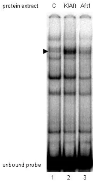
Figure S2 KlAft but not Aft1 binds efficiently to the -531 CACACCC KlAft DNA-binding site of the KLLA0E14652g promoter.
Gel-shift assays were carried out with extracts from aft1Δaft2Δ cells expressing LexA (C, lane 1), LexA-KlAft (lane 2) and
LexA-Aft1 (lane 3). The arrow indicates the KlAft- and Aft1-containing complexes. The DNA probe corresponds to the
sequence of positions -570 to -429 of KLLA0E14652g containing the -530 CACACCC KlAft DNA-binding site and was prepared
as described in (Conde e Silva et al., 2009). The binding reaction mixtures (20 µl) contained 25 mM HEPES, pH 7.6, 60 mM
KCI, 7.5% glycerol, 0.1 mM EDTA, 1 mM dithiothreitol, 5 mM MgCl2. Aliquots of 20 µg of cell extract and 0,5 µg of
poly(dIdC)2 were used. Approximately 10,000 cpm of probe (1.7 ng) was used in each binding mixture. Samples were
incubated for 30 min at room temperature. Samples were loaded onto a 5% polyacrylamide gel as described in MATERIALS
File S1
Inferred iron regulons in ascomycete species