INVESTIGATION
A Novel Strategy for Cell-Autonomous Gene
Knockdown in
Caenorhabditis elegans
De
fi
nes
a Cell-Speci
fi
c Function for the G-Protein
Subunit GOA-1
Kathryn N. Maher,* Aishwarya Swaminathan,†Parth Patel,‡and Daniel L. Chase‡,1 *Molecular and Cellular Biology Program,†Department of Microbiology, and‡Department of Biochemistry and Molecular Biology, University of Massachusetts, Amherst, Massachusetts 01003
ABSTRACT We developed a novel knockdown strategy to examine cell-specific gene function in Caenorhabditis elegans. In this strategy a null mutation in any gene is replaced with a genetically stable transgene that contains a wild-type copy of the gene fused to a 39tag that targets the mRNA transcript for degradation by the host nonsense-mediated decay (NMD) machinery. In NMD-defective animals, tagged transgene mRNA is expressed at levels similar to the endogenous gene it replaced and is translated into wild-type protein that fully rescues gene function. Cell-specific activation of NMD cell autonomously knocks down transgene expression in specific cell types without affecting its expression or function in other cells of the organism. To demonstrate the utility of this system, we replaced thegoa-1gene, encoding the pan-neuronally expressed G-protein subunitGOA-1, with a degradation-tagged transgene. We then knocked down expression of the transgene from only two neurons, the hermaphrodite-specific neurons (HSNs), and showed thatGOA-1acts cell autonomously in the HSNs to inhibit egg-laying behavior.
E
XPERIMENTAL strategies that reduce protein activity, such as the use of genetic mutations or small molecule protein inhibitors, have been very useful in elucidating the functional roles of proteins. However, in multicellular organ-isms these strategies cause global effects on protein activity that can obscure the cell-specific function of a protein. This is a particular concern when investigating the function of pro-teins that are widely expressed or that have different functions in different cell types or at different times during develop-ment. To understand the cell-specific function of these pro-teins it is necessary to reduce or eliminate their activity in individual cell types, without affecting their activity in other cells of the organism.InCaenorhabditis elegans, double-stranded RNA interfer-ence (dsRNAi), using either promoter-driven sense and an-tisense transgenes or hairpin RNAs, has been used to reduce
protein function in specific cell types (Tavernarakis et al.
2000; Johnsonet al.2005; Espositoet al.2007). However, intermediates generated during processing of dsRNAs can be transported between cells of the organism and thus even transgenes driven by cell-specific promoters can cause global knockdown effects (Jose et al. 2009, 2011). In addition, extrachromosomal transgenes typically used to drive expres-sion of dsRNAs are randomly lost during cell diviexpres-sion, lead-ing to mosaic knockdown effects within individual animals of a population (Stinchcombet al.1985).
We have developed a method to knock down the expres-sion of any gene in any cell type inC. elegansthat is both cell autonomous and genetically stable. In this strategy, an endog-enous gene is replaced by an integrated single-copy transgene containing the endogenous gene’s promoter and coding se-quence tagged with a unique 39-UTR that targets transgene mRNA for destruction by the host cell’s nonsense-mediated decay (NMD) machinery. In NMD-defective animals, the tagged transgene is expressed at levels comparable to that of the endogenous gene and is able to restore wild-type gene function. Spatial control of knockdown is achieved by cell-specific restoration of NMD activity. Using appropriate cell-specific promoters to control NMD activity, one can restrict
Copyright © 2013 by the Genetics Society of America doi: 10.1534/genetics.113.149724
Manuscript received January 23, 2013; accepted for publication March 12, 2013 Supporting information is available online athttp://www.genetics.org/lookup/suppl/ doi:10.1534/genetics.113.149724/-/DC1.
the knockdown of transgene expression to individual cell types in the animal without affecting its expression in any other cells. To demonstrate the utility of this strategy, we used a tagged transgene to investigate the cell-specific function of the G-protein subunitGOA-1and found that selective removal ofGOA-1from the two hermaphrodite-specific neurons (HSNs) (but not from other cells of the organism) was sufficient to cause goa-1null egg-laying defects. Thus GOA-1 acts cell autonomously in the HSNs to inhibit egg-laying behavior.
This cell-autonomous method of gene knockdown can be used to examine the cell-specific function of any protein, eliminating the confounding effects caused by the global reduction of protein function typical of other knockdown strategies.
Materials and Methods
Transgenes
To generate let-858 tagged transgenes 4188 bp of let-858 sequence was amplified from pPD118.60 (L3808, Addgene), using primers SbfI-let-858F andXhoI-let-858R and inserted intoSbfI/XhoI sites of pCFJ151 to generate pCL123 (see list of primers in Supporting Information,Table S1). Promoter and coding sequences were then inserted into theAvrII/SbfI sites of pCL123 to generate tagged transgenes. For mCherry, a 2105 bp region including rab-3 promoter and mCherry coding sequence was amplified from pGH8, using primers
AvrII-pRAB-3-F andSbfI-mCherry-R to generate pCL124. For goa-1, a 9618 bp region including 5,972 bp of promoter and the entiregoa-1coding region was amplified from genomic DNA, using primers goa-1-pro-NheI-F and goa-1-SbfI-R to generate pCL136. For unc-4, a 5205 bp region including 2807 bp of promoter and the entire unc-4 coding region was amplified from genomic DNA, using primers unc-4
pro-AvrII-F andunc-4SbfI-R to generate pCL145.
To generate NMD rescue transgenes, a 1971 bp region of smg-5 genomic sequence and 39-UTR was amplified from genomic DNA, using primers smg-5-AvrII-F and smg-5 -39utr-KpnI-R, and inserted into the AvrII/KpnI sites of pCFJ178 (Addgene) to generate pCL119. Cell-specific pro-moters were then inserted into the SbfI/AvrII sites of pCL119. To restore NMD in cholinergic cells, 3249 bp of unc-17 promoter sequence was amplified using primers
unc-17-SbfI-F and unc-17-pro-AvrII-R to generate pCL121. To restore NMD in all goa-1–expressing cells 5972 bp of promotergoa-1sequence was amplified using primers goa-1-pro-SbfI-F and goa-1-pro-NheI-R to generate pCL137. To restore NMD in HSNs, 3124 bp oftph-1promoter sequence was amplified using primerstph-1-pro-SbfI-F andtph-1
-pro-AvrII-R to generate pCL140. To restore NMD in all unc-4– expressing cells 2804 bp of unc-4 promoter sequence was amplified using the primersunc-4-pro-SbfI-F andunc-4
-pro-AvrII-R to generate pCL147.
Sense/antisense unc-4 transgenes were made as de-scribed using 2912 bp of unc-4 promoter (Esposito et al.
2007). The sense promoter used primers unc-4-pro-outer-F andunc-4-pro-fusion-sense-R. The antisense promoter used primers unc-4-pro-outer-F and unc-4-pro-fusion-R-AS. The RNAi target sequence was amplified using unc-4 -seq-exon-4-F and unc-4-ex6-outer-R primers. Using these DNAs as template, the fusion sense transgene was made using pri-mers unc-4-pro-inner-F and unc-4-ex6-inner-R and the fusion antisense transgene was made using primers unc-4-pro-inner-F and unc-4-ex4-inner-F to generate 3525 bp products. PCR transgenes were purified before injection.
C. elegans strains
Worm strains were generated and maintained using standard methods and conditions (Brenner 1974). The wild-type strain was BristolN2. The boundaries of thegoa-1(n363)deletion mutation were determined to be 59 -AGAACAATATAGAAG-TAGTGCTTAG-ACGCAACTTTTCCAATTGGC-39, resulting in a 15,217 bp deletion that removed the entire coding sequence of goa-1. Strains analyzed in this study were as follows:
Figure 1: XP495ndEx135, XP492smg-5(r860)I;ndEx132, XP487 smg-5,(r860) I; ndEx126, XP445 smg-5(r860) I; ndSi1IV;ndEx115.
Figure 2: N2, XP384 smg-5(r860) I, KO98 goa-1(n363) I, XP447goa-1(n363)I;ndSi7II, XP466smg-5(r860) goa-1(n363)I, XP467smg-5(r860)goa-1(n363)I;ndSi7II. Figure 3:N2, KO98goa-1(n363)I, XP467smg-5(r860)
goa-1(n363) I; ndSi7II, XP469 smg-5(r860) goa-1(n363) I; ndSi7 II; ndSi5 IV, XP468 smg-5(r860) goa-1(n363) I; ndSi7II;ndSi6IV.
Figure 4:N2, KO98goa-1(n363)I, XP467smg-5(r860) goa-1(n363) I; ndSi7II, XP469 smg-5(r860) goa-1(n363) I; ndSi7II;ndSi5IV.
Table 1:N2,VC1453unc-4(gk668)II, XP470smg-5(r860)I; unc-4(gk668) II, XP481 smg-5(r860) I; unc-4(gk668)
ndSi19 II, XP503 smg-5(r860) I; unc-4(gk668) ndSi19 II; ndSi20 IV, XA4254 qaIs4254, KP3948 eri-1(mg366)
IV, lin-15b(n744) X, XP508 ndEx149, XP509 ndEx150, XP510ndEx151, XP511ndEx152, XP512ndEx153.
Construction of transgenic strains
Strains expressing extrachromosomal arrays were generated by coinjecting transgenes at the following concentrations: XP495, pRab100 (rab-3p::GFP) (50 ng/ml) and pCL124 (50 ng/ml) into N2 animals (array designation: ndEx135); XP492, pRab100 (50 ng/ml) and pCL124 (50 ng/ml) into XP384 smg-5(r860) animals (array designation: ndEx132); XP487, pCL124 (50 ng/ml) and pCL30 (unc-17p::GFP con-taining 3249 bp ofunc-17 promoter sequence) (50 ng/ml) into XP384smg-5(r860)animals (array designation:ndEx126); and XP445, pCL124 (50 ng/ml) and pCL30 (50 ng/ml) into XP380 (smg-5(r860); ndSi1) animals (array designation:
unc-4transgenes (50 ng/ml each) with pGH8 (10 ng/ml) (array designations:ndEx149,ndEx150,ndEx151,ndEx152, and
ndEx153, respectively).
Transgene integration at Mos1 alleles cxTi10882 or ttTi5605 (strains EG5003 and EG4322, respectively) was done as described in Frøkær-Jensen et al. (2008). Single-copy insertions were confirmed by PCR and enzyme digest, using a protocol developed by M. Nonet (http://thalamus. wustl.edu/nonetlab/ResourcesF/Resources.html). Briefly, genomic DNA was purified from putative integrants and the transgene insertion was amplified with primers that
flanked the recombination region. Primers to verify inser-tions at cxTi10882 were Chr4-SC-Int-OF-2 and Ti10882-Chr4right-OR. Primers for ttTi5605 were NM3880 and NM3884. Appropriately sized PCR products were digested withDraI to confirm their identity.ndSi1,ndSi5,ndSi6, and ndSi20were generated by recombination of pCL121, pCL137, pCL140, and pCL147, respectively at Mos1 allelecxTi10882. ndSi7 and ndSi19 were generated by recombination of pCL136 and pCL145, respectively, at Mos1 allelettTi5605.
Behavioral assays
Locomotion assays were performed as described in Koelle and Horvitz (1996). Briefly, staged L4 animals were placed
on NGM plates containing a thin lawn ofOP50bacteria and allowed to develop for 24 hr at 20°and then assayed. The number of body bends was counted over a 3-min period for 30 animals per strain. Reversal assays were performed on animals staged as L4 larvae 24 hr before the assay. They were then transferred to NGM plates containing a thin lawn ofOP50bacteria and left undisturbed for 3 min before assay. The number of spontaneous reversals made by each of 30 animals was counted for a 3-min period to determine rever-sals per minute. A reversal was scored when an animal changed its direction of movement from forward to reverse and the animal reversed by at least one body bend (a body bend was counted when a point in the body immediately posterior to the pharynx passed through a minimum or max-imum amplitude).
Egg-laying assays were performed as described in Koelle and Horvitz (1996). Briefly, animals were staged as L4 her-maphrodites and assayed after developing an additional 30 hr at 20°. Individual adult animals were incubated in 1% sodium hypochlorite solution to dissolve the bodies and the number of eggs that remained was counted for a total of 30 animals per strain.
For nose touch response, staged L4 animals were placed on NGM plates containing a lawn of OP50 bacteria and
allowed to develop for 24 hr at 20° before assay. Adult animals were moved to an unseeded NGM plate and for-ward-moving animals were tapped lightly on the head with a platinum wire.unc-4(gk668)mutants do not reverse when tapped but instead contract their midbody, causing the ani-mal to coil, defined as the head curling back and touching a region of the body posterior to the head. One hundred animals were assayed per strain. Animals expressing the extrachromosomal unc-4 sense/antisense transgenes were selected by the presence of a coinjected rab-3p::mCherry marker. Nose touch response in these transgenic strains was determined by assaying$50 animals from each offive individually isolated transgenic lines for a total of $250 animals. XA4254 embryos were heat-shocked for 4 hr to induce expression of the unc-4 hairpin RNA as described in Johnson et al.(2005) and assayed for their ability to re-verse as adults. For feeding dsRNA,KP3948eri-1(mg366); lin-15b(n744) animals were fed bacteria expressing either unc-4 dsRNA or the control pL4440 strain as described in Waniet al.(2012) and F1generation animals were assayed
for reversal behavior as young adults.
Quantitative RT-PCR
To prepare total RNA, mixed-staged animals were washed from three 6 cm plates and cleaned using a 30% sucrose solution to generate an100ml worm pellet. Worms were
flash frozen in liquid nitrogen, ground in a mortar, and dis-solved in 1 ml Trizol reagent. RNA was then purified by standard methods (typical yield, 100–250 mg). Five micro-grams total RNA was treated with DNaseI (Promega, Madison, WI) and purified using an RNA concentrator kit (Zymo Re-search). Five hundred nanograms of purified RNA was used to
generate cDNA, using the Superscript IIIfirst-strand synthesis kit (Invitrogen, Carlsbad, CA) with random hexamers. Quan-titative real-time PCR assays were performed using Brilliant III SYBR green and ROX master mix (Agilent Technologies) in a Stratagene (La Jolla, CA) Mx3000P thermocycler. Primers
goa-1-qPCR-F, goa-1-qPCR-R, and cdc-42-qPCR-F, cdc-42 -qPCR-R were used to measuregoa-1andcdc-42expression, respectively.
Worm lysates and Western blotting
Worms were grown in 125 ml liquid cultures as described in Hofler and Koelle (2011). Animals were cleaned using a 30% sucrose solution and washed twice with 0.1 M NaCl. The worm pellet was flash frozen in liquid nitrogen and stored at280°. Worm protein extracts were made by boiling 100ml worm pellets for 5 min in 1 ml sample buffer (100 mM Tris, pH 6.8, 2% SDS, 5%b-mercaptoethanol, 15% glycerol) followed by centrifugation to remove worm debris. Fifty micrograms of total protein of each sample was analyzed by 10% SDS–PAGE. Proteins were transferred to nitrocellulose membrane and the membrane was incubated with rabbit
anti-UNC-64antibody (Ab940, dilution 1:20,000) and rabbit
anti-GOA-1(dilution 1:300). The secondary antibody used was goat anti-rabbit HRP-conjugated antibody (dilution 1:3000; Bio-Rad, Hercules, CA) and proteins were detected using the Immun-Star HRP Chemiluminescence Kit (Bio-Rad).
Statistical analysis
We used a two-tailed Mann–Whitney test to compare behav-iors in Figures 2 and 3 and a two-tailed, unpaired Student’s
t-test to compare RNA abundances between wild-type and transgenic animals shown in Figure 4.
Results
Protein expression from a tagged transgene is greatly reduced by NMD
InC. elegans, at least seven proteins (SMG-1through SMG-7) are required for the degradation of aberrant mRNA tran-scripts that contain a premature termination codon (PTC) (Pulak and Anderson 1993; Caliet al.1999). While the pre-cise signals that trigger NMD in C. elegans are not known, the efficiency with which a PTC-containing transcript is de-graded is correlated with the distance of the PTC from the normal stop codon, with distances .500 nt causing robust degradation (Pulak and Anderson 1993; Longman et al.
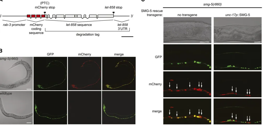
2007). To determine whether NMD could be used to regu-late gene expression, we generated a tripartite mCherry transgene that contained the pan-neuronalrab-3 promoter placed upstream of mCherry coding sequence followed by a degradation tag to act as an NMD trigger (Figure 1A). The degradation tag we chose consisted of several exons and introns from the let-858 gene with its normal stop codon and 39-UTR. This segment oflet-858 has been shown pre-viously to be capable of targeting chimeric mRNA transcripts for NMD-dependent degradation in C. elegans (Link et al.
2003). In transcripts from the tripartite transgene, the stop codon of mCherry should be recognized as a PTC by the NMD machinery as it occurs.500 nt upstream of the stop codon of let-858. We injected equal concentrations of this taggedrab-3p::mCherry::let-858transgene together with an untagged rab-3p::GFP transgene into wild-type andsmg-5
(NMD-defective) mutant animals. Coinjected transgenes concatenate in theC. elegansgonad to form large multicopy arrays and thus coinjection of the mCherry and GFP trans-genes ensured their coexpression in transgenic progeny (Mello et al. 1991). We found that while GFP was easily detected in most or all neurons of both wild-type and smg-5transgenic animals, mCherry protein was detectable only in smg-5 mutant animals, suggesting that the tagged mCherry mRNA was stable in NMD-defective animals and was degraded by NMD in wild-type animals (Figure 1B).
Since the expression of the mCherry protein was de-pendent upon NMD activity, we next determined whether spatial control of NMD activity could be used to reduce mCherry protein expression cell specifically. To cell-specif-ically control NMD activity we generated an unc-17p:: SMG-5 transgene to rescue NMD activity in cholinergic cells (Alfonso et al. 1993). To ensure that NMD activity was restored in all cholinergic cells, we integrated the unc-17p::SMG-5 transgene at single copy at the Mos1 allele cxTi10882 by Mos1-mediated single-copy insertion (MosSCI) (Frøkær-Jensen et al. 2008). cxTi10882 is lo-cated within an intergenic region of chromosome IV and does not disrupt the function of nearby genes or exert ad-verse position effects on gene expression (Frøkær-Jensen
et al.2008). To test for cell-specific knockdown of mCherry, we coinjected smg-5 and smg-5; unc-17p::SMG-5 animals with the pan-neuronally expressed taggedrab-3p::mCherry:: let-858and an untaggedunc-17p::GFP transgene and exam-ined expression of mCherry and GFP in ventral cord motor
neurons (Figure 1C). The cell bodies of only two neuron types are found in the ventral cord in C. elegans: GABAergic and cholinergic neurons. In our transgenic animals GFP expres-sion marks the cholinergic neurons. We found that all smg-5 mutants that lacked the unc-17p::SMG-5 NMD-rescuing transgene expressed mCherry in all neurons, including both GABAergic and cholinergic neurons of the ventral cord (Figure 1C, left). In contrast, mCherry was easily detected only in GABAergic neurons of smg-5; unc-17p:: SMG-5 animals (Figure 1C, right). The absence of mCherry in GFP-expressing cholinergic cells ofunc-17p::SMG-5 res-cued animals indicated that the mCherry mRNA was effi -ciently degraded by NMD in these cells. Thus NMD can be used in C. elegansto restrict knockdown of transgene ex-pression to specific cell types.
A single-copy integrated tagged transgene can rescue function of an endogenous gene
To functionally replace an endogenous gene, a transgene must be expressed at the same levels and in the same cells as the gene it replaces. To ensure that our transgenes met these criteria, we used full-length endogenous promoters to drive expression of all transgenes used in this study and we integrated the transgenes in single copy at well-character-ized Mos1 alleles that did not cause adverse effects on gene expression (Frøkær-Jensen et al.2008).
We chose to determine whether a tagged transgene could replace the function ofgoa-1, which encodes the most abun-dant G-protein subunit (GOA-1) in C. elegans and shares 80% amino acid identity to the human G-protein subunit Gao. We chose thegoa-1 gene for four reasons. First, the
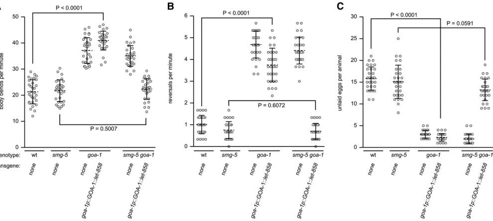
goa-1(n363) null deletion removes the entire coding se-quence ofgoa-1. Because nogoa-1mRNA or protein is made in agoa-1(n363)deletion mutant, it would serve as an ideal genetic background for expression of a degradation-tagged goa-1transgene. Anygoa-1mRNA or protein made in a goa-1(n363) transgenic strain must be from the tagged trans-gene. Second,GOA-1is highly expressed in most or all neu-rons of wild-type animals and is also expressed in many muscle cells (Mendelet al.1995; Ségalatet al.1995). Since our gene replacement strategy would use the endogenous goa-1promoter to drive expression of the tagged transgene, we would expect similarly high levels ofGOA-1expression ingoa-1(n363)null, NMD-defective transgenic animals. Ad-ditionally, robust expression from thegoa-1promoter would allow us to measure large changes in transgene mRNA and protein abundance that might occur as the result of NMD-dependent knockdown. Third,GOA-1functions in different cell types to modulate different worm behaviors, including locomotion and egg laying, and these behaviors are highly reproducible and easily measured (Mendel et al. 1995; Ségalat et al. 1995). Thus we could use behavioral assays as a qualitative measure of the ability of the tagged trans-gene to rescue the function of the goa-1(n363) null muta-tion. goa-1(n363) null mutants move faster than wild-type animals (Mendel et al.1995; Ségalat et al. 1995) (Figure 2A). While the specific cells that control locomotion rate have not been formally identified, GOA-1 is expressed in the motor neurons that innervate body wall muscles and in the body muscles themselves, making these cell types the likely sites ofGOA-1action (Mendelet al.1995; Ségalat
et al.1995). goa-1(n363)mutants reverse more frequently than wild-type animals (Figure 2B). In C. elegans reversal behavior is controlled by the command interneurons and thus GOA-1 likely functions in the command interneurons to control reversal frequency (Chalfie et al. 1985). goa-1
(n363) null mutants lay eggs more frequently than wild-type animals (Mendelet al.1995; Ségalatet al.1995). This leads to a reduction of the steady-state number of eggs retained in the uterus of goa-1(n363) null mutants com-pared to wild-type animals (Figure 2C). Because GOA-1 is
expressed in both the egg-laying muscle cells and the two HSNs that innervate them, the abnormal egg-laying behav-ior of goa-1(n363) mutants could be due to loss ofGOA-1
function in either of these cell types. In experiments designed to determine the site ofGOA-1 function in egg-laying behavior, Moresco and Koelle (2004) expressed a transgene encoding a constitutively activeGOA-1protein separately in the egg-laying muscles and in HSN neurons. They found that expression of the mutantGOA-1protein in the HSN neurons, but not in the egg-laying muscles, inhibited egg-laying behavior. While the mutantGOA-1 pro-tein used in these studies may have inhibited egg-laying behavior by activating nonphysiological signaling pathways, these experiments do suggest thatGOA-1function is neces-sary in the HSN neurons and not in the egg-laying muscles to control egg-laying behavior. The cell-specific effects of
GOA-1 activity on C. elegans behaviors would provide a means for us to evaluate cell-specific gene knockdown, using assays for locomotion rate, reversal frequency, and egg-laying behaviors. The fourth reason for choosing goa-1 was that global overexpression of wild-type GOA-1 causes locomotion and egg-laying defects that are opposite to those observed in the goa-1(n363) null mutant (Mendel et al.
1995; Ségalat et al. 1995). Thus animals that overexpress wild-typeGOA-1move slower and lay eggs less frequently than wild-type animals (Mendel et al. 1995; Ségalat et al.1995). The sensitivity of these behaviors togoa-1dosage would allow a qualitative assessment of tagged transgene expression levels.
We generated a goa-1 transgene (goa-1p::GOA-1::let-858) that included the full rescuinggoa-1promoter (Ségalat
et al.1995) and coding sequence fused to thelet-858 deg-radation tag and integrated this tagged transgene at single copy at the Mos1ttTi5605allele. This integrated strain was then used to generate goa-1; goa-1p::GOA-1::let-858 and smg-5 goa-1; goa-1p::GOA-1::let-858 transgenic animals, whose locomotion and egg-laying behaviors were compared to those of control strains (Figure 2). Importantly, the loss of NMD had no effect on the three behaviors tested as wild-type and smg-5 mutants moved at similar rates, showed similar reversal frequencies, and retained a similar number of eggs in utero(Figure 2, A–C). We found that in NMD-competent animals, the goa-1p::GOA-1::let-858 transgene was unable to replace GOA-1 function to control any of the three behaviors (compare wild-type animals to goa-1 mutants that express the goa-1p::GOA-1::let-858 trans-gene). In contrast, we found that in NMD-defective animals the goa-1p::GOA-1::let-858 transgene was able to fully re-placeGOA-1function for all three behaviors (compare wild-type animals or smg-5 mutants to smg-5 goa-1 double mutants that express the goa-1p::GOA-1::let-858 trans-gene). Because GOA-1 functions in different cell types to control locomotion rate, reversal frequency, and egg-laying behaviors, we conclude that mRNA from the tagged trans-gene was degraded in most or all neurons of NMD-compe-tent animals and, in contrast, was stably expressed in most or all neurons of NMD-defective animals. Notably, no
Table 1 Comparison of knock down efficiency between gene replacement/NMD and dsRNA interference strategies.
Nose Touch Response (% of Prodded Animals)
Genotype and Knock Down Strategy
Defective Backing Behavior
Normal sinusoidal backing behavior
Midbody contraction resulting in partial coil
Midbody contraction resulting in full coil
Total percent animals with backing defect
No knock down(n = 100 for all strains)
1. wildtype 100 0 0 0
2.unc-4 0 8 92 100
3.smg-5; unc-4 0 11 89 100
NMD replacement and knock down (n = 100 for all strains)
4.smg-5; unc-4 ndSi19[unc-4p::UNC-4:: let-858 3’UTR]
100 0 0 0
5.smg-5;unc-4 ndSi19[unc-4p::UNC-4:: let-858 3’UTR];ndSi20[unc-4p::SMG-5]
0 72 28 100
Hairpin dsRNAi(n = 100 for all strains)
6.qaIs4254 94 3 3 6
dsRNAi Feeding(n = 100 for all strains)
7.eri-1; lin-15;pL4440 control 100 0 0 0
8.eri-1; lin-15; unc-4(RNAi) 38 52 10 62
Injection of sense/antisense dsRNA (>50 animals per line)
9. line 1 41 12 47 59
10. line 2 39 4 57 61
11. line 3 18 39 43 82
12. line 4 46 8 46 54
13. line 5 12 49 39 88
intermediate phenotypes were observed in any of the trans-genic animals tested, indicating that transgene expression was tightly controlled by NMD in all animals and in all cell types and that knockdown was robust. Finally, we note that thegoa-1p::GOA-1::let-858transgene did not cause overex-pression phenotypes insmg-5 goa-1double-mutant animals, suggesting that it was expressed at levels that were similar to those of the endogenousgoa-1gene in wild-type animals.
Knockdown of mRNA expressed from a single-copy tagged transgene
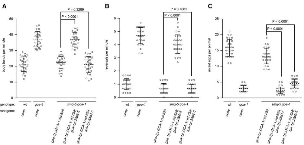
The results described so far showed that expression of a single-copy, integrated tagged transgene could replace the function of an endogenous gene to rescue null mutant phenotypes to wild-type behavior. We also demonstrated that the rescuing activity of the tagged transgene is NMD dependent. We next wanted to determine whether we could knock down the expression of the tagged transgene both globally and in specific cell types by selective activation of NMD. To do this we generated two NMD-rescuing trans-genes. Thefirst was an untaggedgoa-1p::SMG-5 transgene that we integrated at the Mos1 cxTi10882 allele of smg-5 goa-1double mutants that expressed the integratedgoa-1p:: GOA-1::let-858 tagged transgene. Expression of the un-taggedgoa-1p::SMG-5 transgene should restore NMD activ-ity in all cells that express the goa-1p::GOA-1::let-858 tagged transgene, leading to global degradation of its tran-script. Indeed we found that, unlike control strains that lacked the goa-1p::SMG-5 transgene, these animals were defective in locomotion, reversal frequency, and egg-laying behavior likegoa-1(n363)null mutants (Figure 3, A–C).
To demonstrate that cell-specific activation of NMD could restrict transgene knockdown to individual cell types, we generated a second untagged transgene (tph-1p::SMG-5) and integrated it at the Mos1 cxTi10882 allele of smg-5 goa-1 double mutants that also expressed the integrated goa-1p::GOA-1::let-858 tagged transgene. In these trans-genic animals NMD activity would be restored in only four neurons: the two pharyngeal neurosecretory motor (NSM) neurons and the two HSNs where the tph-1 promoter is active (Moresco and Koelle 2004). The NSM neurons are located in the head of the animal and do not affect egg-laying behavior while the HSNs are located near the vulva, express GOA-1, and directly innervate the egg-laying muscles. As expected, we found that expression of SMG-5
in the HSNs had no measurable effect on locomotion rate or reversal behavior (Figure 3, A and B). In contrast, however, expression of SMG-5 in the HSNs caused defects in egg-laying behavior that were as dramatic as those observed in goa-1(n363) null mutants (Figure 3C). That these trans-genic animals showed wild-type locomotion rate and rever-sal frequency, but showed null-like egg-laying behavior, indicates that thegoa-1p::GOA-1::let-858tagged transgene was expressed at wild-type levels in all cells of these trans-genic animals except in the two HSNs where its transcript was specifically degraded by NMD. The ability of our
trans-gene replacement strategy to remove protein expression/ function from individual cell types without affecting func-tion in other cells allows us to unambiguously assign cell-specific function(s) to proteins. For example, these results demonstrate thatGOA-1normally functions in the HSNs to inhibit egg-laying behavior.
Measurement of tagged transgene expression and knockdown efficiency
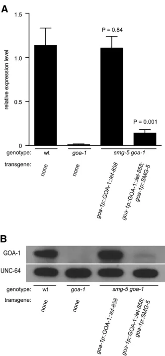
From our behavioral analyses we conclude that the goa-1 tagged transgene was expressed at levels near those of the endogenous gene and that tagged transcripts could be re-duced or eliminated by NMD to generate null-like behav-ioral defects. However, we wanted to directly measure the expression level of the tagged transgene and the efficiency of knockdown by NMD. Therefore, we measured the abun-dance of taggedgoa-1transgene mRNA insmg-5 goa-1 dou-ble-mutant animals by quantitative RT-PCR and compared it to goa-1 mRNA expression in wild-type and goa-1(n363)
null animals. We found that goa-1p::GOA-1::let-858 trans-gene mRNA insmg-5 goa-1animals was expressed at levels that were indistinguishable from those of endogenousgoa-1 mRNA found in wild-type animals (Figure 4A, compare bars 1 and 3). We also found that GOA-1 protein levels were similar in these two strains, confirming that the tagged transgene was translated into wild-typeGOA-1protein (Fig-ure 4B, compare protein levels in lanes 1 and 3). In stark contrast, when we restored NMD activity in smg-5 goa-1 animals to all cells that also expressed the tagged transgene, using an integrated untaggedgoa-1p::SMG-5 transgene, we found that the abundance of tagged transgene mRNA was reduced by .87% compared to goa-1p::GOA-1::let-858 mRNA levels in smg-5 goa-1 double mutants that did not express the untaggedgoa-1p::SMG rescue transgene (Figure 4A, compare bars 3 and 4). We observed a similarly dra-matic reduction inGOA-1protein levels in this strain (Figure 4B, compare protein levels in lanes 3 and 4). Indeed, expres-sion of the untagged goa-1p::SMG-5 transgene reduced
GOA-1 mRNA and protein expression to near null levels, which correlated well with null-like behaviors observed in this strain as shown in Figure 3 (Figure 4A, compare bars 2 and 4 and protein levels in Figure 4B, lanes 2 and 4). Thus NMD is able to degrade tagged transgene mRNAs and main-tain them at near null levels, preventing expression of the encoded protein.
Comparison of NMD-dependent knockdown efficacy to that of other knockdown strategies
null-like phenotypes. To ask whether this strategy could be effectively adapted to other genes we wanted to test the efficacy of NMD-dependent knockdown on a gene that has been difficult to knock down by conventional dsRNA knock-down approaches. We choseunc-4for this test gene as it has proved to be resistant to several dsRNA-based silencing strategies (Simmeret al.2003; Johnsonet al. 2005).unc-4 encodes a homeodomain protein expressed in VA neurons and is required for proper synaptic input choice (Milleret al.
1992; Whiteet al.1992). VA neurons normally receive syn-aptic input from interneurons that control backward loco-motion, but in unc-4 mutants these VA neurons instead receive synaptic input from command interneurons that con-trol forward locomotion (Miller et al. 1992; White et al.
1992). As a result, unc-4 mutants are defective in backing behavior. Whereas wild-type animals back freely in a sinusoi-dal motion when prodded on the head,unc-4null mutants do not back but instead contract their midbody tightly, caus-ing a dorsalflexure that often becomes so extreme that the head and tail of the animal touch, placing the body in a coiled position. (Table 1, lines 1–3, and File S1and File S2). We found that when prodded, 92% of unc-4 null mutants contracted their midbody fully to form a tight coil while the other 8% contracted their midbody but did not fully coil.smg-5had no significant effect on backing behav-ior (Table 1, compare lines 2 and 3). While we do not un-derstand why some unc-4mutants contract their bodies to form a full coil and others do not fully coil when tapped on the head, we suspect that this might be at least partially dependent on the location or strength of the prodding stimulus.
We generated a taggedunc-4p::UNC-4::let-858transgene and integrated it at single copy at the Mos1ttTi5605allele of smg-5; unc-4 double mutants. This transgene fully res-cued unc-4 backing behavior (Table 1, compare lines 3 and 4, File S3). We then integrated an untagged unc-4p:: SMG-5 transgene into this strain to restore NMD activity in all cells that expressed the tagged transgene. We found that unlike single-transgenic animals, 100% of the double-trans-genic animals were defective in backing likeunc-4null ani-mals (Table 1, compare lines 4 and 5,File S4). We note that a higher proportion of double-transgenic animals did not fully coil after midbody contraction when compared to unc-4null mutants (Table 1, compare lines 3 and 5). How-ever, in each case, 100% of the animals were defective in backing. While coiling may be dependent upon the location or strength of the prodding stimulus, the higher levels of noncoiling we observed in the double-transgenic animals compared to unc-4null animals might also be due to resid-ualunc-4expression in the transgenic animals.
To compare the knockdown of unc-4 function that we observed using tagged transgenes to dsRNA knockdown strategies, we delivered unc-4 dsRNA to animals by three different methods. First, we induced the expression of an integrated unc-4 hairpin dsRNA by heat shock. Since this hairpin construct is integrated into the genome, it should
not be susceptible to mosaic effects that might reduce the efficacy of extrachromosomal transgenes. However, as mea-sured by nose touch response, and as described previously (Johnsonet al.2005), we found that hairpin dsRNA was not an effective means to knock down unc-4 expression with only 6% of heat-shocked animals showing a backing defect (Table 1, line 6). Second, we fed animals bacteria expressing unc-4 dsRNA. Previous attempts to knock down unc-4 ex-pression by feeding dsRNA-expressing bacteria failed to cause the expected uncoordinated phenotype (Simmer
et al.2003; Johnsonet al.2005). Becauseunc-4is expressed in neurons, which are refractory to the effects of RNAi, we used an RNAi-sensitive strain for these experiments. Control animals fed bacteria containing empty expression plasmid (pL4440) did not show an uncoordinated phenotype. In contrast, we found that 62% of animals fed unc-4 dsRNA-expressing bacteria showed defects in backing (Table 1, compare lines 7 and 8). Like the knockdown observed using NMD-dependent transgenes, a high proportion of these backing-defective animals showed the contraction but non-coiling phenotype. Finally, we coinjected unc-4 sense and antisense transgenes driven by theunc-4promoter together with arab-3p::mCherry transgene. While extrachromosomal transgenes are often lost during cell division, we attempted to reduce these mosaic effects by selecting transgenic ani-mals that showed strong mCherry fluorescence in the ner-vous system. We recoveredfive independent transgenic lines and found that 69% (range 54–88%) of all mCherry-positive animals from these lines showed backing defects that were similar to those ofunc-4null animals (Table 1, lines 9–13) (.50 animals tested per line). Again, the backing defects were distributed between partial and full coiling behavior. However, 31% (range 12–41%) of sense/antisense-expressing transgenic animals showed wild-type behavior, suggesting that the sense/antisense transgenes were either lost or expressed at low levels in at least some neurons in these animals. From these results we conclude that our gene replacement and NMD-mediated knockdown strategy is capable of causing ro-bust knockdown effects that are sufficient to generate null-like behavioral effects in 100% of treated animals, even for genes that are normally recalcitrant to conventional knockdown strategies.
Discussion
We developed a novel strategy that has the potential to knock down the expression of any gene in any cell type inC. elegans. The strategy combines three emerging experimental tools inC. elegans:
1. NMD-dependent degradation of engineered PTC-contain-ing mRNA transcripts (Linket al.2003): Tagging trans-genes with an artificial 39-UTR creates PTC-containing transcripts that are recognized and degraded by NMD. 2. Mos1-mediated single-copy insertion of transgenes
transgenes are expressed at levels comparable to those of the endogenous gene they replace.
3. Cell-specific promoters: Cell-type-specific restoration of NMD activity provides spatial control of transgene expression.
Together, these tools allowed us to functionally replace an endogenous gene with its tagged counterpart and to knock down the expression of the tagged transgene in either all cells or in specific cell types to generate global or cell-specific null behavioral phenotypes.
Comparison of NMD strategy to knockdown by dsRNA interference
Our knockdown strategy provides several advantages over the three commonly used dsRNA interference strategies, including (1) feeding animals bacteria that express dsRNA (Timmons and Fire 1998), (2) direct gonad injection of dsRNA or dsRNA-encoding constructs to generate extrachromosomal arrays (Fireet al.1998; Espositoet al.2007), and (3)in vivo expres-sion of dsRNA from hairpin constructs (Tavernarakis et al.
2000; Johnsonet al. 2005). First, and most importantly, be-cause NMD is a cell-autonomous process (Weischenfeldtet al.
2008), knockdown by our method is absolutely cell specific. In contrast, dsRNA or intermediates produced during processing of dsRNA are exported from cells in C. elegans to produce systemic knockdown effects (Joseet al.2009, 2011). Second, because our method uses integrated transgenes, we were able to generate large populations of isogenic animals where every animal in the population showed consistent effects of trans-gene expression and knockdown. This is most clearly seen in Figures 2 and 3, where we plot the behavior of 30 individual animals of each genotype. In contrast, dsRNAs encoded on extrachromosomal arrays or delivered by feeding worms bac-teria are not stably inherited. Consistent with the results of others, we found that all three dsRNA approaches caused variable knockdown effects with phenotypes observed in only a portion of all treated animals (Table 1) (Montgomeryet al.
1998; Tavernarakiset al.2000; Simmeret al.2003). We sus-pect that the knockdown effects of dsRNA transgenes could be improved by integrating the transgene to eliminate mosaic knockdown effects; however, the integrated hairpin RNA transgene was the least effective knockdown approach tested in this study. Third, knockdown effects using our strategy are robust. As measured by mRNA abundance, our approach re-duced gene expression by 87%. For the twoC. elegansgenes tested (goa-1andunc-4), this was sufficient to cause null-like behavioral defects in all animals of the population. In compar-ison, all three dsRNA interference strategies caused less dra-matic behavioral defects with many animals in the population showing no knockdown effects (Table 1). This could either be due to reduced efficacy of the RNAi mechanism itself, which we did not assess, or, at least in the case of dsRNA injections, be due to mosaic loss of extrachromosomal transgenes. Fourth, since the cell-specific NMD rescue transgenes are sta-bly integrated in our approach, a collection of different
cell-specific NMD-rescue strains could be easily generated and archived for sharing among the research community. With these strains in hand, the cell-specific function of any gene could be investigated by generating just one MosSCI-integrated transgene containing the gene of interest and the degradation tag and crossing this strain to the shared cell-specific NMD rescue strains. Finally, we note that our strategy could be expanded to also include temporal con-trol of cell-specific knockdown. Since sevensmggenes are required for NMD activity, temporal control could be achieved by placing the expression of a second smggene under inducible control such as that provided by a heat-shock promoter or the Q repressible binary expression sys-tem (Joneset al.1989; Weiet al.2012). Alternatively, tem-poral control could be achieved by regulating the activity of a secondsmggene, using a temperature-sensitivesmgallele such assmg-1(cc546ts)and switching animals from the re-strictive to the permissive temperature (Linket al.2003).
Uses and limitations of the NMD-dependent knockdown strategy
Because the lack of NMD does not affect viability in C. ele-gans, we believe this gene replacement strategy will be use-ful to study the function of most or all proteins in this organism. However, there may be some genes whose func-tion cannot be examined using this strategy. First, it is pos-sible that NMD might not function equally well in all cell types at all stages of development. While not analyzed inC. elegans, it has recently been shown that a developmentally regulated microRNA (miR-128) found in vertebrates can re-press NMD activity in mammalian neural cells, suggesting that NMD activity might be under developmental control at least in some cell types (Brunoet al.2012). Second, some genes normally express PTC-containing isoforms and these isoforms may accumulate in NMD-defective cells, possibly causing expression a truncated protein. In rare cases, such a truncated protein might interfere with the ability to ana-lyze protein function in neighboring, NMD-competent, cells. It has been estimated that13–25% ofC. elegansgenes are alternatively spliced (Zahler 2005; Ramaniet al.2011) and that 34% of these genes express alternatively spliced mRNAs that naturally contain PTCs (Barberan-Soler et al.
2009). Thus it is possible that as many as 4–8% ofC. elegans
genes could not be analyzed by our methods for this reason. We also note that 10% ofC. elegans genes are regulated post-transcriptionally by sequences in their 39-UTR (Lall
et al.2006). Our strategy should still work for these genes if the tagged transgene is modified to include the 39-UTR of the gene under study in place of the 39-UTR oflet-858. Such a tagged transgene would contain the regulatory sequences found in the 39-UTR of the gene under study but would still trigger NMD of transgene mRNA.
different functions in different cell types, and for proteins for which null mutations are lethal.
Acknowledgments
We thank M. Nonet for the long-range PCR protocol, UNC-64 antibodies, and plasmid constructs; E. Jorgensen for MosSCI reagents; M. Koelle for GOA-1 antibody; and J. Lopes for qRT-PCR technical advice. This work was supported by a Faculty Research Grant from University of Massachusetts PIFRG0000000105 and by funding from the National Institutes of Health (NIH) MH097163. Some strains were provided by the CaenorhabditisGenetics Center, which is funded by the NIH Office of Research Infrastructure Pro-grams (P40-OD010440).
Literature Cited
Alfonso, A., K. Grundahl, J. S. Duerr, H. P. Han, and J. B. Rand,
1993 TheCaenorhabditis elegansunc-17 gene: a putative
ve-sicular acetylcholine transporter. Science 261: 617–619. Barberan-Soler, S., N. J. Lambert, and A. M. Zahler, 2009 Global
analysis of alternative splicing uncovers developmental regulation of nonsense-mediated decay inC. elegans. RNA 15: 1652–1660. Brenner, S., 1974 The genetics ofCaenorhabditis elegans. Genetics
77: 71–94.
Bruno, I. G., R. Karam, L. Huang, A. Bhardwaj, C. H. Lou et al., 2012 Identification of a microRNA that activates gene expression by repressing nonsense-mediated RNA decay. Mol. Cell 42: 500–510. Cali, B. M., S. L. Kuchma, J. Latham, and P. Anderson, 1999 smg-7 is required for mRNA surveillance in Caenorhabditis elegans. Genetics 151: 605–616.
Chalfie, M., J. E. Sulston, J. G. White, E. Southgate, J. N. Thomson
et al., 1985 The neural circuit for touch sensitivity in
Caeno-rhabditis elegans. J. Neurosci. 4: 956–964.
Esposito, G., E. Di Schiavi, C. Bergamasco, and P. E. Bazzicalupo, 2007 Efficient and cell specific knock-down of gene function in targetedC. elegansneurons. Gene 395: 170–176.
Fire, A., S. Xu, M. K. Montgomery, S. A. Kostas, S. A. Driveret al., 1998 Potent and specific genetic interference by double-stranded RNA inCaenorhabditis elegans. Nature 391: 806–811. Frøkær-Jensen, C., M. W. Davis, C. E. Hopkins, B. J. Newman, J. M. Thummel et al., 2008 Single-copy insertion of transgenes in
Caenorhabditis elegans. Nat. Genet. 40: 1375–1383.
Hofler, C., and M. R. Koelle, 2011 AGS-3 alters Caenorhabditis elegans behavior after food deprivation via RIC-8 activation of the neural G protein Gao. J. Neurosci. 31: 11553–11562. Johnson, N. M., C. A. Behm, and S. C. Trowell, 2005 Heritable
and inducible gene knockdown in C. elegansusing Wormgate and the ORFeome. Gene 359: 26–34.
Jones, D., D. K. Dixon, R. W. Graham, and E. P. Candido, 1989 Differential regulation of closely related members of the hsp16 gene family inCaenorhabditis elegans. DNA 8: 481–490. Jose, A. M., J. J. Smith, and C. P. Hunter, 2009 Export of RNA
silencing fromC. eleganstissues does not require the RNA chan-nel SID-1. Proc. Natl. Acad. Sci. USA 106: 2283–2288. Jose, A. M., G. A. Garcia, and C. P. Hunter, 2011 Two classes of
silencing RNAs move between Caenorhabditis eleganstissues. Nat. Struct. Mol. Biol. 18: 1184–1188.
Koelle, M. R., and H. R. Horvitz, 1996 EGL-10 regulates G protein signaling in the C. elegans nervous system and shares a conserved domain with many mammalian proteins. Cell 84: 115–125.
Lall, S., D. Grün, A. Krek, K. Chen, Y. L. Wang et al., 2006 A genome-wide map of conserved microRNA targets inC. elegans. Curr. Biol. 16: 460–471.
Link, C. D., A. Taft, V. Kapulkin, K. Duhke, S. Kimet al., 2003 Gene expression analysis in a transgenic Caenorhabditis elegans Alz-heimer’s disease model. Neurobiol. Aging 24: 397–413.
Longman, D., R. H. Plasterk, I. L. Johnstone, and J. F. Cáceres, 2007 Mechanistic insights and identification of two novel fac-tors in theC. elegansNMD pathway. Genes Dev. 9: 1075–1085. Mello, C. C., J. M. Kramer, D. Stinchcomb, and V. Ambros, 1991 Efficient gene transfer inC. elegans: extrachromosomal maintenance and integration of transforming sequences. EMBO J. 12: 3959–3970.
Mendel, J. E., H. C. Korswagen, K. S. Liu, Y. M. Hajdu-Cronin, M. I. Simonet al., 1995 Participation of the protein Goin multiple aspects of behavior inC. elegans. Science 267: 1652–1655. Miller, D. M., M. M. Shen, C. E. Shamu, T. R. Bürglin, G. Ruvkin
et al., 1992 C. elegans unc-4 gene encodes a homeodomain
protein that determines the pattern of synaptic input to specific motor neurons. Nature 355: 841–845.
Montgomery, M. K., S. Xu, and A. Fire, 1998 RNA as a target of double-stranded RNA-mediated genetic interference in
Caeno-rhabditis elegans. Proc. Natl. Acad. Sci. USA 95: 15502–15507.
Moresco, J. J., and M. R. Koelle, 2004 Activation of EGL-47, a Gao-coupled receptor, inhibits function of hermaphrodite-specific motor neurons to regulate Caenorhabditis elegansegg-laying be-havior. J. Neurosci. 24: 8522–8530.
Pulak, R., and P. Anderson, 1993 mRNA surveillance by the
Cae-norhabditis eleganssmg genes. Genes Dev. 10: 1885–1897.
Ramani, A. K., J. A. Calarco, Q. Pan, S. Mavandadi, Y. Wanget al., 2011 Genome-wide analysis of alternative splicing in
Caeno-rhabditis elegans. Genome Res. 21: 342–348.
Ségalat, L., D. A. Elkes, and J. M. Kaplan, 1995 Modulation of serotonin-controlled behaviors by GoinCaenorhabditis elegans. Science 267: 1648–1651.
Simmer, F., C. Moorman, A. M. van der Linden, E. Kuijk, P.V. van den Bergheet al., 2003 Genome-wide RNAi ofC. elegansusing the hypersensitive rrf-3 strain reveals novel gene functions. PLoS Biol. 1: 77–84.
Stinchcomb, D. T., J. E. Shaw, S. H. Carr, and D. Hirsh, 1985 Extrachromosomal DNA transformation of
Caenorhabdi-tis elegans. Mol. Cell. Biol. 12: 3484–3496.
Tavernarakis, N., S. L. Wang, M. Dorovkov, A. Ryazanov, and M. Driscoll, 2000 Heritable and inducible genetic interference by double-stranded RNA encoded by transgenes. Nat. Genet. 2: 180–183. Timmons, L., and A. Fire, 1998 Specific interference by ingested
dsRNA. Nature 395: 854.
Wani, K. A., M. Catanese, R. Normantowicz, M. Herd, K. N. Maher
et al., 2012 D1 dopamine receptor signaling is modulated by
the R7 RGS protein EAT-16 and the R7 binding protein RSBP-1 in Caenorhabditis elegans motor neurons. PLoS ONE 7: e37831. Wei, X., C. J. Potter, L. Luo, and K. Shen, 2012 Controlling gene expression with the Q repressible binary expression system in
Caenorhabditis elegans. Nat. Methods 9: 391–395.
Weischenfeldt, J., I. Damgaard, D. Bryder, K. Theilgaard-Mönch, L. A. Thorenet al., 2008 NMD is essential for hematopoietic stem and progenitor cells and for eliminating by-products of programmed DNA rearrangements. Genes Dev. 22: 1381–1396. White, J. G., E. Southgate, and J. N. Thomson, 1992 Mutations in
theCaenorhabditis elegans unc-4gene alter the synaptic input to
ventral cord motor neurons. Nature 355: 838–841.
Zahler, A. M., 2005 Alternative splicing in C. elegans (Septem-ber 26, 2005),WormBook, ed. TheC. elegansResearch Com-munity, WormBook, doi/10.1895/wormbook.1.31.1, http:// www.wormbook.org.
GENETICS
Supporting Information http://www.genetics.org/lookup/suppl/doi:10.1534/genetics.113.149724/-/DC1
A Novel Strategy for Cell-Autonomous Gene
Knockdown in
Caenorhabditis elegans
De
fi
nes
a Cell-Speci
fi
c Function for the G-Protein
Subunit GOA-1
Kathryn N. Maher, Aishwarya Swaminathan, Parth Patel, and Daniel L. Chase
Files S1-‐S4 Supporting Movies
Available for download at http://www.genetics.org/lookup/suppl/doi:10.1534/genetics.113.149724/-‐/DC1.
File S1: Response of wild-‐type animals to nose touch. Wild-‐type animals reverse normally when tapped on the nose.
File S2: Response of unc-‐4 mutants to nose touch. unc-‐4 mutants are unable to reverse.
File S3: Response of smg-‐5; unc-‐4 ndSi19 [unc-‐4p::UNC-‐4::let-‐858] animals to nose touch. The tagged unc-‐4 transgene restores normal backing behavior in NMD-‐defective animals.