YEASTBOOK
GENOME ORGANIZATION & INTEGRITY
The Composition, Functions, and Regulation
of the Budding Yeast Kinetochore
Sue Biggins
Division of Basic Sciences, Fred Hutchinson Cancer Research Center, Seattle, Washington 98109
ABSTRACT The propagation of all organisms depends on the accurate and orderly segregation of chromosomes in mitosis and meiosis. Budding yeast has long served as an outstanding model organism to identify the components and underlying mechanisms that regulate chromosome segregation. This review focuses on the kinetochore, the macromolecular protein complex that assembles on centromeric chromatin and maintains persistent load-bearing attachments to the dynamic tips of spindle microtubules. The kinetochore also serves as a regulatory hub for the spindle checkpoint, ensuring that cell cycle progression is coupled to the achievement of proper microtubule–kinetochore attachments. Progress in understanding the composition and overall architecture of the kinetochore, as well as its properties in making and regulating microtubule attachments and the spindle checkpoint, is discussed.
TABLE OF CONTENTS
Abstract 817
Introduction 818
Stages of chromosome alignment and segregation 818
Establishing kinetochore biorientation 819
Assays to study yeast chromosome segregation 819
Cytological assays 819
Genetic and genomic assays 820
Biochemical, structural, and biophysical assays 820
The Centromere 821
Centromere structure 821
Centromeric chromatin 822
Composition of the Budding Yeast Kinetochore 823
Inner centromere binding proteins 824
CBF3: 824
CCAN components: 826
Model for the inner kinetochore: 826
Outer kinetochore proteins 827
KMN: 827
Continued
Copyright © 2013 by the Genetics Society of America doi: 10.1534/genetics.112.145276
Manuscript received January 21, 2013; accepted for publication May 14, 2013
CONTENTS,continued
Dam1 complex: 828
Other outer kinetochore proteins: 828
Architecture of the kinetochore 828
Kinetochore–microtubule attachments and coupling activity 829
Regulation of kinetochore attachments 831
Kinetochore biorientation 831
The Spindle Checkpoint 833
Perspectives 836
C
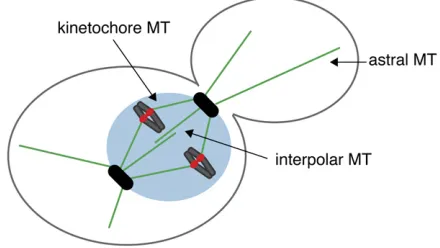
HROMOSOME segregation is mediated by the interac-tion between spindle microtubules and kinetochores, the macromolecular structures that assemble at a unique chro-mosomal locus called the centromere (Westermann et al.2007). Microtubules are dynamic polymers that grow and shrink by the addition and removal of tubulin dimers from their tips (Mitchison and Kirschner 1984). They switch sto-chastically between phases of assembly and disassembly, a behavior called dynamic instability (Mitchison and Kirschner 1984). Microtubules are nucleated by the centrosome, which is called the spindle pole body (SPB) in yeast (Winey and Bloom 2012). Microtubules have an inherent polarity with the minus end embedded in the SPB and the dynamic plus end distal. In yeast, microtubule growth and shrinkage ap-pears to occur exclusively at the plus end (Maddoxet al.
2000). Because the yeast nuclear envelope does not break down, the SPB is embedded in the nuclear envelope throughout the cell cycle. The SPB nucleates three populations of yeast microtubules that facilitate proper chromosome segre-gation (Figure 1). In the cytoplasm, astral microtubules position the nucleus throughout the cell cycle. Within the nucleus, kinetochore microtubules attach to the kineto-chore at their plus ends, and interpolar microtubules in-terdigitate to connect the poles and stabilize the spindle during mitosis. The zone of overlap between interpolar microtubules is called the spindle midzone; a number of proteins specifically localize to the midzone to facilitate spindle assembly and disassembly.
Stages of chromosome alignment and segregation
Yeast kinetochores are assembled and bind to microtubules for almost the entire cell cycle, with the exception of a brief window during S phase when they disassemble and rapidly reassemble (Kitamura et al. 2007). This may be the time when the replication fork travels through the centromere, although this has not yet been directly tested. Yeast kinet-ochores thus cluster near the spindle pole for most of the cell cycle (Heath 1980; Jin et al. 2000; Kitamuraet al. 2007). This proximity led to the initial identification of many kinet-ochore components through SPB purifications (Wiggeet al.
1998). Each budding yeast kinetochore binds to a single microtubule (Winey et al. 1995), which greatly simplifies
studies because a kinetochore is either attached or unat-tached to a microtubule at any given time. In contrast, most eukaryotic kinetochores have from 3 to 30 microtubule bind-ing sites, which can be partially occupied (Walczak et al.
2010). Replication creates sister chromatids, which become physically linked together by protein complexes called cohe-sin (Oliveira and Nasmyth 2010). Proper segregation requires sister kinetochores to biorient and attach to micro-tubules from opposite poles (Tanaka 2010). Once every pair of chromosomes biorients, the linkage between the sister chromatids is destroyed and the spindle physically pulls sis-ter chromatids to opposite poles.
The small size of the yeast nucleus and difficulty in visualizing yeast chromosomes by microscopy makes it difficult to directly examine the steps of chromosome segre-gation. The assays used to examine the process therefore require cells to be arrested in conditions that may or may not reflect the normal course of events (Tanaka et al.
Huffaker 1997; Al-Bassam et al. 2006; Brouhard et al.
2008).Stu2also helps kinetochores nucleate microtubules, a feature that appears to help establish lateral attachments through microtubule–microtubule interactions that are even-tually converted to plus end attachments at the kineto-chores (Kitamura et al. 2010; Tanaka 2010). Once the kinetochores travel back to the pole, the sister kinetochores make bioriented attachments to the tips of microtubules and come under tension due to pulling forces that are op-posed by the linkage between the sisters (Figure 2, C and D). The kinetochores then maintain persistent load-bearing attach-ments to the continually growing and shrinking tips of the microtubules.
Establishing kinetochore biorientation
The process of making bioriented kinetochore–microtubule attachments is inherently error prone (Nicklas 1997). Kinet-ochores can make syntelic attachments where both sisters attach to microtubules from the same pole or monotelic attachments in which one of the two sister kinetochores attaches to a microtubule from one pole (Figure 3). Most eukaryotic kinetochores can also make merotelic attach-ments where a single kinetochore binds to microtubules from both poles (Cimini 2008), but this is not possible in budding yeast where there is only one microtubule-binding site on each kinetochore (Wineyet al.1995). Because syn-telic or monooriented attachments will lead to errors in segregation, the cell has mechanisms to detect and correct inappropriate microtubule attachments. A variety of evi-dence suggests that the cell monitors the tension generated when sister kinetochores achieve biorientation (Nicklas and Koch 1969; Nicklas 1997). When kinetochores lack tension, the conserved Aurora B protein kinase phosphorylates kinet-ochore proteins (discussed below, kinetkinet-ochore biorienta-tion), leading to their release from microtubules so the cell can attempt biorientation again (Biggins et al. 1999; Cheeseman et al. 2002; Tanaka et al. 2002). In addition,
tension prolongs the lifetime of kinetochore–microtubule interactionsin vitro, suggesting that tension directly stabil-izes microtubule attachments (Franck et al.2007; Akiyoshi
et al.2010). Elegant computer modeling supports the role of tension in stabilizing attachments (Gardneret al.2005).
Once all kinetochores biorient, the cohesin between sisters is cleaved, allowing the chromosomes to be separated and moved to the poles at anaphase. If even a single pair of chromosomes lacks tension or attachment, a signal trans-duction system called the spindle checkpoint prevents ana-phase (Zich and Hardwick 2010; Murray 2011; Musacchio 2011). To date, it is still controversial whether there is a single upstream signal that triggers the checkpoint or whether tension and attachment are separately monitored (discussed below,The Spindle Checkpoint).
Assays to study yeast chromosome segregation
Cytological assays
Historically, one of the greatest difficulties in studying yeast chromosome segregation has been that the 16 budding yeast chromosomes cannot be distinguished by classical cytolog-ical techniques. Instead, they appear as a single amorphous nuclear mass that splits into two at anaphase when stained with dyes. This makes it impossible to monitor the fate of sister chromatids at anaphase and to determine whether individual chromosomes are attached or unattached to microtubules. One of the biggest technical advances was the development of a system to fluorescently mark in-dividual chromosomes with a GFP tagin vivo(Straightet al.
1996; Michaeliset al.1997). These systems exploit the abil-ity to integrate tandem arrays of lactose or tetracycline operators from bacteria into the yeast genome into strains-containing GFP fusions to the lactose or tetracycline repress-ors, respectively. The GFP fusions bind to the operators and fluorescently mark the chromosomal locus, and the opera-tors can be easily moved to any genomic position using homologous recombination. This technique revealed that yeast chromosome arms are held in close proximity until anaphase (Straight et al. 1996; Goshima and Yanagida 2000; He et al. 2000; Tanaka et al. 2000; Pearson et al.
2001). However, centromeres transiently separate and reas-sociate prior to anaphase, and this splitting can be detected with probes integrated up to 38 kb from the centromere (Goshima and Yanagida 2000; He et al. 2000; Pearson
et al.2001). Because the splitting depends on microtubules and its frequency increases as the probe is moved toward the centromere, it is presumably generated by microtubule pull-ing forces on bioriented kinetochores. Fluorescently markpull-ing a single centromere is therefore a powerful technique to monitor the kinetics of biorientation and separation to the poles.
GFP fusions to kinetochore proteins have been another major advance in assaying kinetochore function. Although this technique means that all kinetochores are marked Figure 1 Key structures that mediate chromosome segregation. A
rather than individual sisters, it is informative because yeast kinetochores cluster. A tagged kinetochore protein exhibits a single fluorescent focus prior to biorientation that splits into two foci upon biorientation (Goshima and Yanagida 2000; Heet al.2000; Pearsonet al.2001). When anaphase ensues, the foci move to opposite poles as the spindle elongates. However, if kinetochore function is dis-rupted, the GFP foci often have a reduced intensity due to a decreased association with the centromere, and the kinet-ochores often decluster because they detach from micro-tubules (Pinskyet al.2006). In this case, it is obvious that the kinetochores no longer colocalize with microtubules. The disadvantage to this assay is that it is currently impos-sible to know whether a GFP focus represents one or more kinetochores, so the fate of a pair of sister chromatids can-not be monitored.
Genetic and genomic assays
There are also a number of genetic and genomic assays for kinetochore function. Chromatin immunoprecipitation (ChIP) assays and ChIP-sequencing techniques clearly determine if a protein is associated with the centromere, an issue that was difficult to confirm in the past (Meluh and Koshland 1995; Lefrancois et al. 2009; Krassovsky et al. 2012). A fruitful genetic assay exploits the ability to monitor the segregation of a nonessential ectopic chromosome contain-ing a centromere by colony color (Koshland and Hieter 1987; Sheroet al.1991). This sectoring assay has been used in numerous screens to identify segregation genes and to quantify chromosome loss rates in mutant strains (Spencer
et al. 1990; Dohenyet al. 1993; Warrenet al. 2002). An-other useful assay is a conditional dicentric assay where a second centromere is integrated into the chromosome (Hill and Bloom 1987). Although dicentric chromosomes are normally unstable and lost during cell division, the ga-lactose promoter controls the second centromere in this as-say and transcription through the centromere abolishes its function. When the cells are shifted into glucose, transcrip-tion through the centromere is halted allowing a second kinetochore to form, which can subsequently be assayed for thede novoassembly of kinetochore proteins and other kinetochore functions (Tanaka et al. 1999; Mythreye and Bloom 2003; Collinset al.2005). One-hybrid assays can also identify kinetochore proteins (Ortizet al.1999).
Biochemical, structural, and biophysical assays
While studiesin vivohave been essential for the
identifi-cation of kinetochore components and functions, dissect-ing the underlydissect-ing mechanism of chromosome movement depends on experimentsin vitrothat allow individual events to be monitored and manipulated (Akiyoshi and Biggins 2012; Umbreit and Davis 2012). A number of biochemical and biophysical assays for kinetochore function have there-fore been developed. Gel shift assays using centromeric DNA originally identified the inner centromere binding proteins (Lechner and Carbon 1991). “Minimal” kinetochores con-taining centromeric DNA and some inner kinetochore pro-teins have helped to dissect functions (Kingsbury and Koshland 1991; Sorger et al.1994; Bigginset al.1999; Sandallet al.2006), and large kinetochore particles were re-cently isolated (Akiyoshiet al.2010). In the past decade, the development of biophysical assays to analyze the functions of both individual subcomplexes and larger kinetochore as-semblies has led to major mechanistic insights (Gestautet al.
2010). The use of total internal reflection microscopy (TIRF) allows complexes to be visualized at the single particle level in the presence or absence of microtubules. Optical trapping is powerful because tension can be applied to linkages be-tween complexes and microtubules, mimicking the forces that kinetochores sustain in vivo (Asburyet al.2006; Grishchuk
et al. 2008a; Francket al.2010). Finally, structural biology has played a key role in elucidating the organization and Figure 2 Steps leading to bioriented kinetochore attachments. (A)
architecture of many kinetochore assemblies, including the two major microtubule binding complexes in the yeast kinetochore (Mirandaet al.2005; Weiet al.2005, 2006, 2007; Westermann
et al.2005, 2006; Wanget al.2007, 2008; Maskellet al.2010; Hornunget al.2011).
The Centromere
Centromere structure
The budding yeast centromere was first identified by its ability to confer mitotic and meiotic stability to a plasmid (Clarke and Carbon 1980). In contrast to most eukaryotic centromeres that span megabases of DNA (Burrack and Berman 2012), the functional yeast centromere is defined by a 200-bp nuclease resistant region containing a 125-bp
“point”centromere, with regularly spaced nucleosomes po-sitioned on either side (Bloom and Carbon 1982; Fitzgerald-Hayeset al.1982; Clarke and Carbon 1985). There are three conservedcentromere-determiningelements (CDE): an 8-bp palindrome called CDEI, a 78- to 86-bp stretch of AT-rich (.90%) DNA called CDEII, and a conserved 26-bp element called CDEIII (Figure 4) (Clarke 1998). Although most eukaryotic centromeres are maintained epigenetically (Blacket al.2010; Henikoff and Furuyama 2010), yeast
centromeres are genetically specified by DNA sequence. The CDEI consensus sequence (PuTCACPuTG) binds to the helix-loop-helix protein Cbf1 (Cai and Davis 1989; Baker and Masison 1990; Cai and Davis 1990), a transcription factor that also binds to other elements throughout the ge-nome. CDE1 and Cbf1 contribute to kinetochore function but are not essential. The CDEIII consensus (TGTTT(T/A) TGNTTTCCGAAANNNAAAAA) binds to theCBF3complex via a conserved CCG motif that is essential for centromere function (Jehnet al.1991; Lechner and Carbon 1991). The small size and sequence specificity of the budding yeast cen-tromere has made yeast a powerful organism for its study because the sequences can be easily mutated to identify the important functional regions. It also facilitates techniques such as ChIP, which cannot be easily performed on the highly repetitive centromeres in other organisms. In addi-tion, the centromere can be moved to other genomic regions, allowing the construction of artificial chromosomes and plasmids as well as tools such as conditional centromeres (Murray and Szostak 1983; Hill and Bloom 1989).
Like other eukaryotes, the budding yeast centromere replicates early in S phase (McCarroll and Fangman 1988). The early replication is due to the presence of the centro-mere, but it is not yet known what aspect of the centromere or kinetochore dictates early origin activity (Pohl et al.
2012). While it is not yet clear whether early centromere replication is important for subsequent kinetochore func-tion, one possibility is that it ensures that the kinetochore has enough time to assemble prior to mitosis. This might be especially important in budding yeast where there is no clear G2 phase of the cell cycle, resulting in little time for kinetochore assembly prior to mitosis (Kitamuraet al.
2007).
Most eukaryotic centromeres contain arrays of canonical and specialized centromereic nucleosomes that are embed-ded in pericentric heterochromatin (Choo 2001; Kniolaet al.
2001). Budding yeast lack many of the characteristic hall-marks of pericentric heterochromatin, including histone H3– K9 methylation and the associated transcriptional silencing of genes. However, similar to other eukaryotes, cohesin is enriched within a 20- to 50-kb domain around centromeres (Blat and Kleckner 1999; Glynn et al. 2004; Weber et al.
2004). Strikingly, the pericentric cohesins in budding yeast appear to be arranged as a cyclindrical array around the spindle (Yeh et al.2008), which may be due to the forma-tion of an intramolecular C loop on each sister chromatid that extends25 kb (Yehet al.2008). Cohesin would there-fore encircle a single chromatid rather than sisters in this region, resolving the apparent“cohesin”paradox where the highest levels of cohesin reside in the areas that are physi-cally split at metaphase. At least one function of pericentric cohesion is to facilitate kinetochore biorientation by resist-ing the pullresist-ing forces of microtubules and/or by promotresist-ing the architecture of sister kinetochores (Eckert et al. 2007; Fernius and Marston 2009; Nget al.2009; Bloom and Joglekar 2010). Consistent with this, the geometry and elasticity of Figure 3 Types of kinetochore–microtubule attachments. (A) Bioriented
the pericentromere and inner kinetochore can change in response to alterations in microtubule dynamics (Haase
et al. 2012; Stephenset al. 2013). These properties are reg-ulated by the Bub1 and Sgo1proteins as well as various chromatin-remodeling complexes (Haase et al. 2012; Verdaasdonket al.2012). While heterochromatin recruits peri-centric cohesin in some organisms (Bernard et al. 2001; Fukagawaet al.2004), components of the kinetochore itself direct cohesion enrichment in budding yeast (Megee et al.
1999; Tanaka et al. 1999; Weber et al. 2004; Eckert et al.
2007; Fernius and Marston 2009; Ng et al. 2009; Fernius
et al.2013).
The pericentromere also contributes to segregation by localizing key regulators of kinetochore biorientation and the checkpoint. The Bub1 kinase, originally identified as
a spindle checkpoint protein (see below), phosphorylates H2A in the pericentromeres (Hoyt et al.1991; Kawashima
et al. 2010; Yamagishi et al. 2010). This phosphorylation recruits theSgo1protein that facilitates kinetochore biorien-tation and the spindle checkpoint when kinetochores lack tension (Indjeian et al.2005; Kitajimaet al. 2005; Fernius and Hardwick 2007; Indjeian and Murray 2007). In most organisms, the Haspin kinase phosphorylates H3 to recruit the chromosome passenger complex (CPC), which contains the Aurora B protein kinase that regulates biorientation and the checkpoint (Daiet al.2005; Kellyet al.2010). However, the budding yeast Haspin kinases, Alk1 and Alk2, are not known to have a role in chromosome segregation. The CPC may act in a distinct pathway from Bub1andSgo1in bud-ding yeast (Storchovaet al.2011), and it is still unclear how it is recruited to budding yeast pericentromeres.
Budding yeast centromeres have a defined centromeric DNA sequence, leading to the assumption that epigenetic mechanisms do not contribute to their propagation. How-ever, at least twofindings using the conditional centromere suggest there is an epigenetic component. First, cohesin enrichment around centromeres exhibits a greater depen-dence on kinetochore function in newly activated condi-tional centromeres than previously established endogenous centromeres (Tanakaet al.1999). This observation suggests that cohesin levels are maintained at least in part by an epigenetic mechanism. Second, theChl4kinetochore pro-tein is required for the function of a newly established kinetochore but not a previously formed kinetochore (Mythreye and Bloom 2003), suggesting that epigenetic sig-nals allow cells to bypass the need for Chl4 at established kinetochores. The underlying mechanisms for these observa-tions are not yet known.
Centromeric chromatin
A hallmark of all eukaryotic centromeres is a specialized chromatin structure (Carroll and Straight 2006). Classical chromatin mapping experiments showed that the budding yeast centromere contains a 160- to 220-bp nuclease resis-tant core flanked by positioned nucleosomes (Bloom and Carbon 1982; Bloom et al.1984). While most of the chro-mosome contains nucleosomes made of histone octamers composed of two copies of H2A, H2B, H3, and H4 wrapped by two turns of DNA, centromeres contain a specialized nu-cleosome where H3 is replaced by a histone H3 variant originally named CENP-A (Earnshaw and Rothfield 1985; Palmeret al.1987). The budding yeast centromeric histone H3 variant isCse4and was initially shown to localize to the centromere by ChIP experiments (Stoleret al.1995; Meluh
et al. 1998). Higher resolution techniques later determined that there is a single, well-positioned nucleosome containing
Cse4that resides over CDEII (Furuyama and Biggins 2007; Lefrancois et al. 2009; Cole et al. 2011; Krassovsky et al.
2012). There are also additional Cse4 molecules around centromeres (Coffman et al. 2011; Lawrimore et al. 2011; Lefrancois et al. 2013), and a challenge for the field is to determine the properties and number ofCse4nucleosomes that contribute to kinetochore assembly and function.Cse4
can also incorporate into euchromatin, especially at sites of high histone turnover (Collinset al.2004; Lefrancoiset al.
2009; Krassovskyet al.2012).Cse4does not stably incorpo-rate into euchromatin because its protein levels are tightly controlled by proteolysis via thePsh1E3 ubiquitin ligase and additional mechanisms (Collinset al.2004; Hewawasamet al.
2010; Ranjitkaret al.2010; Auet al.2013). In the absence of proteolysis, Cse4 levels increase and its overexpression in these cells leads to mislocalization throughout euchromatin and subsequent lethality.
Like all histones, Cse4 is recognized and deposited into chromatin by a histone chaperone called Scm3in budding yeast (HJURP in human cells) (Stoleret al.2007; Dunleavy
et al.2009; Foltzet al.2009).Scm3recognizesCse4through the centromere-targeting domain (CATD) in the histone fold and mediates its incorporation into chromatin in vivoand
in vitro(Camahortet al.2007; Shivarajuet al.2011).Scm3
timing of Cse4deposition is probably similar. Fluorescence recovery after photobleaching (FRAP) experiments showed it is deposited during late G1 or early S phase (Pearsonet al.
2004). Although it was reported thatCse4is also deposited during anaphase (Shivarajuet al.2012), the marker used for anaphase may not distinguish between late anaphase and G1. Consistent with this, other groups have not observed anaphase incorporation (Pearson et al. 2004; Coffman et al.
2011; Lawrimoreet al.2011).
AlthoughCse4is an essential component of centromeric chromatin, the precise composition of theCse4nucleosome is controversial (Henikoff and Furuyama 2012).Cse4is re-leased from minichromosomes in 0.3 M NaCl, conditions that do not affect the binding of canonical H3 to DNA (Akiyoshiet al.2009b). In addition,Cse4protects a smaller region of DNA at the centromere than a traditional H3 octamer when treated with the enzyme micrococcal nucle-ase (MNnucle-ase), suggesting that the centromeric nucleosome is atypical (Cole et al.2011; Krassovskyet al. 2012). Consis-tent with this, it has been proposed that the centromeric nucleosome might exist as a hemisome (containing a single copy of H2A, H2B, CENP-A, and H4) for at least a portion of the cell cycle (Dalal et al. 2007; Dimitriadis et al. 2010; Shivaraju et al. 2012). This was further supported by the observation that centromeric nucleosomes induce positive supercoiling at centromeresin vivo(Furuyama and Henikoff 2009), which has been observed in archaeal tetrameric nucleosomes and is not compatible with the presence of negatively supercoiled histone octamers (Musgrave et al.
1991). However, alternative structures have also been pro-posed that could explain the smaller protected region of centromeric DNA. One posited that centromeric nucleo-somes completely lack H2A and H2B and instead contain two copies ofScm3(Mizuguchiet al.2007). The demonstra-tion thatScm3is a chaperone forCse4/H4 and thatCse4/ H4 cannot simultaneously bind to DNA and Scm3 elimi-nated this model (Cho and Harrison 2011; Dechassa et al.
2011; Shivaraju et al. 2011; Xiao et al. 2011; Zhouet al.
2011). In a revised model, the centromeric nucleosome was proposed to be a tetramer containing two copies ofCse4and H4 and completely lacking H2A and H2B (Xiaoet al.2011). However, H2A and H2B have been detected at centromeres making this model less likely (Krassovskyet al.2012; Loch-mann and Ivanov 2012; Shivarajuet al.2012). Although H3 was also reported to localize to centromeres in budding yeast (Lochmann and Ivanov 2012), the region of DNA an-alyzed was large enough to contain two nucleosomes so the H3 detected may be in the neighboring nucleosome rather than the centromeric nucleosome. Consistent with this, de-pletion of H3 in budding yeast has little effect on kineto-chore function compared to H4 depletion or H2A mutations that lead to defects in kinetochore–microtubule attachments (Pinto and Winston 2000; Bouck and Bloom 2007; Verdaasdonk
et al.2012). Together, these data suggest that H3 does not reside at the point centromere, although it is important for accurate segregation through its role in recruitingSgo1to
the pericentromere and facilitating inner kinetochore func-tion (Luoet al.2010; Verdaasdonket al.2012). Finally, it was argued that Cse4is part of an octameric nucleosome at the centromere based on sequential immunoprecipitation experiments, but the starting material for these experiments was not pure mononucleosomes (Camahort et al. 2009). Therefore, the ability to detect octamers could be due to
Cse4incorporation into neighboring euchromatin. Recently, it was reported thatCse4exists as a hemisome for most of the cell cycle and then transitions into an octamer at ana-phase (Shivarajuet al.2012). Although this model is attrac-tive because it would reconcile different findings, none of the experiments in this manuscript directly measure
Cse4incorporation into nucleosomes. Instead, these conclu-sions are based on fluorescence correlation microscopy measurements that may reflect changes in the positioning of kinetochores at anaphase, as well as sequential immuno-precipitations that were not internally consistent because doubling of the H2A histone was not observed when Cse4
doubled (Shivarajuet al.2012). In sum, the composition of the centromeric nucleosome is still unclear although many of its properties are clearly different from canonical nucleo-somes. Because kinetochores may alter the accessibility of the centromeric nucleosome to MNase, affect crosslinking accessibility, or change the wrap of DNA, settling the debate requires that assays be performed on the centromeric nucle-osome in vivoin the absence of the kinetochore.Cse4 octa-meric nucleosomes and hemisomes can both be assembled
in vitro (Mizuguchi et al. 2007; Camahort et al. 2009; Dechassa et al.2011; Kingstonet al.2011; Furuyamaet al.
2013). Resolving the structure therefore requires studies on centromeric nucleosomes isolated from cells, but there is no current way to isolate them in the absence of the kineto-chore. In the future, it will be critical to apply higher reso-lution techniques to assay the material at the centromere or to develop a method to isolate the centromeric nucleosomes specifically from the kinetochore, to fully understand their composition. Ultimately, the key issue is to understand how the structure of the centromeric nucleosome speci-fies and contributes to the assembly and functions of the kinetochore.
Composition of the Budding Yeast Kinetochore
spindle checkpoint proteins, which associate with kineto-chores depending on the purification conditions and cell cycle stage. Because distinct subcomplexes can be individu-ally purified, it has been suggested that the kinetochore is assembled in a hierarchal manner on centromeric DNA (De Wulfet al.2003). However, it is still unclear how and where the various subcomplexes assemble into larger complexes to form a kinetochore. Because artificial kinetochores can be formed by tethering the Dam1or CENP-T complexes to ec-topic sites (in the absence of a centromeric nucleosome) (Kiermaier et al. 2009; Lacefield et al. 2009; Schleiffer
et al. 2012), the minimal requirements for kinetochore as-sembly are unclear. While the yeast kinetochore is often suggested to contain three domains (inner, middle, and out-er), I refer to proteins as either“inner”to reflect those close to the chromatin or “outer” to reflect roles in mediating microtubule attachment.
Inner centromere binding proteins
The “inner centromere” proteins are those that are most closely associated with centromeric chromatin. Purification of CENP-A and other inner centromere proteins in verte-brates identified a network of associated components that were collectively termed the constitutivecentromerea sso-ciatednetwork (CCAN) (Obuseet al.2004; Foltzet al.2006; Izutaet al.2006; Okadaet al.2006; Horiet al.2008). The CCAN consists of various subcomplexes that include the following proteins: CENP-C, CENP-H/I/K, CENP-L/M/N, O/P/Q/R/U, and the histone fold complexes CENP-T/W and CENP-S/X (McAinsh and Meraldi 2011; Perpelescu and Fukagawa 2011; Takeuchi and Fukagawa 2012). As discussed below, budding yeast inner centromeres contain orthologs of most of these CCAN proteins as well as a yeast-specific complex calledCBF3. The composition and deposi-tion of the Cse4 centromeric nucleosome are discussed above.
CBF3: The CBF3 complex was the first yeast kinetochore subcomplex identified due to its sequence-specific binding activity for centromeric DNA sequences containing CDEIII (Ng and Carbon 1987; Lechner and Carbon 1991; Sorger
et al. 1995). The complex contains four essential proteins that are most commonly referred to as Ndc10 (Cbf3a/
Cbf2/Ctf14/p110) (Dohenyet al.1993; Goh and Kilmartin 1993; Jiang et al. 1993), Cep3 (Cbf3b/p64) (Lechner 1994; Strunnikovet al.1995),Ctf13(Cbf3c/p58) (Doheny
et al. 1993), and Skp1 (Cbf3/p19) (Connelly and Hieter 1996; Stemmann and Lechner 1996).Cep3has a Zinc-cluster motif found in transcription factors (Dhawale and Lane 1993; Strunnikov et al. 1995; Schjerling and Holmberg 1996) and Ndc10 was recently shown to have structural similarity to tyrosine DNA recombinases (Cho and Harrison 2012; Perriches and Singleton 2012), although it does not exhibit catalytic activity or DNA base sequence specificity. Consistent with this, Ndc10 (in the absence of CBF3) can also bind to the CDEII element in vitroas well as
other genomic regions that are AT rich, although these activities are not known to be relevant to CBF3assembly
in vivo (Espelinet al.2003). The stoichiometry of theCBF3
complex bound to centromeres appears to consist of aCep3
homodimer, a Skp1-Ctf13 heterodimer, and an Ndc10
homodimer (Espelin et al. 1997; Pietrasanta et al. 1999; Russell et al. 1999; Cho and Harrison 2012) (Figure 4). The minimal CBF3binding region in vitro is a 57-bp core that covers CDEIII and additional base pairs on the right side of the element (Ng and Carbon 1987; Lechner and Carbon 1991; Sorger et al. 1995; Cho and Harrison 2012). Cep3 appears to contact the essential CCG motif in CDEIII, consistent with its similarity to transcription factors containing Zn2Cys6 clusters (Espelin et al. 1997; Purvis and Singleton 2008). Recent structural studies on
Ndc10 reveal that the dimer binds to independent DNA fragments, leading to the model that it might stabilize a loop at the centromere (Cho and Harrison 2012 and see below). This is consistent with the observation of bending of the DNA upon CBF3 binding by atomic force microscopy (Pietrasantaet al.1999).
The assembly of the CBF3complex is highly regulated
in vivoand there has been more work on its assembly than any other yeast kinetochore subcomplex. Data suggest that the complex assembles prior to binding to DNA (Lechner and Carbon 1991; Russellet al.1999).Ctf13must be“ acti-vated” to form a functionalCBF3 complex. The activation process requires binding toSkp1, a protein that is also a com-ponent of the SCF ubiquitin ligase complex (Baiet al.1996; Connelly and Hieter 1996; Kaplanet al.1997). Although the activation process was initially thought to require Ctf13
phosphorylation by a Skp1-interacting kinase (Kaplanet al.
1997), later work showed that phosphorylation is not required for CBF3 assembly on centromeres (Stemmann
et al.2002). Instead, anHsp90-Sgt1co-chaperone complex binds toSkp1, which enhancesSkp1binding toCtf13(Stemmann
et al. 2002; Bansal et al. 2004; Rodrigo-Brenni et al.
2004). Hsp90 and Sgt1 are not core kinetochore compo-nents and only transiently associate with Ctf13. Although a variety of complexes containing these components exist, the relevant intermediate complexes that form in vivo to generate activatedCtf13are not known. OnceCtf13is acti-vated bySkp1, a complex containingCep3,Ctf13, andSkp1
assembles rapidlyin vivo, and the rate-limiting step inCBF3
formation is the addition of Ndc10 (Russell et al. 1999; Rodrigo-Brenniet al.2004). At this time, it is still not known what precise changes occur to activate Ctf13to allow it to form inCBF3. Because cells can form activeCBF3complexes throughout the cell cycle (Rodrigo-Brenniet al.2004), there is careful control over the total levels of the complex via
Ctf13 proteolysis (Kaplan et al. 1997). When Ctf13 does not form a complex with Cep3, it is degraded in a Skp1 -dependent manner (Kaplan et al. 1997; Russell et al.
Table 1 Kinetochore proteins in budding yeast
Complex Components Human names
CBF3 Ndc10 (Lechner and Carbon 1991; Goh and Kilmartin 1993) Cep3 (Lechner and Carbon 1991)
Ctf13 (Lechner and Carbon 1991)
Skp1 (Connelly and Hieter 1996; Stemmann and Lechner 1996)
CCAN Mif2 (Meluh and Koshland 1995) CENP-C
Cse4 (Meluhet al.1998) CENP-A
Ctf19 (Ortizet al.1999) CENP-P
Okp1 (Ortizet al.1999) CENP-Q
Mcm21 (Ortizet al.1999) CENP-O
Ame1 (De Wulfet al.2003) CENP-U
Chl4 (Mythreye and Bloom 2003; Potet al.2003) CENP-N
Cnn1 (De Wulfet al.2003) CENP-T
Wip1 (Schleifferet al.2012) CENP-W
Mhf1 (Schleifferet al.2012) CEN-S
Mhf2 (Schleifferet al.2012) CENP-X
Mcm16 (Measdayet al.2002) CENP-H
Ctf3 (Measdayet al.2002) CENP-I
Mcm22 (Measdayet al.2002) CENP-K
Iml3/Mcm19 (Potet al.2003) CENP-L
Nkp1 (Cheesemanet al.2002) Nkp2 (Cheesemanet al.2002) Ybp2 (Ohkuniet al.2008) Cbf1
CPC Ipl1(Biggins and Murray 2001) Aurora B
Sli15 (Widlundet al.2006) INCENP
Nbl1 (Nakajimaet al.2009) Borealin
Bir1 (Widlundet al.2006) Survivin
Mis12 Mtw1 (Goshima and Yanagida 2000) Mis12
Dsn1 (De Wulfet al.2003; Nekrasovet al.2003; Pinskyet al.2003) Dsn1 Nnf1 (De Wulfet al.2003; Nekrasovet al.2003) Nnf1 Nsl1 (De Wulfet al.2003; Nekrasovet al.2003) Nsl1 Ndc80 Ndc80 (Jankeet al.2001; Wigge and Kilmartin 2001) Ndc80
Nuf2 (Jankeet al.2001; Wigge and Kilmartin 2001) Nuf2 Spc24 (Jankeet al.2001; Wigge and Kilmartin 2001) Spc24 Spc25 (Jankeet al.2001; Wigge and Kilmartin 2001) Spc25
Spc105 Spc105 (Nekrasovet al.2003) KNL-1
Ydr532 (Nekrasovet al.2003) Zwint
Dam1 Ask1 (Cheesemanet al.2001a; Jankeet al.2002) Dad1 (Enquist-Newmanet al.2001)
Dad2 (Cheesemanet al.2001a; Jankeet al.2002) Dad3 (Cheesemanet al.2002)
Dad4 (Cheesemanet al.2002) Dam1 (Enquist-Newmanet al.2001) Duo1 (Enquist-Newmanet al.2001)
Spc19 (Cheesemanet al.2001a; Jankeet al.2002) Spc34 (Cheesemanet al.2001a; Jankeet al.2002)
Hsk1 (Cheesemanet al.2001a; Cheesemanet al.2002; Liet al.2002)
Spindle Checkpoint Mad1 (Gillettet al.2004) Mad1
Mad2 (Gillettet al.2004) Mad2
Bub1 (Gillettet al.2004) Bub1
Bub3 (Gillettet al.2004) Bub3
Mps1 (Joneset al.2001) Mps1
Motor proteins Kip1 (Tytell and Sorger 2006) BimC family
Kip3 (Tytell and Sorger 2006) Kinesin-8
Cin8 (Heet al.2001) Kinesin-5
Kar3 (Tanakaet al.2005) Kinesin-14
MAPS Slk19 (Zenget al.1999)
Bik1 (Heet al.2001) CLIP-170
Stu1 (Ortizet al.2009) CLASP
Stu2 (Heet al.2001) XMAP215
prevents accumulation of misassembled complexes, a behav-ior associated with manyHsp90clients.
Once CBF3 associates with the centromere, it is stably bound (Espelin et al. 1997). In fact, even when soluble
CBF3complexes cannot form due to defects in the assembly pathway, previously associated centromere-bound CBF3 is stable (Rodrigo-Brenni et al. 2004). Consistent with this, the ndc10-1 mutation that is commonly used to prevent kinetochore assembly requires that cells go through S phase to remove the mutantCBF3complexes from the centromere (Poddar et al. 2004). At this time, there is no additional structural data on larger assemblies of the CBF3complex. A major challenge for the future is to understand precisely how the components of the CBF3 complex interact with each other and how the entire complex binds to DNA to nucleate kinetochore assembly.
CCAN components: Mif2 Additional budding yeast inner centromere proteins include many orthologs of the verte-brate CCAN (see Table 1). Because the sequence identity is very low, many of these proteins were not identified as CCAN components until very recently (Schleiffer et al.
2012). One of the major conserved components is Mif2, the budding yeast ortholog of CENP-C, an essential inner kinetochore protein (Earnshaw and Rothfield 1985; Meeks-Wagner et al. 1986; Brown 1995; Meluh and Koshland 1995). Mif2 dimerizes andfluorescence measurements
in vivosuggest that a singleMif2 dimer binds to each cen-tromere at CDEIII (Meluh and Koshland 1995, 1997; Ortiz
et al. 1999; Joglekaret al.2006; Cohen et al.2008). Mif2
can bind to CDEIII directlyin vitroin a manner that requires a stretch of A:T bases instead of the CCG motif required for
CBF3binding (Cohenet al.2008). While vertebrate CENP-C binds to CENP-A nucleosomesin vitro(Carrollet al.2010), less is known about the precise manner in whichMif2binds to the yeast centromere. The Cse4 nucleosome co-purifies
with Mif2 (Westermann et al. 2003) and the centromere localization of Mif2 requires both Cse4 and CBF3 (Meluh and Koshland 1997; Westermann et al. 2003), consistent with the possibility thatMif2 recognizes an aspect of yeast centromeric nucleosome structure.Mif2also requires a func-tional Mis12 complex for centromere localization (Westermann
et al.2003), similar to vertebrate requirements for CENP-C localization (Fukagawaet al.2001).
COMA and interacting proteinsAdditional components of the yeast inner kinetochore include the COMA subcomplex (Ctf19, Okp1, Mcm21 and Ame1), as well as many addi-tional interacting proteins (see Table 1) (Krollet al.1996; Sanyal et al. 1998; Hyland et al. 1999; Ortizet al. 1999; Poddaret al.1999; Cheesemanet al.2002; Measdayet al.
2002; De Wulf et al. 2003; Pot et al. 2003; Ohkuni et al.
2008; Schleiffer et al. 2012). With the exception of Okp1
andAme1, most of these proteins are nonessential and may have redundant functions. ACtf19/Mcm21crystal structure of recombinant Kluyveromyces lactis proteins has been solved and shows that each protein contains double
“RWD”domains that are interaction motifs in a variety of proteins (Nameki et al. 2004; Schmitzberger and Harrison 2012). Four of the CCAN components contain histone fold domains (HFD) that form two subcomplexes: Cnn1/Wip1
(orthologs of CENP-T/W) and Mhf1/Mhf2 (orthologs of CENP-S/X) (Bock et al. 2012; Schleiffer et al. 2012). In vertebrates, these two complexes form a heterotetramer that contacts DNA, suggesting it may be a novel nucleosome-like structure at the centromere (Horiet al.2008; Nishinoet al.
2012). However, it is not yet known whether these com-plexes form nucleosome-like structures in budding yeast, nor how they might be positioned relative to the centro-meric nucleosome. In contrast to other organisms and yeast kinetochore proteins, the copy number of these proteins at the kinetochore appears to increase at anaphase (Bocket al.
2012; Schleiffer et al.2012).Cnn1interacts with the outer kinetochore complex Ndc80, and recent evidence suggests that it may be a receptor forNdc80in anaphase (Bocket al.
2012; Schleiffer et al.2012; Malvezziet al.2013). Fluores-cence microscopy measurements suggest there are approxi-mately three COMA complexes that constitutively associate with the centromere (Joglekaret al.2006), but the relative stoichiometry of most of the other inner kinetochore CCAN components has not been analyzed.
Model for the inner kinetochore: Combined data from many studies has led to a potential model for inner kinetochore structure in yeast (Figure 5) (Yeh et al.2008; Cho and Harrison 2012). A key aspect of the model is based on the observation that anNdc10dimer binds to indepen-dent DNA segments as well as to multiple kinetochore pro-teins through other domains (Cho and Harrison 2012). Because Ndc10 binds to CDEIII as well the CDEI binding protein Cbf1, an attractive idea is thatNdc10can loop the centromeric DNA so that CDEI and CDEIII are in proximity (Cho and Harrison 2012). Cbf1 is nonessential, so Ndc10
may maintain this structure even in its absence.Ndc10binds to theCse4chaperone,Scm3, through a different domain to localize the centromeric nucleosome (Camahortet al.2007; Stoleret al.2007; Cho and Harrison 2012), andNdc10 loop-ing may help to position the centromeric DNA around the nucleosome. Cbf3 also recruits Mif2 and the other CCAN components, but their precise locations relative to CBF3
and the centromeric nucleosome core are not yet known. Together, these data explain whyCBF3is a key nucleating factor for the yeast kinetochore. Although other organisms do not haveCBF3, the overall conservation of inner kineto-chore proteins suggests similar functions. One possibility is that CCAN components have acquired CBF3 activities in other organisms. Consistent with this, the requirement for CBF3 function to stabilize minichromosomes can be bypassed in yeast by artificially tethering theCnn1 (CENP-T) kinetochore protein to the minichromosome (Schleiffer
et al.2012).
the Ipl1 protein kinase (Aurora B), Sli15 (INCENP), Bir1
(Survivin), andNbl1(Borealin) proteins, this complex asso-ciates with kinetochores from G1 until anaphase (Widlund
et al.2006; Carmenaet al.2012). The CPC association with the inner kinetochore is mediated via its interaction with
CBF3through Bir1 (Yoon and Carbon 1999; Sandallet al.
2006), and a separate pool may be localized through binding COMA via Sli15 (Knockleby and Vogel 2009). At anaphase, the CPC dissociates from the kinetochore and localizes to the spindle and spindle midzone. The dynamic localization of the CPC reflects its numerous functions in chromosome segregation, including kinetochore biorienta-tion and spindle funcbiorienta-tion (Carmenaet al.2012 and below). While CPC association with the spindle requires dephos-phorylation of the Sli15 microtubule-binding domain (Pereira and Schiebel 2003), the mechanisms that control the timing of its localization to kinetochores and pericentro-meric chromatin in budding yeast have not been elucidated.
Outer kinetochore proteins
The outer kinetochore contains the microtubule-binding activity and consists of the essential subcomplexes Mtw1/ Mis12/MIND,Spc105/Knl-1/Blinkin,Ndc80,Dam1/DASH/ DDD, as well as nonessential proteins such as motors and checkpoint components (Table 1). For simplicity, I use the most common yeast complex names, Mis12, Spc105, and
Dam1.
KMN:The Mis12 (composed ofMtw1,Dsn1,Nnf1, andNsl1
at a 1:1:1:1 stoichiometry (Euskirchen 2002; De Wulfet al.
2003; Nekrasovet al.2003; Pinskyet al.2003; Westermann
et al. 2003; Maskell et al. 2010; Hornung et al. 2011),
Spc105 (composed of Spc105 and Ydr532/Kre28at a 1:2
Spc105:Kre28 ratio (Nekrasov et al. 2003; Pagliuca et al.
2009) and Ndc80 (composed of a 1:1:1:1 ratio of Ndc80,
Nuf2,Spc24, andSpc25(Jankeet al.2001; Wigge and Kilmartin 2001; Ciferri et al. 2005; Weiet al. 2005) subcomplexes
form a larger, highly conserved network called KMN that contains the core microtubule binding activity of the kinetochore (Cheeseman et al. 2006). Consistent with this, yeast mutants in KMN fail to make kinetochore–microtubule attachments (Wiggeet al.1998; Nekrasovet al.2003; Pinsky
et al.2006; Pagliucaet al.2009). The entire KMN complex is likely a 1:1:1 stoichiometry of Mis12, Ndc80, and
Spc105 subcomplexes (Cheeseman et al. 2006; Joglekar
et al.2006), although this has not been precisely determined in any organism. The Mis12 complex is composed of hetero-dimers of Mtw1/Nnf1andDsn1/Nsl1(Maskellet al.2010; Hornunget al.2011) and does not exhibit microtubule bind-ing activity on its own (Cheeseman et al. 2006; Hornung
et al. 2011). The complex is a 21–25 nm long elongated bilobed complex (Maskell et al. 2010; Hornung et al.
2011). All four components appear to contribute to a larger globular domain at the head that is connected to an ex-tended rod most likely composed of the Nnf1 and Mtw1
subunits (Maskellet al.2010). TheDsn1/Nsl1heterodimer interacts directly with the globular C-terminal domains of the Ndc80 complex Spc24/25 heterodimer (Maskell et al.
2010). The Spc105complex has not been reconstituted so structural work on recombinant proteins has not been per-formed.Spc105purified from yeast exhibits weak
microtu-bule binding activity (Pagliucaet al.2009). The microtubule binding activity within the ortholog KNL1 appears to be mediated by its N terminus (Cheesemanet al.2006; Kiyomitsu
et al.2007; Pagliucaet al.2009; Welburnet al.2010). The C terminus ofSpc105interacts with the Mis12 complex, likely through multiple Mis12 components (Maskell et al.
2010). In addition to contributing to KMN function,
Spc105also appears to be a scaffold for other outer kineto-chore proteins. It recruits theBub1andBub3proteins to the kinetochore, and it may be a regulatory subunit for PP1at the kinetochore (Kiyomitsu et al. 2007, 2011; Liu et al.
2010; Rosenberget al.2011) (discussed below,The Spindle Checkpoint). The Ndc80 complex has two globular head domains that are connected by a long rod (Wei et al.
2005; Ciferri et al. 2008). One head contains Nuf2 and
Ndc80, which each contain positively charged calponin-homology domains (CH) that facilitate binding to the nega-tive microtubule surface (Weiet al.2005; Cheesemanet al.
2006; Weiet al.2007; Ciferriet al.2008). CH domains have diverse functions and have been identified in other microtu-bule binding proteins (Hayashi and Ikura 2003; Dougherty
et al. 2005). An unstructured N-terminal tail on Ndc80
enhances the microtubule binding activity of the complex (Weiet al.2005; DeLucaet al.2006; Weiet al.2007; Ciferri
et al.2008; Milleret al.2008; Alushinet al.2010), although it is not essential for yeast viability due to redundancy with
Dam1(Kemmler et al.2009; Demirelet al.2012; Lampert
et al. 2013). The interaction between Ndc80 and microtu-bules is largely electrostatic and requires the C-terminal tails of tubulin (Ciferri et al.2008).Spc24 andSpc25 fold into a single globular domain that links theNdc80complex to the kinetochore through the Mis12 complex (Wei et al. Figure 5 Model for the inner kinetochore. One possible model, based on
2006). TheNdc80coiled-coil rod is interrupted by a stretch of residues that are not predicted to form a coiled coil and appear to loop out, possibly facilitating a geometry needed for microtubule binding, tension sensing, and/or serving as a protein interaction motif (Wang et al.2008). The loop is required to recruit theDam1complex to kinet-ochoresin vivo(Maureet al.2011), but it is not necessarily a direct binding site and the requirement may be due to a structural change that occurs when the loop is deleted. In other organisms, the loop has been implicated in inter-acting with the Ska1 complex, theDis1/TOG/Stu2protein, and the Cdt1 replication factor (Hsu and Toda 2011; Varma et al. 2012; Zhang et al.2012), so its precise role is unclear.
The major microtubule binding activity within KMN is via the Ndc80 globular N-terminal domain and its extension (Cheeseman et al. 2006; DeLuca et al. 2006; Wei et al.
2007; Ciferri et al.2008; Powerset al.2009; Alushinet al.
2010; Hornung et al.2011; Sundinet al. 2011). Although theCaenorhabditis elegansKMN enhances microtubule bind-ing of the individual components in a cooperative manner, this has not been directly tested with yeast proteins due to the inability to purify recombinantSpc105and reconstitute yeast KMN (Cheesemanet al.2006). The microtubule bind-ing activity within the nematode KNL1 appears to be impor-tant for spindle checkpoint silencing in vivo rather than kinetochore–microtubule coupling activity (Espeut et al.
2012). A goal for the future is therefore to determine how the Mis12 andSpc105subcomplexes contribute to enhanc-ing microtubule-bindenhanc-ing activity.
Dam1 complex:TheDam1complex is an essential 10 com-ponent yeast-specific complex (Ask1, Dad1, Dad2, Dad3,
Dad4, Dam1, Duo1, Hsk3, Spc19, and Spc34 (Hofmann
et al. 1998; Jones et al. 1999; Cheeseman et al. 2001a,b; Enquist-Newman et al. 2001; Janke et al. 2002; Li et al.
2002; De Wulf et al. 2003; Li et al. 2005; Miranda et al.
2005; Westermann et al. 2005) that requires the function of KMN and microtubules for kinetochore localization (Jankeet al.2002; Liet al.2002; Tanakaet al.2005; Maure
et al.2011). Consistent with this,Ndc80has been implicated in its localization and microscopy studies show thatDam1is the outermost kinetochore complex (Shang et al. 2003; Joglekaret al.2006; Maureet al.2011; Gonenet al.2012; Lampert et al. 2013). TheDam1 complex can be recon-stituted by coexpression of all components in bacteria (Miranda et al. 2005). Each protein is present at a single copy per complex and 16 complexes can assemble into a ring around microtubules in vitro in either orientation relative to the plus end (Mirandaet al.2005; Westermann
et al.2005; Wanget al.2007; Rameyet al.2011). However, small oligomers and other largerDam1structures can also attach to microtubules, making it unclear which structures are relevant to activity in vivo (Gestaut et al. 2008; Grishchuk et al. 2008b). At low concentrations, the Dam1
complex prefers to interact with microtubules through the
C-terminal E-hook regions of tubulin (Westermann et al.
2005; Rameyet al.2011). The diameter of the ring is50 nm and appears to interact with microtubules through elec-trostatic interactions via“arms”that extend from theDam1
complex (Miranda et al. 2005; Westermann et al. 2005). These interactions are at least partly mediated through the N terminus ofDam1and possibly theDuo1subunit, which also exhibits microtubule-binding activity (Hofmann et al.
1998; Cheeseman et al. 2001b; Wang et al.2007; Ramey
et al.2011). Although there are no atomic structures for any
Dam1components, cryo-EM analyses indicate that the com-plex does not appear to undergo major rearrangements upon forming a ring around the microtubule (Rameyet al.
2011).
Other outer kinetochore proteins:Additional proteins that localize to the outer kinetochore include theStu1andStu2
proteins (orthologs of the vertebrate CLASP and XMAP215/
Dis1 proteins) (Heet al.2001; Ortizet al.2009; Kitamura
et al. 2010), theSlk19protein (Zenget al.1999), the Bik1
protein (He et al. 2001), and the four nuclear motor pro-teins,Kar3,Cin8,Kip1, andKip3(Tanakaet al.2005; Tytell and Sorger 2006; Pagliuca et al.2009). The localization of these proteins to kinetochores has been assayed by ChIP and/or microscopy, so it is difficult to determine how closely associated each protein is with the core kinetochore. In ad-dition, these proteins are not core proteins that are part of the constitutive structure, but are instead regulatory pro-teins that associate transiently. Some of these propro-teins may reside at microtubule plus ends rather than bind directly to the kinetochore (Shimogawaet al.2006, 2010), but resolu-tion limits make it difficult to directly test this in budding yeast. Because many of these proteins may affect microtu-bule dynamics and/or kinetochore–microtubule interac-tions, it will be important to understand their roles at the kinetochore in the future. Additional regulatory proteins, such as the checkpoint proteinsMps1,Mad1,Mad2,Bub1, and Bub3, also associate with the outer kinetochore (see below,The Spindle Checkpoint).
Architecture of the kinetochore
inner kinetochore complexes range from 1–2 copies (Mif2) to up to 16 copies of the outer kinetochore complexes (Dam1) (Joglekar et al. 2006). KMN is estimated to be at 5–8 subcomplexes/kinetochore, consistent with EM data on isolated kinetochores showing 5–7 globular domains that may represent KMN (Joglekar et al. 2009; Gonen et al.
2012). However, if a differentfluorescence standard is used, all kinetochore components are present at two- to threefold higher numbers, which greatly changes the overall size of the kinetochore (Lawrimoreet al.2011).
A model of overall kinetochore organization was proposed, which was based on the wealth of existing biochemical and genetic interaction data, combined with elegant microscopy experiments that measured the average distances between kinetochore subcomplexes (Figure 6A) (Joglekar et al. 2009). First, the kinetochore is built upon a centromeric chromatin base that containsCBF3,Mif2, and other CCAN components. CENP-C/Mif2 interacts with the Mis12 subcomplex in other eukaryotes (Petrovic et al.
2010; Przewloka et al. 2011), and this appears to be true in yeast (S. Westermann, personal communication). The COMA inner kinetochore complex also binds to Mis12, pro-viding an additional bridge between centromeric chromatin and the outer kinetochore (Hornunget al.2011). The exis-tence of multiple inner kinetochore receptors for the Mis12 complex may explain how the copy number of the outer complexes increases relative to the inner kinetochore com-ponents. There are also multiple receptors for the Ndc80
complex, because it binds to both the Mis12 andCnn1 com-plexes (Bock et al. 2012; Schleiffer et al. 2012; Malvezzi
et al.2013). TheSpc24/25 proteins within theNdc80 com-plex interact with similar motifs in Cnn1 and the Mis12 component Dsn1 (Malvezzi et al. 2013). The interaction with Dsn1 is essential and Dsn1 is the major receptor throughout the bulk of the cell cycle.Cnn1may inhibit this interaction at anaphase, suggesting a potential change in KMN receptors for unknown reasons (Bock et al. 2012; Schleiffer et al. 2012; Malvezzi et al. 2013). Spc105 also binds to Mis12 (Maskell et al. 2010), but its localization
in vivodoes not depend on subcomplexes other thanCBF3
(Pagliucaet al.2009). The connections betweenSpc105and the kinetochore are still not completely understood.Ndc80
orients and localizes the Dam1complex, which is the out-ermost complex and may form a ringin vivo. In meiosis I, the kinetochore must change its behavior to coorient sister kinetochores rather than biorient to ensure that sister chro-matids travel to the same pole. The Csm1/Lrs4monopolin complex forms a clamp-like structure that binds to theDsn1
protein (Corbettet al.2010), leading to the idea that sister kinetochores can be crosslinked to behave as a single unit.
While the precise number and arrangement of subcom-plexes within the kinetochore are still unknown, isolated kinetochores were recently visualized by EM (Figure 6B) (Gonen et al. 2012). They appear to have a central hub surrounded by 5–7 globular domains that appear to contact the microtubule, consistent with their identity as KMN or
a part of KMN. In support of this possibility, an extension that contains a kink and is the length of theNdc80complex extends from the globular domains. In some kinetochore particles, the extension is connected to a ring that encircles the microtubule and that depends on the presence of functionalDam1complex (Gonenet al.2012). Partial rings around microtubules were also recently visualized within cells by EM (McIntoshet al.2013). Together, these data are consistent with the idea that the central hub represents the inner kinetochore and the surrounding globular domains represent KMN connected to aDam1ring. Although it has been difficult to visualize yeast kinetochores within cells by EM, puck-like structures at the end of microtubules that may correspond to kinetochores were recently described (McIntosh et al. 2013). In the future, higher resolution structural information will be critical to determining pre-cisely where each kinetochore component exists in the kinetochore.
Kinetochore–microtubule attachments and coupling activity
One of the most outstanding questions in the field is the mechanistic basis for the persistent load-bearing attachment of kinetochores to the tips of dynamic microtubules. A variety of evidence suggests that an end-on attachment is different from a lateral attachment close to the tip of the microtubule (Asbury et al. 2006; Powers et al. 2009; Akiyoshiet al.2010; Tanaka 2010). While the discovery of motor proteins at the kinetochore immediately suggested a mechanism to couple kinetochores to microtubules, we now know that motor proteins do not provide the major coupling activity (McIntosh 2012). Some motor proteins lo-calize to yeast kinetochores and facilitate segregation, but the genes are all nonessential and the phenotypes of dele-tions do not lead to defects in kinetochore–microtubule attachments (Tanaka et al. 2005; Tytell and Sorger 2006; Pagliuca et al.2009; Wargackiet al.2010).
In the budding yeast kinetochore,Ndc80andDam1are the major subcomplexes currently known to bind to micro-tubules (Cheesemanet al.2001a; Jankeet al.2002; Miranda
et al.2005; Tanakaet al.2005; Weiet al.2005; Westermann
et al. 2005; Asbury et al. 2006; Westermann et al. 2006; Gestaut et al. 2008; Powers et al. 2009). Unlike Ndc80, the Dam1 complex requires microtubules for kinetochore association (Li et al. 2002). Consistent with this, the
Ndc80 complex is required for both lateral attachments and end-on attachments, while Dam1is only required for proper end-on attachments (Tanakaet al.2005; Shimogawa
et al. 2006; Akiyoshi et al. 2010). Elegant experiments
in vitro showed that Dam1 enhances the microtubule tip tracking activity of theNdc80complex under load, suggest-ing that it is a processivity factor for Ndc80(Lampertet al.
currently unclear how the other budding yeast subcom-plexes contribute to microtubule binding. Spc105 purified
from yeast exhibits weak binding activity but the lack of recombinant complex has made it difficult to study its pre-cise contributions (Pagliuca et al. 2009). Although kineto-chore particles purified from spc105 mutant cells are defective in microtubule attachment, the particles also have substantially reduced Ndc80 levels (Akiyoshi et al. 2010). Similarly, although many kinetochore components are re-quired for kinetochore–microtubule attachments in vivo
(Tanaka et al.2005), this may be a secondary effect of al-tered kinetochore composition. For example, mutants in COMA components (Okp1 and Ame1) exhibit segregation defectsin vivo(Ortizet al.1999; Tanakaet al.2005; Knock-leby and Vogel 2009), but this may reflect their role in recruiting outer kinetochore proteins.
Because it is difficult to distinguish direct effects on microtubule binding from secondary effects on kinetochore compositionin vivo, elucidating the mechanism of kineto-chore–microtubule attachments requires studies in vitro
(Akiyoshi and Biggins 2012). Considerable progress in reconstituting the kinetochore–microtubule interfacein vitro
has provided experimental support for two major coupling models (Asbury et al. 2011). The first proposes a biased diffusion mechanism in which the kinetochore contains multiple weak microtubule binding elements that to-gether have enough total energy to maintain an attachment (Hill 1985). As long as the elements are able to quickly dif-fuse along the microtubule, they can maintain kinetochore
attachment to the dynamic microtubule tip and har-ness the energy of microtubule dynamics to move the chromosome. Any motion that brings more of the binding elements into contact with the microtubule will favor the attachment and thus provide a biased direction for the dif-fusion. A variety of data support this model. First, both the
Ndc80and theDam1complexes are able to diffuse rapidly along the microtubule lattice in vitro (Westermann et al.
2006; Gestautet al.2008; Powerset al.2009). Additionally, these complexes (alone or in combination) as well as puri-fied kinetochore particles maintain load-bearing attach-ments to dynamic microtubule tips (Westermann et al.
2006; Francket al.2007; Grishchuk et al.2008a,b; Powers
et al. 2009; Akiyoshiet al. 2010; Lampertet al.2010; Tien
et al.2010; Volkovet al.2013). Second, kinetochores con-tain numerous copies of the Ndc80and Dam1complexes, consistent with a multivalent attachment mechanism (Joglekar
et al. 2006; Gonenet al. 2012). Although it is still not known how many of these elements within a single kineto-chore might contact a microtubule at one time, static EM images show that multiple domains within a single kineto-chore particle can contact a microtubule (Gonen et al.
2012). Although isolated kinetochore particles do not dif-fuse on the lattice (Akiyoshiet al.2010), the rate of diffusion for a multivalent coupler is slower on the lattice than on a disassembling tip. Depending on the number of binding elements, the lattice rate can be negligibly slow, but the tip rate will remain fast enough to support tip tracking (Hill 1985; Powerset al.2009).
The other major mechanism that has been supported by both theoretical considerations and experimental evidence is referred to as the“conformational wave”model (Koshland
et al. 1988; Molodtsov et al. 2005; McIntosh et al. 2008). This model and a variation called the“forced walk”theorize that a portion of the kinetochore forms a ring orfibrils that are pushed on by depolymerizingfilaments in the microtu-bule to move the kinetochore. The conformational wave model proposes a ring structure with sufficient diameter that it could freely slide along the microtubule, while the forced walk model suggests that fibrils would harness protofila-ment peeling. In either case, the underlying mechanism is similar. Support came from the exciting discovery thatDam1
complexes can self-assemble into rings with a 16-fold sym-metry around microtubules in vitro (Miranda et al. 2005; Westermannet al.2006). In addition, isolated kinetochore particles bound to microtubules sometimes contain rings encircling the microtubule (Gonen et al.2012). TheDam1
complex exhibits a preference for the GTP-bound tips of microtubules (Westermann et al. 2005; Gestaut et al.
2008) and moves along microtubules in a processive man-ner, consistent with the sliding of a ring (Westermannet al.
2005, 2006; Asburyet al.2006; Grishchuk et al.2008a). It can also maintain load-bearing attachments to dynamic microtubules (Franck et al.2007; Grishchuk et al. 2008a; Volkov et al.2013), and quantitative fluorescence data in-dicate that there are sufficient Dam1complexes at kineto-chores in vivo to form rings (Joglekar et al. 2006). When
Dam1 is tethered to beads in a manner that might mimic fibrils, it can maintain much greater load in vitro (Volkov
et al.2013), andfibril-like connections have been observed by tomography on mammalian cells (McIntoshet al.2008). While these data support the conformational wave model, a singleDam1complex is sufficient to diffuse along a
micro-tubule and to attach to disassembling tipsin vitro(Gestaut
et al. 2008; Grishchuk et al. 2008b). A potential unifying view is that rings likely do exist in vivo, but that they are involved in a biased diffusion mechanism. This is supported by data showing that the Dam1 complex exhibits electro-static interactions with the C-terminal tails of tubulin that likely promote biased diffusion rather than a forced-walk model (Westermann et al. 2005; Ramey et al. 2011). In addition, the conformational wave model is based on curved, peeling protofilaments and therefore predicts that kinetochores would be more stably attached to disassem-bling tips than assemdisassem-bling tips. However, isolated kineto-chores as well as the Dam1 and Ndc80 subcomplexes all detach from disassembling tips more readily than assem-bling tips (Asbury et al. 2006; Franck et al. 2007; Powers
et al.2009; Akiyoshiet al.2010; Tienet al.2010). In addi-tion, theDam1complex exhibits autonomous tracking with assembling tips, a property that is consistent with its prefer-ence for the GTP-bound microtubule tip but not with a re-quirement for peeling protofilaments (Westermann et al.
2006). EM data within cells and with purified kinetochores bound to microtubules also support the possibility of
multi-valent attachments, a basis for biased diffusion (Donget al.
2007; Akiyoshi et al. 2010; McCwen and Dong 2010). In sum, experiments suggest that elements of both biased dif-fusion and the conformational wave may contribute, and further defining the coupling mechanism will be a major focus of future research.
Regulation of kinetochore attachments
Kinetochore biorientation
Accurate chromosome segregation requires pairs of sister kinetochores to biorient so that they attach to microtubules from opposite poles. Biorientation is a complicated process that requires cells to both detect and correct kinetochore– microtubule attachment errors. Biorientation generates ten-sion on kinetochores due to the microtubule pulling forces on sister chromatids linked by cohesin. Consistent with this, cohesin is highly enriched in a 50-kb domain around yeast centromeres, presumably to resist the pulling forces of microtubules (Megeeet al.1999; Tanakaet al.1999; Glynn
et al.2004). The kinetochore is required to recruit pericen-tromeric cohesin, and the COMA subcomplex has been spe-cifically implicated in this process (Tanaka et al. 1999; Weberet al.2004; Eckertet al.2007; Fernius and Marston 2009; Ng et al. 2009; Fernius et al. 2013). However, the details of how cohesin spreads from kinetochores to a large domain around the centromere are still unknown.
Cells appear to monitor biorientation via the level of tension generated on the kinetochore. Attachments lacking tension in vivo are highly unstable, while those that come under tension are stably maintained (Nicklas 1997). A pio-neering experiment that directly tested the effects of tension was performed in grasshopper spermatocytes cells by apply-ing tension to a monooriented chromosome (Nicklas and Koch 1969). Once the chromosome came under tension, it maintained a stable attachment to the pole. Similarly ele-gant in vivoexperiments were performed in budding yeast and showed that minichromosomes lacking tension destabi-lized their microtubule attachments and continued to reor-ient between spindle pole bodies, while those under tension were stably attached (Tanaka et al. 2002; Dewar et al.
2004). Direct supportin vitrofor the stabilization of attach-ments by tension came from thefinding that isolated bud-ding yeast kinetochores maintain attachments to microtubules for longer periods of time at higher forces (Akiyoshiet al.
2010).
One can imagine a variety of mechanisms that could regulate kinetochore biorientation, and there is support for at least three. Two mechanisms involve the selective destabilization of kinetochore attachments lacking tension, thereby giving the cell another chance to make a proper attachment. First, tension directly stabilizes attachments
in vitro by modulating microtubule tip dynamics (Franck