University of Windsor University of Windsor
Scholarship at UWindsor
Scholarship at UWindsor
Electronic Theses and Dissertations Theses, Dissertations, and Major Papers
1-1-2006
Functional analysis of enolase in Toxoplasma gondii.
Functional analysis of enolase in Toxoplasma gondii.
Xiang Wang
University of Windsor
Follow this and additional works at: https://scholar.uwindsor.ca/etd
Recommended Citation Recommended Citation
Wang, Xiang, "Functional analysis of enolase in Toxoplasma gondii." (2006). Electronic Theses and Dissertations. 6948.
https://scholar.uwindsor.ca/etd/6948
Functional Analysis of Enolase in
Toxoplasma gondii
by Wang Xiang
A Thesis
Submitted to the Faculty o f Graduate Studies and Research through Chemistry and Biochemistry
in Partial Fulfillment o f the Requirements for the Degree o f Master o f Science at the
University o f Windsor
Windsor, Ontario, Canada
2006
1*1
Library and Archives CanadaPublished Heritage Branch
Bibliotheque et Archives Canada
Direction du
Patrimoine de I'edition
3 9 5 W ellington Street Ottawa ON K1A 0N 4 C anad a
3 9 5 , rue W ellington Ottawa ON K1A 0N 4 C an ad a
Your file Votre reference ISBN: 978-0-494-34919-9 Our file Notre reference ISBN: 978-0-494-34919-9
NOTICE:
The author has granted a non exclusive license allowing Library and Archives Canada to reproduce, publish, archive, preserve, conserve, communicate to the public by
telecommunication or on the Internet, loan, distribute and sell theses
worldwide, for commercial or non commercial purposes, in microform, paper, electronic and/or any other formats.
AVIS:
L'auteur a accorde une licence non exclusive permettant a la Bibliotheque et Archives Canada de reproduire, publier, archiver,
sauvegarder, conserver, transmettre au public par telecommunication ou par I'lnternet, preter, distribuer et vendre des theses partout dans le monde, a des fins commerciales ou autres, sur support microforme, papier, electronique et/ou autres formats.
The author retains copyright ownership and moral rights in this thesis. Neither the thesis
nor substantial extracts from it may be printed or otherwise reproduced without the author's permission.
L'auteur conserve la propriete du droit d'auteur et des droits moraux qui protege cette these. Ni la these ni des extraits substantiels de celle-ci ne doivent etre imprimes ou autrement reproduits sans son autorisation.
In compliance with the Canadian Privacy Act some supporting forms may have been removed from this thesis.
While these forms may be included in the document page count,
their removal does not represent any loss of content from the thesis.
Conformement a la loi canadienne sur la protection de la vie privee, quelques formulaires secondaires ont ete enleves de cette these.
ABSTRACT
Toxoplasma gondii is an intracellular parasite o f humans in the phylum
Apicomplexa. In an intermediate host (including human and other mammals), T. gondii
occurs in an actively dividing form (tachyzoite) and a dormant form (bradyzoite). My
research is focused on the function o f enolase, which is present in two isoforms known as
EN O l and E N 02. Previous study showed that enolase is expressed in stage-specific
manner. E N 02 is expressed only in tachyzoites, and EN O l is specifically expressed in
bradyzoites. Moreover, both isoforms o f enolase are be localized in nucleus as well as in
cytoplasm o f T. gondii. This research is aimed to dissect the role o f E N 0 2 in the growth
o f T. gondii. I used homologous dsRNA to induce EN O l or E N 0 2 gene silencing. I
generated two transgenic parasite lines expressing EN O l or E N 0 2 dsRNA, and used
them in the loss-of-function study. Although the reduction o f E N 02 expression was
observed, I did not detect any changes in the growth o f parasites, as monitored by two
different methods, vacuole counting and plaque assay. I postulated that another protein or
ACKNOWLEDGEMENTS
First o f all, I would like to thank my supervisor, Dr. Sirinart Ananvoranich for her
excellent supervision throughout my study. I appreciate and applaud her integrity, hard
work, and ingenuity. My progress up to this point would be impossible without her help. I
also would like to thank Dr. Siyaram Pandey and Dr. Lisa Porter to be members o f my
committee, and for their help and suggestions during my study. I would like to send out a
special thanks to Fatme Al-Anouti, Ahmad Al-Riyahi, Kevin Renaud and Ghazala
Shamim, for all the assistance and guidance during my experiments. I also want to thank
Marwan Al-Rayes, Usama Al-Riyahi, Karen Cozens, Ashley Dresser, Emona Kraja, Kate
McDowell and Vanell Iltass for their friendships. I also would like to thank Dr. Lana Lee,
Dr. Panayiotis Vacratsis and all the biochemistry students for their supports. In addition, I
would like to express my appreciati onto wards University o f Windsor, and the
Department o f Chemistry and Biochemistry faculty for giving me an opportunity to
complete my M aster’s study.
Finally, I would like to thank my family and friends for their support and help in
TABLE OF CONTENTS
ABSTRACT... iii
ACKNOW LEDGEM ENTS... iv
LIST OF FIG U R E S... x
ABBREVIATION... xi
CHAPTER 1 ... 1
INTRODUCTION...1
1.1 T o x o p l a s m a g o n d i i... 1
1.1.1 Life cycle ofT. gondii...2
1.1.2 Differences between tachyzoites and bradyzoites... 6
1.1.3 Interconversion between tachyzoite and bradyzoite... 8
1.1.4 T. gondii culture conditions and parasite strains...9
1.1.5 T. gondii as an experimental model fo r apicomplexan p a ra site s...10
1.1.6 Genetic manipulation o f l gondii......11
1.1.6.1 Transient and stable tr a n sfo r m a tio n ... 11
1.1 .6.2 S electa b le markers for stable tra n sfo rm a tio n ... 13
1.1 .6.3 R N A to o ls in T.gondii... 17
1.2 En o l a s e... 18
1.2.1 Enolase isoforms in T. gondii...18
1.2.2 Structural features o f enolase...20
1.2.3 Multiple functions o f enolase...25
1.2.3 Multiple functions o f enolase...25
1.3 Ge n e sil e n c in g in T. g o n d i i... 2 6 1.3.1 Gene targeting in T. g o n d ii...26
1.3.2 RNA interference...28
1 .3 .2 .1 T he d isc o v ery o f R N A in terferen ce... 2 9 1 .3 .2 .2 T he m ech a n ism o f R N A in terfere n c e... 3 0 1 .3 .2 .3 T he ap plication o f R N A in te r fe r e n c e ...36
1 .3 .2 .4 R N A i in p rotozoa p a r a s ite s ... 37
Ob j e c t iv e s... 39
CHAPTER 2 ... 40
MATERIALS AND M ETHODS... 40
2.1 Pa r a s it e St r a in s... ...4 0 2 .2 Ma t e r ia l sa n d Ch e m ic a l s... 40
2.3 Appa r a tu sa n din s t r u m e n t a t io n... 43
2 .4 Ce l lc u l t u r e s...4 4 2.4.1 Human foreskin fibroblasts (HFF) culture...44
2.4.2 T. gondii culture...45
2 .5 Co n s t r u c t io n ofd sR N A e x p r e s s io np l a s m i d... 4 6 2.5.1 RNA extraction from T. g o n d ii...47
2 .5.1.1 A g a r o se g e l e le c tr o p h o r e sis ... 48
2.5.2 Reverse transcription and polym erase chain reaction (RT-PCR)...48
2 .5.2.1 R e v erse transcription ( R T ) ... 49
2.5.3 Preparation o f vector...50
2.5.3.1 Plasmid miniprep... 50
2.5.3.2 Restriction enzyme digestion... 51
2.5.3.3 Mung Bean Nuclease treatm ent... 52
2.5.3.4 C alf Intestinal Alkaline phosphatase (CIAP) treatm ent... 52
2.5.4 Gel purification ofD N A fragm ents...53
2.5.5 Ligation... 53
2.5.6 Transformation ofE . coli with plasm ids...53
2.5.6.1 Preparation o f competent bacteria...54
2.5.6.2 Transformation o f competent bacteria with plasm ids... 54
2.5.7 Screening fo r the correctly constructed plasm ids...55
2.5.8 Insertion ofH X G P R T cassette into pla sm id...55
2.6 Tr a n s i e n t dsRNA e l e c t r o p o r a t i o n... 56
2.6.1 dsRNA synthesis by in vitro transcription...56
2.6.2 Electroporation o f dsRNA into T. gondii...57
2.6.3 RT-PCR to study mRNA expression level...58
2.6.4 Sodium dodecyl sulfate-Polyacrylamide gel electrophoresis (SDS-PAGE) and western blot...59
2.6.4.1 Protein quantification by Bradford assay ... ...59
2.6.4.2 SDS-PAGE... 59
2.6.4.3 Protein transfer and imm uno-detection... 60
2.7 Ge n e r a t i o n o ft r a n s g e n i c p a r a s i t el i n e s... 61
2.7.2 T. gondii transformation using electroporation and selection...63
2.8 Ch a r a c t e r iz a t io no ft r a n s g e n ic p a r a sit el i n e s... 63
2.8.1 Genomic DNA extraction and P C R...64
2.8.2 Detection o f dsRNA expression by RT-PCR...65
2.9 Im m u n o f l u o r e s c e n c e As s a y...65
2 .1 0 Me a s u r e m e n t o fpa r a sit eg r o w t h... 6 6 2 .\l I n v i t r o d if f e r e n t ia t io n of T. g o n d i i...68
CHAPTER 3 ... 69
RESULTS... 69
3.1 Tr a n s ie n t k n o c k d o w n ofe n o l a s e e x p r e s s i o n... 69
3.2 Ge n e r a t io nofs t a b l e E N O l a n d E N 0 2 k n o c k d o w np a r a s it e l i n e s... 74
3.3 Ch a r a c t e r iz a t io n oft h et r a n s g e n ic p a r a sit el i n e s... 78
3.3.1 Confirmation o f transgene integration...78
3.3.2 Expression o f dsRNA in the transgenic pa ra sites...82
3 .4 Th es t e a d ystatel e v e lofe n o l a s e t r a n sc r ip t s int r a n s g e n icpa r a sit e s t r a i n s... 86
3.5 Th el o c a l iz a t io nofe n o l a s e... 94
3 .6 Ph e n o t y p ica n a l y s is oft r a n s g e n ic p a r a s it es t r a i n s... 97
CHAPTER 4 ... 102
DISCUSSIO N...102
Ph e n o t y p i c s t u d y o fe n o l a s e k n o c k d o w n... 105
Fu t u r ew o r k... 106
R EFERENCE... 108
APPEN D IC ES... 119
APPENDIX I Ge n e r a lb u f f e r sa n d s o l u t i o n s u s e d i n t h e s t u d y...119
APPENDIX II Ol i g o n u c l e o t i d e p r i m e r s u s e d i nt h e s t u d y...121
APPENDIX III Nu c l e o t i d e s e q u e n c e o f c o n s t r u c t e d p l a s m i d s u s e d i n t h e s t u d y 122 APPENDIX IV Nu c l e o t i d e s e q u e n c e o f o t h e r c o n s t r u c t e d p l a s m i d s...127
LIST OF FIGURES
Figure Title Page
Figure 1 .1 Life cycle o f Toxoplasma gondii 4
Figure 1. 2 Purine salvage pathway o f Toxoplasma gondii 15
Figure 1.3 Sequence Alignment o f E N O l and E N 0 2 22
Figure 1 .4 The mechanism of RNA interference 34
Figure 3 .1 Transient down regulation of E N 0 2 expression 71
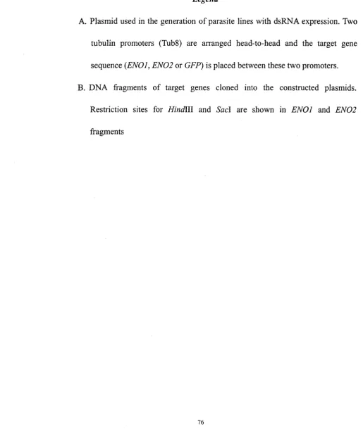
Figure 3. 2 Schematic representation o f construction strategies 76
Figure 3 .3 Confirmation of transgene integration 80
Figure 3 .4 Confirmation of dsRNA expression 84
Figure 3 .5 Gene expression in transgenic strains 88
Figure 3 .6 Localization o f E N 0 2 95
ABBREVIATIONS
ATP Adenosine triphosphate
bp base pair
BAG1 bradyzoite surface antigen 1
BSA bovine serum albumin
CAT chloramphenicol acetyl tranferase
cDNA complementary DNA
DAPI 4 ’, 6 diamidino-2-phenylindole
DEPC diethlypyrocarbonate
DHFR-TS dihydrofolate reductase-thymidylate synthase
DMEM Dulbecco’s Modified Eagle Medium
DMSO dimethylsulfoxide
DNA deoxyribonucleic acid
DNase deoxyribonuclease
dNTP deoxyribonucleoside triphosphate
DPBS Dulbecco’s phosphate-buffered saline
dsRNA Double-stranded RNA
EDTA ethylenediaminetetra-acetic acid disodium salt
ENO enolase
FBS fetal bovine serum
FITC Fluorescein iso-thiocyanate
GFP Green fluorescent protein
G6PI glucose-6-phosphate isomerases
HFF human foreskin fibroblasts
HXGPRT Hypoxanthine-guanine phosphoribosyltransferase
INF Interferon
LDH lactate dehydrogenase
LacZ B-galactosidase
M-MLV RT Moloney murine leukemia virus reverse transcriptase
MPA mycophenolic acid
mRNA Messenger RNA
miRNA Micro-RNA
nt nucleotide
rNTP ribonucleoside triphosphate
Oligo Oligonucleotide
PAGE polyacrylamide gel electrophoresis
PBS phosphate buffered saline
PCR polynerase chain reaction
RNA ribonucleic acid
RNAi RNA interference
R O Pl rhoptry protein 1
RT reverse transcription
RT-PCR reverse transcription PCR
RISC RNA induced silencing complex
siRNA small interfering RNA
SAG1 surface antigen 1
SDS sodium dodecyl sulphate
SSC sodium chloride-sodium citrate
Taq Thermus Aquaticus
TEMED N,N,N',N'-Tetramethylethylenediamine
TUB Tubulin
6TX 6 thioxanthine
UPRT uracil phosphoribosyltransferase
CHAPTER 1
INTRODUCTION
1.1 Toxoplasma gondii
Toxoplasma gondii is an intracellular pathogenic protozoa parasite. It belongs to
the phylum Apicomplexa and is the only known member o f the genus Toxoplasma. It was
first discovered in a rabbit, by Splendore in Brazil in 1908, and also by Nicolle and
Manceaux in a northern African rodent Ctenodactylus gundi (Black and Boothroyd,
2000). As most o f the Apicomplexa parasites, T. gondii is an important pathogenic agent
o f humans and animals. Its hosts include the vast majority o f warm-blooded animals. In
2000, it was reported that, 15.8% o f the population between 12 to 49 years old in United
States were infected with T. gondii (Jones et ah, 2003). The infection causes the disease
called toxoplasmosis, which is usually asymptomatic in healthy individuals. However, for
immuno-compromised patients, such as AIDS patients and those who have recently
received organ transplantation, infection by T. gondii can cause severe complications
such as hepatitis, pneumonia, blindness, and neurological disorders and may lead to
behavioral alteration. Furthermore, an acute infection during pregnancy can cause in
utero infection as well, resulting in a spontaneous abortion, a stillborn child, or a child
that is bom with some degree o f mental or physical retardation (Dubey, 1996).
While residing in the hosts, T. gondii converts between tachyzoite, the rapid
proliferating stage, and bradyzoite, the slowly replicating stage. Normally,
infection. In most cases o f human toxoplasmosis, tachyzoites can differentiate into
bradyzoites, which stay within the tissue cysts. Parasites are thus able to escape the host
immune response for the remainder o f the host’s lifespan, leading to the chronic infection.
When the patients become immuno-compromised, bradyzoites are released from the cysts
and differentiate into tachyzoites, resulting in a recurrent acute infection (Gross et al.,
1996). Current treatment o f toxoplasmosis using a cocktail o f pulfonamide
(dihydropteroate synthetase inhibitor) and pyrimethamine (dihydrofolate reductase
inhibitor) is only effective for the acute infection and may cause harsh side effects (Black
and Boothroyd, 2000). Although a combination o f atovaquone and clindamycin is
effective for treatment o f the chronic toxoplasmosis in mouse and some other animals,
their effects have not been re-produced in human (Alves and Vitor, 2005;
Djurkovic-Djakovic et al., 2002). To develop new drug targets and strategies to eradicate
its infection, we need to learn more about the parasite, especially its metabolism and
interaction with host organisms.
1.1.1 Life cycle o f T. gondii
Toxoplasma gondii multiplies by sexual and asexual cycles. The sexual life cycle
occurs exclusively in the intestine o f its definite host, felines. The cycle begins when a cat
ingests food containing T. gondii-tissue cysts. The parasites are released from the cyst in
the small intestine and infect epithelial cells, initiating gametogenesis and differentiation
later secreted in feces. Millions o f oocysts can be generated within 2 weeks after oocyst
formation. Within 2-3 days after being released from the intestine, oocysts undergo
sporogony and generate the infective haploid stage known as sporozoites. The mature
sporozoites are very stable in the environment and can survive for months in cold and dry
climates (Dubey, 1994). When the sporozoites are ingested by the felines, they continue
to propagate by the sexual life cycle. Ingested by other animals, referred to intermediate
hosts, the parasites will multiply using their asexual life cycle.
The asexual life cycle occurs in any infected warm-blooded animals. The
transmission can usually begin following the ingestion o f sporulated oocysts or
bradyzoites in tissues cysts in raw or contaminated meat products. Then the parasites
transform into tachyzoites and reproduce asexually within infected cells. After several
rounds o f synchronous division o f the parasites, the infected cells that cannot support the
parasite load will be destroyed. Newly formed tachyzoites are then released to infect
other cells. During this phase o f infection, the tachyzoites can spread to every organ o f
the host. Approximately 2-3 weeks following infection and in response to the host
immunity, the tachyzoites convert to slow-replicating bradyzoites within the tissue cysts
Figure 1.1
Life cycle o f Toxoplasma gondii
Legend
The life cycle o f T. gondii consists o f two phases: sexual and asexual. The sexual
phase occurs only in the intestine o f felines (definite host) while the asexual phase takes
place in birds and animals (Yang and Parmley, 1997). The figure is obtained from Black
Figure 1.1
Life cycle o f Toxoplasma gondii
Sexual Cycle
(feline intestine)
S ch izo g o n y and
g am e to g e n e s is Z y g o tefusion
c a>
E
> c h iIngestion of infected tissue
i
O ocyst M aturation
t
A ccid en tal ingestion
C hronic infection: brad yzo ites
Im m u n o s u p p re s s io n ( s a m e h o st)
Asexual Cycle
(m am m als/b ird s)
A cu te infection: tach yzo ites
1.1.2 Differences between tachyzoites and bradyzoites
The conversion between tachyzoites and bradyzoites is accompanied by
morphological and molecular biological changes, including the expression o f
stage-specific antigen and the alterations o f basal metabolism (Ferguson and Hutchison,
1987; Denton et al., 1996).
The fast-growing tachyzoite is usually surrounded by a sac-like membrane, called a
parasitophorous vacuole, which originates from both the host and parasite cell
membranes (Gross et al., 1996). The tachyzoites within a single vacuole usually divide
synchronously, leading to the formation o f a rosette structure. The bradyzoites stay in the
tissue cysts that can be as large as 100pm in length and are mainly localized in the brain
and muscle tissues. The tissue cysts are surrounded by an elastic thin wall which contains
P-(l, 4) linkaged N-acetyl-glucosamine residues called chitin without glycogen and other
polysaccharides (Gross et al., 1996). The cyst wall is able to bind with lectins such as
those found in the seed o f Dolichos biflorus and wheat-germ (Boothroyd et al., 1997;
Tomavo, 2001; Cleary et al., 2002). Thus these lectins are used as the detection reagents
for the tissue cysts and bradyzoites (Ferguson et al., 2002). Within the cyst wall, the
bradyzoites are less susceptible proteolytic enzymes and gastric acid in the stomach
(Freyre, 1995). Within the cytosol o f the bradyzoites, several amylopectin granules are
located and supposedly act as energy source. Having non-functional mitochondria,
bradyzoites lack a functional TCA cycle and respiratory chain. As a result, bradyzoites
the other hand, tachyzoites have functional mitochondria and both aerobic and anaerobic
respiration (Denton et al., 1996; Dando et al., 2001). Thus glycolysis is essential for both
tachyzoites and bradyzoites. Therefore, the enzymes catalyzing the reactions in the
glycolysis pathway, such as enolase, glucose-6-phosphate isomerases and
glyceraldehyde-3-phosphate dehydrogenase, have been considered as promising drug
targets in T. gondii and many other apicomplexa parasites (Verlinde et al., 2001; Roos,
2005).
Differential expressions o f heat shock proteins (HSP) were reported in the
tachyzoite and bradyzoite stages. For example, two differentially spliced forms o f HSP60
are present in both life cycle stages. Using polyclonal antibodies raised against HSP60, it
was found that HSP60 is localized in the mitochondria in tachyzoite stage, while in two
vesicular bodies during the stage o f bradyzoites (Toursel et al., 2000). Bradyzoite specific
antigen 1 (BAG1) or HSP30, which is homologous to small plant HSP, is only expressed
in bradyzoites, but not in tachyzoites (Bohne and Roos, 1997; Parmley et al., 1995). In
addition, many surface antigens, including CST1, SAG2A and SAG2B, are stage-specific,
suggesting they might be important for the interaction between the parasites and their
host cells (Zhang et al., 2001; Cleary et al., 2002; Lekutis et al., 2000). Moreover, a
number o f metabolic enzymes, including those o f glycolysis and gluconeogenesis such as
lactate dehydrogenase (Dando et al., 2001), glucose-6-phosphate isomerases (G6PI) and
enolase (ENO), were reported to be differentially expressed in tachyzoite and bradyzoite,
adaptation o f the parasites (Yang and Parmley, 1997; Tomavo, 2001; Dzierszinski et al.,
2001; Dzierszinski et al., 1999).
1.1.3 Interconversion between tachyzoite and bradyzoite
The conversion between tachyzoite and bradyzoite is essential for T. gondii and
plays a key role in its pathogenesis. A better understanding o f the interconversion
mechanism could greatly facilitate a design for new chemotherapeutic agents capable o f
eliminating tissue cysts (Boothroyd et al., 1997). In vivo, the stage conversion is related
to the host inmmune responses. It has been suggested that the switch between tachyzoites
and bradyzoites is related to the expression o f heat shock proteins and parasites’ stress
responses.
Most experimental strains o f T. gondii can switch between tachyzoites and
bradyzoites in cell cultures. About 10-20% o f cysts are formed by spontaneous
conversion. External stresses mimicking the host immune responses can induce tissue
cyst formation and increase the formation o f cyst in vitro (Soete et al., 1993). The
cyst-induction includes growing the parasites in the alkaline environment (pH 8.2-8.4) or
acid conditions (pH 6.6-6.8), increasing culture temperature (42 °C instead o f 37 °C).
These methods rely on the stress response o f the parasites (Soete et al., 1993; Weiss et al.,
1995; Weiss and Kim, 2000). Inhibitors o f mitochondrial function, such as oligomycin,
antimycin A, atovaquone and rotenone, as well as inducers o f oxidative stress including
encystment o f in vitro cultures (Soete et al., 1993; Bohne et al., 1994). It was also
shown that the elevation o f cAMP or cGMP could play a role in the bradyzoite induction
(Kirkman et al., 2001). Immunological factors, including interferon (IFN)-y and tumour
necrosis factor (TNF)-a, play a role in controlling tachyzoite growth and could indirectly
control stage conversion. Although evidence suggest the indirect effect o f NO-induced
stress, the underlining mechanism has yet been determined (Alexander and Hunter,
1998).
Additionally, it was reported that conditions favouring the formation o f bradyzoites
were associated with the up-regulation o f heat shock proteins (HSPs), suggesting that
HSPs are essential during stress-induced stage conversion (Weiss et al., 1998). Knockout
o f the BAG1 {HSP30) gene can decrease, but not completely prevent in vivo cyst
formation o f the parasites. This evidence further validates the involvement o f HSPs in the
process o f stage conversion (Bohne et al., 1998).
1.1.4 T. gondii culture conditions and parasite strains
Toxoplasma gondii can be in vitro cultured in different cell types. Human foreskin
fibroblasts (HFF) are the most widely used because they quickly propagate and survive in
the presence o f many growth-inhibiting drugs, such as those used during the selection o f
transgenic parasites. Additionally, they allow the parasites to replicate for several cycles
Hela cells and T cells, are commonly used in the culture of T. gondii (Cleary et al., 2002;
Boothroyd et al., 1997).
There are several experimental strains o f T. gondii. Each strain exhibits different
replication rates, virulence and ability to form in vitro cysts and are thus suitable for
different experimental design (Freyre, 1995). An experimental strain called RH is highly
virulent and fast-duplicating and the most commonly used in biochemical experiments,
particularly in the characterization o f parasite virulence and production drug-resistant
mutant lines (Roos et a l, 1994). However, the RH strain exhibits less ability to form
tissue-cysts, thus produces few tissue cysts in mouse when cultured in vitro. Moreover
the RH strain can not undergo the sexual replication in the definite hosts (Freyre, 1995).
Other strains, such as ME49 produce more tissue cysts and are thus ideal for study on
bradyzoite development and differentiation. A subclone o f ME49 strain, referred to as the
PLK strain, shows slower replicating rates and is widely used for the study o f life cycle
and stage conversion o f T. gondii, due to its ability to undergo complete sexual life cycle
and to convert between tachyzoites and brayzoites (Soete et al., 1993; Boothroyd et al.,
1997).
1.1.5 T. gondii as an experimental model for apicomplexan parasites
Many members o f Apicomplexa are pathogenic. For example, Plasmodium
falciparum is the causing agent o f malaria. It is difficult to culture and maintain in the
hand, can be easily maintained in vitro using standard cell culture techniques. In addition,
this parasite is amenable for molecular transformation and is thus ideal for genetic
manipulation and analysis. Genetic manipulation by molecular transformation, including
both transient transformation and stable transformation, has been widely employed as a
tool to verify gene function and molecular events at various stages o f T. gondii (Kim and
Weiss, 2004). The modulations o f T. gondii gene expressions by molecular tools such as
insertional mutagenesis, tetracycline-repressor-based inducible systems, are extensively
utilized to analyze gene functions (Donald and Roos, 1995; Nakaar et al., 2000; Meissner
et al., 2001). T. gondii has an 80 Mb haploid genome consisting o f 14 chromosomes
during its asexual life cycle, which facilitates the generation o f loss-of-function mutants
(Black and Boothroyd, 1998; Khan et al., 2005). Additionally, most o f the genome
sequence o f T. gondii has been revealed (ToxoDB, http://www.toxodb.org, (Kissinger et
al., 2003). Several genes o f T. gondii are representive o f related genes found in other
related Apicomplexans. Thus this parasite is widely used as an experimental model (Roos
et al., 1994; Black and Boothroyd, 2000; Kim and Weiss, 2004).
1.1.6 Genetic manipulation o f T. gondii
1.1.6.1 Transient and stable transformation
The development o f molecular transformation o f T. gondii has made genetic
manipulation a highly employed tool to study gene functions and molecular events o f the
current, is the most efficient technique to introduce nucleic acids into T. gondii. In
transient transformation, several reporter genes such as P-galactosidase (LacZ) and
chloramphenicol acetyltransferase (CAT) have been successfully employed. Their
expressions are efficient when driven by T. gondii promoters such as those o f the major
surface antigen (Lekutis et al., 2000), the rhoptry protein (Soldati et al., 1995) and the
P-tubulin (TUB1) (Roos et al., 1994; Soldati et al., 1995)
Stable transformation, resulting in the generation o f parasite lines that permanently
express transgenes by directed (homologous) or random integration (non-homologues
recombinants) into the parasite genome, has widely been used (Donald and Roos, 1994).
Moreover, Black and Boothroyd (1998) developed an episomal vector, which is able to
replicate autonomously in T. gondii without integrating into the genome. They found that,
when they randomly used DNA fragments isolated from the Toxoplasma genomic DNA
to construct into transforming plasmids, some o f these sequences permit the episomal
maintenance o f the plasmid in the parasites. These episomal vectors avoid the possibility
o f introducing mutations into the genome by non-homologous recombination. Episomal
vectors also offer the advantage o f allowing easy analysis o f the activity attributed to the
transformed DNA. This is accomplished by isolating the episome to re-transform the
parental strain or by selecting against the episom e using a negative selectable marker. The
development o f this molecule tool may help to understand the events critical to T. gondii
such as invasion, intracellular replication, and differentiation (Black and Boothroyd,
1.1.6.2 Selectable markers for stable transformation
Selectable markers are required for gene transformation that cannot be selected
directly. Two non-essential enzymes involved in the nucleotide pathways o f T. gondii,
uracil phosphoribosyl transferase (Al-Anouti et a l, 2003) and
hypoxanthine-xanthine-guanine phosphoribosyl transferase (HXGPRT), as well as other
enzymes like dihydrofolate reductase-thymidylate synthase (DHFR-TS) and
chloramphenicol acetyltransferase (CAT) are utilized efficiently as the selectable markers
for T. gondii transformation (Donald and Roos, 1994).
HXGPRT is important for the purine salvage pathway o f T. gondii. It catalyzes the
conversion from hypoxanthine, xanthine, and guanine into inosine monophosphate (IMP),
xanthosine monophosphate (XMP) and guanine monophosphate (GMP) respectively.
XMP can be further utilized for the synthesis o f GMP, and can also be generated from
IMP by IMP dehydrogenase in the absence o f xanthine or HXGPRT (Fig. 1.2). HXGPRT
can also catalyze the conversion from 6-thioxanthine (6-TX) to
6-thioxanthosine-5'-phosphate, which is toxic to the parasites. However, 6-TX cannot be
recognized by mammalian host cells that lack the XPRT activity. Thus HXGPRT can be
utilized as a negative selectable marker using 6-TX as the selection (Chaudhary et al.,
2004; Donald et al., 1996). The generation o f H XGPRT knockout T. gondii strains (RHA
HXGPRT, FLKAHXGPRT) makes it possible to use HXGPRT as a positive selectable
mycophenic acid (MPA) and xanthine. MPA inhibits the activity o f IMP dehydrogenase
and thus prevents the formation o f XMP and subsequent GMP, which is essential for T.
gondii survival using this pathway. Parasites with HXGPRT expression can utilize
HXGPRT to generate XMP from xanthine and maintain their lives, while those without
HXGPRT expression would be killed (Donald et al., 1996). The H XG PRT knockout
strains can be used in transformation with plasmids that express HXGPRT and the
medium supplemented with MPA and xanthine is effective to select transformed parasites
Figure 1.2
Purine Salvage Pathway o f Toxoplasma gondii
Legend
Schematic representation o f purine salvage pathway o f T. gondii. Enzymes are indicated
by arrows labeled AK (adenosine kinase), HXGPRT, or with numbers in the Toxoplasma
panel (unless otherwise indicated): 1, adenosine deaminase; 2, purine nucleoside
phosphorylase; 3, adenine deaminase; 4, AMP deaminase; 5, IMP dehydrogenase
(inhibition target o f MPA); 6, GMP synthetase; 7, adenylosuccinate synthetase; 8,
adenylosuccinate lyase. HC, host cell cytoplasm; PV, parasitophorous vacuole; PC,
Figure 1.2
Purine Salvage Pathway o f Toxoplasma gondii
A d e
Ado
Hyp Xan
Gua
G uo
A d o - ^ l n o , /
T
t
?
-?Guo
Hyp Xan
Gua
HXGPRT
T
AMP^-^rlfvlP 4 -XM
p
V
g
MP
S
7
T
MPA
1.1.6.3 RNA tools in T.gondii
RNA tools, including ribozyme and antisese RNA, have been used for the
down-regulation o f gene expression in many organisms including T. gondii (Al-Anouti
and Ananvoranich, 2002; Sheng et al., 2004; Nakaar et al., 2000; Nakaar et al., 1999).
They are efficient for the study on essential genes functions, because these RNA tools
have the advantage that they do not disrupt gene expression at the DNA level as the DNA
transformation (Lamond and Sproat, 1993). Moreover, these RNA tools can be used in
suppressing gene expression and in phenotypic studies.
Antisense RNA functions by base-pairing with target mRNA to interrupt the
downstream process (i.e. translation) or by destroying the target mRNA by RNase H
(Lamond and Sproat, 1993; Hostomsky et al., 1994). It was employed to modify the
expression o f triphosphoate hydrolase (NTPase) as well as HXGPRT in T. gondii (Nakaar
et a l, 1999). Ribozyme, first discovered by Cech and Altman in the 1980s, are RNA
molecules that can catalyze RNA cleavage in a site-specific manner (Tanner, 1999). In T.
gondii, engineered delta ribozymes were successfully utilized to reduce the expression of
UPRT and HXGPRT (Sheng et al., 2004).
RNA interference (RNAi), an endogenous machinery o f gene regulation, is
functional in many eukaryotic organisms (Hannon, 2002; Fire et ah, 1998). In T. gondii,
when dsRNA homologous to UPRT is introduced into the parasites, the expression level
and delta ribozyme are similar and are much higher than that o f antisense RNA
(Al-Anouti and Ananvoranich, 2002).
1.2 Enolase
Enolase (2 phospho-D-glycerate hydrolase, EC 4.2.1.11) is a glycolytic
metal-activated enzyme. It belongs to the enolase superfamily comprising among other
carboxyphosphonoenolpyruvate synthase. The enzyme catalyzes the Mg2+-dependent
removal o f one water molecule from 2-phosphoglycerate (2PGA) to yield
phosphoenolpyruvate (PEP), the penultimate step in the conversion o f glucose to
pyruvate. This reaction step is the only dehydration reaction in the glycolysis pathway.
Moreover, enolase also catalyzes the reverse reaction during the process o f
gluconeogenesis. This glycolytic enzyme usually exists as a dimer o f 45-48 kDa subunits,
in which one magnesium ion is required for each subunit (Lebioda and Stec, 1991).
Moreover, this enzyme is found to be highly conserved among different species from
archaebacteria to mammals.
1.2.1 Enolase isoforms in T. gondii
In T. gondii, two isoforms o f enolases are known as E N O l and E N 02. Genes
encoding these two enolase isoforms, showing 65.8% in open reading frame (ORF)
nucleotide sequence identity, are localized on Chromosome VIII o f T. gondii, residing
expressed only in bradyzoites and E N 02 is specifically expressed in tachyzoites.
Compared to enolases from other organisms, the T. gondii enolase isoforms carry a
pentapeptide EWGY(W)S(C) (103-107 aa) insertion and a dipeptide E(D)K (263 and 264
aa) and are thus considered highly related to those o f plants (Dzierszinski et al., 1999).
During differentiation between tachyzoites and bradyzoites, enolase is important for
glycolysis and anaerobic respiration. It has thus been hypothesized that EN O l and E N 02
might be a promising target for developing drugs for chronic toxoplasmosis.
Comparing characterizations o f these two enolase isoforms using recombinant
proteins, it was found that EN O l and E N 02 exhibit similar kinetic parameters: Michaelis
constant (Kirkman et al., 2001) o f 76.8pM vs. 77.7pM. However, E N 02 had threefold
higher specific activity (Vmax - 89.2 mmol x m in'1 x m g '1) than EN O l (Fmax. = 34.1 mmol
x m in'1 x m g '1). Both isoforms have a similar optimum pH at pH 7.2. But EN O l is
slightly more stable than E N 02. The denaturation temperature o f EN O l was estimated at
64.0 °C, while that o f E N 02 at 57.0 °C (Dzierszinski et al., 2001). The different
expression patterns as well as different enzymatic characterizations o f EN O l and E N 02
suggest that these two isoforms might be related to the different metabolism rates o f
tachyzoites and bradyzoites. E N 02, which is more active and less stable, is expressed
only in the virulent tachyzoite stage, supposedly important for rapid development. E N O l,
less active but more stable than E N 02, is more suitable for the encysted dormant
bradyzoite with less energetic requirements. EN O l and E N 02 are distinct in their
with E N 0 2 , vice versa, despite the high degree o f amino acid homology (Dzierszinski et
al., 2001). It hypothesized that the difference in the expression pattern o f enolase
isoforms, as well as other stage-specific metabolism enzymes such as LDH and GP6I, is
necessary for environmental adaptation and metabolic requirements o f the two different
parasitic stages. However, little is known about how the expressions o f these stage
specific enzymes are regulated during the stage conversion.
1.2.2 Structural features o f enolase
Similar to enolases in other organisms, the two enolase isoforms o f T. gondii
contain conserved amino acid residues important for their catalytic activity. As indicated
in Figure 1.3, these amino acids are G lul74, Glu217 and Lys355, which are involved in
the dehydratation step; Asp252, Glu303 and Asp330, whose carboxylate groups
coordinate the conformational metal ion ligand (Mg2+) required for substrate binding;
Arg384 interacting with the phosphate group o f 2-PGA; and His383 and Lys406
interacting with the carboxylic group o f 2-PGA (Dzierszinski et al., 2001; Lebioda et al.,
1989; Lebioda and Stec, 1991). The different characterization o f EN O l and E N 02 might
be due to the difference in the residues surrounding or neighboring those residues directly
involved in the substrate binding or catalysis.
The deletion o f the two plant-like motifs in EN O l decreases its activity
(Dzierszinski et al., 2001). These two plant-like peptide insertions are positioned in
264 aa is in one o f the connection loops intruding into the active site o f E N 02 and EN O l.
The pentapeptide insertion found at position 103-107 aa increases the length o f a
connecting loop in the C-terminal domain and makes it close to the active site o f the
enzyme. These structural features provide possible explanation to the significance o f
these two insertions in the activity o f enzyme. These plant-like motifs are also found in
enolase o f Plasmodium falciparum, another member o f Apicomplexa family (Read et al.,
1994). This feature may suggest that Apicomplexa and plants are derived from a common
ancestor, or Apicomplexa acquired their enolase by an endosymbiotic gene transfer from
Figure 1.3
Sequence Alignment of E N O l and E N 0 2
Legend
The amino acid sequnce o f T. gondii EN O l and EN 02 is aligned using LALIGN
program (http://www.ch.embnet.org/software/LALIGN form.htmQ. Two plant-like
motifs (103-107 aa EW G Y (W )S(C ), 263 and 264 aa E(D)K) are shown in green colour.
And conserved amino acids (G lul74, Glu217, Asp252, Glu303, Asp330, Lys355, His383,
Figure 1.3 Sequence alignment of E N O l and E N 0 2
1 0 2 0 3 0 4 0 5 0 6 0 E N O l M V V I K D I V A R E I L D S R G N P T I E V D V S T E G G V F R A A V P S G A S T G I Y E A L E L R D K D P K R Y L G
E N 0 2 M V A I K D I T A R Q I L D S R G N P T V E V D L L T D G G C F R A A V P S G A S T G I Y E A L E L R D K D Q T K F M G 1 0 2 0 3 0 4 0 5 0 6 0
7 0 8 0 9 0 1 0 0 1 1 0 1 2 0 E N O l K G V L N A V E I V R Q E I K P A L L G K D P C D Q K G I D M L M V E Q L D G T K N E W G Y S K S K L G A N A I L G V S
E N 0 2 K G V M K A V E N I H K I I K P A L I G K D P C D Q K G I D K L M V E E L D G T K N E W G W C K S K L G A N A I L A V S 7 0 8 0 9 0 1 0 0 1 1 0 1 2 0
1 3 0 1 4 0 1 5 0 1 6 0 1 7 0 1 8 0 E N O l I A C C R A G A A S K G L P L Y K Y I A T L A G K T I D K M V M P V P F F N V I N G G E H A G N G L A L Q E F L I A P V
E N 0 2 M A C C R A G A A A K G M P L Y K Y I A T L A G N P T D K M V M P V P F F N V I N G G S H A G N K V A M Q F . F M I A P V 1 3 0 1 4 0 1 5 0 1 6 0 1 7 0 1 8 0
1 9 0 2 0 0 2 1 0 2 2 0 2 3 0 2 4 0 E N O l G A P N I R E A I R Y G S E T Y H H L K N V I K N K Y G L D A T N V G D E G G F A P N V A T A E E A L N L L V E A I K A
E N 0 2 G A S T I Q E A I Q I G A E V Y Q H L K W I K K K Y G L D A T N V G D E G G F A P N I S G A T E A L D L L M E A I K V 1 9 0 2 0 0 2 1 0 2 2 0 2 3 0 2 4 0
2 5 0 2 6 0 2 7 0 2 8 0 2 9 0 3 0 0 E N O l A G Y E G K I K I A F D A A A S E F Y K Q D E K K Y D L D Y K C K T K N A S K H L T G E K L K E V Y E G W L K K Y P I I
E N 0 2 S G H E G K V K I A A D V A A S E F F L Q D D K V Y D L D F K T P N N D K S Q R K T G E E L R N L Y K D L C Q K Y P F V 2 5 0 2 6 0 2 7 0 2 8 0 2 9 0 3 0 0
3 1 0 3 2 0 3 3 0 3 4 0 3 5 0 3 6 0 E N O l S V E D P F D Q D D F A S F S A F T K D V G E K T Q V I G D D I L V T N I L R I E K A L K D K A C N C L L L K V N Q I G
E N 0 2 S I E D P F D Q D D F H S Y A Q L T N E V G E K V Q I V G D D L L V T N P T R I E K A V Q E K A C N G L L L K V N Q I G 3 1 0 3 2 0 3 3 0 3 4 0 3 5 0 3 6 0
ENO l S V T E A I E A C L L A Q K S G W G V Q V S H R S G E T E D S F I A D L W G L R C G Q I K S G S P C R S E R L C K Y N
E N 0 2 T V S E S I E A C Q L A Q K N K W G V M V S H R S G E T E D S F I A D L V V G L R T G Q I K T G A P C R S E R L C K Y N 3 7 0 3 8 0 3 9 0 4 0 0 4 1 0 4 2 0
4 3 0 4 4 0 E N O l Q L M R I E E S L G A D C V Y A G E S F R H P K
1.2.3 Multiple functions o f enolase
As a glycolytic enzyme, enolases localize in the cytoplasm, where they can be free
or easy to associate with the cytoskeleton and other glycolytic enzymes. In T. gondii,
EN O l and E N 02 are localized in the cytoplasm and nuclei, although there is no classic
nuclear localization signal found in their amino acid sequences (Ferguson et a l, 2002).
This discovery suggests that, T. gondii enolase might have some additional functions,
such as nuclear activities (i.e. division) and regulation o f gene expression.
In other organisms, enolase has also been reported to perform multiple functions.
For example, a-enolase, one o f the three isoforms o f enolase found in animals, is
identified as the eye lens crystallin in reptiles and birds (Piatigorsky and Wistow, 1989).
In HeLa cells, a-enolase acts as a component o f the centrosome, and one o f its alternative
transcript products known as rayoprom oter binding protein 1 (Feo et a l, 2000) can bind
to c-myc promoter and negatively regulate transcription o f this protooncogene (Johnstone
et al., 1992; Ghosh et al., 1999). In human peripheral blood cells, a-enolase functions as a
plasminogen receptor (Redlitz et a l, 1995), and is one o f the hypoxia-inducible proteins
(Semenza et a l, 1996). In fusion yeast Saccharomyces cerevisiae, enolase is the heat
shock protein HSP48 and involved in thermal tolerance and growth control in this
organism. Thus yeast enolase is recruited as a cofactor o f tRNA targeting toward
Apart from their metabolitic functions, it is not yet known whether T. gondii
enolase isoforms have additional functions. One o f the focuses o f this study is to reveal
the physiological functions o f EN O l and E N 0 2 in the development o f T. gondii.
1.3 Gene silencing in T. gondii
In order to study the roles o f E N 02 in T. gondii, I chose to suppress or silence the
expression o f E N 02 in the parasites. Upon the silencing o f E N 0 2 , resultant
loss-of-function phenotypes may provide useful information on the functions o f this
enzyme. In this section, I will review some techniques used in loss-of-functions
phenotype analysis in T. gondii.
1.3.1 Gene targeting in T. gondii
Gene targeting, including gene knockout and knockin, has been widely used in
many organisms such as yeast, Arabidopsis, Drosophila and mouse for analysis o f gene
functions. Gene targeting is based on homologous recombination, which involves the
exchange o f DNA between sequences o f perfect or near perfect homology over several
hundreds o f base pairs. The process o f homologous recombination plays essential roles in
the mitotic and meiotic cell cycles o f most eukaryotic organisms. However, in most o f the
eukaryotes, the opportunity o f homologous recombination is extremely low when
compared with non-homologous recombination, rendering the efficiency o f gene
Toxoplasma gondii, which has haploid genome during its asexual life cycle, is very
amendable for gene silencing and replacement. In gene targeting, constructs carrying the
desired mutated allele o f the interested gene with selectable markers are used in the
transformation o f parasites, and the homologous DNA fragment targets would guide and
direct the homologous recombination to occur at corresponding chromosome locus. Thus,
researchers can disrupt or delete the target gene or part o f it (so-called knockout, KO), or
replace the original gene with either a mutated one or another gene (so-called knockin, KI)
(Koller and Smithies, 1992; Hanin and Paszkowski, 2003; Rong, 2002). Up to now,
several genes o f T. gondii, including HXGPRT, UPRT, dihydrofolate
reductase-thymidylate synthase (DHFR-TS) and BAG1 were reported to be knocked-out
or replaced (Donald and Roos, 1998; Bohne et al., 1998; Bohne et al., 1994; Donald and
Roos, 1994).
One o f the often-utilized methods to increase the efficiency o f gene targeting is
increasing the length o f homologous regions required for homologous recombination. At
the locus o f T. gondii DHFR-TS, when genomic fragments o f 8 kb homology were used
as a circular plasmid in the transformation, ~50% o f the transformed parasites harbored
transgenes integrated by homologous transformation. When a 16 kb genomic fragment
was utilized, >80% homologous recombination was observed (Donald and Roos, 1994).
Furthermore, advancements o f positive-negative selection system and efficient
molecular screening methods have made gene targeting more effective in many
targeting including tissue- or cell-type specific gene targeting and temporal-inducible
gene targeting has rendered gene targeting a more flexible and promising tools for the
study o f various genes (Sauer, 1998; Utomo et ah, 1999).
It is possible to generate null mutants by gene knockout in T. gondii, which enables
the study o f gene function that can lead to the identification o f potential drug targets.
Although the frequency o f homologous recombination in T. gondii is relatively high, it is
not convenient to silence enolase isoforms by gene targeting becausetheir genomic loci
are almost overlapped. To create a knockout or a knockin o f either E N O l or E N 0 2 would
interfere with the other. Moreover, it is highly likely that the null mutant would be lethal.
To circumvent these potential difficulties, RNA interference is used. The introduction o f
dsRNA would attenuate the expression o f enolases and allow us to dissect their functions.
1.3.2 RNA interference
RNAi is one o f the most remarkable biological discoveries in the last 20 years. It is
an evolutionary conserved mechanism in which double-stranded RNA (dsRNA) or small
interference RNA (siRNA) initiates the specific silencing o f homologous genes. It is a
mechanism that is widely found in eukaryotes, including protozoa, insects and mammals
(Fire et al., 1998). It may function as a defensive mechanism to block the expression o f
aberrant or harmful genes originating from viruses and transposons (Hannon, 2002).
RNAi also plays a fundamental role in genome rearrangement, chromosome remodeling
Gorovsky, 2005). Most importantly, RNAi is one o f the most powerful and convenient
tools to knock down specific gene expression and has a potential for gene therapy
(Novina and Sharp, 2004; Harper et al., 2005).
1.3.2.1 The discovery of RNA interference
The first observed phenomenon o f RNAi took place in 1990 in plant research when
scientists introduced a transgene designed to overexpress the enzyme chalcone synthase
in petunias aimed to deepen floral color. Unexpectedly these researchers found white
patchy flowers instead o f the expected increased flower pigmentation (Napoli et al., 1990;
van der Krol et al., 1990). They thought that it was the transgene that silenced the
expression o f the plant purple-flower genes. The phenomenon was named as
co-suppression, because the exogenous transgene suppressed the expression o f itself as
well as its endogenous homolog. The phenomenon remained obscure until similar gene
silencing effects (known as quelling) was observed in Caenorhabditis elegans upon
injection o f dsRNA (Fire et al., 1998). It was found that it was the dsRNA that lead to the
degradation o f target mRNA, known as post-transcriptional gene silencing (PTGS). Later
it was reported that RNAi was also functional in other organisms including fusion yeast
Schizosaccharomyces pombe, Drosophila melanogaste, Arabidopsis thaliana,
Neurospora crassa as well as mammals (Hannon, 2002). Furthermore, it was also
reported that RNAi activity can persist through cell division rounds and growth, and that
1.3.2.2 The mechanism o f RNA interference
It is suggested that the suppressive effects o f RNAi can operate on several different
levels. The co-suppression effects in plants is triggered by DNA methylation o f the genes
which are homologous to dsRNA at cytosine residues, and thus these modified genes are
prevented from being transcribed (Wassenegger, 2005). However, the cleavage o f mRNA
homologous to dsRNA in post-transcriptional level is usually thought to be the most
essential pathway for the silencing effects o f dsRNA.
Genetic and biochemical studies have contributed tremendously to the
understanding o f the mechanism o f RNAi. Using extracts from Drosophila embryos and
tissue-cultured cells transfected with dsRNA, a nuclease capable o f degrading exogenous
homologous mRNA, was partially purified. This nuclease was co-purified with small
RNAs o f 21-25 nt long consisting o f sense and antisense sequences derived from within
the used regions (Tuschl et al., 1999; Elbashir et al., 2001; Yang et al., 2000). Incubation
o f a cell-free system from Drosophila embryos with both the dsRNA and its homologous
mRNA exhibited RNAi activity as found in vivo. And preincubation o f the cell-free lysate
with the dsRNA potentiated the degrading activity o f the lysate on the target mRNA in
vitro (Hammond et al., 2000; Zamore et al., 2000). These series o f experiments led to a
model o f RNAi pathways consisting o f initiation and effecter steps.
In the initiation step, an RNase III like enzyme known as Dicer recognizes and
a highly conserved protein found in worms, flies, plants, fungi and mammals, basically
consists o f a dsRNA binding domain and one or two RNase III nuclease domains
(Elbashir et al., 2001). These 21-25 nt siRNA, RNA duplex carrying 5 ’-phosphate and
3 ’-hydroxyl termini with 2-nt overhanging 3 ' end, can later incorporates into a -3 6 0 kDa
ribonucleoprotein nuclease-complex called the RNA induced silencing complex (RISC)
(Hannon, 2002). The exact components o f RISC are not clear now, with the exception o f
the Argonaute family protein that is supposed to be the essential element in the complex.
The Argonaute proteins are divided into two sub-families, known as Argonaute 1-like
(after the Arabidopsis Argonaute 1) and Piwi-like (after the Drosophila Piwi), depending
on the existence o f N-terminal PAZ domain in addition to the highly conserved
C-terminal Piwi domain found in all Argonaute proteins (Liu et al., 2004). Argonaute
proteins were reported to be able to bind with Dicer, suggesting that their interaction is
important for the loading o f siRNA into RISC. In the effecter step, the siRNA is first
unwound in an ATP-dependent process. The formed single-stranded small RNA guides
the RISC to homologous mRNA and subsequently cleaves it at 10-11 nt away from the 3 ’
terminus o f the guide RNA. The 5 'phosphate group on the siRNA is essential for
assembly with the RISC and subsequent target cleavage (Elbashir et al., 2001; Hannon,
2002). Moreover, heterochromatin formation is also one possible pathway to inhibit
target gene expression (Wassenegger, 2005).
Furthermore, Dicer can digest hairpin RNA precursors into micro RNA (miRNA),
incorporate into a protein complex known as miRNA-containing ribonucloprotein
(miRNP) complex, which also contains a Argonaute family protein. In some organisms
such as human, miRNP can guide miRNA to bind with homologous mRNA and inhibit its
translation without affecting mRNA levels (Nelson et al., 2004).
Other components o f RNAi machinery, including R2D2, a Dicer interacting protein
found to be important to load siRNA into RISC, were also reported. Among them, RNA
dependent RNA polymerase (RdRP), which was reported to be able to amplify RNAi
signals, is suggested to be essential for RNAi in some organisms, such as Neurospora
crassa, and Schizosaccharomyces pom be (Cogoni and Macino, 1999; Hall et al., 2002).
The siRNA can bind with the target transcript as primer, and RdRP can extend the
primers and generate the complementary RNA strand, resulting in a dsRNA which may
serve as a new substrate for Dicer (Martienssen, 2003). Moreover, in S. pombe, it was
also shown that RdRP was essential for RNAi-mediated heterochromatin assembly
(Sugiyama et al., 2005; Hall et al., 2002). However, RdRP may be not required for RNAi
in some other organisms. In Caenorhabditis elegans, it was reported that the mutant for
the gene ego-1 encoding its RdRP homologue showed similar RNAi activity when
compared to the wild type (Sijen et al., 2001). Drosophila and mammals, containing no
RdRP homologues in its genome, still exhibit gene-silencing efficiency at the
post-transcriptional level (Cottrell and Doering, 2003). It appears that RdRP is not a
However, it is also possible that these organisms have distinct or divergent RdRP
Figure 1.4
The mechanism o f RNA interference
Legend
The first commintting step o f the RNA interference pathway is catalyzed by Dicer
which digests endogenous and exogenous dsRNA and generates siRNAs. The siRNA is
loaded into RISC and directs the complex to silence gene by mRNA degradation,
translational inhibition, or chromatin remodeling. In some organisms, RdRP-dependent
synthesis o f new dsRNA may be used to amplify RNAi signals. The figure is adopted
Figure 1.4
The mechanism of RNA interference
1.3.2.3 The application o f RNA interference
RNAi is a powerful tool to suppress specific gene expression for reverse genetic
studies and to elucidate the functions o f genes in different organisms. The emergence o f
high throughput RNAi microarray chips has provided an important tool to analyze the
functions o f new genes coming from high throughput sequencing centers (Silva et al.,
2004). Moreover, RNAi technology is frequently employed in identify novel drug targets
and is a potential therapy for some diseases. Many pharmaceutical companies and
research centers have launched RNAi-therapy programs. Actually, any diseases could be
potential targets for RNAi-therapy. For example, in mice, siRNA homologous tumor
necrosis factor a (TNFa) was reported to be able to inhibit joint inflammation caused by
collagen induced arthritis (CIA) (Schiffelers et al., 2005). In addition, by using siRNA
expression vector to inhibit the expression o f HIV cellular receptor CD4 and coreceptors
CXCR4 and CCR5, HIV infection on T cells can be significantly reduced (Anderson and
Akkina, 2005). Specific siRNA is also reported to be able to inhibit growth o f cancer
cells (Takei et al., 2004; Leng and Mixson, 2005).
For the application o f RNAi in research, many parameters need to be considered
including specificities and length o f the dsRNA utilized, types o f cells employed, as well
as the method o f dsRNA delivery. In tissue culture, methods such as electroporation,
microinjection, and lipid mediated gene delivery are often used to deliver dsRNA or
siRNA to cells (Parrish and Fire, 2001; Elbashir et al., 2001). For Caenorhabditis elegans,
successfully deliver dsRNA into cells (Timmons and Fire, 1998; Tabara et al., 1998).
Moreover, stable transformations, which can produce stable RNAi effects, with vectors
expressing dsRNA and siRNA, have been put into practice. The target sequence can be
inserted into a plasmid as inverted repeats so that the dsRNA is expressed as a hairpin
dsRNA in vivo (Sui et al., 2002; Paddison et al., 2002). A target sequence can also be
placed between two promoters arranged in a head-to-head fashion (Tschudi et al., 2003;
Al-Anouti et al., 2004; LaCount et al., 2000). In addition, inducible expression systems
such as tetracycline-inducible vectors, and stage- and tissue-specific expression systems,
were reported to be utilized in RNA silencing experiment and may have great prospect in
the future study (Cottrell and Doering, 2003; Anderson and Akkina, 2005).
However, there is some concerns about using dsRNA in mammalian systems due to
a protective antiviral response system which leads to a non-specific inhibition o f host
gene expression (Huppi et al., 2005). But this difficulty can be avoided by using
embryonic cells instead o f non-embryonic cells, or by using siRNA instead o f long
dsRNA (Yang et al., 2002; Elbashir et al., 2001; Huppi et al., 2005).
1.3.2.4 RNAi in protozoa parasites
When RNAi was first put into practice for the down-regulation o f gene expression,
it was used in the protozoa Trypanosome brucei (Ngo et al., 1998). Genome searching
has shown that T. brucei contains members o f Argonaute family proteins but no Dicer
Trypanosomatidae family, Trypanosoma congonlese was found to have RNAi functions,
while T. cruzi and Leishmania major, were revealed to be RNAi negative and their
genome databases show no homologues o f Dicer and Argonaute (Ullu et al., 2004). In
addition, RNAi was reported to be functional in T. gondii, as well as in Plasmodium
falciparum , another member o f Apicomplexa family (Malhotra et al., 2002; McRobert
and McConkey, 2002; Al-Anouti et al., 2003). The presence o f classical RNAi genes,
including potential homologues o f Argonaute, Dicer and RdRP, were reported from
database mining o f the T. gondii predicted coding regions. The presence o f Argonaute
family protein was reported in T. gondii, further supporting the functioning o f RNAi in
Objectives
Stage-specific expression and nuclear localization o f enolase isoforms in
Toxoplasma gondii suggest additional important functions o f this glycolotic enzyme in
the parasites and provide potential drug targets for toxoplasmosis treatment. But limited
knowledge has been acquired about enolase up to now. The objective o f this study was to
investigate the possible role o f E N 02 in the development o f T. gondii, including their
involvement in parasite growth and stage differentiation. We would also like to
investigate the effectiveness and specificity o f dsRNA induced gene silencing in T.