GENETICS | INVESTIGATION
Quantitative Changes in
Gimap3
and
Gimap5
Expression Modify Mitochondrial DNA Segregation
in Mice
Riikka Jokinen,* Taina Lahtinen,* Paula Marttinen,* Maarit Myöhänen,* Pilvi Ruotsalainen,* Nicolas Yeung,* Antonina Shvetsova,†Alexander J. Kastaniotis,†J. Kalervo Hiltunen,†Tiina Öhman,‡
Tuula A. Nyman,‡Hartmut Weiler,§and Brendan J. Battersby*,1 *Research Programs Unit—Molecular Neurology, Biomedicum Helsinki, and‡Institute of Biotechnology, University of Helsinki, 00290 Helsinki, Finland,†Department of Biochemistry and Biocenter Oulu, University of Oulu, 90014 Oulu, Finland, and§Blood Research Institute, Blood Center of Wisconsin, Milwaukee, Wisconsin 53266
ABSTRACTMammalian mitochondrial DNA (mtDNA) is a high-copy maternally inherited genome essential for aerobic energy metabolism. Mutations in mtDNA can lead to heteroplasmy, the co-occurence of two different mtDNA variants in the same cell, which can segregate in a tissue-specific manner affecting the onset and severity of mitochondrial dysfunction. To investigate mechanisms regulating mtDNA segregation we use a heteroplasmic mouse model with two polymorphic neutral mtDNA haplotypes (NZB and BALB) that displays tissue-specific and age-dependent selection for mtDNA haplotypes. In the hematopoietic compartment there is selection for the BALB mtDNA haplotype, a phenotype that can be modified by allelic variants ofGimap3. Gimap3 is a tail-anchored member of the GTPase of the immunity-associated protein (Gimap) family of protein scaffolds important for leukocyte development and survival. Here we show how the expression of two murineGimap3alleles fromMus musculus domesticusandM. m. castaneusdifferentially affect mtDNA segrega-tion. The castaneus allele has incorporated a uORF (upstream open reading frame) in-frame with theGimap3 mRNA that impairs translation and imparts a negative effect on the steady-state protein abundance. We found that quantitative changes in the expression ofGimap3and the paralogue Gimap5, which encodes a lysosomal protein, affect mtDNA segregation in the mouse hematopoietic tissues. We also show that Gimap3 localizes to the endoplasmic reticulum and not mitochondria as previously reported. Collectively these data show that the abundance of protein scaffolds on the endoplasmic reticulum and lysosomes are important to the segregation of the mitochondrial genome in the mouse hematopoietic compartment.
KEYWORDSmitochondria; mitochondrial DNA; mice; segregation; Gimap
M
AMMALIAN mitochondrial DNA (mtDNA) is a mater-nally inherited small circular multicopy genome that encodes 13 proteins that are essential subunits of four of thefive complexes required for mitochondrial oxidative phos-phorylation. Germline or somatic-cell mtDNA mutations lead to the co-occurrence of two or more sequence variants in a cell, a state known as heteroplasmy. In the absence of selection, the segregation of mtDNA sequence variants is neutral and can bemodeled as a random walk (Chinnery and Samuels 1999); however, in some cases there is preferential selection for a mtDNA sequence variant that is dependent upon the nucle-otide sequence, tissue, and nuclear background (Battersby and Shoubridge 2001; Battersby et al. 2003, 2005; Jokinen and Battersby 2013; Burgstalleret al.2014). The majority of pathogenic mtDNA mutations are heteroplasmic and some mutations display skewed segregation patterns in somatic tissues. (Larsson et al. 1990; Bouletet al. 1992; Kawakami
et al.1994; Dunbaret al.1995; Fuet al.1996; Chinneryet al.
1997, 1999; Weber et al.1997), which can affect the onset and severity of mitochondrial dysfunction. Currently, the mo-lecular basis for this regulation of the mitochondrial genome is largely unknown (Jokinen and Battersby 2013).
To study mtDNA segregation in mammals we use a heter-oplasmic mouse model with two neutral mtDNA variants
Copyright © 2015 by the Genetics Society of America doi: 10.1534/genetics.115.175596
Manuscript received February 17, 2015; accepted for publication March 20, 2015; published Early Online March 25, 2015.
Supporting information is available online athttp://www.genetics.org/lookup/suppl/ doi:10.1534/genetics.115.175596/-/DC1.
derived from two old inbred mouse strains, BALB/c and NZB (Jenuth et al. 1996, 1997). These mtDNA haplotypes, re-ferred to as BALB and NZB, differ at 90 nucleotide positions (Hagstrom et al. 2014) and have been stably transmitted through the female germline of this mouse model for 20 years (Jenuth et al. 1996). There is no selection for either mtDNA haplotype during transmission as the heteroplasmy level in the offspring follows a Gaussian distribution (Jenuth
et al. 1996; Wai et al. 2008). However, postnatally there is age-dependent selection of one haplotype over the other in three tissue types (liver, kidney, and hematopoietic tissues), while all other tissues in these mice are neutral with respect to mtDNA segregation (Jenuthet al.1997). In the liver and kid-ney there is selection for the NZB haplotype tofixation (Jenuth
et al. 1997; Battersby and Shoubridge 2001; Battersbyet al.
2003, 2005). In contrast, the hematopoietic tissues (bone mar-row, blood, thymus, and spleen) select against the NZB mtDNA haplotype, which can be modeled as an exponential decay (Battersbyet al.2005). This tissue-specific mtDNA segregation
is best treated as a quantitative genetic phenotype and one that cannot be explained by detectable functional differences in the mitochondrial respiratory chain or by differential rates in mtDNA replication (Battersby and Shoubridge 2001).
To uncover the molecular basis of this mtDNA regulation we have used forward genetic approaches in mice. On the nuclear background ofM. m. domesticusin several different mouse strains (BALB/c, C3H, C57BL/6J, DBA, NZB, and 129Sv) there are no differences in these tissue-specific mtDNA segregation phenotypes (Battersbyet al.2003, 2005). In con-trast, crosses onto aM. m. castaneusnuclear background have a significant effect on these mtDNA segregation phenotypes (Battersbyet al.2003, 2005). This allowed us to identify three nuclear loci that affect mtDNA segregation in a tissue-specific manner (Battersbyet al.2003). At one of these loci we have successfully clonedGimap3(Jokinenet al.2010), which can modulate the segregation of mtDNA in leukocytes.
GTPase of the immunity-associated proteins (Gimap) are encoded in a conserved cluster of seven to eight genes found only in vertebrates with an ortholog in plants (Kruckenet al.
2004). In mammals, Gimap expression appears to be re-stricted to hematopoietic tissues and is important for leuko-cyte development and survival, although the molecular basis for these functions is poorly understood (Kruckenet al.2004; Nitta and Takahama 2007; Schulteiset al.2008; Barneset al.
2010; Chenet al.2011). Gimaps can be tail anchored or soluble and are structurally related to Septins, Tocs, and Dynamins (Schwefelet al.2010). Based on structural studies the membrane anchored Gimaps form GTP-dependent homo-oligomers with low inherent GTP hydrolysis activity that sta-bilizes these oligomers allowing them to act as scaffolds (Schwefelet al.2010, 2013).
Gimap3 andGimap5 are paralogues in this gene cluster and 84% identical at the amino acid level, differing only at the N and C termini (Kruckenet al.2004). Both have a short transmembrane domain at the C terminus for insertion into a lipid bilayer. The intracellular localization of Gimap5 has
been controversial with data reporting insertion into many different organelles. However, data using species-specific monoclonal antibodies against Gimap5 in T cells robustly demonstrate that in humans, mice, and rats the protein is anchored into the lysosomal membrane (Wonget al.2010). So far there is only one report for the localization of Gimap3 suggesting it is mitochondrial (Daheronet al.2001). In mice, both genes are expressed and appear to be important for T-cell development (Nittaet al.2006) and possibly in a cooper-ative way (Yanoet al.2014). In contrast,GIMAP3in humans appears to be a pseudogene (Kruckenet al.2004). Complete loss ofGimap5 function in mice appears to be catastrophic for the hematopoietic compartment, producing a decrease of lineage-committed hematopoietic progenitors leading to a reduction of T and B lymphocytes, NK and NK T cells, altered erythropoiesis, and early lethality (Schulteis et al.
2008; Barneset al.2010; Chenet al.2011). In rats the loss of Gimap5 function is milder, as a premature termination in
Gimap5 of the BioBreeding rat results only in a T-cell lym-phopenia, which is a susceptibility factor for autoimmunity in this diabetic rodent model (Hornumet al.2002; MacMurray
et al.2002). Both Gimap3 and Gimap5 have been shown to interact with Bcl2 family members (Nitta et al.2006; Chen
et al. 2011), although the function of these interactions on membrane surfaces has yet to be elucidated.
Here, we investigated in more detail the basis by which
Gimap3 is important for mtDNA segregation in the mouse hematopoietic compartment. We show that the tail-anchor sequence of Gimap3 localizes the protein to the endoplasmic reticulum (ER) and not the mitochondrial outer membrane as previously reported. The steady-state abundance of Gimap3 in leukocytes appears to be a critical factor for the regulation of mtDNA segregation in mouse hematopoietic tissues. Our data also implicate a role for the paralogueGimap5in mtDNA segregation. Together our findings demonstrate that the abundance of two membrane-bound protein scaffolds on the endoplasmic reticulum and lysosomes are important mod-ulators to the segregation of the mitochondrial genome in mouse leukocytes.
Materials and Methods
Animals
The Regional State Administrative Agency of Southern Fin-land (ESAVI) approved all mouse work. The Gimap5tm1Wlr
knockout mouse and the heteroplasmic mice were main-tained on the BALB/c nuclear background and housed under standard conditions applied by the University of Helsinki Lab-oratory Animal Center. The animals in experiments were housed and sampled in random groups. Both male and fe-male mice were used in the analyses.
Cell culture
from BALB/c mice (Battersbyet al.2005). MG132 (20mM) (Sigma) was dissolved in DMSO.
cDNAs, plasmids, and retroviral expression
Full-length cDNAs (BALB Gimap3, BALB Gimap5, CAST
Gimap3, BALB Gimap3-omp25,YFP-Gimap3,YFP-(263-301) Gimap3,RFP-Gimap3, pGFP-omp25 (Nemoto and De Camilli 1999) (gift from Hans Spelbrink) were cloned into Gateway (Invitrogen) converted retroviral expression vectors (pBABE-puro, pMYS-IRES-Neo, or pMXs-IRES-Blasticidin) or pcDNA3.1 for transient transfections. Retroviral vectors were trans-fected (Jetprime, Polyplus) into the Phoenix amphotropic packaging line for virus production to infect recipient cells (MEFs and COS-7). Following antibiotic selection, cells were used in experiments. For transient expression, plas-mids were transfected (Jetprime, Polyplus) into COS-7 and HEK293 cells. The GFP–Sec61b plasmid was a gift from Benoit Kornmann).
Immunoblotting and antibodies
Cells or tissue samples were solubilized in phosphate buffered saline, 1% dodecyl-maltoside, 1 mM PMSF (phenylmethyl-sulfonylfluoride), complete protease inhibitor (Roche). Pro-tein concentrations were measured by the Bradford assay (Bio-Rad). Equal amounts of proteins were separated by Tris-glycine SDS–PAGE and transferred to nitrocellulose by semi-dry transfer. Primary antibodies were incubated overnight at +4°and detected the following day with secondary HRP conjugates (Jackson ImmunoResearch) using enhanced chemiluminescence (Cell Signaling) with film or a Bio-Rad chemi-doc imaging station. Gimap3 antibodies were gener-ated in rabbits with the following peptides: BALB variant, amino acids 2–16 (ETLQNVVTGGKKGGC) and amino acids 259–272 (C-EGSWVLKVLPIGKK) and CAST variant, amino acids 32–45 (RIPVYTTDHLRCPDS). Sources for commercial antibodies: Chromotek (anti-GFP); Mitosciences (Sdha); Millipore (Porin); Proteintech Group (Atp5B, Calnexin, and RPL18a); Sigma (anti-HA and Atg5), Santa Cruz (Tom40). Representative immunoblotting data from multiple indepen-dent biological experiments were cropped in Photoshop with only linear corrections applied. When quantified, Western blots were analyzed with Bio-Rad chemi-doc imaging station and Image Lab software.
In vitro protein synthesis
Proteins were synthesized with TNT T7-coupled reticulocyte lysate system (Promega) according to manufacturer’s instructions using plasmids with Gimap3 or YFP cDNA con-structs cloned into pcDNA3.1. Each reaction also included Superase RNAse inhibitor (Ambion) and [35S]Met-Cys
(EasyTag, Perkin Elmer) and incubated for 25 min at 30°. The reaction time was chosen based upon linear radiolabel incorporation for YFP (Figure S2). Equal aliquots from each reaction mix were separated by SDS–PAGE with the gel dried and exposed to a Phosphoscreen for scanning with a Typhoon 9400 (GE Healthcare) for quantification.
Histodenz gradients
Mouse spleens were homogenized in buffer (200 mM mannitol, 70 mM sucrose, 10 mM HEPES, 1 mM EGTA pH 7.5) with a Teflon pestle and then centrifuged twice at 6003gfor 10 min. The post-nuclear suspension was mixed with an equal volume of 50% Histodenz (Sigma) (10 mM HEPES, 1 mM EGTA pH 7.5). A discontinuous Histodenz gradient was then formed (10, 15, 20, 25, and 30%) to sep-arate organelles (Okado-Matsumoto and Fridovich 2001). Samples were centrifuged in a SW32.1Ti rotor for 90 min at 52 000 3 g at +4°. Fractions were collected from the top, precipitated with TCA, and then resuspended in 13 Laemmli buffer. Samples were then subjected to SDS–PAGE and immunoblotting.
Sucrose gradient centrifugation
HEK293 cells from a 150-mm plate were treated with 100
mg/ml cycloheximide upon harvesting and lysed in 450ml of lysis buffer (20 mM Tris-HCl pH 7.5, 150 mM NaCl, 5 mM MgCl2, 1 mM DTT, 100mg/ml cycloheximide, 1% Triton
X-100) with Superase RNAse inhibitor (Ambion) for 20 min and centrifuged for 5 min at 13,0003gat 4°. The supernatant was loaded on a 16-ml linear 10–50% sucrose gradient (20 mM HEPES pH 7.6, 100 mM KCl, 5 mM MgCl2) and
centri-fuged for 200 min at 28,000 rpm at 4°(Beckman, SW 32.1 Ti). Eighty-two equal volume (200 ml) fractions were col-lected from the top. A total of 100 ml of each fraction was used for measuring RNA absorbance at 254 nm. The remain-ing fractions were pooled, combinremain-ing five consecutive frac-tions, for TCA precipitation, heated at 95° for 5 min and separated by 12% SDS–PAGE for immunoblotting.
Mitochondrial DNA analysis
All sampled mouse tissues were immediately frozen in liquid nitrogen and stored at280°. Total DNA was extracted with phenolchloroform and mtDNA heteroplasmy level deter-mined as described in Battersby and Shoubridge (2001). Modeling selection for mtDNA in the hematopoietic tissues of heteroplasmic mice was as described in Battersby et al.
(2005). Data sets were testedfirst for normality then subject to parametric statistics (two-tailed t-test).
RNA analysis
All tissues sampled were frozen in liquid nitrogen and stored at 280°. Total RNA was extracted with Trizol (Invitrogen) then treated with DNaseI to eliminate potential DNA contam-ination. Allelic expression ofGimap3was measured from total RNA amplified by semiquantitative RT–PCR compared to a standard curve with equimolar mixtures of CAST/Ei and BALB/c RNA (r2 = 0.97) and quantified with a Typhoon
9400. Data were analyzed statistically using a Mann–Whitney
U-test.
25 sec. Total RNA was isolated by Trizol from stable retroviral cell line controls and treated with DNaseI (New England Biolabs). A total of 15mg of RNA for tissue samples and 5mg for the cell line controls (concentration determined with a Nanodrop1000 spectrophotometer) were separated on a 1.2% agarose–formaldehyde gel and transferred by neutral transfer to Hybond-N+membrane (GE healthcare).
The membrane was UV cross-linked and consecutively probed with oligonucleotide probes for Gimap3andAtp5B. The probes were 59radiolabeled with T4 PNK (New England Biolabs) and purified with Illustra ProbeQuant G-50 micro-column (GE Healthcare). Hybridization (25% formamide, 7% SDS, 1% BSA, 0.25 M sodium phosphate pH 7.2, 1 mM EDTA pH 8.0, 0.25 M NaCl2) was performed for 16–
20 hr at 37°. Membranes were washed (23SSC/0.1% SDS and 0.23 SSC/0.1% SDS) and then dried for exposure on Storage Phosphor screens (GE Healthcare), which were scanned and quantified with a Typhoon 9400 (Amersham).
Microscopy
Cells were grown on glass coverslips andfixed in fresh 4% paraformaldehyde. Coverslips were mounted with DABCO/ MOWIOL on glass slides for imaging. Confocal images were taken on a Zeiss META 510 with the 633Plan-Apochromat (1.4 NA) objective using the Argon (488 nm) and HeNe1 (543 nm) lasers or a Leica TCS Sp8 CARS with a 633APO (1.2 NA) water objective using the Argon (488 nm) and DPSS (561 nm) lasers. Fluorescence microscopy was carried out on a Zeiss Axioplan2 using a 633 (1.25 NA) or 1003 (1.3 NA) objective. Image processing and line profiles were performed using Image J. Only linear corrections were ap-plied to images.
iTRAQ analysis
Three mouse spleens per genotype were homogenized (200 mM mannitol, 70 mM sucrose, 10 mM HEPES, 1 mM EGTA pH 7.5) with a Teflon pestle. Differential centrifugation was used to enrich for the membrane compartment containing Gimap3 and Gimap5. The postnuclear lysate (two 600 3g
for 10 min) wasfirst centrifuged at 50003gfor 10 min and the resulting pellet was discarded. The supernatant was then centrifuged for 20 000 3 g for 40 min. This pellet was solubilized and a total of 120mg of protein per sample was precipitated using 2-D Clean Up Kit (GE Healthcare). The precipitated proteins were dissolved into 30 ml of iTRAQ dissolution buffer, and 2ml of each sample was sub-jected to SDS–PAGE and silver staining to control for protein amount in the samples after precipitation. Protein alkyl-ation, trypsin digestion and labeling of the resulting peptides were performed according to manufacturer’s instructions (Applied Biosystems). After labeling, the sam-ples were pooled and dried, and the peptides were fraction-ated by strong cation exchange chromatography using an Ettan HPLC system (Amersham Biosciences) connected to a Polysulfoethyl A column. Each selected SCX fraction con-taining labeled peptides was then processed with C18 Spin
Columns (Pierce) according to the manufacturer instruc-tions before being analyzed twice with nano-LC-ESI-MS/ MS using Ultimate 3000 nano-LC (Dionex) and QStar Elite mass spectrometer (Applied Biosystems) as previously de-scribed (Lietzenet al.2011). MS data were acquired auto-matically using Analyst QS 2.0 software. iTRAQ analyses were performed on three biological replicate samples.
Protein identification and relative quantitation were per-formed with Paragon search algorithm (Shilov et al.2007) using ProteinPilot 4 interface (Applied Biosystems). Datafiles from both technical replicates of an iTRAQ sample set were processed together. Database searches were performed against mouse protein sequences in UniProt database (v. 2012-08-13 with 16 540 mouse entries). The search criteria were cysteine alkylation with MMTS, trypsin digestion, bio-logical modifications allowed, thorough search and detected protein threshold of 95% confidence (Unused ProtScore
.1.3). Additionally, automatic bias correction was used to correct for uneven protein loading. The false discovery rates were calculated as previously described (Elias and Gygi 2007) and were,1% for all biological replicates.
Relative quantity fold change of 0.66 or 1.5 was used as a threshold for changed proteins, and proteins were in-cluded in the data set only if they either exhibited similar change in at least two of the biological replicates, or a statistically significant quantitation change in one of the replicates and no changes in the others. Biological process for the identified proteins was retrieved from UniProtKB.
Results
Previously we identified M. musculus allelic variants of
Gimap3that affect mtDNA segregation in hematopoietic tis-sues (Jokinenet al.2010). In the common lab mouse strains with a M. m. domesticus background, such as BALB/c,
Gimap3 containsfive exons and transcription produces an mRNA with two AUG codons as potential start codons for translation initiation (Figure 1A). The first AUG encodes an upstream open reading frame (uORF) of 60 amino acids, while translation initiation on the second AUG generates a 301-amino-acid protein (Figure 1A and Supporting Information,Figure S1) that contains the conserved GTPase domain that defines the Gimap family. Surprisingly, the uORF has a greater Kozak consensus sequence around the AUG than that seen for the downstream reading frame of Gimap3 (Figure 1B). Unless indicated otherwise, hereafter Gimap3
refers to the allele found among the common lab strains with aM. m. domesticusbackground that encodes the 301-amino-acid protein (Jokinenet al.2010). In contrast, the subspecies
M. m. castaneushas a mutation in the splice acceptor site of exon 4 causing a splicing defect leading to the absence of exon 4 from the CASTGimap3mRNA (Figure 1A) (Jokinen
all of the known functional domains and differ only at the N terminus, which has an additional 58 amino acids in the CAST/Ei version (Figure 1A) (Jokinen et al.2010). Linkage mapping and transgenic expression of the CAST Gimap3
cDNA established that this allele significantly decreases the rate of mtDNA segregation in the mouse hematopoietic com-partment (Jokinenet al.2010).
In the annotated genomes of most mammals Gimap3
appears to be absent from the Gimap gene cluster. In humans,GIMAP3appears to be a pseudogene (Kruckenet al.
2004) and it is not clear whether this gene function has been completely lost or taken over byGIMAP5. However, the high identity in nucleotide sequence between Gimap3 and
Gimap5 can be a complicating factor in the robustness of the genome annotations with respect to the presence of
Gimap3 in these taxa. Thus, it is still an open question whetherGimap3is specific to mice. Aligning the C-terminal
amino acid sequence of mouse Gimap3 against that of Gimap5 from a number of mammalian taxa demonstrates that the rodent Gimap5 possesses a difference in the charge balance around the tail-anchor sequence and in the amino
acid composition of the transmembrane domain (Figure 1D). These features are critical for intracellular localization of tail-anchor proteins (Rapaport 2003). Based upon this protein alignment of the C terminus it would appear that Gimap5 in Rodentia has diverged in sequence from other taxa of the Mammalia lineage.
Gimap3 is anchored into the endoplasmic reticulum
The intracellular localization of tail-anchored Gimap family members has been controversial (Wong et al. 2010). The original localization study for Gimap3 demonstrated that the tail-anchor sequence of the protein was required for membrane insertion and was proposed to be the outer mi-tochondrial membrane (Daheronet al.2001). Unfortunately closer examination of the data suggests that the findings could not properly distinguish between organelles. We de-cided to revisit the intracellular localization for Gimap3 in more detail.
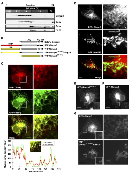
The first test was to determine with which organelles endogenous Gimap3 would sediment on a Histodenz gradi-ent. A postnuclear spleen lysate was separated in a stepwise
Figure 1 The organization of the
Gimap3 gene in mice. (A) A graphical illustration of theGimap3gene, mRNA, and protein derived from BALB/c and CAST/Ei alleles. Red indicates the main coding sequence for Gimap3, while pur-ple indicates the sequence of the uORF. CC, coiled coil; TM, transmembrane; AIG, Gimap family domain. (B) Alignment of the predicted starter methionine from
discontinuous Histodenz gradient to determine the sedi-mentation profile of Gimap3. Fractions were collected and used for immunoblotting using polyclonal antisera against Gimap3 (Figure S1). The sedimentation profile for
endoge-nous Gimap3 was similar to the ER chaperone Calnexin but not to mitochondrial proteins in the outer mitochondrial membrane (Porin) and the matrix (Sdha) (Figure 2A).
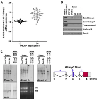
Unfortunately, our polyclonal antibody against Gimap3 was ineffective for indirect immunofluorescence and thus unable to be used for determining intracellular localization by light microscopy. Therefore, we generated a chimeric Gimap3 with RFP fused at the N terminus of the protein to circumvent this problem (Figure 2B). This RFP construct should not affect the membrane targeting of the protein since tail-anchored proteins require only the C-terminal transmem-brane domain andflanking residues for membrane insertion and retention (Rapaport 2003). Stable retroviral transduc-tion or transient expression of this Gimap3 construct consis-tently localized to the ER and not mitochondria (Figure 2, C and D). Only the 39 amino acids at the C terminus were required for this ER localization (Figure 2, E and F). Further-more, replacing the last 32 C-terminal amino acids with a tail-anchor sequence from Omp25, abonafideouter mito-chondrial membrane protein (Nemoto and De Camilli 1999) redirects Gimap3 to the outer mitochondrial membrane (Fig-ure 2G). Collectively, these data suggest that Gimap3 local-izes to the ER.
The expression level of Gimap3 modulates mtDNA segregation
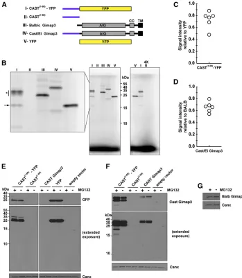
To investigate how allelic expression ofGimap3modulates mtDNA segregation in the spleen, we first tested whether there were quantitative differences in the expression of these two murine alleles. The mRNA expression was quan-tified of both the CAST/Ei and BALB/c alleles in 3-month-old mice from an F2 (BALB/c3CAST/Ei) cross, which were heterozygous for these twoGimap3 alleles. At 3 months of age most F2 mice display quantitative differences in the mtDNA segregation rate in hematopoietic tissues and are considered positive for segregation (Jokinen et al. 2010). However, there is also a subset of mice in this F2 cross that at 3 months of age can be identified while mtDNA segrega-tion cannot yet be detected in hematopoietic tissues (Jokinen
et al.2010). These mice were considered negative for segre-gation. A semiquantitative RT–PCR approach was used to quantify the expression differences of these two alleles in the F2 mice utilizing a primer set that bind in exons 3 and 5 to amplify across exon 4, which is only 35 nucleotides long with a stretch of poly(A)’s and the only unique feature within the Gimap3mRNA to distinguish between the BALB/c and CAST/Ei alleles. The mRNA expression was quantified against a standard curve generated with equimolar ratios of BALB/c and CAST/Ei RNA. In mice positive for mtDNA selec-tion there was a significantly stronger expression from the BALB Gimap3 allele relative to the CAST Gimap3allele (P,0.001) (Figure 3A).
Next, we investigated whether the difference in Gimap3
mRNA expression resulted in a corresponding effect on the steady-state protein levels. Spleen homogenates were pre-pared from BALB/c, CAST/Ei, and F2 (BALB/c3CAST/Ei) mice heterozygous at theGimap3locus for immunoblotting. Both Gimap3 protein variants can be expressed in cultured cells and detected by immunoblotting using our allele-specific polyclonal antibodies (Figure 3B andFigure S1). To our sur-prise there was no detectable Gimap3 protein (Figure 3B) in spleen lysates from CAST/Ei mice. This is in contrast to the abundant protein expression of the BALB/c Gimap3 (Figure 3B). In F2 (BALB/c 3CAST/Ei) mice we also could not de-tect the CAST Gimap3 protein. Moreover, in these F2 mice the abundance of the BALB Gimap3 protein was reduced50% compared to spleens from BALB/c mice (Figure 3B), suggest-ing a dosage effect on the steady-state protein abundance of Gimap3.
To confirm the expression of the CAST/EiGimap3mRNA, we analyzed total spleen RNA by Northern blotting. Using three independent oligonucleotide probes that hybridize against sequences from exons 1–2, exon 3, and exons 4–5, we detected three transcripts (Figure 3C). All three probes were selected against sequence unique to Gimap3and not found inGimap5(Figure 3C). As a positive control we used total RNA from murine embryonic fibroblasts ectopically expressing the BALB/c Gimap3 cDNA consisting only of exons 4 and 5, which can be recognized only by probe c (Figure 3C). Probe c bound weakly to the CAST/Ei mRNA, which was not surprising, considering half of the probe rec-ognizes nucleotides in exon 4, a region missing from the CAST/Ei mRNA. Assessing Gimap3expression using probe b, which hybridizes to exon 3, showed robust expression of both the BALB/c and CAST/Ei alleles in the spleen. These results confirm that although both Gimap3 alleles were expressed at the mRNA level, only the BALB/c variant could be detected in mouse spleens as a mature protein by immunoblotting.
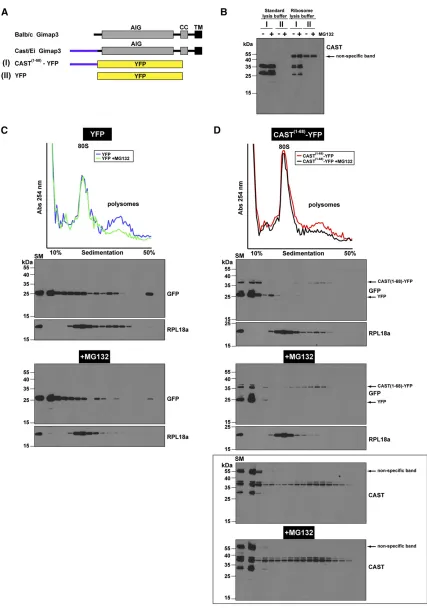
The N terminus of CAST Gimap3 impairs protein synthesis
The inability to detect the CAST Gimap3 proteinin vivomay be due to repressed synthesis of the polypeptide chain or in post-translational processing, such as cleavage of the N ter-minus and differential turnover. We pursued the hypothesis that protein synthesis of the CASTGimap3was impaired be-cause this allele has incorporated a uORF into the coding sequence for this protein. The presence of a uORF within a gene can act as a potentin cispost-transcriptional regulator of the main downstream ORF whereby protein synthesis of the uORF stalls ribosomes repressing translation of the down-stream ORF (Morris and Geballe 2000; Calvoet al.2009).
polypeptides represents the only unique amino acids at the N terminus between the BALB and CAST Gimap3 (Jokinen
et al.2010). The objective was to express the constructs both
in vitroand in cultured cells to test if there was difficulty in
the synthesis of this polypeptide sequence and whether there was any post-translation regulation in the cell to mod-ulate the steady-state protein abundance. We used a T7-pro-moter-coupled reticulocyte lysate assay with radiolabeled [35S]methionine and cysteine to express these proteins in vitro. The linearity for the 35S-radiolabel incorporation
was established using YFP as a template cloned into the same vector backbone (Figure S2). Hereafter, the same con-ditions were used for expressing all constructs off the T7 promoterin vitro. Strikingly there was no detectable expres-sion of the CAST1–68fragment alone (Figure 4B). Moreover,
fusion of this CAST1–68fragment to YFP reduced thein vitro
expression of the full-length protein compared to YFP (Fig-ure 4, B and C). A prominent feat(Fig-ure with the synthesis of CAST1–68–YFP was consistent smearing of the mature
pro-tein (Figure 4B), which may be suggestive of stalled synthe-sis. In addition, there were smaller fragments generated with the CAST1–68–YFP template, one of which comigrated
with the full-length YFP (Figure 4B). Next we assessed the
in vitrosynthesis of full-length CASTvs.BALB Gimap3 (Fig-ure 4, A and B). There was a reduction in the synthesis of the CAST Gimap3 compared to the BALB allele using the same T7-expression-coupled assay (Figure 4D). These data
suggest that the N terminus of CAST Gimap3 exerts a nega-tive effect on protein synthesisin vitro.
To evaluate the role of the uORF on protein synthesis in cultured cells we transiently transfected the plasmids into HEK293 cells followed by immunoblotting 24 hr post-trans-fection. For the CAST1–68–YFP construct, most of the
immu-nodetectable protein with an anti-GFP antibody comigrated through SDS–PAGE with full-length GFP and not near the expected molecular size of 35 kD as seenin vitro(Figure 4, B and E). Extended exposure with the anti-GFP antibody could detect a weaker band with the approximate size of the mature CAST1–68–YFP (Figure 4E), showing that this
pro-tein can be synthesizedin vivo. In contrast, using the CAST-specific polyclonal antibody we detected two prominent bands approximately of equal intensity for CAST1–68–YFP that
were more abundant than the steady-state level of full-length CAST Gimap3 (Figure 4F). There also was no detectable pro-tein for the CAST1–68, a finding consistent with thein vitro
expression (Figure 4, B and F). Since the abundance of the full-length CAST–YFP differs between in vitro and in vivo, synthesis suggests that addtional factors are important for the steady-state level of the protein.
Stalled protein synthesis on ribosomes generates a stress to the cell, which typically activates mRNA surveillance pathways (Lykke-Andersen and Bennett 2014). As part of this stress response, polypeptide chains on stalled ribosomes are rapidly degraded by the proteasome (Lykke-Andersen
Figure 3 Gimap3expression affects mtDNA segrega-tion in mouse spleen. (A) The expression level of the BALB/cGimap3mRNA relative to CAST/EiGimap3by semiquantitative RT–PCR in F2 (BALB/c3 CAST/Ei) mice heterozygous at theGimap3locus. Mice were either positive (+) (n= 51) or negative (2) (n= 19) for mtDNA segregation. Mann–Whitney Test,P, 0.001. (B) Immunoblotting of total cell lysates from the spleen of BALB/c, CAST/Ei or F2 (BALB/c 3 CAST/Ei) mice. A NIH3T3 cell line was transduced with retroviruses expressing HA-tagged version of both the BALB/c Gimap3 and CAST/Ei Gimap3
and Bennett 2014) and thus stabilized only in the presence of proteasomal inhibitors, such as MG132. To test for evi-dence of the proteasome in modulating the abundance of the uORF we incubated cells with MG132. In cells express-ing the CAST1–68–YFP cDNA we observed
smaller-molecular-weight fragments when treating with MG132 and using the CAST-specific antibody, but not the monoclonal anti-GFP antibody that detects only the mature protein (Figure 4, E and F). We also observed a shift with MG132 in the pro-portion of the two immunodetectable fragments expressed by CAST Gimap3 (Figure 4F). In contrast, MG132 had no effect on the steady-state abundance of Gimap3 (Figure 4G). Since the CAST-specific antibody recognizes a stretch of 15 amino acid residues found only in the N terminus of the protein (Figure S1), the immunodetection of stable smaller-molecular-weight fragments when the proteasomal function is blocked may be attributable to the partial synthesis of the CAST1–68–
YFP because of ribosome stalling.
Next, we wanted to confirm this interpretation by testing whether the synthesis of CAST1–68–YFP on polysomes was
affected following the treatment of cells with MG132. Iso-kinetic sucrose density gradients were used to separate ribo-somes and polyribo-somes from treated cells (Figure 5). We found an increase in the CAST1–68–YFP on polysomes
fol-lowing MG132 treatment (Figure 5) suggesting that the uORF fused to YFP leads to proteasomal turnover. Moreover, this analysis demonstrates that the predominant protein generated from the CAST1–68–YFP cDNA comigrates with
the molecular size of YFP when we used the GFP anti-body for immunoblotting (Figure 5, C and D). This is con-sistent with our previousfindings (Figure 4E) and indicates the potential for ribosomes scanning through the uORF se-quence to initiate translation on the AUG of YFP. Using the CAST antibody we also detected another truncated protein that was.25 kD (Figure 5D, bottom) and must contain an N terminus with the CAST polypeptide sequence. Collec-tively, these data suggest that the uORF inGimap3mediates a negative effect on protein synthesis, possibly by stalling synthesis on ribosomes, and may account for the decreased abundance of CAST Gimap3 in mouse spleen.
Figure 4 The N terminus of CAST Gimap3 impairs protein synthesis. (A) A graphical il-lustration of the Gimap3 constructs used. AIG, Gimap family domain; CC, coiled coil; TM, transmembrane domain. (B) SDS–PAGE ofin vitro synthesis of proteins from a T7 promoter with [35S]methionine and cysteine. Lanes numbered according to the constructs in A. (*) Consistent smearing of labeled pro-tein. Arrow indicates labeled protein from CAST(1–68)–YFP that comigrates with full-length YFP (lane V). Right: Four times excess sample was loaded for construct II. (C) Quan-tification of CAST1–68–YFP in vitro produc-tion relative to YFP from six independent experiments. For the quantification, the smeared signal for CAST(1–68)–YFP (*in B) was included along with the mature protein. (D) Quantification of CAST Gimap3in vitro
production relative to BALB Gimap3 from six independent experiments. (E and F) Immu-noblotting of total cell lysates from HEK293 cells transfected with the indicated cDNA constructs and treated with the indicated antibodies. Cells were treated with or with-out MG132 7 hr prior to collection. (G) Im-munoblotting of total cell lysates from HEK293 cells transfected with a Gimap3
Gimap3 and Gimap5 are important for mtDNA segregation
Our original cloning ofGimap3as a modifier of mtDNA
segre-gation (Jokinenet al.2010) was unable to determine whether other Gimap family members, in particular the paralogue
Gimap5, may also contribute to this mtDNA phenotype. Recent evidence points to a cooperative genetic function between
Gimap3andGimap5in lymphocyte development (Yanoet al.
2014). To test Gimap5for a role in mtDNA segregation, we took advantage of a germline knockout mouseGimap5tm1Wlr
(Schulteiset al.2008). HomozygousGimap5tm1Wlrmice exhibit
a severe hematopoietic stem cell defect, which leads to a re-duced number of mature lymphocytes and lack of detectable NK cells (Schulteiset al.2008). InGimap5tm1Wlr/WTthere are
no reported defects in hematopoiesis or on the mRNA expres-sion of otherGimapfamily members in the thymus; these mice were described only as having a 50% reduction in Gimap5
expression (Schulteis et al. 2008). This dosage effect on
Gimap5expression in heterozygotes has been consistently observed with two other independent murine Gimap5 null mutations at both the mRNA and protein level in Barneset al.
(2010) and Yanoet al. (2014), suggesting a tight correlation betweenGimap5gene expression and protein abundance.
To test whether changes in the expression ofGimap5affect mtDNA segregation, we crossed our heteroplasmic mouse model (NZB and BALB mtDNA haplotypes) toGimap5tm1Wlr.
Since both mouse lines were already established on the BALB/c nuclear background, no further backcrossing was re-quired. In the spleen of homozygousGimap5tm1Wlrmice there
are severe cellular and morphological abnormalities: a nine-fold reduction in splenic T cells and a twonine-fold reduction in total splenic cellularity (Schulteis et al.2008). These patho-logical features in the spleens ofGimap5tm1Wlrhomozygote
mice complicate the analysis of mtDNA segregation because the cellular composition of the organ is dramatically altered, which in turn affectsGimap3expression in this tissue (Figure 6A and Figure S3). Therefore, we focused our mtDNA segre-gation analysis on Gimap5tm1Wlr/WT because these mice are
phenotypically indistinguishable from wild type and display no signs of pathology or cellularity defects in the spleen, but have a 50% decrease in Gimap5 abundance.
In the hematopoietic compartment of wild-type heteroplas-mic heteroplas-mice mtDNA segregation can be modeled as an exponential decay, so that by 100 days of post-natal life there is an50% decrease in the proportion of NZB mtDNA in leukocytes of both myeloid and lymphoid lineages (Battersbyet al.2005). We found a significant difference in mtDNA segregation of
Gimap5tm1Wlr/WTin both the spleen and bone marrow compared
to wild-type littermate controls (Figure 6B). The effect is more modest in the spleen compared to the bone marrow (Figure 6B) because within the spleen the supporting stromal cells are neu-tral with respect to mtDNA segregation (Battersbyet al.2005).
Since we found that changes in Gimap3 abundance were accompanied with an effect on mtDNA segregation, we wanted to test whether in Gimap5tm1Wlr/WT there was any
effect on the abundance of Gimap3. Total cell lysates were prepared from isolated splenocytes for immunoblotting demonstrating a 50% reduction in Gimap3 protein abun-dance compared to wild type (Figure 6C) even though there was no difference in the expression of the Gimap3 mRNA (Figure 6A). Thesefindings provide further support for a co-operative function between Gimap3 and Gimap5 and indi-cate that the steady-state abundance of these two proteins is an important factor for the segregation of mtDNA haplo-types in mouse leukocytes.
A 50% reduction of Gimap3 and Gimap5 protein levels was sufficient to significantly modulate mtDNA segregation in leukocytes and suggests that the identification of other factors dependent upon Gimap5 may provide insight into the molecular connection to the segregation of the mito-chondrial genome. To identify such factors we used a quan-titative proteomic approach with iTRAQ (isobaric tags for relative and absolute quantification) labeling followed by LC-MS/MS analysis of postnuclear membrane enrichments generated from spleens of wild type andGimap5tm1Wlr
het-erozygote littermates (File S1 and File S2)(Lietzen et al.
2011). Three independent biological replicates for each ge-notype were used for the iTRAQ analysis, which led to the identification of.1100 distinct proteins (File S1). We were unable to quantify Gimap3 and Gimap5 using this approach with any confidence because the sequence homology between these two proteins is very high (84% identity) and the num-ber of unique peptide sequences detected by LC-MS/MS was inadequate for confident identification and iTRAQ quantifica-tion. In Gimap5tm1Wlr/WTwe detected robust changes in the
abundance of five proteins that all have a connection to the regulation of the cytoskeleton and cell motility via small GTPase signaling (Table1). The abundance of these proteins was also further affected inGimap5tm1Wlrhomozygotes (File S2). Since Gimap5 is a membrane-anchored lysosomal pro-tein, this points to a quantitative effect ofGimap5expression on the interaction between organelles and the cytoskeleton during cell motility as an important factor in mtDNA segre-gation of hematopoietic cell lineages in mice.
Discussion
The mechanisms governing the segregation of mitochondrial genomes are still poorly understood despite considerable efforts since the discovery of pathogenic mtDNA mutations (Jokinen and Battersby 2013). We followed up our cloning of Gimap3 as a modifier of mtDNA segregation in mice to
determine how allelic variants of this gene influence the phenotype. The most important finding from this study is
that expression differences inGimap3andGimap5appear to be a critical factor to the segregation of the mitochondrial genome in mouse hematopoietic tissues.
Regulation ofGimap3expression is complex and possibly involves cell-specific modulators. The incorporation of a uORF into the coding sequence of theM. m. castaneus Gimap3has a negative effect on protein synthesis potentially through translation repression. Our data are consistent with a variable penetrance of the uORF on the CAST Gimap3, as the expres-sion and stability of the mature protein decreases steadily from in vitrosynthesis to ectopic expression in nonhemato-poietic cell types to the undetectable levelsin vivoin mouse leukocytes. This is in contrast to the BALB Gimap3 that can be robustly detected in the mouse. Since our polyclonal antibod-ies do not recognize both protein alleles, we cannot exclude the possibility of N-terminal processing of CAST Gimap3 in mice. UORFs have been described as translation regulatory sequences and are estimated to be present in.40% of human and mouse transcripts (Calvoet al.2009). The mechanism of action for uORFs often involves repressing translation of the downstream (main) open reading frame (Morris and Geballe 2000; Calvoet al.2009). Such a regulatory mechanism allows for rapid changes in the protein abundance upon stress con-ditions, whentransfactors can release the uORF repression of
translation (Barbosaet al.2013). Our data also indicate that ribosomes may scan through this uORF sequence and initiate translation on a downstream AUG even though there is stron-ger sequence conservation to the Kozak consensus in the uORF. Further work is required to elucidate the regulatory role of the uORF on the expression ofGimap3alleles in mice. We show that Gimap3 localizes to the ER and not mitochondria as previously reported. How then could a resi-dent protein of the ER affect mtDNA segregation? Interac-tions between the ER and mitochondria play a key role in many aspects of mitochondrial homeostasis, such as mor-phology of the mitochondrial reticulum (division and tether-ing of the organelle), calcium handltether-ing, lipid transfer, and transmission of the mitochondrial genome among others (de Brito and Scorrano 2008; Kornmann et al. 2009; Friedman
et al.2011; Hoppinset al.2011; Connerthet al.2012; Rowland and Voeltz 2012; Korobovaet al.2013; Hajnoczkyet al.2014). Any number of these functions could be important to mtDNA segregation.
Why the need to restrict the expression of these Gimap protein scaffolds to the hematopoietic compartment? Leu-kocytes have a relatively small cytosolic volume with few organelles by comparison to most cell types and are one of the most dynamic cell types in mammals as they migrate
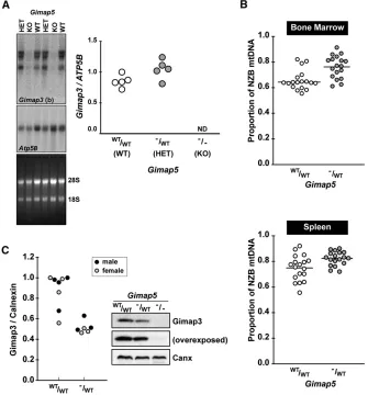
Figure 6 Genetic loss ofGimap5affects Gimap3 and mtDNA segregation. (A) Northern blot of spleen total RNA from wild-type Gimap5,
Gimap5tm1Wlr/WT, and homozygoteGimap5tm1Wlr
mice hybridized withGimap3(b) probe and con-trol probeAtp5B. Right: Quantification ofGimap3
mRNA compared toAtp5B. (B) Decreased selec-tion against the NZB mtDNA genome in the bone marrow and spleen ofGimap5tm1Wlr/WTcompared
to wild-type littermate controls.n= 18 for each group; (*)P,0.01 two-tailedt-test; means are indicated by the line through the scatter plot. Mice were 70 days of postnatal age at the time of sampling. (C) A representative immunoblotting of Gimap3 from whole-cell lysates from isolated splenocytes of wild type,Gimap5tm1Wlr/WTand
ho-mozygoteGimap5tm1Wlrmice. Left: Quantification
throughout the body by chemotaxis. Chemotaxis and re-ceptor signaling at the immunological synapse both require dynamic and coordinated reorganization of organelle dis-tribution, especially mitochondria (Campello et al. 2006; Quintanaet al.2007). During chemotaxis, leukocytes become polarized with mitochondria relocalized to the uropod. Dis-rupting this mitochondrial relocalization impairs the ability of a leukocyte to migrate (Campello et al.2006; Quintana
et al.2007). Recent reports from mammalian systems sug-gest that interactions with the actin cytoskeleton are needed for mitochondrial division (Korobova et al. 2013) and dy-namics during formation of the immunological synapse (Schwindling et al. 2010). The stoichiometric changes in
Gimap5tm1Wlrheterozygotes of factors interacting with the
actin cytoskeleton could suggest that these Gimap scaffolds are important for the dynamic organization and coordina-tion of organelle movement during chemotaxis. Consistent with this interpretation is the recent work on Gimap4 show-ing that this soluble member of the Gimap family mediates an interaction between the trans Golgi network and the actin cytoskeleton to sustain for interferon gamma secretion (Heinonenet al.2014). Since Gimap3 and Gimap5 reside in the membrane of different organelles, it is tempting to hy-pothesize that these proteins could help mediate organelle distribution, organization, and contact within the cell, which in turn would affect segregation of the mitochondrial ge-nome in leukocyte lineages.
The absence ofGimap3in most current mammalian genome annotations could indicate that the gene function has been lost or taken over byGimap5, or could be due to poor resolution of current genome annotations because of the high sequence sim-ilarity betweenGimap3andGimap5. Therefore the overall im-portance ofGimap3across mammals, let alone vertebrates, is at present difficult to conclude, as it might be restricted to mice. However, analysis of the C-terminal protein sequence of Gimap5 across mammalian taxa suggests the evolution ofGimap5in Rodentia differs considerably from other mammalian clades. Clearly, further research is required to resolve this issue.
The regulation of mtDNA segregation in metazoans remains a complex genetic problem. In the majority of cases, selection for specific mtDNA haplotypes in mammals is not based upon detectable differences in mitochondrial oxidative phosphorylation or on the replication rate of the genome (Jokinen and Battersby 2013). Thus, in mammals the identity of regulatory mechanisms that coordinate the segre-gation of the mitochondrial genome remain intractable de-spite considerable effort by thefield (Jokinen and Battersby
2013). The forward genetic approaches we have taken have provided a degree of insight into the nuclear control of mtDNA segregation. Nonetheless, progress remains slow as the problem can be investigated only in mice because cul-tured cells cannot faithfully replicate the mtDNA segregation patterns observed in mammals (Jokinen and Battersby 2013). In the end, selection for a particular mtDNA haplotype within a specific tissue can occur only at three levels: the cell, organ-elle, or genome. Two fundamental components are required for such a mechanism to work: signal generation that distin-guishes mtDNA variants or mutations and the molecular ma-chinery to act on or interpret those signals. Such a signal can be generated only at the sequence level, nucleic acid and or polypeptide, or by the functional consequences derived from those sequences. There appears to be tissue-specific differen-ces in either the generation or interpretation of these signals in our mouse model. Based upon our findings, the segrega-tion of mtDNA haplotypes in the mouse hematopoietic com-partment may be dependent upon the regulation of an intrinsic property of these cell types to coordinate organelle interactions as they migrate throughout the body.
Acknowledgments
We thank Uwe Richter, Anu Wartiovaara, and Howy Jacobs for discussion. We thank the Biomedicum Imaging Unit (BIU) for the confocal facilities. The Academy of Finland Center of Excellence—FinMIT, the University of Helsinki, and the Jane and Aatos Erkko Foundation support research in the BJB lab-oratory. R.J. was supported by Helsinki Biomedical Graduate Program, Emil Aaltonen Foundation, Biomedicum-Helsinki Foundation, the Waldemar von Frenckell Foundation, and the Finnish Cultural Foundation. H.W. was supported by Na-tional Institute of Allergy and Infectious Diseases AI42380. T.A.N.’s laboratory is supported by the Academy of Finland (135628 and 140950) and the University of Helsinki. A.J.K. and J.K.H. were supported by grants from the Academy of Finland and the Sigrid Juselius Foundation. The authors de-clare no conflict of interest.
Literature Cited
Barbosa, C., I. Peixeiro, and L. Romao, 2013 Gene expression regulation by upstream open reading frames and human dis-ease. PLoS Genet. 9: e1003529.
Barnes, M. J., H. Aksoylar, P. Krebs, T. Bourdeau, C. N. Arnoldet al., 2010 Loss of T cell and B cell quiescence precedes the onset of Table 1 Proteins whose abundance in mouse splenocytes is dependent upon Gimap5: iTRAQ analysis comparing Gimap5WT/WT to
Gimap5tm1Wlr/WTlittermates
Protein Fold change het/wt Biological process
Cyfip2 0.55 Component of WAVE complex for assembly of F-actin complex
Arhgdia 0.57 Rho protein signal transduction, regulation of protein localization
Vasp 0.57 Actin cytoskeleton reorganization
Elmo1 1.83 Actinfilament-based process, phagocytosis
microbialflora-dependent wasting disease and intestinal infl am-mation in Gimap5-deficient mice. J. Immunol. 184: 3743–3754. Battersby, B. J., and E. A. Shoubridge, 2001 Selection of a mtDNA sequence variant in hepatocytes of heteroplasmic mice is not due to differences in respiratory chain function or efficiency of replication. Hum. Mol. Genet. 10: 2469–2479.
Battersby, B. J., J. C. Loredo-Osti, and E. A. Shoubridge, 2003 Nuclear genetic control of mitochondrial DNA segregation. Nat. Genet. 33: 183–186.
Battersby, B. J., M. E. Redpath, and E. A. Shoubridge, 2005 Mitochondrial DNA segregation in hematopoietic line-ages does not depend on MHC presentation of mitochondrially encoded peptides. Hum. Mol. Genet. 14: 2587–2594.
Boulet, L., G. Karpati, and E. A. Shoubridge, 1992 Distribution and threshold expression of the tRNA(Lys) mutation in skeletal muscle of patients with myoclonic epilepsy and ragged-red fi -bers (MERRF). Am. J. Hum. Genet. 51: 1187–1200.
Burgstaller, J. P., I. G. Johnston, N. S. Jones, J. Albrechtova, T. Kolbeet al., 2014 MtDNA segregation in heteroplasmic tissues is common in vivo and modulated by haplotype differences and developmental stage. Cell Rep. 7: 2031–2041.
Calvo, S. E., D. J. Pagliarini, and V. K. Mootha, 2009 Upstream open reading frames cause widespread reduction of protein ex-pression and are polymorphic among humans. Proc. Natl. Acad. Sci. USA 106: 7507–7512.
Campello, S., R. A. Lacalle, M. Bettella, S. Manes, L. Scorranoet al., 2006 Orchestration of lymphocyte chemotaxis by mitochon-drial dynamics. J. Exp. Med. 203: 2879–2886.
Chen, Y., M. Yu, X. Dai, M. Zogg, R. Wenet al., 2011 Critical role for Gimap5 in the survival of mouse hematopoietic stem and progenitor cells. J. Exp. Med. 208: 923–935.
Chinnery, P. F., and D. C. Samuels, 1999 Relaxed replication of mtDNA: a model with implications for the expression of disease. Am. J. Hum. Genet. 64: 1158–1165.
Chinnery, P. F., N. Howell, R. N. Lightowlers, and D. M. Turnbull, 1997 Molecular pathology of MELAS and MERRF: the rela-tionship between mutation load and clinical phenotypes. Brain 120(10): 1713–1721.
Chinnery, P. F., P. J. Zwijnenburg, M. Walker, N. Howell, R. W. Taylor et al., 1999 Nonrandom tissue distribution of mutant mtDNA. Am. J. Med. Genet. 85: 498–501.
Connerth, M., T. Tatsuta, M. Haag, T. Klecker, B. Westermannet al., 2012 Intramitochondrial transport of phosphatidic acid in yeast by a lipid transfer protein. Science 338: 815–818. Daheron, L., T. Zenz, L. D. Siracusa, C. Brenner, and B. Calabretta,
2001 Molecular cloning of Ian4: a BCR/ABL-induced gene that encodes an outer membrane mitochondrial protein with GTP-binding activity. Nucleic Acids Res. 29: 1308–1316. de Brito, O. M., and L. Scorrano, 2008 Mitofusin 2 tethers
endo-plasmic reticulum to mitochondria. Nature 456: 605–610. Dunbar, D. R., P. A. Moonie, H. T. Jacobs, and I. J. Holt,
1995 Different cellular backgrounds confer a marked advan-tage to either mutant or wild-type mitochondrial genomes. Proc. Natl. Acad. Sci. USA 92: 6562–6566.
Elias, J. E., and S. P. Gygi, 2007 Target-decoy search strategy for increased confidence in large-scale protein identifications by mass spectrometry. Nat. Methods 4: 207–214.
Friedman, J. R., L. L. Lackner, M. West, J. R. DiBenedetto, J. Nunnari
et al., 2011 ER tubules mark sites of mitochondrial division.
Science 334: 358–362.
Fu, K., R. Hartlen, T. Johns, A. Genge, G. Karpatiet al., 1996 A novel heteroplasmic tRNAleu(CUN) mtDNA point mutation in a sporadic patient with mitochondrial encephalomyopathy seg-regates rapidly in skeletal muscle and suggests an approach to therapy. Hum. Mol. Genet. 5: 1835–1840.
Hagstrom, E., C. Freyer, B. J. Battersby, J. B. Stewart, and N. G. Larsson, 2014 No recombination of mtDNA after heteroplasmy
for 50 generations in the mouse maternal germline. Nucleic Acids Res. 42: 1111–1116.
Hajnoczky, G., D. Booth, G. Csordas, V. Debattisti, T. Golenaret al., 2014 Reliance of ER-mitochondrial calcium signaling on mito-chondrial EF-hand Ca binding proteins: miros, MICUs, LETM1 and solute carriers. Curr. Opin. Cell Biol. 29C: 133–141. Heinonen, M. T., K. Kanduri, H. J. Lahdesmaki, R. Lahesmaa, and
T. A. Henttinen, 2015 Tubulin- and actin-associating GIMAP4 is required for IFN-gamma secretion during Th cell differentia-tion. Immunol. Cell Biol. 93: 158–166.
Hoppins, S., S. R. Collins, A. Cassidy-Stone, E. Hummel, R. M. Devayet al., 2011 A mitochondrial-focused genetic interaction map reveals a scaffold-like complex required for inner mem-brane organization in mitochondria. J. Cell Biol. 195: 323–340. Hornum, L., J. Romer, and H. Markholst, 2002 The diabetes-prone BB rat carries a frameshift mutation in Ian4, a positional candidate of Iddm1. Diabetes 51: 1972–1979.
Jenuth, J. P., A. C. Peterson, K. Fu, and E. A. Shoubridge, 1996 Random genetic drift in the female germline explains the rapid segregation of mammalian mitochondrial DNA. Nat. Genet. 14: 146–151.
Jenuth, J. P., A. C. Peterson, and E. A. Shoubridge, 1997 Tissue-specific selection for different mtDNA genotypes in heteroplas-mic heteroplas-mice. Nat. Genet. 16: 93–95.
Jokinen, R., and B. J. Battersby, 2013 Insight into mammalian mitochondrial DNA segregation. Ann. Med. 45: 149–155. Jokinen, R., P. Marttinen, H. K. Sandell, T. Manninen, H. Teerenhovi
et al., 2010 Gimap3 regulates tissue-specific mitochondrial DNA
segregation. PLoS Genet. 6: e1001161.
Kawakami, Y., R. Sakuta, K. Hashimoto, O. Fujino, T. Fujitaet al., 1994 Mitochondrial myopathy with progressive decrease in mitochondrial tRNA(Leu)(UUR) mutant genomes. Ann. Neurol. 35: 370–373.
Kornmann, B., E. Currie, S. R. Collins, M. Schuldiner, J. Nunnari
et al., 2009 An ER-mitochondria tethering complex revealed
by a synthetic biology screen. Science 325: 477–481.
Korobova, F., V. Ramabhadran, and H. N. Higgs, 2013 An actin-dependent step in mitochondrial fission mediated by the ER-associated formin INF2. Science 339: 464–467.
Krucken, J., R. M. Schroetel, I. U. Muller, N. Saidani, P. Marinovski
et al., 2004 Comparative analysis of the human gimap gene
cluster encoding a novel GTPase family. Gene 341: 291–304. Larsson, N. G., E. Holme, B. Kristiansson, A. Oldfors, and M. Tulinius,
1990 Progressive increase of the mutated mitochondrial DNA fraction in Kearns–Sayre syndrome. Pediatr. Res. 28: 131–136. Lietzen, N., T. Ohman, J. Rintahaka, I. Julkunen, T. Aittokallio
et al., 2011 Quantitative subcellular proteome and secretome
profiling of influenza A virus-infected human primary macro-phages. PLoS Pathog. 7: e1001340.
Lykke-Andersen, J., and E. J. Bennett, 2014 Protecting the pro-teome: Eukaryotic cotranslational quality control pathways. J. Cell Biol. 204: 467–476.
MacMurray, A. J., D. H. Moralejo, A. E. Kwitek, E. A. Rutledge, B. Van Yserlooet al., 2002 Lymphopenia in the BB rat model of type 1 diabetes is due to a mutation in a novel immune-associated nucleotide (Ian)-related gene. Genome Res. 12: 1029–1039.
Morris, D. R., and A. P. Geballe, 2000 Upstream open reading frames as regulators of mRNA translation. Mol. Cell. Biol. 20: 8635–8642.
Nemoto, Y., and P. De Camilli, 1999 Recruitment of an alterna-tively spliced form of synaptojanin 2 to mitochondria by the interaction with the PDZ domain of a mitochondrial outer mem-brane protein. EMBO J. 18: 2991–3006.
Nitta, T., M. Nasreen, T. Seike, A. Goji, I. Ohigashiet al., 2006 IAN family critically regulates survival and development of T lympho-cytes. PLoS Biol. 4: e103.
Okado-Matsumoto, A., and I. Fridovich, 2001 Subcellular distribution of superoxide dismutases (SOD) in rat liver: Cu,Zn-SOD in mitochondria. J. Biol. Chem. 276: 38388– 38393.
Quintana, A., C. Schwindling, A. S. Wenning, U. Becherer, J. Rettig
et al., 2007 T cell activation requires mitochondrial
transloca-tion to the immunological synapse. Proc. Natl. Acad. Sci. USA 104: 14418–14423.
Rapaport, D., 2003 Finding the right organelle: targeting signals in mitochondrial outer-membrane proteins. EMBO Rep. 4: 948– 952.
Rowland, A. A., and G. K. Voeltz, 2012 Endoplasmic reticulum-mitochondria contacts: function of the junction. Nat. Rev. Mol. Cell Biol. 13: 607–625.
Schulteis, R. D., H. Chu, X. Dai, Y. Chen, B. Edwards et al., 2008 Impaired survival of peripheral T cells, disrupted NK/ NKT cell development, and liver failure in mice lacking Gimap5. Blood 112: 4905–4914.
Schwefel, D., C. Frohlich, J. Eichhorst, B. Wiesner, J. Behlkeet al., 2010 Structural basis of oligomerization in septin-like GTPase of immunity-associated protein 2 (GIMAP2). Proc. Natl. Acad. Sci. USA 107: 20299–20304.
Schwefel, D., B. S. Arasu, S. F. Marino, B. Lamprecht, K. Kochert
et al., 2013 Structural insights into the mechanism of GTPase
activation in the GIMAP family. Structure 21: 550–559. Schwindling, C., A. Quintana, E. Krause, and M. Hoth,
2010 Mitochondria positioning controls local calcium influx in T cells. J. Immunol. 184: 184–190.
Shilov, I. V., S. L. Seymour, A. A. Patel, A. Loboda, W. H. Tanget al., 2007 The Paragon Algorithm, a next generation search engine that uses sequence temperature values and feature probabilities to identify peptides from tandem mass spectra. Mol. Cell. Pro-teomics 6: 1638–1655.
Wai, T., D. Teoli, and E. A. Shoubridge, 2008 The mitochondrial DNA genetic bottleneck results from replication of a subpopula-tion of genomes. Nat. Genet. 40: 1484–1488.
Weber, K., J. N. Wilson, L. Taylor, E. Brierley, M. A. Johnson et al., 1997 A new mtDNA mutation showing accumulation with time and restriction to skeletal muscle. Am. J. Hum. Genet. 60: 373–380. Wong, V.W.Y., A. E. Saunders, A. Hutchings, J. C. Pascall, C. Carter
et al., 2010 The autoimmunity-related GIMAP5 GTPase is
a lysosome-associated protein. Self Nonself 1: 259–268. Yano, K., C. Carter, N. Yoshida, T. Abe, A. Yamada et al.,
2014 Gimap3 and Gimap5 cooperate to maintain T-cell num-bers in the mouse. Eur. J. Immunol. 44: 561–572.
GENETICS
Supporting Information http://www.genetics.org/lookup/suppl/doi:10.1534/genetics.115.175596/-/DC1
Quantitative Changes in
Gimap3
and
Gimap5
Expression Modify Mitochondrial DNA Segregation
in Mice
Riikka Jokinen, Taina Lahtinen, Paula Marttinen, Maarit Myöhänen, Pilvi Ruotsalainen, Nicolas Yeung, Antonina Shvetsova, Alexander J. Kastaniotis, J. Kalervo Hiltunen, Tiina Öhman, Tuula A. Nyman, Hartmut Weiler, and Brendan J. Battersby
Files S1‐S2
Available for download as Excel files at http://www.genetics.org/lookup/suppl/doi:10.1534/genetics.115.175596/‐/DC1
FileS1 Complete LC‐MS/MS data analysis