R E S E A R C H
Open Access
Complete genome analysis of a frog virus 3 (FV3)
isolate and sequence comparison with isolates of
differing levels of virulence
Elizabeth A Morrison
1, Shawn Garner
1,2†, Pierre Echaubard
3†, David Lesbarrères
2, Christopher J Kyle
1and Craig R Brunetti
1*Abstract
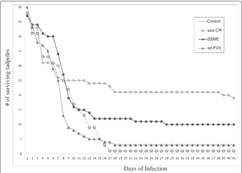
Background:Frog virus 3 (FV3) is the type species of the genusRanavirus, and in the past few decades, FV3 infections have resulted in considerable morbidity and mortality in a range of wild and cultivated amphibian species in the Americas, Europe, and Asia. The reasons for the pathogenicity of FV3 are not well understood. Findings:We investigated three FV3 isolates designated SSME, wt-FV3, and aza-Cr, and reported that our wt-FV3 and aza-Crstrains showed similar levels of virulence, while SSME was the least virulent in anin vivostudy with
Lithiobates pipienstadpoles. Using 454 GS-FLX sequencing technology, we sequenced SSME and compared it to the
published wt-FV3 genome. SSME had multiple amino acid deletions in ORFs 49/50L, 65L, 66L, and 87L, which may explain its reduced virulence. We also investigated repeat regions and found that repeat copy number differed between isolates, with only one group of 3 isolates and 1 pair of isolates being identical at all 3 locations. Conclusions:In this study we have shown that genetic variability is present between closely related FV3 isolates, both in terms of deletions/insertions, and even more so at select repeat locations. These genomic areas with deletions/insertions may represent regions that affect virulence, and therefore require investigation. Furthermore, we have identified repeat regions that may prove useful in future phylogeographical tracking and identification of ranaviral strains across different environmental regions.
Keywords:FV3, 454 GS-FLX technology, Viral isolates, Genetic variation, Virulence
Background
Global amphibian populations have declined consider-ably in recent years, in part due to habitat fragmenta-tion, pollufragmenta-tion, and the chytrid fungusBatrachochytrium dendrobatidis [1,2]. More recently, certain members of the family Iridoviridae have also been associated with amphibian decline. The family Iridoviridaeis comprised of large, cytoplasmic, double stranded DNA viruses with icosahedral capsids [3], and is divided into five genera: Iridovirus,Chloriridovirus,Lymphocystivirus, Megalocyti-virus, and Ranavirus [4]. Specifically linked to amphib-ian mortality in this family are infectious diseases caused by members of the genus Ranavirus. In past research,
ranaviruses received little attention as most infections were deemed subclinical; however, recent ranavirus in-fections have resulted in considerable morbidity and mortality in a range of wild and cultivated amphibian species in the Americas, Europe, and Asia [5-8]. It has been reported that 43% of known amphibian die-offs in the USA from 2000 to 2005 were due to ranaviruses [9], and that from 1996–2001 ranaviruses were isolated from most of the amphibian mortality events in North America [10]. Detection of these outbreaks could be due to better surveillance, increased environmental awareness, the mu-tation of viral species creating highly pathogenic strains, or environmental changes resulting in host immune sup-pression [11]. Ranaviruses have become a significant cause of death and disease in amphibians, and thus investigation into these viruses is warranted from a virological, com-mercial, and ecological standpoint [11].
* Correspondence:[email protected] †Equal contributors
1
Department of Biology, Trent University, 1600 East Bank Dr., Peterborough, Ontario K9J 7B8, Canada
Full list of author information is available at the end of the article
Frog virus 3 (FV3) is the type species of the genus Ranavirus [4]. FV3′s genome is 105,903 base pairs (bp) comprised of 98 open reading frames (ORFs) [12]. De-pending on factors such as strain virulence and host im-mune response, infection with FV3 may or may not lead to mortality. However, in susceptible amphibians, FV3′s necrotic and apoptotic effects cause systemic, chronic cell death in multiple internal organs, resulting in death of the host within a few days to several weeks [2,13,14]. FV3 infection is also marked by cutaneous signs, includ-ing ulceration of the skin, and erythema and swellinclud-ing of the limbs and body. In fatal cases, intracoelomic lesions are often present, including haemorrhages of the kidneys and reproductive organs, and pale, swollen livers [15]. While our understanding of ranavirus pathogenicity has improved over the last decade, there is still a need for the research community to more fully describe the de-terminants of virulence variation. Elucidation of this area will likely heavily rely on the genetic analysis and com-parison of ranaviruses that differ in host range and virulence.
Genetic comparison of related DNA viruses has proven to be an important tool in classifying viral strains and understanding the epidemiology and evolution of differ-ent genotypes [16]. By analyzing genetic differences, re-searchers can link clinically significant alterations with molecular changes, and better understand viral origins and evolution. Increasingly, new viral strains are being identified based on the systematic analysis of sequence data, including short amino acid insertions, transla-tional stop codons, and single amino acid deletions [17]. Analysis of viral isolates has led to the discovery of new viral genotypes, as well as better understanding of the functional genetic differences among strains [17]. For instance, the entire genome of a virulent strain of duck entiritis virus was recently sequenced and com-pared to the genomes of an attenuated strain and an-other virulent strain [18]. The results indicated several nucleotide insertions/deletions and frame-shift mutations effecting ORF initiation or termination [18]. These find-ings allowed the researchers to identify possible virulence factors and provided information on ORFs that are chan-ged during serial passage.
In addition to nucleotide insertions/deletions, variation between viral genomes may occur at repeat regions. Eaton et al. [19] identified repetitive sequences in the ge-nomes of various ranaviruses with high copy number variation. Repetitive sequences are commonly classified into one of three groups: macro, mini, or micro satellites [20]. The repeats that we will examine in this study con-tain less than 400 bp comprised of 9-19 bp repeating units. Thus, we suggest that these repeats are some vari-ation between micro and mini satellites, and for the pur-poses of our analysis, will be referred to as short tandem
repeats (STRs). A selection of these repeat regions will be used to analyze FV3 isolates in order to further inves-tigate the fine scale, genetic differences present in vari-able regions.
Past studies on the genetic variation between DNA viral strains have allowed for the detection of minute genetic changes that would otherwise have gone un-noticed. These kinds of changes have proved useful in explaining phenotypic differences and evolutionary his-tories. The purpose of the present investigation is to nar-row our focus even further by comparing the genomes of closely related FV3 isolates, including those with vary-ing levels of virulence. This will be done in an attempt to explore the genetic diversity present in strains of FV3, with the ultimate goal of further elucidating the possible genetic basis behind FV3′s unpredictable infectious behaviour.
Results
Genomic sequencing of SSME
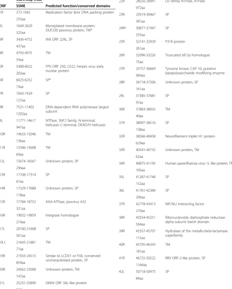
In order to better understand the possible genetic basis of phenotypic variation among strains, we sequenced the SSME strain using 454 GS-FLX technology. Our se-quenced genome showed high similarity to the published FV3 sequence (wt-FV3) [12], with a sequence identity of 98.79% and average genome sequencing coverage of 51x. Results revealed that SSME differed from the wt-FV3 genome length of 105,903 bps and ORF number of 98; instead, SSME had a length of 105,070 bps, and a total of 95 predicted ORFs (Table 1). Despite high overall se-quence identity between SSME and FV3, marked differ-ences were noted in several regions of the genome (Table 2).
Gene variation between ranaviruses
In order to investigate the possible genetic variation within coding regions and regions of high variability be-tween closely related viral isolates, we compared our se-quenced SSME genome with four published ranavirus
genomes. The genomes compared included: SSME, wt-FV3 [17], rana grylio iridovirus (RGV) [23], soft-shelled turtle iridovirus (SSTIV) [24], and tiger frog virus (TFV) [25]. We began by focusing our analysis on predicted coding regions, as variation in these areas may have con-sequences in pathogenicity or the kind of disease a virus causes.
Upon analysis of selected ranaviral genomes, a 757 bp deletion was discovered in SSME, deleting the entire 65L coding region and the majority of the 66L coding region (Figure 2). This deletion was not present in wt-FV3 or in related ranaviruses RGV, SSTIV, and TFV. However, se-quence alignments indicated reorganization of 66L in RGV, SSTIV, and TFV, with a 139 bp insertion found in their corresponding 66L regions (Figure 2). Therefore, there are at least 3 different genomic presentations of this region in ranaviral genomes, making it highly variable among isolates.
Another coding region demonstrating variation was 50L. An in frame deletion of 13 amino acids was found
#
o
f surviving t
ad
p
o
le
s
n
o
i
t
c
e
f
n
I
f
o
s
y
a
D
aza-CR Control
SSME
wt-FV3
Table 1 Description of nucleotide start/stop locations and amino acid length of ORFs in SSME, along with
predicted functions
Start/stop (#aa)
ORF SSME Predicted function/conserved domains
1R 272-1042 Replication factor &/or DNA packing protein
255aa
2L 1649-2629 Myristylated membrane protein, DUF230 poxvirus protein, TM* 325aa
3R 3436-4752 IIV6 ORF 229L, SP
437aa
4R 4793-4975 TM
59aa
5R 5408-6022 FPV ORF 250, US22, herpes virus early nuclear protein
203aa
6R 6025-6252 SP*
74aa
7R 7043-7429 SP
127aa
8R 7521-11402 DNA-dependent RNA polymerase largest subunit
1292aa
9L 11771-14617 NTPase, SNF2 family, N-terminal, helicases C-terminal, DEAD/H helicases 947aa
10R 14633-15046 TM
136aa
11R 15396-15608 TM
69aa
12L 15674-16567 Unknown protein, SP
296aa
13R 17108-17314 SP
67aa
14R 17329-17688 Unknown protein, SP
118aa
15R 17784-18752 AAA-ATPase, poxvirus A32
321aa
16R 19032-19859 Integrase homologue
274aa
17L 20100-21608 SP
501aa
18 L 21645-21881 TM
77aa
19R 21933-24515 Similar to LCDV1 or f10L conserved uncharacterized protein, SP 859aa
20R 24562-25008 Unknown protein, TM
147aa
21L 25231-25890 ISKNV ORF 56L-like protein
218aa
Table 1 Description of nucleotide start/stop locations and amino acid length of ORFs in SSME, along with
predicted functions(Continued)
22R 26020-28941 D5 family NTPase, ATPase
972aa
23R 29319-30467 SP
381aa
24Rº 30877-31947 SP
355aa
25R 32141-32929 P31K protein
261aa
26R 32996-33226 Truncated elf-2αhomologue 75aa
27R 33757-36669 Tyrosine kinase, CAP 10, putative lypopolysaccharide modifying enzyme 969aa
28R 36718-37206 Unknown protein, SP
161aa
29L 37385-37681 SP
97aa
30R 37883-38035 TM
49aa
31R 38097-38516 SP
138aa
32R 38566-40458 Neurofilament triplet H1 protein
629aa
33R 40541-40732 Unknown protein, TM
62aa
34R 40875-41195 Human parainfluenza virus 1L like protein, TM
105aa
35L 41287-41748 SP
152aa
36L 41761-42384 SP
206aa
37R 42778-43413 NIF/NLI interacting factor
210aa
38R 43554-45251 Ribonucleoside diphosphate reductase alpha subunit barrel domain 564aa
39R 45357-45707 Hydrolase of the metallo-beta-lactamase superfamily
115aa
40R 45795-46343 TM
181aa
41R 46725-50222 RRV ORF-2-like protein, SP
1164aa
42L 50718-50975 SP
Table 1 Description of nucleotide start/stop locations and amino acid length of ORFs in SSME, along with
predicted functions(Continued)
43R 50974-51491 TM
171aa
44R 51512-51697 SP
60aa
45L 51973-52383 LCDV1 ORF-88-like protein, SP
135aa
46L§ 52437-53000 RGI 47L-like protein, SSTIV 049L-like protein, Neurofilament triplet H1-like protein, microneme/rhoptry antigen
49/50L¶ 53904-55451 LCDV1 ORF 58-like protein, RGI ORF 50L-like protein, SSTIV ORF 052-like protein, SAP DNA binding domain 514aa
51R 55531-57216 Unknown, SP
560aa
52L 57473-58540 3-beta-hydroxy-delta 5-C27 steroid oxidoreductase-like protein, TM 354aa
53R 58878-60446 LCDV1 ORF-20-like protein, SP
521aa
54L 60677-60907 Nuclear calmodulin-binding protein
75aa
55L 60945-62240 Helicase-like protein
430aa
55R 61089-62228 FV3 40-kDa protein, SP
378aa
56R 62328-62765 SP
144aa
57R 62879-64375 Phosphotransferase, S-TKc, Serine/ Threonine protein kinase 497aa
58R 64700-65413 SP
236aa
59L 65964-67022 RGV 9807 unknown protein, SP
351aa
60R 67184-70225 DNA polymerase-like protein, DNA polymerase family B exonuclease 1012aa
61L 70234-70416 SP
59aa
62L 70859-74524 DNA-directed RNA polymerase II second largest subunit RNA polymerase domain 6, 7, 3, 2 beta subunit
1220aa
63R 74903-75397 dUTPase-like protein
163aa
Table 1 Description of nucleotide start/stop locations and amino acid length of ORFs in SSME, along with
predicted functions(Continued)
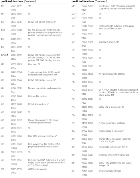
64R 75522-75809 Interleukin-1 beta convertase precursor, Caspase-recruitment domain/DEATH 94aa
65L# N/A SP
66L# 76106-76157 SP
16aa
67L 76212-77375 Ribonucleoside-reductase diphosphate beta subunit-like protein
386aa
68R 77657-77944 SP
94aa
69R 78080-78346 Unknown protein, TM
87aa
73L 80232-81206 NTPase/helicase-like protein
323aa
74L 81381-82493 SP
369aa
75L 82525-82779 LITAF/PIG7 possible membrane associated motif in LPS-induced tumor necrosis factor alpha factor, TM
83aa
76R 82842-83063 SP
72aa
77L 83060-83407 LCDV ORF 2-like protein, SP
114aa
78L 84007-84645 SP
211aa
79R 84781-86499 ATPase-dependent protease
571aa
80L 87122-88237 Ribonuclease III-like protein
370aa
81R 88293-88571 Transcription elongation Factor SII, C2C2 zinc finger
91aa
82R 88700-89173 Immediate-early protein ICP-18
156aa
83R 89623-90267 Cytosine DNA methyl-transferase
213aa
84R 90652-91389 LCDV 1-like proliferating cell nuclear antigen, SP
244aa
85R 91464-92051 Deoxynucleoside kinase
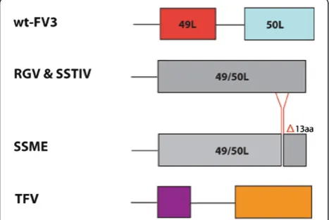
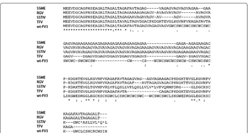
in 50L when compared to wt-FV3, present only in the SSME genome (Figure 3). In addition, 50L had a single nucleotide deletion resulting in a frameshift mutation in SSME, RGV, and SSTIV when compared to wt-FV3. This frameshift mutation resulted in the loss of 50L’s stop codon, which led to the combination of 49L and 50L into a single ORF. These two ORFs merged in frame, creating 49/50L. In contrast, TFV did not have this single nucleotide deletion, nor did it display the merger of its 49L and 50L equivalent ORFs (Figure 3). This is consistent with TFV being the most divergent virus of the group.
Further ranaviral gene variation was discovered in 43R in the form of a single nucleotide deletion, resulting in a frameshift mutation. Similar deletions were found in SSME, RGV, SSTIV, and TFV, leading to pronounced differences in amino acid sequences when compared to wt-FV3 (Figure 4). It is worth noting that although SSME, RGV, and SSTIV share the same frameshift muta-tion the genes are not identical as there are several sin-gle amino acid changes that lead to modest variation (Figure 4). In contrast, despite having a similar nucleo-tide deletion, TFV’s proposed amino acid sequence was markedly different from other analyzed ranaviruses. An-other single nucleotide deletion was found in 46L (Tables 1 and 2), which led to the loss of the original stop codon and the extension of the ORF by 319 bps (Tables 1 and 2). This was found in all genomes other than TFV and wt-FV3. These data demonstrate that even between very similar viral genomes, there is consid-erable gene variation, and that when viruses differ in pathogenicity, it could be related to variation in coding regions.
Repeat regions can vary in copy number between viral isolates
Genomic sequencing, and subsequent analysis, of SSME in comparison to wt-FV3, provided a general overview of the genetic variation between closely related FV3 iso-lates, particularly in coding regions. We then decided to explore possible genetic variation in highly variable Table 1 Description of nucleotide start/stop locations and
amino acid length of ORFs in SSME, along with predicted functions(Continued)
86L 92441-92626 SP
60aa
87L** 92979-94778 Unknown protein, SP
598aa
88R 94811-95263 Evrl-air-augmenter of liver regeneration
149aa
89R 95331-96497 SP
388aa
90R 96590-97981 Major capsid protein
462aa
91R 98105-99292 Immediate-early protein ICP-46
394aa
92R 99627-99872 SP
80aa
93L 100054-100221 SP
54aa
94L 100331-100798 Regina ranavirus P8.141 C-like protein, TM
154aa
95R 100891-101982 DNA repair protein RAD2, Xeroderma pigmentosum G N-region, Helix-hairpin-helix, Class 2 (Pol I) family
362aa
96R 102784-103455 Unknown protein, SP
222aa
97R 103538-103951 Myeloid cell leukemia protein, MCL-1, TM
136aa
98R 104649-104849 SP
65aa
*Note: SP-surface protein, TM-transmembrane domain. ºLoss of original, predicted start codon in SSME; new start codon 27 bp downstream.
§Loss of original, predicted stop codon in SSME; new stop codon 319 bp upstream.
¶39 bp deletion in original 50L ORF of SSME. Loss of ORF 50L’s original, predicted stop codon in SSME; ORFs 49L and 50L are now combined as 49/50L.
#Deletion of 65L ORF and majority of 66L ORFs in SSME. **18 bp deletion in 87L ORF of SSME.
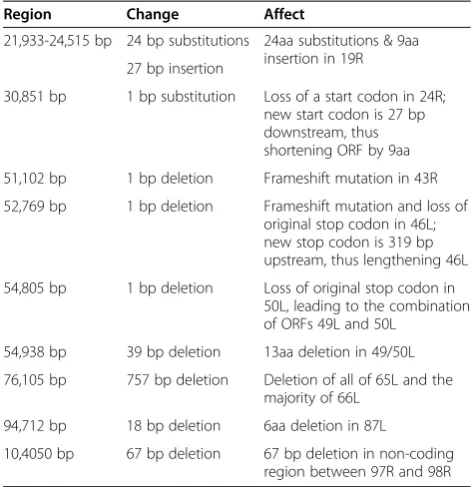
Table 2 Genetic variation present within the SSME genome as compared to the wt-FV3 reference genome
Region Change Affect
21,933-24,515 bp 24 bp substitutions 24aa substitutions & 9aa insertion in 19R 27 bp insertion
30,851 bp 1 bp substitution Loss of a start codon in 24R; new start codon is 27 bp downstream, thus shortening ORF by 9aa
51,102 bp 1 bp deletion Frameshift mutation in 43R
52,769 bp 1 bp deletion Frameshift mutation and loss of original stop codon in 46L; new stop codon is 319 bp upstream, thus lengthening 46L
54,805 bp 1 bp deletion Loss of original stop codon in 50L, leading to the combination of ORFs 49L and 50L
54,938 bp 39 bp deletion 13aa deletion in 49/50L
76,105 bp 757 bp deletion Deletion of all of 65L and the majority of 66L
94,712 bp 18 bp deletion 6aa deletion in 87L
10,4050 bp 67 bp deletion 67 bp deletion in non-coding region between 97R and 98R
repeat regions, and thus define these genetic regions with greater inter-strain variability. To achieve this, we chose three repeat regions (suspected to be polymorphic in copy number) to investigate. These regions included: Region 1 (22,499-22,574 bps); Region 2 (52,443-52,747 bps); and Region 3 (54,948-54,986 bps), based on the wt-FV3 se-quence. We performed a repeat analysis on our sequenced SSME genome that we had previously evaluated for cod-ing region variability, and reference genomes RGV, SSTIV, and TFV. We also decided to analyze the repeat regions of wt-FV3 and aza-Cr in order to check for repeat number stability between the two strains over the multiple viral passages they have undergone since the creation of aza-Cr in 1987 [22]. In addition, we expanded our analysis to in-clude 6 environmental samples isolated from the same waterway in Manitoulin, Kagawong, ON, Canada. These
isolates were designated as: E3, E4, E5, F4, F6, and G4; a lab FV3 sample (ATCC® VR-567™) was also used [26].
The information acquired through analyzing these re-peat regions indicated that variation in rere-peat copy num-ber is present between ranaviral isolates, including those from one geographic location (Figure 5). In the first re-gion, repeat copy number ranged from 2–5 copies, with 2 copies being the most common and 5 copies being unique to isolate F6. Wt-FV3 and aza-Cr shared a dis-tinctive repeat sequence, containing a G rather than an A at the 9th nucleotide position (Figure 5). Region 2 proved to be more variable than region one, with repeat copy number ranging from 4–18 copies, while Region 3 was found to consist of two repeat sequences. These Region 3 repeats varied in copy number between iso-lates, with a maximum copy number of 4. However, in some instances, the first sequence of nucleotides charac-teristic of this region did not repeat (Figure 5).
Although copy number variation was present amongst isolates, when samples were compared across all three repeat regions, some were found to be identical; such isolates included F4, E5, and E3 (Figure 5), which were isolated from the same geographic region. In addition, isolates wt-FV3 and aza-Crwere also found to be identi-cal across repeat regions (Figure 5), which is consistent with their common origin [22]. This data taken as a whole suggests that viral isolates, even those from one geographic location, can display both variation and iden-tical patterns across repetitive regions of the genome. Thus, these repeats could act as a fingerprint to discrim-inate between viral isolates.
Discussion
Our analysis of related ranaviruses presents a novel ap-proach to genomic comparison that differs from other studies. We analyzed the genomes of closely related iso-lates of FV3 through 454 GS-FLX technology and STR
Figure 3Variation in ORF 49/50L.Sequence alignment of the 49/50L region across ranaviruses. A 39 bp deletion is present in the SSME genome, spanning from 54,909-54,947 bp in wt-FV3. A single nucleotide deletion is present in SSME, RGV, and SSTIV, leading to the loss of 50L’s original stop codon and the merger of 49L and 50L into one ORF. Different colours are used to represent the portions of TFV’s ORFs that are homologous to parts of 49L and 50L respectively.
65L 66L
64R
64R wt-FV3
SSME
RGV, SSTIV &
TFV 64R 65L 66L
comparison. The scale (fully sequenced genomes), and the nature of comparison (using viral isolates of FV3), set our investigation apart from past studies. We found that the 3 strains we examined displayed slightly differ-ent levels of virulence duringin vivostudies. By sequen-cing the one genome yet to be sequenced, we were able to highlight areas that may be important in generating infectious phenotypes of FV3. This kind of analysis has never been done for FV3 or related ranaviruses: thus, it provides greater insight into the genetic variation among
these closely related DNA viruses and the possible gen-etic basis of ranaviral virulence.
Our results demonstrate that genetic variation is present between closely related FV3 isolates in both coding and non-coding regions. The SSME genome was sequenced and compared to the published wt-FV3 genome, along with related ranaviral genomes RGV, SSTIV, and TFV. Comparisons revealed that SSME was divergent from wt-FV3, and aza-Cr. This variation could be due to the fact that SSME was isolated from a spotted salamander rather
Figure 4Variation in 43R ORF.Amino acid sequence alignment of the 43R ORF in ranaviruses. A single nucleotide deletion in SSME, RGV, SSTIV, and TFV led to frameshift mutations, causing significant amino acid variability amongst genomes.
Viral Isolate Region 1 Region 2 Region 3
F4, E5, & E3 x2
-CTGCGAGAAAGTCTC-x17
-CTTACGCTTCACAGGGGA-x2 x4 -
TCAGGCTGC-TCAGGCTGT-E4 x2
-CTGCGAGAAAGTCTC-x15
-CTTACGCTTCACAGGGGA-x2 x4 -
TCAGGCTGC-TCAGGCTGT-G4 x4
-CTGCGAGAAAGTCTC-x14
-CTTACGCTTCACAGGGGA-x2 x4 -
TCAGGCTGC-TCAGGCTGT-wt-FV3 and aca-Cr x3
-CTGCGAGAAGGTCTC-x16
-CTTACGCTTCACAGGGGA-x2 -
TCAGGCTGC-TCAGGCTGT-SSME x3
-CTGCGAGAAAGTCTC-x16
-CTTACGCTTCACAGGGGA-x2 -
TCAGGCTGC-TCAGGCTGT-F6 x5
-CTGCGAGAAAGTCTC-x18
-CTTACGCTTCACAGGGGA-x2 -CTTACGCTTCACAGGGGA-x2 -
TCAGGCTGC-TCAGGCTGT-VR-567 x4
-CTGCGAGAAAGTCTC-x4
-CTTACGCTTCACAGGGGA-x2 -CTTACGCTTCACAGGGGA-x2 -
TCAGGCTGC-TCAGGCTGT-RGV x2
-CTGCGAGAAAGTCTC-x10
-CTTACGCTTCACAGGGGA- -
TCAGGCTGC-TCAGGCTGT-SSTIV x2
-CTGCGAGAAAGTCTC-x18
-CTTACGCTTCACAGGGGA- -
TCAGGCTGC-TCAGGCTGT-TFV x3
-CTGCGAGAAAGTCTC- -TGAAACAGACTTGCTGTCTC-
than anurans, and so the strain may have evolved in order for it to better adapt to its novel host, as is seen during serial passage [27]. For example, the pathogen-icity of Dengue virus was altered by serial passaging Dengue virus 27 times which resulted in 25 nucleotide changes between 2 strains [27]. This finding is interest-ing given the fact that our results also showed that SSME was the least virulent of our 3 strains during an in vivostudy in anurans (Figure 1). Thus, genetic muta-tions observed in SSME, such as delemuta-tions in ORFs 65L, 66L and 49/50L, could have had an effect on strain virulence in anuran hosts. Also, past sequencing results [12] as well as our own genomic sequencing of wt-FV3 and aza-Crgenomes revealed only 13 nucleotide differ-ences (data not shown) between the strains, supporting ourin vivofinding that the two strains cause essentially the same amount of tadpole mortality (97% and 96% respectively).
One of the most significant genetic variations found in SSME was a 757 bp deletion that deleted all of the 65L coding region and most of the 66L coding region (Figure 2). Among other ranaviruses, the functions of these genes have not been determined. Further sequen-cing of ranaviruses has shown that 65L is present in both RGV and SSTIV as 69L and 68L respectively [23,28], while this form of 66L appears to be unique to FV3, as other ranaviruses have a 139 nucleotide inser-tion in their related 66L regions (Figure 2). Another genetic variation specific to SSME was a 13 amino acid deletion in ORF 50L (Figure 3). Interestingly, a second variation was found in this area in the form of single nucleotide deletion. This deletion lead to the merger of ORFs 49L and 50L in all examined ranaviruses other than TFV and wt-FV3 (Figure 3). In terms of function, 49L has multiple SAP motifs, which are DNA/RNA binding domains predicted to be involved in chromo-somal organization and DNA replication [29]. Thus, this new 49/50L ORF may function in viral replication.
Single nucleotide deletions were found in multiple rana-viruses within the 43R genes (Figure 4) and 46L (Tables 1 and 2) genes. In 43R, the deletion was present in all rana-viruses analyzed other than wt-FV3, and resulted in a frameshift mutation (Figure 4). In 46L, the original stop codon was lost, leading to the extension of the ORF by 319 bps (Tables 1 and 2). This was found in all genomes other than TFV and wt-FV3. Supposedly, 46L encodes for a neurofilament triplet H1-like protein [12]. However, the extended version of 46L that we discovered has a putative conserved domain known as a microneme/rhoptry anti-gen in the area previously thought to be non-coding. Micronemes and rhoptries are organelles possessed by Apicomplexa protozoans that secrete proteins involved in parasite entry into a host cell, specifically possessing protein-binding motifs that recognize ligands on the host
cell surface [30]. Although usually associated with proto-zoan parasites, these microneme/rhoptry antigens found in 46L could give further indication as to 46L’s function.
The changes present in 43R, 46L, 49/50L, 65L, and 66L represent the main variable regions amongst the re-lated ranaviral isolates we analyzed. This suggests that these are changeable areas across ranaviruses, and could be used in the future to help explain variable infectious phenotype. Moreover, multiple amino acid deletions present in 65L, 66L, and 49/50L, were limited to the SSME strain, which displayed the lowest level of viru-lence during tadpole infection. Thus, changes that are unique to SSME may present areas of the genome that are particularly effective in viral attenuation, specifically in an alternative host. As FV3 mortality and morbidity continues to worsen and fluctuate across environmental regions, examination of these genomic areas may prove useful as an initial way to investigate the genetic basis behind infective changes [6]. Further research could be used to explain variations in the virulence of different FV3-like isolates.
furthering our understanding of genetic variation be-yond coding regions.
The STR analysis we performed in our study has the potential to contribute to our understanding of FV3 tracking and strain designation. Surveillance and phylo-geographical analysis of FV3 are pivotal in understand-ing how the pathogen varies between different habitat sites and amphibian species, as well as for revealing pos-sible sources of a disease outbreak [31]. It can also have direct effects on conservation by aiding in strategy de-velopment to minimize die-offs in high-risk areas, and in creating vaccines through knowledge of the FV3 genome itself [14,31]. However, exact taxonomic identification of viruses in amphibian populations has been difficult given the lack of detailed molecular data on FV3 and other ranaviruses. The methodology used to classify these vi-ruses in the past has been through comparing the major capsid protein (MCP) of different viral isolates [3]. How-ever, the use of the MCP as a tool to distinguish between different ranaviruses, as well as between different strains of the same virus, has been called under scrutiny [24,32,33]. Thus, there is a need to develop new methods of strain tracking for ranaviral isolates.
The use of STRs for ranavirus strain identification has precedence in other virus studies. In one such study, 12 isolates of human cytomegalovirus (HCMV) were iso-lated from various individuals infected with the virus. The isolates were then tested for variable repeats in 24 polymorphic regions, and based on this analysis, each viral isolate was designated as an individual strain of HCMV [34]. Many of the HCMV repeats used in this study were found in non-coding regions of the genome, similarly to the ones used in our study. The study sug-gested that these changes in repeats are evolutionarily neutral and so appropriate for strain identification, not only in HCMV, but in other similar, large genome DNA viruses [34].
Other studies have used coding instead of non-coding repeat regions to identify viral strains [35,36]. In our study, Region 1 is found in the 19R ORF, unlike the non-coding areas of Regions 2 and 3. There are many examples of functional microsatellites that are known to affect viral characteristics based on copy number, includ-ing hepatitis C virus and vesicular stomatitis virus [35,36]. Therefore, in addition to being potentially useful in viral tracking, STRs from the Region 1 coding region may have functional significance in FV3.
Conclusions
In this study we have been able to uncover fine scale genetic variation between closely related ranaviral iso-lates that have different levels of virulence. We have shown that substantial genetic variability is present be-tween closely related FV3 isolates, both in terms of
deletions/insertions, and even more so at select STR lo-cations. These genomic areas with deletions/insertions may present regions that affect viral infectious pheno-type, and therefore require investigation. Furthermore, we have identified STR regions that may prove useful in future phylogeographical tracking and identification of ranaviral strains across different environmental regions. As FV3 leads to more unexplained lethal infections in amphibian populations, studies such as this are neces-sary. The genetic insight that they provide into ranaviral genomes will prove invaluable when seeking to explain the variability of FV3 infectious phenotype in wild popu-lations, and in preventing the devastation that ensues.
Material and methods Reagents, viruses
FV3 isolate SSME, wt-FV3, and aza-Cr, were analyzed in this study and were provided by Professor Gregory Chinchar from the University of Mississipi Medical Center (Jackson, MS, USA). SSME was isolated from a wild, spot-ted salamander population in Maine, USA, while aza-Cr was derived from the laboratory wt-FV3 strain through se-lection with azacytidine [22]. Both wt-FV3 and aza-Cr have been previously sequenced and found to be identical, with wt-FV3′s nucleotide sequence deposited into Gen-Bank [12]. Amphibian renal cells (A6 cells) were supplied by Niels Bols of the University of Waterloo and main-tained in Leibovitz’s L-15 media (Invitrogen, Burlington, ON) supplemented with 10% fetal bovine serum (FBS; Invitrogen, Burlington, ON), penicillin (100 U/ml), and streptomycin (100μg/ml).
Infection of tadpoles with FV3
plastic containers filled with 1 L of dechlorinated water (aged for three days) for the rest of the experiment. Containers were held in a climatic chamber (Thermo Incubator Model 3740) where the temperature was set to remain at 22°C with a 12 h:12 h dark:light cycle. Tadpoles were fed on a weekly basis after the water was changed with standard tadpole food (Carolina Biological Supply Company, Burlington, NC) at 30 mg/tadpole for week 1, 60 mg/tadpole for week 2, and 120 mg/tadpole for week 3 until the end of the experiment [38]. Starting on week 3 the water in each tank was replaced once a week with clean dechlorinated aged (24 h) water. As a result, exposed tadpoles were held in virus-containing water for 3 weeks, a period which is long enough for tadpoles to be in close proximity with residual infection [38]. Tanks were moni-tored on a daily basis. Dead tadpoles were removed to pre-vent any scavenging, and stored at −25°C in individual plastic vials with ethanol for subsequent analyses.. The ex-periment terminated when all the individuals died or reached metamorphosis. The procedures used in this ex-periment follow protocol #2010-04-02 approved by the Laurentian University Animal Care Committee.
Screening of tadpoles for FV3
In order to check for ranavirus infection, all animals (including euthanized ones) were dissected to remove the liver that was then crushed into a 1.5 ml Eppendorf tube. The resulting tissue mixture was used for DNA extraction. DNA was extracted using QIAmp DNeasy Kit following the standard protocol (Qiagen). After ex-traction, a double blind PCR was performed using a primer set known to successfully amplify a portion of the major capsid protein within the FV3 genome: MCP-ranavirus-F (5′-GACTTGGCCACTTATGAC-3′) and MCP-ranavirus-R (5′- GTCTCTGGAGAAGAAGAA), following the PCR conditions listed in Mao et al. [33], using 1.5μl of template DNA and cycled 40 times. Indi-viduals showing two positive amplifications for both PCRs were considered infected. We analyzed host sur-vival using a sursur-vival analysis and failure time analysis following the Kaplan & Meier product limit method as-sociated with Chi square and Gehan’s Wilcoxon tests (multiple and two sample comparisons respectively) [39]. Individuals surviving to the end of the experiment were censored to account for our lack of information about their true time of death [40].
Viral DNA isolation
The SSME viral isolate was propagated on a confluent monolayer of A6 cells, and grown in a 75 cm2 flask, with cells infected at a multiplicity of infection (MOI) of 1 PFU/cell. The cells were harvested 5 days post in-fection (once cytopathic effect appeared), and viral DNA was extracted using the Purelink Viral RNA/DNA
Mini Kit according to manufacturer’s protocol (Invitro-gen, Burlington, ON).
Viral genome sequencing
Standard kits and protocols developed by the manufacturer were used to sequence the SSME sample on a 454 GS-FLX platform (Roche Diagnostics Corporation). Briefly, a Rapid Library Preparation Kit (Roche, Mississauga, ON) was used to mechanically shear 500 ng of template DNA into short fragments. A universal sequencing primer that included a short DNA sequence unique to the sample (MID tag) was then annealed to both ends of each DNA fragment. A GS Junior Titanium Emulsion PCR Kit (Roche, Mississauga, ON) was used to amplify the sample library, which was sequenced using a GS Junior Titanium Sequencing Kit (Roche, Mississauga, ON). In order to assemble a full genomic sequence, the short sequences produced by 454 sequencing were aligned with the reference FV3 genome, wt-FV3, using GS Reference Mapper (Roche, Mississauga, ON). Any gaps in the assembled genome were then sequenced using custom PCR primers spe-cific to each gap, with sequencing performed by the Robarts Sequencing Facility (London, ON). The final genomic sequence was deposited in GenBank accession number KJ175144.
FV3 sample collection
FV3 environmental samples were collected from frogs caught by hand at various sites along a lakeshore in Manitoulin, Kagawong, ON, Canada (Latitude: 45.86418, Longitude:−82.27150). The frogs were caught using dis-posable gloves which were changed between each animal inspection. This method is preferred to the‘net-catching method’ as it has been suggested that cross contamin-ation can occur via the net. Each individual was toe clipped following the protocol #2009-03-04 approved by the Laurentian University Animal Care Committee for tissue sample collection. DNA was then isolated from toe clippings using the DNeasy Blood & Tissue Kit according to the manufacturer’s protocol for total DNA extraction from animal tissues (Qiagen, Mississauga, ON). Samples then underwent PCR with primers designed to amplify specific repeat regions identified in Eaton et al. [19]. Primers used included: Region 1-F: CGTGGTCAGACT GGTCCTCG; Region 1-R: CACCTCTGTCTCTGAATC GG; Region 2-F: GAGTTTACTTGGTGGCCATG; Region 2-R: TCCTGTCAAGAGATCCCCTC; Region 3-F: CTTG CTGCTGCCGTTCAGGC; and Region 3-R: AGAGTGA AAAAGGTAAAGGC.
TAQ DNA polymerase (Invitrogen, Burlington, ON), 10 mM deoxyribonucleotide triphosphates (dNTPs), 0.1 mM primer, 2.5 ng DNA, and water to a final volume of 50μl. The reactions were then placed in a thermocy-cler under the following conditions: 94°C for 3 minutes, 94°C for 30 seconds, 56°C for 1 minute, and 72°C for 1 minute for 30 cycles. Sequences of PCR products were determined by Robarts Research Institute DNA Sequen-cing Facility in London, ON, and were analyzed using BioEdit v7.0.5.
Competing interests
The authors declare that they have no competing interests.
Authors’contributions
EAM participated in the design of the study, isolated viral strainsin vitro, carried out the molecular genetic studies, participated in 454 GS-FLX sequence alignment, and drafted the manuscript. SG participated in 454 GS-FLX sequence alignment. PE collected viral isolates from the field, carried out the immunoassays, and participated in the design of the study. DL participated in the design of the immunoassays. CJK oversaw the 454 GS-FLX sequencing. CRB conceived of the study, and participated in its design and coordination and helped to draft the manuscript. All authors have read and approved the final manuscript.
Acknowledgements
This work was supported by Discovery Grants (Natural Science and Engineering Research Council (NSERC) of Canada) to CRB, CJK, and DL. EAM is the recipient of a NSERC postgraduate scholarship and past recipient of a NSERC Undergraduate Research Award.
Author details
1Department of Biology, Trent University, 1600 East Bank Dr., Peterborough,
Ontario K9J 7B8, Canada.2Current address: Department of Biology, Western University, 1151 Richmond Street, London, Ontario N6A 5B7, Canada. 3
Genetics and Ecology of Amphibians Research Group (GEARG), Department of Biology, Laurentian University, Sudbury, Ontario P3E 2C6, Canada.
Received: 22 October 2013 Accepted: 6 March 2014 Published: 12 March 2014
References
1. Berger L, Speare R, Daszak P, Green DE, Cunningham AA, Goggin CL, Slocombe R, Ragan MA, Hyatt AD, McDonald KR, Hines HB, Lips KR, Marantelli G, Parkes H:Chytridiomycosis causes amphibian mortality associated with population declines in the rain forests of Australia and Central America.Proc Natl Acad Sci U S A1998,95:9031–9036. 2. Daszak P, Berger L, Cunninham AA, Hyatt AD, Green DE, Speare R:
Emerging infectious diseases and amphibian population declines.
Emerg Infect Dis1999,5:735–748.
3. Tidona CA, Schnitzler P, Kehm R, Darai G:Is the major capsid protein of iridoviruses a suitable target for the study of viral evolution?Virus Genes 1998,16:59–66.
4. King A, Adams M, Carstens E, Lefkowitz E:Family Iridoviridae, In: Virus Taxonomy: Ninth Report of the International Committee on Taxonomy of Viruses.San Diego: Academic Press; 2012. pp 193–207.
5. Daszak P, Cunningham A, Hyatt H:Infectious disease and amphibian population declines.Divers Distrib2003,9:141–150.
6. Lesbarrères D, Balseiro A, Brunner J, Chinchar VG, Duffus A, Kerby J, Miller DL, Robert J, Schock DM, Waltzek T, Gray MJ:Ranavirus: past present and future.Biol Lett2012,8:481–483.
7. Marsh IB, Whittington RJ, O’Rourke B, Hyatt AD, Chisholm O:Rapid differentiation of Australian, European and American ranaviruses based on variation in major capsid protein gene sequence.Mol Cell Probes2002,
16:137–151.
8. Stuart SN, Chanson JS, Cox NA, Young BE, Rodrigues AS, Fischman DL, Waller RW:Status and trends of amphibian declines and extinctions worldwide.Science2004,306:1783–1786.
9. Muths E, Gallant AL, Campbell Grant EH, Battaglin WA, Green DE, Staiger JS, Walls SC, Gunzburger MS, Kearney RF:The amphibian research and monitoring initiative (ARMI): 5-year report. US Geological Survey.
Sci Invest Rep2006,5224:77.
10. Green DE, Converse KA, Schrader AK:Epizootiology of 64 amphibian morbidity and mortality events in the USA, 1996–2001.Ann N Y Acad Sci 2002,969:323–339.
11. Chinchar V, Ranavirus (family Iridoviridae):Emerging cold-blooded killers.
Arch Virol2002,147:447–470.
12. Tan W, Barkman T, Chinchar V, Essani K:Comparative genomic analyses of frog virus 3, type species of the genus Ranavirus (family Iridoviridae).
Virology2004,323:70–84.
13. Chinchar VG, Bryan L, Wang J, Long S, Chinchar GD:Introduction of apoptosis in frog virus 3-infected cells.Virology2003,306:303–312. 14. Williams T, Barbosa-Solomieu V, Chinchar GD:A decade of advances in
iridovirus research.InAdvances in virus research, Volume 65. Edited by Maramorosch K, Shatkin A. New York: Academic Press; 2005:173–248. 15. Cunningham AA, Langton TE, Bennett PM, Lewin JF, Drury SE, Gough RE,
Macgregor SK:Pathological and microbiological findings from incidents of unusual mortality of the common frog (Rana temporaria).Philos Trans R Soc Lond B1996,351:1539–1557.
16. Kidd-Ljunggren K, Miyakawa Y, Kidd A:Genetic variability in hepatitis B viruses.J Gen Virol2002,83:1267–1280.
17. Stuyver L, De Gendt S, Van Geyt C, Zoulim F, Fried M, Schinazi RF, Rossau R:
A new genotype of hepatitis B virus: complete genome and phyogenetic relatedness.J Gen Virol2000,81:67–74.
18. Wang J, Hoper D, Beer M, Osterrieder N:Complete genome sequence of virulent duck enteritis virus (DEV) strain 2085 and comparison with genome sequences of virulent and attenuated DEV strains.Virus Res 2011,160:316–325.
19. Eaton HE, Ring BA, Brunetti CR:The genomic diversity and phylogenetic relationship in the familyIridoviridae.Viruses2010,2:1458–1475. 20. Kass D, Batzer M:Genome organization/human.Life Sciences: Encyclopedia of;
2001:1–8.
21. Chinchar V, Granoff A:Temperature-sensitive mutants of frog virus 3: biochemical and genetic characterization.J Virol1986,58:192–202. 22. Essani K, Goorha R, Granoff A:Mutation in a DNA-binding protein
reveals an association between DNA-methyltransferase activity and a 26,000-Da polypeptide in frog virus 3-infected cells.Virology1987,
161:211–217.
23. Lei X, Ou T, Zhu R, Zhang Q:Sequencing and analysis of the complete genome of Rana grylio virus (RGV).Arch Virol2012,157:1559–1564. 24. Hyatt AD, Gould A, Zupanovic Z, Cunningham AA, Hengstberger S,
Whittington RJ, Kattenbelt J, Coupar BE:Comparative studies of piscine and amphibian iridoviruses.Arch Virol2000,145:301–331.
25. He J, Lu L, Deng M, He H, Weng S, Wang XH, Zhou SY, Long QX, Wang XZ, Chan SM:Sequence analysis of the complete genome of an iridovirus isolated from the tiger frog.Virology2002,292:185–197.
26. Granoff A, Came P, Breeze D:Viruses and renal carcinoma of Rana pipiens.Virology1966,29:133–148.
27. Puri B, Nelson WM, Henchal EA, Hoke CH, Eckels KH, Dubois DR, Porter KR, Hayes CG:Molecular analysis of dengue virus attenuation after serial passage in primary dog kidney cells.J Gen Virol1997,78:2287–2291. 28. Huang Y, Huang X, Liu H, Gong J, Ouyang Z, Cui H, Cao J, Zhao Y, Wang X,
Jiang Y, Qin Q:Complete sequence determination of a novel reptile iridovirus isolated from soft-shelled turtle and evolutionary analysis of Iridoviridae.BMC Genomics2009,10:224.
29. Boyle K, Arps L, Traktman P:Biochemical and genetic analysis of the vaccinia virus D5 protein. Multimerization-dependent APTase activity is required to support viral DNA replication.J Virol2007,81:844–89. 30. Striepen B, Soldati D, Garcia-Reguet N, Dubremetz JF, Roos D:Targeting of
soluble proteins to the rhoptries and micronemes inToxoplasma gondii.
Mol Biochem Parasitol2001,113:45–53.
31. Jancovich JK, Davidson EW, Parameswaran N, Mao J, Chinchar VG, Collins JP, Jacobs BL, Storfer A:Evidence for emergence of an amphibian iridoviral disease because of human-enhanced spread.Mol Ecol2005,14:213–224. 32. Gray MJ, Miller DL, Hoverman JT:Ecology and pathology of amphibian
ranaviruses.Dis Aquat Organ2009,87:243–266.
33. Mao J, Hedrick RP, Chinchar VG:Molecular characterization, sequence analysis, and taxonomic position of newly isolated fish iridoviruses.
34. Davis C, Field D, Metzgar D, Saiz R, Morin P:Numerous length polymorphisms at short tandem repeats in human cytomegalovirus.
J Virol1999,73:6265–6270.
35. Barr JN, Whelan SPJ, Wertz GW:cis-acting signals involved in termination of vesicular stomatitis mRNA synthesis include the conserved AUAC and the U7 signal for polyadenylation.J Virol1997,71:8718–8725.
36. Yamada N, Tanihara K, Takada A, Yorihuzi T, Tsutsumi M, Shimomura H, Tsuji T, Date T:Genetic organization and diversity of the 3′non-coding region of the hepatitis C virus genome.Virology1996,223:255–261.
37. Gosner KL:A simplified table for staging anuran embryos and larvae with notes on identification.Herpetologica1960,16:183–190.
38. Echaubard P, Little K, Pauli B, Lesbarrères D:Context-dependent effects of ranaviral infection on Northern Leopard frog life history traits.
PLoS One2010,5:e13723.
39. Gehan EA:A generalized wilcoxon test for comparing arbitrarily singly-censored samples.Biometrika1965,52:203–223.
40. Leung KM, Elashoff RM, Afifi AA:Censoring issues in survival analysis.
Annu Rev Public Health1997,18:83–104. doi:10.1186/1743-422X-11-46
Cite this article as:Morrisonet al.:Complete genome analysis of a frog virus 3 (FV3) isolate and sequence comparison with isolates of differing levels of virulence.Virology Journal201411:46.
Submit your next manuscript to BioMed Central and take full advantage of:
• Convenient online submission
• Thorough peer review
• No space constraints or color figure charges
• Immediate publication on acceptance
• Inclusion in PubMed, CAS, Scopus and Google Scholar
• Research which is freely available for redistribution