R E S E A R C H
Open Access
Molecular interplay between T-Antigen and
splicing factor, arginine/serine-rich 1
(SRSF1) controls JC virus gene expression
in glial cells
Michael Craigie, Patrick Regan, Yolanda-Lopez Otalora and Ilker Kudret Sariyer
*Abstract
Background:Human polyomavirus JCV is the etiologic agent of progressive multifocal leukoencephalopathy (PML), a fatal demyelinating disease characterized by lytic infection of glial cells in the central nervous system. PML is seen primarily in immunosuppressed patients and is mainly classified as an AIDS-defining disease. In addition to
structural capsid proteins, JCV encodes multiple regulatory proteins, including T-antigen and agnoprotein, which are required for functional lytic infection. Previous studies have suggested that molecular interaction between viral proteins and host factors play an important role in reactivation of JCV and progression of the viral life cycle in glial cells. Recently, serine/arginine rich splicing factor 1 (SRSF1), a cellular alternative splicing factor, was identified as a strong negative regulator of JCV in glial cells. SRSF1 inhibits JCV gene expression and viral replication by directly interacting with viral promoter sequences. Here, we have investigated possible impact of JCV regulatory proteins, T-antigen and agnoprotein, on SRSF1-mediated suppression of JCV gene expression in glial cells.
Results:Reporter gene analysis has suggested that T-antigen rescues viral transcriptional suppression mediated by SRSF1. Further analyses have revealed that T-antigen promotes viral gene expression by suppressing SRSF1 gene transcription in glial cells. A subsequent ChIP analysis revealed that T-antigen associates with the promoter region of SRSF1 to induce the transcriptional suppression.
Conclusions:These findings have revealed a molecular interplay between cellular SRSF1 and viral T-antigen in controlling JCV gene expression, and may suggest a novel mechanism of JCV reactivation in patients who are at risk of developing PML.
Introduction
JC virus (JCV) is a human polyomavirus and the etio-logical agent of progressive multifocal leukoencephalo-pathy (PML), a fatal demyelinating disease of the white matter [1–3]. PML is primarily found in AIDS-patients, with between 3 % and 5 % of patients developing PML [4, 5]. However, PML has recently been diagnosed in patients undergoing immunomodulatory therapy with monoclonal antibodies against immune cells, such as Natalizumab, Rituximab, and Efalizumab, which indi-cates an immune component to the reactivation of the
virus from latent reservoirs [6–9]. JCV is a non-enveloped polyomavirus with a genome comprised of 5 kb of double-stranded circular DNA with a bidirectional non-coding control region separating the early and late coding regions [10]. The early coding region of JCV encodes regulatory proteins, T-antigen, small t-antigen, and T’splice variants expressed upon alternative splicing of the primary viral transcript [11]. The late coding region of JCV encodes the viral capsid proteins, VP1, VP2, and VP3, which are required for the formation of the viral capsid, as well as the small regulatory protein agnoprotein.
Previous studies have demonstrated that JCV under-goes cell-type specific activation, primarily in glial cells, which is proposed to be regulated at the transcriptional level [12]. Cellular proteins, such as SRSF1, play a major
* Correspondence:[email protected]
Department of Neuroscience, Center for Neurovirology, Temple University Lewis Katz School of Medicine, 3500 North Broad Street, 7th Floor, Philadelphia, PA 19140, USA
role in controlling JCV infection in glial cells [10]. We have recently identified alternative splicing factor SRSF1 as a negative regulator of JCV gene expression and repli-cation in glial cells [10]. SRSF1 functions by targeting the JCV promoter and strongly inhibits JCV early and late gene transcription. Likewise, the down regulation of SRSF1 in astrocytes increases levels of viral gene expres-sion and replication [10]. SRSF1 is a strong negative regulator of JCV gene expression as it suppresses both early and late gene transcription in glial cells [10, 13, 14]. Moreover, SRSF1 interaction with the CR3 region of JCV promoter sequences has been shown to be required for the ability of SRSF1 to regulate JCV gene expression and replication [14]. In order to proceed to a productive replication cycle, JC virus has to initiate the transcription of viral early and late genes by releasing the transcrip-tional silencing caused by host factors, such as SRSF1.
While cellular factors hold a negative pressure on viral gene expression in infected cells, viruses have evolved and developed strategies to bypass host defense mechanisms by utilizing mainly their regulatory proteins [15–22]. Here we investigated possible role of JCV regulatory proteins, T-antigen and agnoprotein, in regulation of viral transcriptional suppression mediated by SRSF1. Our results suggest that T-antigen but not agno-protein is able to rescue and initiate viral transcription suppressed by SRSF1. These results have revealed a novel interaction between T-antigen and SRSF1 in controlling JCV gene transcription and replication, which may suggest a unique mechanism of JCV reactivation in patients who are at risk of developing PML.
Results
Effect of JCV regulatory proteins, T-antigen and agnoprotein, on SRSF1-mediated suppression of JCV transcription
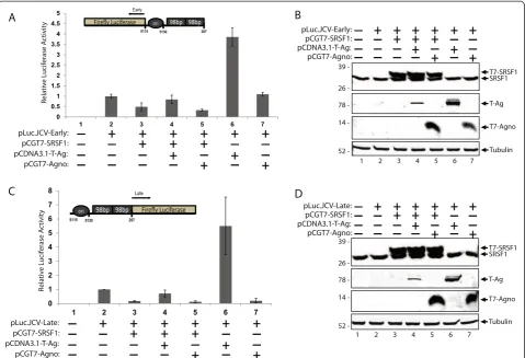
In order to investigate the role of JCV regulatory pro-teins T-antigen and agnoprotein in SRSF1-mediated sup-pression of JCV gene exsup-pression, primary human fetal astrocytes (PHFA) were transiently transfected with a lu-ciferase reporter construct in both early and late orienta-tions (pLuc.JCV-Early or pLuc.JCV-Late). In addition to transfection with the luciferase reporter construct, PHFA cells were co-transfected with plasmids encoding SRSF1 (pCGT7-SRSF1), T-antigen (pCDNA3.1-T-ag), and agno-protein (pCGT7-agno) in varying combinations. At 48 h post transfections, JCV early and late transcription was analyzed by luciferase reporter assay as described in ma-terials and methods. Luciferase assay of JCV-early gene transcription showed a strong suppression with SRSF1, which was rescued by co-expression of T-antigen, as well as a significant increase in transcription with the expres-sion of T-antigen alone (Fig. 1a). However, there was no observed effect of agnoprotein on transcriptional
suppression by SRSF1. Similarly, luciferase assay of JCV-late gene transcription yielded strong suppression of transcription with SRSF1, and a significant increase in transcription with T-antigen. When compared with the early gene transcription, SRSF1 possessed a much stronger suppression of late gene transcription (compare panel A, lane 3 with panel C, lane 3). Consistent with early gene transcription, T-antigen was also able to rescue the SRSF1-mediated transcriptional suppression of late pro-moter (Fig. 1c). Western blot analysis of the whole cell protein extracts were completed, demonstrating overex-pression of SRSF1 and the exoverex-pression of the viral proteins that were transfected in parallel to luciferase assays (Fig. 1b, d). These results suggest a novel role of viral T-antigen in suppression of cellular SRSF1 in glial cells.
T-Antigen inhibits SRSF1 expression in glial cells
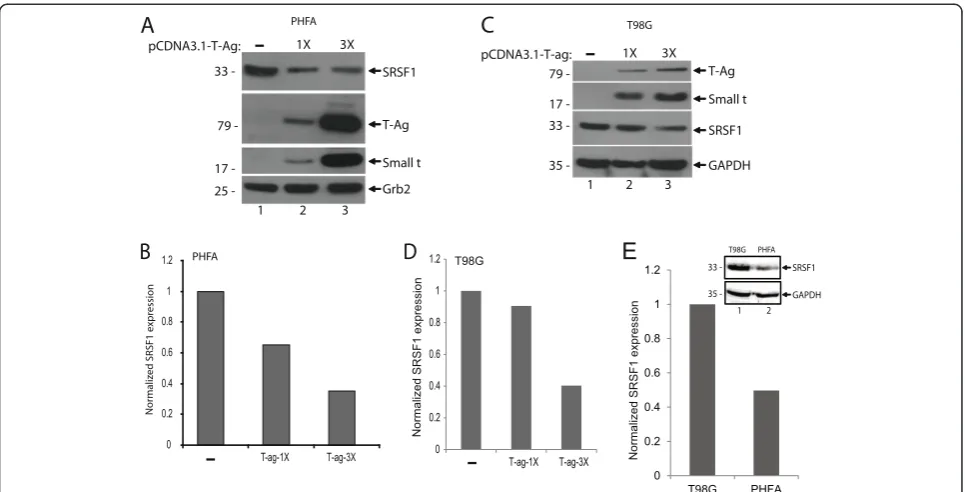
To determine possible impact of T-antigen on cellular ex-pression of SRSF1, both primary human fetal astrocytes (PHFA) and a human glioblastoma cell line (T98G) were transiently transfected with an expression vector encoding T-antigen in increasing concentrations. After 48 h post-transfection, cells were harvested and the whole cell pro-tein extracts were analyzed by Western blotting for the detection of SRSF1 expression. As shown in Fig. 2a and b, expression of T-antigen caused a significant reduction in the basal expression levels of SRSF1 in PHFA cells in a dose dependent manner. On the other hand, consistent with primary glial cultures, expression of T-antigen also caused a significant reduction in SRSF1 levels in T98G cells (Fig. 2c and d). The early gene product small t anti-gen was also expressed by alternative splicing of T-antianti-gen pre-mRNA and detected in the same immunoblots. These results suggest that JCV T-antigen can target and down-regulate the expression of cellular SRSF1.
T-Antigen targets SRSF1 promoter and inhibits its transcription
In order investigate the molecular mechanism of T-antigen-mediated suppression of SRSF1 expression in glial cells, a luciferase assay reporting the transcriptional activ-ity of the SRSF1 promoter was utilized. T98G cells were transiently transfected with a luciferase reporter construct consist of −1000 to +48 bp promoter region of SRSF1. Along with luciferase reporter construct, cells were co-transfected with plasmids encoding T-antigen or agnopro-tein to determine the impact of these viral proagnopro-teins on SRSF1 transcription. Cells were also transfected with an expression plasmid encoding green fluorescein protein (GFP) to access the transfection efficiencies. After 48 h, cells were harvested and processed for luciferase assay. It was observed that T-antigen, but not agnoprotein, sup-pressed the transcriptional activity of SRSF1 promoter (Fig 3a and b). Similar to our previous experiments (Fig. 2),
it was found that higher levels of T-antigen correspond to a greater decrease in the transcriptional activity of SRSF1, which accounts for the decreased expression of SRSF1 in T-antigen expressing cells. To better understand the events associated with T-antigen suppression of SRSF1 promoter activity, we designed a series of experiments to assess the ability of T-antigen to bind to the SRSF1 pro-moter sequences. T98G cells were transfected with ex-pression plasmid encoding T-antigen, cross-linked and possible interaction of T-antigen with SRSF1 promoter was analyzed by ChIP assay using antibodies to T-antigen as described in Materials and Methods. ChIP analysis of the cells demonstrated the association of T-antigen with SRSF1 promoter sequences (Fig. 3b).
Discussion
HCMV is detected by cells, an interferon gamma response is induced, resulting in the expression of multiple antiviral proteins, including the iron-sulfur cluster-binding antiviral protein viperin [15–17]. During the infection cycle, pre-expression of viperin results in inhibition of HCMV infec-tion, however, the virus also is able to induce viperin ex-pression independently of the IFN-dependent pathway [17, 18]. Although viperin contains antiviral activity, HCMV-induced viperin functions to disrupt cellular me-tabolism through localization to the mitochondria in infected cells, which results in enhancement of the HCMV infection process [18–22]. HCMV encodes a viral protein, viral mitochondrial inhibitor of apoptosis (vMIA), which interacts with viperin during HCMV infection, thus facilitating viral replication [18].
Similar to HCMV, JCV encodes viral regulatory pro-teins which are required for the progression of the lytic viral life cycle. We recently identified that cellular SRSF1 can overcome JCV reactivation by suppressing the ex-pression of the viral genes [10, 13, 14]. Here, we investi-gated the molecular interplay between SRSF1 and JCV regulatory proteins T-antigen and agnoprotein in regula-tion of the viral transcripregula-tion. Our results demonstrate
that T-antigen is able to rescue the SRSF1-mediated transcriptional suppression seen in both early and late coding regions of the JCV genome. Likewise, it was found that T-antigen alone significantly increased viral transcription, however, a second viral regulatory protein, agnoprotein, had no effect on JCV transcription. To de-termine the mechanism behind T-antigen rescue of tran-scriptional suppression mediated by SRSF1, the impact of T-antigen expression on SRSF1 expression was ana-lyzed. T-antigen expression decreased the expression of SRSF1, thus allowing for increased viral transcription. This step is hypothesized to be highly important within the JCV infection process, as T-antigen acts as a tran-scription factor for viral trantran-scription and is able to autoregulate the viral promoter to drive the required viral processes for a productive infection cycle: in-cluding DNA replication and transcription of the late region allowing for agnoprotein and the capsid pro-teins to be expressed [23, 24]. The observed suppres-sion of SRSF1 levels within cells expressing T-antigen was then demonstrated to be due to the T-antigen interaction with the SRSF1 promoter region leading to transcriptional inhibition.
Fig. 2T-Antigen suppresses SRSF1 expression in glial cells.a. PHFA cells were transfected with increasing concentration of T-antigen expression plasmid. After 48 h post-transfection, whole cell protein extracts were collected. Western blot analyses were performed to detect levels of SRSF1, T-antigen, and small t antigen expressions. Grb2 served as a loading control.b. Bar graph representation of relative SRSF1 expression from panel a. Quantification of the intensity of the bands of SRSF1 from panelawere normalized to Grb2 band intensities and used to calculate relative expression.c. T98G cells were transfected with increasing concentration of T-antigen expression plasmid. After 48 h post-transfections, whole cell protein extracts were collected. Western blot analyses were performed to detect levels of SRSF1, T-antigen, and small t antigen expressions. GAPDH served as a loading control.d. Bar graph representation of relative SRSF1 expression from panelc. Quantification of the intensity of the bands of SRSF1 were normalized to GAPDH band intensities and used to calculate relative SRSF1 expression levels.e. Whole cell protein lysates from PHFA and T98G cells were prepared and analyzed by Western blotting for the detection of SRSF1 expression. GAPDH served as a loading control. Band intensity of the bands of SRSF1 were quantified, normalized to GAPDH, and shown as bar graph
Conclusion
In summary, our results demonstrate a novel role of T-antigen in SRSF1-mediated suppression of JCV gene expression and provide basis for the further in-vestigation of the role of the molecular interplay be-tween cellular SRSF1 and viral T-antigen in JCV reactivation in glial cells leading to initiation of
productive viral life cycle in patients who are at risk of developing PML.
Materials and methods Cell lines and culture
(ATCC) and grown in Dulbecco’s Modified Eagle’s Medium (DMEM) containing 5 % heat-inactivated fetal bo-vine serum (FBS) and penicillin/streptomycin (100 ug/ml). T98G cells were maintained at 37 °C in a humidified environment with 5 % CO2. Primary human fetal as-trocytes were cultured from the fetal human brain of
a donor and provided by the Comprehensive
NeuroAIDS Core facility at Temple University and cul-tured in Dulbecco’s Modified Eagle’s Medium/Nutrient Mixture F-12 (DMEM/F-12) containing 10 % heat-inactivated fetal bovine serum (FBS) penicillin/strepto-mycin (100 μg/ml), GlutaMax (100 μg/mL) and insulin (100 μg/mL). Cultured PHFA cells were maintained at 37 °C in a humidified atmosphere with 5 % CO2.
Plasmid constructs
The T-antigen gene was cloned into the eukaryotic ex-pression vector pcDNA3.1 (+) at the EcoRI restriction enzyme site and designated as pcDNA3.1-T-antigen pre-viously [25]. pCGT7-SF2/ASF (SRSF1) expression plas-mid was kindly provided by Javier F. Cáceres (Medical Research Council Human Genetics Unit, Western General Hospital, Edinburgh EH4 2XU, Scotland, United Kingdom) and was described previously [26]. The pCGT7-agno plasmid was described previously [27]. Briefly, agnoprotein coding sequence was cloned into the eukaryotic expression plasmid pCGT7 at Xba1/ BamH1 restriction enzyme sites and designated as pCGT7-agno. The luciferase reporter construct pLuc-JCV-Early and pLuc-JCV-Late were made by blunt end cloning of the full-length Mad-1 NCCR into the SmaI site immediately upstream of the luciferase gene in the plasmid pGL3 (Promega, Madison WI) and described previously [28]. The luciferase reporter plasmid pLuc-SRSF1 was made by cloning the −1000 to +49 promoter region of SRSF1 gene into pGL3 vector at BamH1 site.
Luciferase reporter assay
T98G cells were plated in 6-well tissue culture dishes and transiently transfected with JCV-Early, pLuc-JCV-Late, or pLuc-SRSF1-1000 bp reporter plasmids in the presence or absence of expression plasmids for T-antigen and agnoprotein or pCDNA3.1 as control. At 48 h post-transfection, cells were extracted and lysed using reporter lysis buffer for the luciferase re-porter system provided by the manufacturer Promega. After cell lysis, luciferase activity of samples was de-termined through the use of luciferase assay reagent (LAR). The luciferase activities were then corrected for protein concentrations and normalized to the basal levels of transcription, allowing for determin-ation of the fold changes.
Chromatin Immunoprecipitation Assay (ChIP)
T98G cells were transfected with pCDNA3.1-T-antigen. At 48 h post-transfection, proteins were cross-linked to DNA using formaldehyde at a final concentration of 1 %. After cross-linking, cells were lysed and subject to sonication to fragment the chromatin. After sonication, cells were incubated overnight with Protein G beads and anti-T antigen antibody (pAb-416, Calbiochem) for immunoprecipiation. After immunoprecipiation, the beads were washed and bound proteins were eluted using ChIP elution buffer (1 % SDS, 100 mM NaHCO3). After elution, cross-linking was reversed by using 5 M NaCl followed by Proteinase and RNase treatment. The sample was then purified for DNA using phenol/chloro-form extraction followed by ethanol precipitation. Obtained DNA fragments were analyzed by PCR using primers; SRSF1-Promoter-Forward (−1000 to +47): 5’-A
CCTTCCAAAGCTTTCCAGATTTCAG-3’ and
SRSF1-Promoter-Reverse (+47 to +27): 5’-ACCTTCCACTCG AGGAAGGAAACAGC-3’. The PCR conditions were as follows: denaturing at 95° for 30 s, annealing at 58 °C for 40 s, and extension at 72 °C for 65 s. The PCR products were then resolved on a 1 % agarose gel.
Western blot
Whole cell protein extracts were washed with PBS and lysed with TNN lysis buffer with protease inhibitors. Purified protein extracts were then heated to 95 °C for 5 min and resolved through sodium dodecyl sulfate polyacrylamide gel electrophoresis (SDS-PAGE). After resolution, the gel was transferred to a nitrocellulose membrane (Whatman, Germany) for three hours at 250 mÅ at 4 °C in transfer buffer (25 mM Tris pH 7.4, 200 mM glycine, 20 % methanol). After transfer, mem-branes were blocked for thirty minutes at room temperature with 5 % non-fat dry milk in 1X phosphate-buffered saline containing 0.1 % Tween-20 (PBST). After blocking, membranes were washed and incubated with primary antibodies at a 1:1000 dilution overnight at 4 °C. After primary antibody incubation, membranes were washed three times in PBST and incubated with second-ary antibodies at a 1:5000 dilution at room temperature. Following secondary antibody incubation, membranes were visualized with the Odyssey CLx Imaging System (LI-COR). Primary antibodies used were anti-T-antigen (pAb-416, Calbiochem), anti-SRSF1 (ab12910, Abcam), anti-agnoprotein (Pab7903, house raised), anti-β-Tubulin (LI-COR), and anti-GAPDH (Cell Signaling Technol-ogy). Secondary antibodies used were IRDye 800CW goat anti-mouse (LI-COR) and IRDye 680RD goat anti-rabbit (LI-COR).
Competing interests
None of the authors have any competing interests in the manuscript.
Authors’contributions
Conceived and designed the experiments: IKS and MC. Performed the experiments: MC, PR, and YLO. Analyzed the data: IKS and MC. Wrote the paper: IKS and MC. All authors read and approved the final manuscript.
Authors information
Michael Craigie, graduate student, Department of Neuroscience, Center for Neurovirology, Temple University Lewis Katz School of Medicine. Patrick Regan, graduate student, Department of Neuroscience, Center for Neurovirology, Temple University Lewis Katz School of Medicine. Yolanda-Lopez Otalora, research assistant, Department of Neuroscience, Center for Neurovirology, Temple University Lewis Katz School of Medicine. Ilker Kudret Sariyer, DVM, PhD (corresponding author), Assistant Professor, Department of Neuroscience, Center for Neurovirology, Temple University Lewis Katz School of Medicine.
Acknowledgments
The authors thank the past and present members of the Department of Neuroscience/Center for Neurovirology for sharing their ideas and reagents, particularly Drs. Kamel Khalili and Jennifer Gordon. Research reported in this publication was supported by the National Institute of Allergy and Infectious Diseases of the National Institutes of Health under Award number R01AI101192. This study utilized services offered by core facilities of the Comprehensive NeuroAIDS Center (CNAC NIMH Grant Number P30MH092177) at Temple University Lewis Katz School of Medicine. The funding organizations played no role in the design of the study, in the collection, analysis, and interpretation of the data and in the decision to submit the manuscript for publication.
Received: 10 August 2015 Accepted: 15 November 2015
References
1. Wollebo HS, Woldemichaele B, Khalili K, Safak M, White MK. Epigenetic regulation of polyomavirus JC. Virol J. 2013;23:264–73.
2. Berger JR, Khalili K. The pathogenesis of progressive multifocal leukoencephalopathy. Discov Med. 2011;12:495–503.
3. Weber T. Progressive multifocal leukoencephalopathy. Neurol Clin. 2008;26:833–54.
4. Holman RC, Janssen RS, Buehler JW, Zelasky MT, Hooper WC. Epidemiology of progressive multifocal leukoencephalopathy in the United States: analysis of national mortality and AIDS surveillance data. Neurol. 1991;41:1733–6. 5. San-Andres FJ, Rubio R, Castilla J, Pulido F, Palao G, de Pedro I, et al.
Incidence of acquired immunodeficiency syndrome-associated opportunistic diseases and the effect of treatment on a cohort of 1115 patients infected with human immunodeficiency virus. Clin Infect Dis. 2003;36:1177–85.
6. Sandborn WJ, Colombel JF, Enns R, Feagan BG, Hanauer SB. Natalizumab induction and maintenance therapy for Crohn’s disease: International efficacy of Natalizumab as active Crohn’s therapy, evaluation of natalizumab as continuous therapy trial group. N Engl J Med. 2005;353:1912–25. 7. Langer-Gould A, Atlas SW, Green AJ, Bollen AW, Pelletier D. Progressive
multifocal leukoencephalopathy in a patient treated with natalizumab. N Engl J Med. 2005;353:375–81.
8. Kleinschimdt-DeMasters BK, Tyler KL. Progressive multifocal leukoencephalopathy complicating treatment with natalizumab and interferon beta-1a for multiple sclerosis. N Engl J Med. 2005;353:369–74. 9. Carson KR, Focosi D, Major EO, Petrini M, Richey EA, West DP, et al. Monoclonal
antibody-associated progressive multifocal leukoencephalopathy in patients treated with rituximab, natalizumab, and efalizumab: A review from the research on adverse drug events and reports project. Lancet Oncol. 2009;10:816–24.
10. Sariyer IK, Khalili K. Regulation of human neurotropic JC virus replication by alternative splicing factor SF2/ASF in glial cells. PLoS ONE. 2011;6, e14630. 11. Trowbridge PW, Frisque RJ. Identification of three new JC virus proteins
generated by alternative splicing of the early viral mRNA. Neurovirol. 1995;1:195–206.
12. White MK, Safak M, Khalili K. Regulation of gene expression in primate polyomaviruses. J Virol. 2009;83:10846–56.
13. Uleri E, Beltrami S, Gordon J, Dolei A, Sariyer IK. Extinction of tumor antigen expression by SF2/ASF in JCV-transformed cells. Genes and Cancer. 2011;2:728–36.
14. Uleri E, Regan P, Dolei A, Sariyer IK. SF2/ASF binding region within JC virus NCCR limits early gene transcription in glial cells. Virol J. 2013;10:147. 15. Duschene KS, Broderick JB. The antiviral protein viperin is a radical SAM
enzyme. FEBS Lett. 2010;584:1263.
16. Shaveta G, Shi J, Chow VT, Song J. Structural characterization reveals that viperin is a radical S-adenosyl-L-methionine (SAM) enzyme. Biochem Biophys Res Commun. 2010;391:1390.
17. Chin KC, Cresswell P. Viperin, an INF-inducible antiviral protein directly induced by human cytomegalovirus. Proc Natl Acad Sci U S A. 2001;98:15125.
18. Seo JY, Yaneva R, Hinson ER, Cresswell P. Human cytomegalovirus directly induces the antiviral protein viperin to enhance infectivity. Science. 2011; 332:1093–7.
19. Goldmacher VS, Bartle LM, Skaletskaya A, Dionne CA, Kedersha NL, Vater CA, et al. A cytomegalovirus-encoded mitochondria-localized inhibitor of apoptosis structurally unrelated to Bcl-2. Proc Natl Acad Sci U S A. 1999;96:12536. 20. Goldmacher VS. vMIA, a viral inhibitor of apoptosis targeting mitochondria.
Biochimie. 2002;84:177.
21. Al-Barazi, H.O., Colberg-Poley, A.M. The human cytomegalovirus UL37 immediate-early regulatory protein is an integral membrane N-glycoprotein which traffics through the endoplasmic reticulum and Golgi apparatus. J Virol. 1996;70:7198–7208.
22. Colberg-Poley AM, Patel MB, Erezo DP, Slater JE. Human cytomegalovirus UL37 immediate-early regulatory proteins traffic through the secretory apparatus and to mitochondria. J Gen Virol. 2000;81:1779–89. 23. Saribas AS, Ozdemir A, Lam C, Safak M. JC virus induced progressive
multifocal leukoencephalopathy. Future Virol. 2010;5:313–23. 24. Lashgari MS, Tada H, Amini S, Khalili K. Regulation of JCVL promoter
function: transactivation of JCVL promoter by JCV and SV40 early proteins. Virol. 1989;170:292–5.
25. Khalili K, Sariyer IK, Safak M. Small tumor antigen of polyomaviruses: role in viral life cycle and cell transformation. J Cell Physiol. 2008;215:309–19. 26. Caceres JF, Misteli T, Screaton GR, Spector DL, Krainer AR. Role of the
modular domains of SR proteins in subcellular localization and alternative splicing specificity. J Cell Biol. 1997;118:225–38.
27. Otlu O, De Simone F, Lopez-Otalora Y, Khalili K, Sariyer IK. The agnoprotein of Polyomavirus JC is Released by Infected Cells: Evidence for Its Cellular Uptake by Uninfected Neighboring Cells. Virology. 2014;468–470:88–95. 28. Wollebo HS, Safak M, Del Valle L, Khalili K, White MK. Role for tumor necrosis
factor-αin JC virus reactivation and progressive multifocal leukoencephalopathy. J Neuroimmunol. 2011;233(1–2):46–53.
• We accept pre-submission inquiries
• Our selector tool helps you to find the most relevant journal • We provide round the clock customer support
• Convenient online submission • Thorough peer review
• Inclusion in PubMed and all major indexing services • Maximum visibility for your research
Submit your manuscript at www.biomedcentral.com/submit