R E S E A R C H
Open Access
CDH1
polymorphisms and haplotypes in sporadic
diffuse and intestinal gastric cancer: a case
–
control
study based on direct sequencing analysis
Chi-Ming Chu
1†, Cheng-Jueng Chen
2†, De-Chuan Chan
2†, Hurng-Sheng Wu
3, Yao-Chi Liu
2*, Chen-Yang Shen
4,
Tzu-Ming Chang
5, Jyh-cherng Yu
2, Horng-Jyh Harn
6, Cheng-Ping Yu
7and Ming-Hsin Yang
2Abstract
Background:Findings related to the influence of the−160C→A promoter polymorphism and haplotypes of the E-cadherin(CDH1) gene have not been consistent in previous studies regarding the risk for sporadic gastric cancer. Investigators in most previous studies detected those genotypes using restriction fragment length polymorphism analysis. Therefore, we conducted a case–control study to investigate the association of theCDH1−160C→A promoter polymorphism and haplotypes for cancer risk related to sporadic diffuse and intestinal gastric cancer by direct sequencing analysis.
Methods:We included 107 diffuse gastric cancer cases, 60 intestinal gastric cancer cases and 134 controls. The genotypic polymorphisms in the−160 promoter region, exons and intron–exon boundaries ofCDH1were detected by direct sequencing analysis. Genotype frequencies were compared. TheCDH1−160C→A promoter
polymorphism and four polymorphisms (48 + 6 T→C, 2076C→T, 2253C→T and 1937–13 T→C) were included in the haplotype analyses, which were estimated using the expectation–maximization algorithm.
Results:Compared to controls, the frequency of the−160A allele was significantly higher in diffuse gastric cancer cases (P= 0.005), but it was not significantly different in intestinal gastric cancer cases (P= 0.119). Two sets of three-marker haplotypes (−160C→A, 48 + 6 T→C, 2076C→T and−160C→A, 1937–13 T→C, 2253C→T) were associated with the risk of diffuse gastric cancer (P= 0.011 andP= 0.042, respectively).
Conclusion:Based on direct sequencing analysis, our findings suggest that theCDH1−160C→A promoter
polymorphism and haplotypes play significant roles in cancer risk for sporadic diffuse gastric cancer, but not for intestinal gastric cancer, in a Taiwanese population.
Keywords:CDH1, Direct sequencing, Haplotypes, Single-nucleotide polymorphisms, Sporadic diffuse and intestinal gastric cancer
Background
E-cadherin (encoded by the CDHI gene [dbSNP:
NC_000016.9]) is a member of a family of transmembrane glycoproteins expressed in epithelial cells and is responsible for calcium-dependent cell-to-cell adhesion [1-3]. Loss of cell adhesion may contribute to loss of growth contact in-hibition, which is an early step in the neoplastic process
[4,5]. Furthermore, loss of cadherin activity may result in cancer cell detachment and metastasis [6]. HenceCDHI is considered to be a tumor suppressor gene.
A C/A single-nucleotide polymorphism (SNP) at
the −160 position of the CDH1 promoter region has
been reported to result in downregulation of the tran-scription of this gene in a prostate cancer cell line, DU145, such that the A allele at this site decreased transcriptional efficiency by 68% compared with the C allele [7]. Therefore, the−160A allele variant has been considered to be a potential genetic marker for susceptibility to cancer. However, findings related to the influence of the−160C→
* Correspondence:[email protected]
†Equal contributors
2Division of General Surgery, Department of Surgery, Tri-Service General
Hospital, National Defense Medical Center, No. 325, Sec. 2, Cheng-Kung Road, Neihu, Taipei 11490, Taiwan
Full list of author information is available at the end of the article
A promoter polymorphism and haplotypes of the E-cad-herin (CDH1) gene have not been consistent in previous studies regarding the risk for sporadic gastric cancer [8-16]. Some studies have reported that the CDH1−160C→A promoter polymorphism was not found to be associated with the risk of gastric cancer [8-11], and others have re-ported that the−160A allele increased the risk for diffuse gastric cancer [12,13]. Conversely, two studies in Asian populations reported that the −160A allele decreased the risk of gastric cancer [15,16].
The genotypes of the−160C→A promoter polymorph-ism in most of the previous studies were detected by re-striction fragment length polymorphism (RFLP) analysis [8-13,15,16]. RFLP analysis is an indirect method applied extensively in the past [17]. However, it involves a manual process that is based on gel-based processing techniques and the results it produces are subjectively evaluated by direct observation. In contrast, direct sequencing is a stan-dardized procedure, and the results are processed by a computer [18].
By literature review, we compared the CDH1−160A allele frequency in controls in studies in which RFLP analysis was used, and the −160A allele frequency in healthy samples reported by HapMap based on direct sequencing derived from the National Center for Bio-technology Information dbSNP Short Genetic Varia-tions online database [19]. The frequency of the−160A allele was calculated using the formula CA% × ½ + AA%, where CA is the C and A alleles and AA is the double A alleles. Humar et al. reported that the fre-quency of the −160A allele in controls in an Italian population was 23.6% [12], and Corsoet al. reported a frequency of 32.05% in a different Italian population [11]. In a study by Pharoah et al., the frequency of the −160A allele was found to be 30% for Canadian, German and Portuguese populations [8]. Park et al. reported that the frequency of the−160A allele in con-trols in a Korean population was 23% [9]. Lu et al. reported that the frequency of the−160A allele was 24.3% for Chinese individuals in Jiangsu Province, China [10]. Medina-Franco et al. reported that the frequency of the
−160A allele was 24.35% in a Mexican population [13]. Wu et al. reported that the frequency of the -160A allele was 33.67% in a Taiwanese population [15]. Kuraoka et al. re-ported that the frequency of the−160A allele was 35% in a Japanese population [16]. The HapMap database, however, gives information based on direct sequencing that the fre-quency of the−160A allele in healthy samples is 26.0% for Europeans in Italy, 25.2% for Han Chinese in Beijing, 19.9% for Japanese in Tokyo and 15.0% for Africans in Nigeria [19]. In aforementioned studies in which RFLP analysis was used, the reported frequencies of the−160A allele in controls were diverse within the same ethnic groups, and some were much higher than the values in the
HapMap database. Therefore, genotype detection by RFLP analysis in some of previous studies might be inaccur-ate, which might contribute to the inconsistency of re-sults in previous studies. Tanahashi et al. and Davis
et al. also reported that RFLP analysis was less accurate than direct sequencing in their studies [20,21]. How-ever, researchers in many studies have suggested that environmental factors, lifestyles and ethnic differences might account for opposite directions in associations
of the CDH1−160C→A polymorphism with gastric
cancer among some Asian and Caucasian studies [8-14,22,23]. Gene-based haplotypes, which are collec-tions of SNPs located throughout the functional re-gions of candidate genes, may have greater power than any individual polymorphism in influencing a clinical response [24].
In one study of haplotype analysis using RFLP methods, the investigators suggested that the CDH1−160C→A polymorphism might be in linkage disequilibrium with other distinct CDH1 polymorphisms in sporadic diffuse gastric cancer [12] Researchers in another study using direct sequencing reported a statistically nonsignificant risk of −160C→A polymorphism containing haplotypes associated with gastric cancer without differentiation of histopathologic subgroups [14]. Furthermore, diffuse and intestinal types of gastric cancer are different in terms of their epidemiology, etiology, pathogenesis and behavior [25]. Therefore, we investigated the influence of the
CDH1−160C→A promoter polymorphism and
haplo-types on risk for diffuse and intestinal gastric cancer sep-arately by direct sequencing.
Methods
Patients and samples
Immunohistochemical staining and evaluation of E-cadherin expression
Specimens from paraffin blocks were cut into 5-μm sections and stained with hematoxylin and eosin for histological diagnosis. E-cadherin expression was evalu-ated using a monoclonal antibody (Cappel, Aurora, OH, USA) followed by a secondary antibody [26]. The signal was detected using an avidin-biotin complex and a 3,3′-diaminobenzidine (DAB) kit (Vector Laboratories, Burlingame, CA, USA). DAB produced a yellowish brown stain if a sample was positive. If more than 90% of the tumor cells showed intense membranous staining similar to that of normal cells, the result was considered positive (++). If the staining intensity was demonstrably reduced relative to that of normal cells and/or if the staining pattern was heterogeneous (10% to 90% positive), the result was re-corded as weakly positive (+). If immunohistochemical ex-pression was completely absent or positive in less than 10% of the cells, the result was defined as negative (−).
Genotyping
DNA samples from all cases were extracted from non-cancerous gastric epithelia. Control DNA samples were isolated from peripheral blood lymphocytes by proteinase K digestion and phenol-chloroform extraction. All CDH1 exons and the corresponding intron–exon boundaries of case DNA and control DNA were amplified using primers developed by Berxet al. [27]. The primers used to amplify the−160 promoter region were described by Liet al. [7]. PCR was carried out in volumes of 25μl containing 20 ng
of genomic DNA as a template, 2 mM MgSO4, 0.4 μM
sense and antisense primers, 0.25 mM deoxyribonucleotide triphosphate, and buffer containingTaqDNA polymerase. The amplification program was as follows: 40 cycles with a denaturing temperature of 95°C for 30 seconds, annealing for 30 seconds and extension at 72°C for 30 seconds in a DNA thermal cycler. All amplified products were puri-fied, and direct sequencing was performed using an ABI PRISM 377 automated sequencer and an ABI PRISM Dye Terminator Cycle Sequencing Kit (PerkinElmer, Greenville, SC, USA).
Statistical analysis
Stata 8 software (StataCorp, College Station, TX, USA) was used to manage data and for statistical analyses. The observed genotype frequencies were compared between cases and controls using aχ2test. Fisher’s exact test was used to assess the genotype and allele distributions in diffuse and intestinal type cases. The allelic distribution was in Hardy-Weinberg equilibrium in the case and control groups (P> 0.82). Genotype-specific risks were estimated as the odds ratio (OR) with associated 95% confidence interval (CI) by unconditional logistic regres-sion. ORs were adjusted according to age and gender. A
P-value less than 0.05 was considered statistically signifi-cant. The study power was 0.7758 to 0.9509 with anαof 0.05 [28]. The haplotype frequencies for various marker combinations were estimated separately for the two type cases and the controls by using an expectation-maximization algorithm.
Results
Characteristics of diffuse and intestinal type cases
The 134 controls were composed of 86 males and 48 fe-males. Their mean age was 51.06 ± 13.04 years (range = 19 to 89 years). The 167 gastric cancer cases comprised 114 males and 53 females. Their mean age was 69.11 ± 13.04 years (range = 27 to 90 years). According to Laurén’s classification system, 107 cases were of the diffuse type and 60 cases were of the intestinal type. The mean age of the diffuse type cases was 66.87 ± 13.81 years (range = 27 to 90 years) and that of the intestinal type cases was 73.82 ± 9.76 years (range = 48 to 88 years). The mean age of the dif-fuse type cases was approximately 7 years younger than that of the intestinal type cases. No differences were ob-served with respect to gender or TNM stages I to III in these two types. There were significantly more diffuse gas-tric cancer cases classified as stage IV. Reduced E-cadherin expression (Figure 1) was more frequent in the diffuse type cases than in the intestinal type cases. Comparison of the characteristics between these two case types is summarized in Table 1.
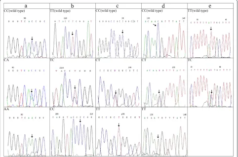
Genotype detected by direct sequencing
The genotypes of theCDH1−160C→A (dbSNP:rs16260),
48 + 6 T→C (dbSNP:rs3743674), 2076C→T (dbSNP:
rs1801552), 2253C→T (dbSNP:rs33964119) and 1937– 13 T→C (dbSNP:rs2276330) polymorphisms detected by
direct sequencing in the cases and controls are shown in Figure 2.
Single-locus analysis in diffuse type cases
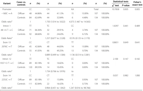
For promoter polymorphism −160C→A, the frequency of the −160A allele was significantly higher in diffuse type cases compared to the controls (P= 0.005). The OR associated with the A allele was 1.750 (95% CI = 1.014 to 3.022) for CA heterozygotes and 4.375 (95% CI = 1.467 to 14.565) for AA homozygotes. For other four polymor-phisms (48 + 6 T→C, 2076C→T, 1937–13 T→C and 2253C→T), the genotype frequencies were not signifi-cantly different between the diffuse type cases and the controls (P= 0.441, 0.649, 0.147 and 0.982, respectively) (Table 2).
Single-locus analysis in intestinal type cases
In the intestinal type cases, no significant association with disease was found for any of the five polymorphisms (−160C→A, 48 + 6 T→C, 2076C→T, 1937–13 T→C and 2253C→T; P= 0.119, 0.329, 0.185, 0.889 and 0.375, respectively).
Table 1 Comparison of clinicopathologic characteristics between intestinal and diffuse type tumors
Characteristics Intestinal type,
n(%) (N= 60)
Diffuse type,
n(%) (N= 107) P-value
Gender
Male 46 (76.7%) 68 (63.6%) 0.081a
Female 14 (23.3%) 39 (36.5%)
Age (yr)
Mean ± SD 73.82 ± 9.76 66.87 ± 13.81 0.0001b
TNM staging
Stage I 16 (26.7%) 16 (15.0%) 0.065c
Stage II 8 (13.3%) 24 (22.4%) 0.152c
Stage III 28 (46.7%) 35 (32.7%) 0.074c
Stage IV 8 (13.3%) 32 (30.0%) 0.016c
Reduced E-cadherin expression
32 (53.3%) 93 (86.9%) <0.001c
aχ2
test.b t-test.c
Proportion test.
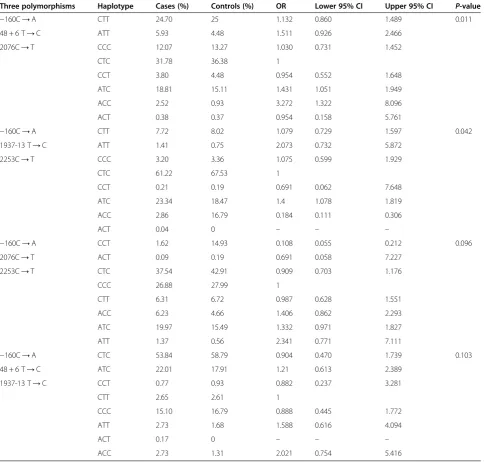
Haplotype analysis in diffuse type cases
A three-marker haplotype (−160C→A, 48 + 6 T→C, 2076C→T) showed a significant association with disease and the ATC, ACC haplotypes were found to be associated with increased risk for diffuse type cancer (Table 3). A differ-ent three-marker haplotype (−160C→A, 1937–13 T→C, 2253C→T) also exhibited a significant association with dis-ease and the ATC haplotypes was associated with incrdis-eased risk for diffuse gastric cancer. The two other sets of three-marker haplotypes (−160C→ A, 2076C→T, 2253C→T and−160C→A, 48 + 6 T→C, 1937–13 T→C) exhibited no significant association with disease (Table 3).
Haplotype analysis in intestinal type cases
In the intestinal type cases, no significant association with disease was found for any of the four sets of three-marker haplotypes (−160C→A, 48 + 6 T→C,
2076C→T; −160C→A, 1937–13 T→C, 2253C→
T; −160C→A, 2076C→T, 2253C→T and −160C→ A, 48 + 6 T→C, 1937–13 T→C;P= 0.164, 0.319, 0.408 and 0.607, respectively).
Discussion
To detect SNP variations, both direct sequencing and RFLP are suitable. RFLP is subject to experimental problems,
however, such as incomplete digestion and relatively poor resolution, resulting in misclassification of genotypic status. Nevertheless, many previous studies have depended on RFLP because it is cheaper and easy to perform. However, the cost of direct sequencing has decreased significantly, and regular automatic sequencing platforms are able to provide reliable results easily. The use of sequencing in the present study can be considered a major strength.
In this study, specimens were obtained from 167 Taiwanese patients with sporadic gastric cancer who underwent surgical treatment. The patients with intes-tinal gastric cancer are generally older, have more co-morbidities and tend not to undergo surgical treatment, which accounts for more diffuse gastric cancer cases than intestinal gastric cancer cases. Although we used an acceptable number of cases with statistical power, the sample size was still relatively small. Further studies including more cases are needed.
The frequency of the A allele in the CDH1−160C→ A promoter polymorphism in this study was significantly greater in diffuse type cases than in the controls. Humar
et al. [12] and Medina-Francoet al. [13] reported similar results.
It has been suggested that CDH1 may contribute to gastric cancer risk in a complex manner due to multiple
Table 2 Genotype frequencies ofCDH1polymorphisms in diffuse gastric cancer cases and controls
Variant Cases vs.
controls n (%) n (%) n (%) n (%)
Statistical tests Fisher’s exact test
χ2
test P-value
Promoter CC CA AA Total 10.7858 0.005 0.005
−160C→A Diffuse 48 44.86% 44 41.12% 15 15.00% 107 100.00% Controls 84 62.69% 44 32.84% 6 4.48% 134 100.00%
Odds ratioa 1 1.750 (1.014 to 3.022) 4.375 (1.467 to 14.565)
Intron 1 TT TC CC 1.6397 0.441 0.489
48 + 6 T→C Diffuse 71 66.36% 32 29.91% 4 3.74% 107 100.00%
Controls 92 68.66% 33 24.63% 9 6.72% 134 100.00%
Odds Ratioa 1 1.257 (0.677 to 2.328) 0.576 (0.125 to 2.170)
Exon 13 CC CT TT 0.8651 0.649 0.641
2076C→T Diffuse 45 42.06% 48 44.83% 14 13.08% 107 100.00%
Controls 55 41.05% 66 49.25% 13 9.70% 134 100.00%
Odds ratioa 1 0.889 (0.499 to 1.584) 1.136 (0.514 to 3.381)
Intron 12 TT TC CC 2.100 0.147 0.182
1937-13 T→C Diffuse 89 83.18% 18 16.82% 0 0.00% 107 100.00%
Controls 120 89.55% 14 10.45% 0 0.00% 134 100.00%
Odds ratioa 1 1.734 (0.766 to 3.976) to
Exon 14 CC CT TT 0.037 0.982 1.000
2253C→T Diffuse 89 83.18% 17 15.89% 1 0.93% 107 100.00%
Controls 111 82.84% 22 16.42% 1 0.75% 134 100.00%
Odds ratioa 1 0.964 (0.451 to 1.842) 1.247 (0.016 to 98.766)
a
polymorphic variants [12,14]. Humaret al. confirmed that the three-marker haplotype (−160C→A, 48 + 6 T→C, 2076C→T) was associated with diffuse gastric cancer and suggested that haplotype ATT (−160A, 48 + 6 T, 2076 T) was a marker for diffuse gastric cancer susceptibil-ity, whereas haplotype CTT had a protective effect [12].
We also found that the three-marker haplotype (−160C→A, 48 + 6 T→C, 2076C→T) was associated with diffuse gastric cancer and the ATC and ACC hap-lotypes were associated with increased risk. However, the haplotype CTT had no protective effect on diffuse gastric cancer which differs from that reported by Humar
et al. [12]. Furthermore, we found another three-marker
haplotype (−160C→A, 1937–13 T→C, 2253C→T) that was associated with diffuse gastric cancer, and the ATC haplotypes showed increased risk.
In the three-marker haplotype in this study (−160C→ A, 48 + 6 T→C, 2076C→T), the haplotype ATC, with
only one polymorphic variant (−160A) showed
in-creased diffuse gastric cancer risk. Therefore, with
re-spect to the CDH1−160C→A polymorphism, the A
allele might result in a defect in gene transcription and increased risk for diffuse type cancer. The ACC haplo-type, with two polymorphic variants (−160A and 48 + 6C), exhibited a higher OR. This finding indicates that the presence of two polymorphic variants in this
Table 3CDH1three-marker haplotype analysis in diffuse gastric cancer cases
Three polymorphisms Haplotype Cases (%) Controls (%) OR Lower 95% CI Upper 95% CI P-value
−160C→A CTT 24.70 25 1.132 0.860 1.489 0.011 48 + 6 T→C ATT 5.93 4.48 1.511 0.926 2.466
2076C→T CCC 12.07 13.27 1.030 0.731 1.452
CTC 31.78 36.38 1
CCT 3.80 4.48 0.954 0.552 1.648
ATC 18.81 15.11 1.431 1.051 1.949
ACC 2.52 0.93 3.272 1.322 8.096
ACT 0.38 0.37 0.954 0.158 5.761
−160C→A CTT 7.72 8.02 1.079 0.729 1.597 0.042 1937-13 T→C ATT 1.41 0.75 2.073 0.732 5.872
2253C→T CCC 3.20 3.36 1.075 0.599 1.929
CTC 61.22 67.53 1
CCT 0.21 0.19 0.691 0.062 7.648
ATC 23.34 18.47 1.4 1.078 1.819
ACC 2.86 16.79 0.184 0.111 0.306
ACT 0.04 0 – – –
−160C→A CCT 1.62 14.93 0.108 0.055 0.212 0.096 2076C→T ACT 0.09 0.19 0.691 0.058 7.227
2253C→T CTC 37.54 42.91 0.909 0.703 1.176
CCC 26.88 27.99 1
CTT 6.31 6.72 0.987 0.628 1.551
ACC 6.23 4.66 1.406 0.862 2.293
ATC 19.97 15.49 1.332 0.971 1.827
ATT 1.37 0.56 2.341 0.771 7.111
−160C→A CTC 53.84 58.79 0.904 0.470 1.739 0.103 48 + 6 T→C ATC 22.01 17.91 1.21 0.613 2.389
1937-13 T→C CCT 0.77 0.93 0.882 0.237 3.281
CTT 2.65 2.61 1
CCC 15.10 16.79 0.888 0.445 1.772
ATT 2.73 1.68 1.588 0.616 4.094
ACT 0.17 0 – – –
haplotype produces synergic effects related to cancer risk.
Conclusions
We conclude that, based on direct sequencing analysis, the −160C→A promoter polymorphism and the investi-gated haplotypes play significant roles in the risk for spor-adic diffuse gastric cancer, but not for intestinal gastric cancer, in a Taiwanese population. In the future, additional polymorphisms in other regions of the same gene, such as the promoter region and splicing sites, should be evaluated, and haplotype analysis should include additional polymor-phisms for the purpose of increasing knowledge about how combinations of polymorphisms can influence cancer risk.
Abbreviations
RFLP:Restriction fragment length polymorphism; SNP: Single-nucleotide polymorphism.
Competing interests
The authors declare that they have no competing interests.
Authors’contributions
LYC and SCY designed the research. LYC, CCJ, CDC, CCM, WHS, CTM, YJC, HHJ, YCP and YMH performed the research and analyzed the data. CCM performed the statistical analysis. LYC, CCJ and CDC wrote the paper. All authors read and approved the final manuscript.
Acknowledgements
This study was supported by the Clinical Research Fund of the Tri-Service General Hospital, Taipei, Taiwan (TSGH-C97-15). The authors are grateful to Ye-Chien Mau for her wonderful technical assistance.
Author details
1Division of Biostatistics and Informatics, Department of Epidemiology,
School of Public Health, National Defense Medical Center, No. 161, Sec. 6, Min-Quan E. Road, Neihu, Taipei 11490, Taiwan.2Division of General Surgery,
Department of Surgery, Tri-Service General Hospital, National Defense Medical Center, No. 325, Sec. 2, Cheng-Kung Road, Neihu, Taipei 11490, Taiwan.3Division of General Surgery, Department of Surgery, Show Chwan
Memorial Hospital, No. 6-1, Lugong Road, Lukang Township, Changhua 50005, Taiwan.4Institute of Biomedical Sciences, Academia Sinica, No. 128,
Sec. 2, Academy Road, Nangang, Taipei 11529, Taiwan.5Division of General
Surgery, Department of Surgery, Tungs’Taichung MetroHarbor Hospital, No. 699, Sec. 8, Taiwan Blvd., Wuqi District, Taichung City 43502, Taiwan.
6Department of Pathology, China Medical University Hospital, No. 2, Yuhder
Road, Taichung 40447, Taiwan.7Department of Pathology, Tri-Service General
Hospital, National Defense Medical Center, No. 325, Sec. 2, Cheng-Kung Road, Neihu, Taipei 11490, Taiwan.
Received: 18 October 2013 Accepted: 10 February 2014 Published: 31 March 2014
References
1. Takeichi M:Cadherin cell adhesion receptors as a morphogenetic regulator.Science1991,251:1451–1455.
2. Takeichi M:Cadherins: a molecular family important in selective cell–cell adhesion.Annu Rev Biochem1990,59:237–252.
3. Grunwald GB:The structural and functional analysis of cadherin calcium-dependent cell adhesion molecules.Curr Opin Cell Biol1993,
5:797–805.
4. Hirohashi S:Inactivation of the E-cadherin-mediated cell adhesion system in human cancers.Am J Pathol1998,153:333–339.
5. Pignatelli M, Vessey CJ:Adhesion molecules: novel molecular tools in tumor pathology.Hum Pathol1994,25:849–856.
6. Bussemakers MJ, van Moorselaar RJ, Giroldi LA, Ichikawa T, Isaacs JT, Takeichi M, Debruyne FM, Schalken JA:Decreased expression of E-cadherin in the progression of rat prostatic cancer.Cancer Res1992,52:2916–2922. 7. Li LC, Chui RM, Sasaki M, Nakajima K, Perinchery G, Au HC, Nojima D, Carroll P,
Dahiya R:A single nucleotide polymorphism in the E-cadherin gene promoter alters transcriptional activities.Cancer Res2000,60:873–876.
8. Pharoah PDP, Oliveira C, Machado JC, Keller G, Volgelsang H, Laux H, Becker KF, Hahn H, Paproski SM, Brown LA, Caldas C, Huntsman D:CDH1 c-160a
promoter polymorphism is not associated with risk of stomach cancer.
Int J Cancer2002,101:196–197.
9. Park WS, Cho YG, Park JY, Kim CJ, Lee JH, Kim HS, Lee JW, Song YH, Park CH, Park YK, Kim SY, Nam SW, Lee SH, Yoo NJ, Lee JY:A single nucleotide polymorphism in the E-cadherin gene promoter-160 is not associated with risk of Korean gastric cancer.J Korean Med Sci2003,18:501–504. 10. Lu Y, Xu YC, Shen J, Yu RB, Niu JY, Guo JT, Hu X, Shen HB:E-cadheringene
C-160A promoter polymorphism and risk of non-cardia gastric cancer in a Chinese population.World J Gastroenterol2005,11:56–60.
11. Corso G, Berardi A, Marrelli D, Pedrazzani C, Garosi L, Pinto E, Roviello F:
CDH1 C-160A promoter polymorphism and gastric cancer risk.Eur J Cancer Prev2009,18:46–49.
12. Humar B, Graziano F, Cascinu S, Catalano V, Ruzzo AM, Magnani M, Toro T, Burchill T, Futschik ME, Merriman T, Guilford P:Association ofCDH1
haplotypes with susceptibility to sporadic diffuse gastric cancer.
Oncogene2002,21:8192–8195.
13. Medina-Franco H, Ramos-De la Medina A, Vizcaino G, Medina-Franco JL:
Single nucleotide polymorphisms in the promoter region of the E-cadherin gene in gastric cancer: case–control study in a young Mexican population.
Ann Surg Oncol2007,14:2246–2249.
14. Al-Moundhri MS, Al-Khanbashi M, Al-Kindi M, Al-Nabhani M, Burney IA, Al-Farsi A, Al-Bahrani B:Association ofE-cadherin(CDH1) gene polymorphisms and gastric cancer risk.World J Gastroenterol2010,16:3432–3436.
15. Wu MS, Huang SP, Chang YT, Lin MT, Shun CT, Chang MC, Wang HP, Chen CJ, Lin JT:Association of the−160C→A promoter polymorphism of E-cadheringene with gastric carcinoma risk.Cancer2002,94:1443–1448. 16. Kuraoka K, Oue N, Yokozaki H, Kitadai Y, Ito R, Nakayama H, Yasui W:
Correlation of a single nucleotide polymorphism in the E-cadherin gene promoter with tumorigenesis and progression of gastric carcinoma in Japan.Int J Oncol2003,23:421–427.
17. de Martinville B, Wyman AR, White R, Francke U:Assignment of first random restriction fragment length polymorphism (RFLP) locus (D14S1) to a region of human chromosome 14.Am J Hum Genet1982,
34:216–226.
18. Guo LH, Yang RC, Wu R:An improved strategy for rapid direct
sequencing of both strands of long DNA molecules cloned in a plasmid.
Nucleic Acids Res1983,11:5521–5540.
19. National Center for Biotechnology Information: dbSNP Short Genetic Variations (Online Database).Available at http://hapmap.ncbi.nlm.nih.gov/ cgi-perl/snp_details_phase3?name=rs16260&source=hapmap28_B36&tmpl= snp_details_phase3 (accessed 03 March 2014).
20. Tanahashi T, Kita M, Kodama T, Sawai N, Yamaoka Y, Mitsufuji S, Katoh F, Imanishi J:Comparison of PCR-restriction fragment length polymorphism analysis and PCR-direct sequencing methods for differentiating
Helicobacter pylori ureBgene variants.J Clin Microbiol2000,38:165–169. 21. Davis AH, Wang J, Tsang TC, Harris DT:Direct sequencing is more accurate
and feasible in detecting single nucleotide polymorphisms than RFLP: using human vascular endothelial growth factor gene as a model.
Biol Res Nurs2007,9:170–178.
22. Gao L, Nieters A, Brenner H:Meta-analysis: tumour invasion-related genetic polymorphisms and gastric cancer susceptibility.Aliment Pharmacol Ther2008,
28:565–573.
23. Wang GY, Lu CQ, Zhang RM, Hu XH, Luo ZW:TheE-cadheringene polymorphism−160C→Aand cancer risk: a HuGE review and meta-analysis of 26 case–control studies.Am J Epidemiol2008,167:7–14. 24. Judson R, Stephens JC, Windemuth A:The predictive power of haplotypes
in clinical response.Pharmacogenomics2000,1:15–26.
25. Laurén P:The two histological main types of gastric carcinoma: diffuse and so-called intestinal-type of carcinoma. An attempt at histo-clinical classification.Acta Pathol Microbiol Scand1965,64:31–49.
and tumour progression: a comparative study with CD44.
Histopathology1999,34:25–34.
27. Berx G, Cleton-Jansen AM, Nollet F, de Leeuw WJ, van de Vijver M, Cornelisse C, van Roy F:E-cadherin is a tumour/invasion suppressor gene mutated in human lobular breast cancers.EMBO J1995,14:6107–6115. 28. Faul F, Erdfelder E, Lang AG, Buchner A:G*Power 3: A flexible statistical
power analysis program for the social, behavioral, and biomedical sciences.Behav Res Methods2007,39:175–191.
doi:10.1186/1477-7819-12-80
Cite this article as:Chuet al.:CDH1polymorphisms and haplotypes in sporadic diffuse and intestinal gastric cancer: a case–control study based on direct sequencing analysis.World Journal of Surgical Oncology201412:80.
Submit your next manuscript to BioMed Central and take full advantage of:
• Convenient online submission
• Thorough peer review
• No space constraints or color figure charges
• Immediate publication on acceptance
• Inclusion in PubMed, CAS, Scopus and Google Scholar
• Research which is freely available for redistribution