301
Green synthesis of copper nanoparticles obtained from
Pedalium Murex
.
L (Yanai Nerunjil)
and their antimicrobial activity
Geetha Paramasivam
1, 3*, Ponsindhuja Karnan
2, Sankaralingam Subbiah
3,
Sivasankara Narayani Somasundaram
4, Sugapriya Menaga Paulraj
11
PG Department of Biotechnology, Ayya Nadar Janaki Ammal College, Sivakasi.
2II M.Sc., Biotechnology, PG Department of Biotechnology, Ayya Nadar Janaki Ammal College, Sivakasi. 3PG and Research Department of Botany, Saraswathi Narayanan College, Madurai.
4PG Department of Microbiology, Ayya Nadar Janaki Ammal College, Sivakasi.
*Corresponding Author e-mail: [email protected]
Abstract: Pedalium murex is useful in treatment for urinary diseases. It is distributed in the coastal area of south
India. In the present study, we investigated the biosynthesis of copper nanoparticles from Pedalium murex. The synthesized nanoparticles were characterized through the UV-Vis Spectrophotometer, SEM, EDAX and FT-IR. The antibacterial activity of copper nanoparticles tested against disease causing five bacterial pathogen like Escherichia coli, Klebsiella sp., Serratia sp., Pseudomonas sp., and Staphylococcus sp. and antifungal species like Aspergillus niger and Aspergillus nidulans. The preliminary phytochemical analysis showed the presence of six compounds namely Tannins, Saponins, Flavonoids, Carbohydrates, Proteins and Steroids. Synthesis of copper nanoparticles from Pedalium murex leaf extractwas non-toxic and environmental friendly.
Index Terms:Pedalium murex,UV-Vis, SEM, EDAX, FT-IR and Antibacterial activity
1. INTRODUCTION
Nanotechnology is a multidisciplinary field. It has been used in various fields like biotechnology, physics, chemistry, material sciences, engineering medicine [1]. In the last fifteen years we assisted to the massive advance of nanomaterials in materials science. Every object possessing at least one characteristic dimension between 1 and 100nm can be defined as a “nanoparticles”. When dealing with such small structures, the ratio between surface or interface and inner atoms became significant [2].Nanoparticles exhibit completely new or improved properties based on specific characteristics such as size, distribution and morphology. Among the various nanoparticles, assume special importance because they are easier and cheaper to synthesize and are the most promising in applications [3]. Copper nanoparticle (CuNPs) synthesis has been targeted now-a-days compared with other NPs. Because their production cost is comparatively lower than that of silver and gold [4]. Research into CuNPs has made significant progress in the areas of nanotechnology and nanomedicine within the last decade due to their excellent catalytic, optical, electrical and antifungal/antibacterial applications [5, 6]. Copper and CuNPs have been synthesized by a variety of plant extracts. Plant extracts from magnolia leaf, Syzygium aromaticum, Euphorbia nivulia, Sterculia urens and latex of Euphorbiaceae were reported to have biomolecules, which reduce Cu ions to CuNPs [7, 8, 9].
1.1 Pedalium Murex
Pedalium murex is most useful traditional medicinal plant in India. It grows abundantly on the sea coasts in South India, Srilanka, Ceylon, Mexico and Tropical Africa. In and around Visakhapatnam the plant is very prolific after summer rains. This plant has medicinal attributes [10]. Pedalium murex leaves and stems were reported to be used in the treatment of gonorrhoea and dysurea, puerperal diseases, digestive tonics, ulcers, fevers, wounds, etc. Pedalium murex have many other names, such as Telugu-Yenugu Palleru, Sanskr-Brihat gokshur, Hindi-Bada goshur, English-Large caltrops [11]. Nowadays Pharmacological studies are the most important studies by using Pedalium murex plant. Pharmacological studies are, Anti-feedent effect activity (impact of ethanol extract of P.murex root), Anti-hyperlipidemic activity (the anti Anti-hyperlipidemic potential of the ethanolic extract from the fruits of
P.murex), Anti-nephrolithiatic activity (the ethanolic extract from the P.murex plant), Nephroprotective activity (the ethanolic extract of dried fruits of
P.murex), Antiulcer activity (the aqueous extract from leaves of P.murex), Anti-inflammatory activity (the ethanolic extract from the P.murex root, leaves and seeds), Antioxidant activity (the methanol extract from fruits of P.murex), Antibacterial activity (the methanol extract from fruits and leaves of P.murex) [12].
2. MATERIALS AND METHODS
2.1 Collection of the plant materials
302 Tamilnadu, India (It is located 10.806624 latitude and
79.071393longitude) (Fig. 1).
Fig. 1. Photography of Pedalium murexLeaves
2.2 Preparation of the extract
The Pedalium murex leaves were washed several time with water to remove dust particles and then dried for 14 days to remove the residual moisture. The Pedalium murex leaf extract was used for the biosynthesis of copper nanoparticles. The Pedalium murex leaves were collected and shadow dried. Then it was grounded using mixer grinder. 5g of powder was taken and dissolved in 100ml of distilled water. Then allowed to boil at 500C for 30 min after boiling it was subjected to cooled down to room temperature. The extract was filtered by Whatmann No.1 filter paper to get clear solution [13].
2.3 Cleaning of glassware’s and sterilization
Glassware’s (borosil and schott brands) were washed with detergents, rinsed in tap water and shocked in chromic solution. They were rinsed in tap water and in double distilled water and kept for sterilization in an autoclave at 121°C for 45 minutes. Distilled water of reverse grade, from a reverse osmosis unit (Millipore, USA) was used for preparation of all solutions and media.
2.3.1 Chemicals
All analytical grade chemicals were purchased from Merck (India) and Hi-Media (Mumbai, India) were used for synthesis of copper nanoparticles and tested its antimicrobial activity.
2.4 Taxonomic classification
Kingdom : Plantae
Order : Caryophyllales
Family : Pedaliaceae
Genus : Pedalium
Species : P. murex L
2.5 Synthesis of Copper nanoparticles
1mM aqueous solution of copper sulphate was prepared and used for the synthesis of copper nanoparticles. 20ml of Pedaliummurex leaf extract was added into 80ml of aqueous solution of 1mM copper sulphate (It was purchased from HiMedia) for reduction to copper ions
and kept at room temperature for 24 hours. As a result the brown colour of the leaf extract was changed in to green colour which will indicate the formation of copper nanoparticles.
2.6 Characterization of Copper nanoparticles
Initial characterization of copper nanoparticles was carried out by using UV-Visible spectrophotometer. Changes in colour was visually observed in the copper sulphate solution incubated with leaf extracts of
pedalium murex. The bio reduction of precursor copper ions were monitored by sampling of aliquots (Copper nanoparticles diluted with distilled water) at different intervals. Absorption measurements were carried out on UV-Visible spectrophotometer using UV-1700 Pharma spec, at a resolution of 1nm between 200-800 nm. Distilled water was used as blank.
2.7 Fourier Transform Infra-Red Spectroscopy (FTIR)
FTIR measurements, the copper nanoparticles solution was centrifuged at 10,000 rpm for 30 minutes. The pellet was washed three times with 20ml of de-ionized water to get rid of the free proteins or enzymes that are not capping the copper nanoparticles. The samples were dried and grinded with KBr pellets and analyzed on a SHIMADZU model in the diffuse reflectance mode operating at a resolution of 4cm-1. Background correction was made using a reference blank KBr pellet.
2.8 Scanning Electron Microscope (SEM)
Scanning Electron Microscope (SEM) analysis was done by using Hitachi S-4500 SEM machine. Thin films of synthesis and stabilized copper nanoparticles were prepared on a carbon coated copper grid by just dropping a very small amount of the sample on the grid, extra solution was removed by using a blotting paper and then the film on the SEM grid were allowed to dry by putting it under a mercury lamp for 5 minutes and sample was analyzed for morphology and size of the copper nanoparticles.
2.9 Energy Dispersive Absorption X-ray
spectroscopy (EDAX)
Pedalium murex leaf extract reduced copper solution was dried and drop coated on to carbon film and tested using Hitachi S-340 N SEM instrument equipped with a Thermo EDAX attachments. Energy Dispersive X-ray Spectroscopy (EDAX) analysis for the confirmation of elemental copper was carried out for the detection of elemental Copper. EDAX was analysed for sample composition of the synthesized nanoparticles.
2.10 Collection of Test Organisms
303 4°C for determining antimicrobial activity of these
medical plants.
2.11 Antibacterial Activity Assay
The antimicrobial activity of selected medical plant against clinical pathogen was determined by using agar well diffusion method [14]. The Muller Hinton Agar medium was prepared and poured on to the petriplates and allowed to solidify. After solidification, 24 hours nutrient broth grown pathogenic cultures were swabbed on the molten medium using sterile cotton swabs. Well of 6mm diameter were punched over the agar plate using a sterile puncher. 50 µl of each leaf extract was poured in to the wells and the plates were incubated at 37°C for 24 hours. After incubation, the antibacterial activity was assayed by measuring the diameter of the inhibition zone formed around the well.
2.12 Anti-Fungal Activity
The synthesized nanoparticles were screened for antifungal activity by agar well diffusion method [14].The cultures of 48 hours old grown on potato dextrose agar (PDA) were used for inoculation of fungal strains such as Aspergillus niger and Aspergillus nidulans. Aliquots of inoculum was introduced to molten PDA and spread into a petriplates by spread plate technique. After solidification, the appropriate wells were made on agar plate by sterile well cutter. Keep plate at room temperature for 2 hours to allow diffusion of the synthesized copper nanoparticles and controls in to the nutrient agar medium. Incubation period of 24-48hours at 28°C was maintained for observation of antifungal activity of the synthesized copper nanoparticles. The antifungal activity was evaluated by measuring zones of inhibition of fungal growth surrounding the synthesized copper nanoparticles extracts. The complete antifungal analysis was carried out under aseptic conditions. The zones of inhibition were measured with antibiotic zone scale in mm.
2.13 Phytochemical Screening
The preliminary phytochemical screening of Pedalium murex leaf sample was investigated and the presence of bioactive compounds [15]. The samples were screened for alkaloids, flavonoids, steroids, tannins, saponin, carbohydrates and proteins.
2.13.1 Test for protein
Ninhydrin test
Leaf extract was when boiled with 2 ml of 0.2 % solution of ninhydrin, violet colour appeared suggesting the presence of amino acids and proteins.
2.13.2 Test for carbohydrates
Molish test
Leaf extract was mixed with 2ml of H2SO4 a reddish
brown indicates the presence of carbohydrate.
2.13.3 Test for tannins
1 ml of leaf extract was added with 2 ml of 5 % ferric chloride. Formation of dark blue or greenish black indicates the presence of tannins.
2.13.4 Test for saponins
5-10 ml of distilled water was added with 1 ml of leaf extract for 15 min lengthwise. Formation of 1 cm layer of foam indicates the presence of saponin.
2.13.5 Test for flavonoids
2 ml of leaf extract was added with1 ml of 1N aqueous NaOH solution. The formation of yellow-orange colouration showed the presence of flavonoids.
2 ml of dis H2O was added with 2 ml of of leaf extract
and the formation of orange colour indictes the presence of flavonoids.
2.13.6 Test for steroids and triterpenes
Lieberman’s test
1 ml of leaf extract was dissolved in acetic anhydride. In that mixture 1ml of conc.H2SO4 .The formation of black
ring indicated the presence of steroids and triterpenes.
3.RESULTS AND DISCUSSION
The copper nanoparticles were successfully synthesized using leaf extract of Pedalium murex plant. The visible observation confirms the synthesis of copper nanoparticles. Initially the leaf extract was brown in colour and later it was changed to green colour after the addition of copper sulphate. It indicates the formation of copper sulphate in the reaction mixture. The colour changes were observed after 24hrs of incubation Fig.2 (A, B). The copper nanoparticles were synthesized by using the Gloriosa superb L extract at 24 hrs incubation after the colour change was light green to dark green [16].
Fig. 2. Leaf extract of
Pedalium murex
304 Delonix elata and their catalytic activity for ligand-free
Ullmanncoupling reaction and reduction of 4-nitrophenol, had subjected the synthesized copper nanoparticles to UV-visble spectrometry at the range of 200-800nm and recorded the absorption peak at 328nm after 24 hours [17].
Fig. 3. UV-Vis spectrum of Pedalium murex leaf
extract
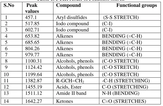
FTIR analysis has become an important tool in understanding the involvement of functional groups in interactions between metal particles and biomolecules. The synthesized copper nanoparticles were confirmed by changes that occurred in FTIR spectra after analysis. This spectrum showed lot of absorption bands indicates the presence of active functional groups in the synthesized copper nanoparticles. Figure. 4 and Table. 1 shows the absorption at 457.1cm-1, 517.85cm-1, 602.71cm-1, 653.82cm-1, 685.65cm-1, 804.26cm-1, 979.77cm-1, 1100.31cm-1, 1124.42 cm-1, 1199.64cm-1, 1382.87cm-1, 1455.19cm-1, 1511.12cm-1, 1642.27cm-1,
[image:4.612.72.296.169.328.2]1744.49cm-1, 2307.67cm-1, 2854cm-1, 2925cm-1, 3358.8cm-1. Through this peak absorption the functional groups are determined using the FTIR interpretation table 1, the aryl disulfides (S-S stretch), iodo compound C-I, bending =C-H alkene, (C-O stretch) alcohol, phenols, -C-H bending alkane, carboxylic group and N-H bending, C=O ketones (stretches), C-N, C-O stretching, C-H (stretches) alkane, O-H stretch. This indicates that the copper nanoparticles synthesized using
[image:4.612.327.539.307.450.2]Pedalium murex leaf extract was proved to have all these functional groups bound in it. Synthesis of copper nanoparticles from Gloriosa superba Lleaf extract. The FTIR characterization is used to find the molecules and their functional group present in the synthesized copper nanoparticles 1740, 1410, 1250, 1051, 885 and 765 cm-1. The FTIR spectra revealed the presence of different functional groups like alcohol, alkane, amine, nitro compounds, acid and ester [16].
Fig. 4. FTIR spectra of Pedalium murex leaf extract
Table 1. FTIR result of Pedalium murex
S.No Peak
values
Compound Functional groups
1 457.1 Aryl disulfides (S-S STRETCH) 2 517.85 Iodo compound (C-I)
3 602.71 Iodo compound (C-I)
4 653.82 Alkenes BENDING (=C-H)
5 685.65 Alkenes BENDING (=C-H)
6 804.26 Alkenes BENDING (=C-H)
7 979.77 Alkenes BENDING (=C-H)
8 1100.31 Alcohols, phenols (C-O STRETCH) 9 1124.42 Alcohols, phenols (C-O STRETCH) 10 1199.64 Alcohols, phenols (C-O STRETCH)
11 1382.87 R-GCH=CH2 -C-H (STRETCHING)
12 1455.19 Acids, Ester C-O (STRETCHING) 13 1511.12 Amide II band N-H (BENDING)
[image:4.612.151.464.518.722.2]305 The synthesized copper nanoparticle by leaf extract
was lyophilized and pelletized. It was examined by SEM. The magnification was done in 5.60x. From the SEM image more or less crystal in shape and the range was 5 µm. The surface morphology and interior
cross sectional structure of leaf extract was visible and the results were shown in Fig 5. The synthesis and characterization of copper nanoparticle using
Saraca indica spherical in shape and confirmed at the size ranging from 40-70nm [18].
Fig. 5. SEM analysis of Pedalium murex leaf extract
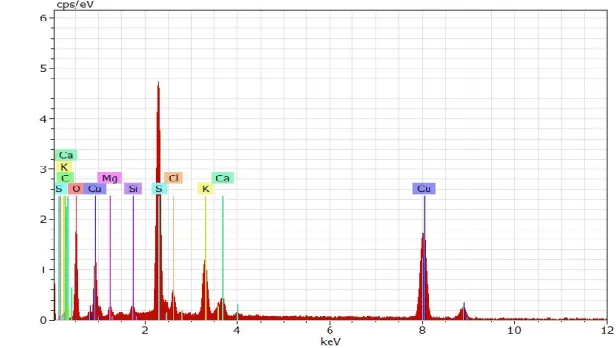
Energy Dispersive Absorption Spectroscopy (EDAX) of synthesized copper nanoparticles of Pedalium murex was showed in the Fig.6. The presence of expected element in the final products Cu was confirmed. The present analysis revealed that the nano-structures were formed as peak (Table 2) at 2.3 keV solely of copper nanoparticles for pedalium
murex. EDAX analysis Energy-dispersive X-ray report confirms the elemental composition for the synthesized CuNPs from the leaf extract of Passiflora foetida. This analysis also gave information about the weight percentage of CuNPs, depicting 49.95 weight % of Cu and 50.05 weight % of oxygen [19].
.
[image:5.612.168.476.521.695.2]\
Fig. 6. EDAX analysis of Pedalium murex leaf extract
15 1744.49 Ketones C=O (STRETCHES)
16 2307.67 Amide III band C-N (STRETCHES)
17 2854.45 Alkanes C-H (STRETCHES)
18 2925.81 Alkanes C-H (STRETCHES)
306
Table 2. EDAX result of Pedalium murex
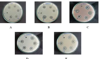
The antibacterial study was performed by Agar-well diffusion method. The effect of Pedalium murex leaf extract on standard strain of five human pathogenic
Escherica coli, Klebsiellae sp., Seratia sp., Pseudomonas sp., and Staphylococcus sp. were investigated for the antibacterial assays. Tetracycline was used as standard reference antibacterial compounds respectively. In the disc diffusion method, the diameter of the inhibition zone was measured (Table 3). The copper nanoparticles at different concentration such as 5μl, 10μl and 15μl
showed antimicrobial activity against human pathogens. Pedalium murex was most sensitive against Seratia sp., and Staphylococcus sp. with a maximum zone of inhibition in diameter (19mm) compared with other strains (Figure.7). The antibacterial activity of synthesized copper nanoparticles using Vitis vinifera leaf extract was evaluated against E. coli, S. aureus and K. pneumonia
with zone of inhibition of 14 mm, 16mm and 12 mm respectively [20].
Table 3. Antibacterial activity of Pedalium murex (Zone of inhibition)
S.No Particulars Concentration(µg) Zone of inhibition (mm)
1
Control
--- ---
2
Antibiotic
5 E.coli Kleb S.a Pseu Stapy
14 14 20 20 14
3 Plant (leaf) extract 5
13 12 18 15 15
4
Copper Nanoparticle extract (T1)
5
12 12 15 15 15
5
Copper Nanoparticle extract (T2)
10
14 13 17 16 16
6
Copper Nanoparticle extract (T3)
15
15 16 19 16 19
ELEMENTS SERIES UNN.
[wt %]
Cnorm. [wt %]
CAtom. [wt %]
C Error
(1Sigma) [wt%]
Carbon 6 K series 8.56 9.65 17.39 3.04
Oxygen 8 K series 39.14 44.09 59.69 6.79
Potassium 19 K series 3.58 4.03 2.23 0.16
Sulphur 16 K series 11.99 13.51 9.13 0.49
Copper 29 K series 20.94 23.60 8.04 0.61
[image:6.612.67.552.393.574.2]307
Fig.7. Antibacterial activity of leaf extract of Pedalium murex against(A). E.coli, (B). Klebsiella, (C). Serratia,
(D). Pseudomonas putida and (E).Staphylococcus, C – Control; A – Antibiotic; P – Plant (leaf) extract; Test sample (CuNPs) (T1, T2, T3)
Antifungal activity of Pedalium murex
derived copper nanoparticles were performed by Agar-well diffusion method. In this study, two fungal cultures were used namely Aspergillus niger and
Aspergillus nidulans. The copper nanoparticles were taken in different concentration such as 5μl and 10μl showed antifungal activity with zone of inhibition of
18mm and 17mm Figure. 8 and Table 4. Similarly, the antifungal activity was determined by using the agar well diffusion method for different concentration of copper nanoparticles against
Aspergillus niger with zone of inhibition in 25µl concentration at 8mm in Persea Americana seeds [21].
Fig. 8. Antifungal activity of leaf extract of Pedalium murex against(A). Aspergillus niger, (B). Aspergillus
[image:7.612.118.439.471.626.2]308
Table 4. Antifungal activity of Pedalium murex (Zone of inhibition)
S.No Particulars Concentration(µg) Zone of inhibition (mm)
1 Control --- ---
2 Antibiotic 5 A.niger A.nidulans
15 20
3 Plant (leaf) extract 5
14 18
4 Copper Nanoparticle extract (T1) 5
16 16
5 Copper Nanoparticle extract (T2) 10
18 17
The phytochemical screenings of Pedalium murex
[image:8.612.205.406.336.447.2]were investigated and the results were plotted in the Table 5. In the preliminary phytochemical analysis of the Pedalium murex leaf chloroform extracts were found to show the presence of tannins, saponins, flavonoids, carbohydrates, proteins and steroid Fig.9.
Similarly, Ziziphus mauritiana L. leaves revealed and highlighted the presence of tannins, saponins, steroid, protein and carbohydrates which may be responsible for the efficient stabilizing agent of nanoparticles [22].
Fig. 9.Phytochemical analysis of Pedalium murex Leaf extracts
Table 5. Phytochemical investigation of Pedalium murex
S.No Phyto
chemical screenings
Reagents Inference Result
1 Tannin Ferric
chloride test
Green colour
positive
2 Saponins Distilled water
Foam formation
Positive
3 Flavinoids NaOH Yellow
colour
positive
4 Protein Ninhydrin test Blue colour positive
[image:8.612.160.454.522.695.2]309
4.CONCLUSION
In conclusion, here we reported that the biosynthesis of copper nanoparticles using Pedalium murex aqueous extract provides a simple, efficient and environment friendly method for the synthesis of nanoparticles. UV– Vis showed that maximum peak was obtained at 218nm. SEM image showed the synthesized copper nanoparticles were relatively crystal in shape and it enhances antibacterial and antifungal activity which is compared with standard antibiotics. Qualitative analysis of phytochemicals revealed the presence of six compounds namely tannins, saponins, flavinoids, carbohydrates, proteins and steroid. Presence of these compounds suggests the medicinal properties of this plant and their biological effects which could be beneficial in the treatment of various diseases.
REFERENCE
[1] Parashar, V., Praashar, R., Sharma, B. & Pandey, A.C. (2009). Bioinspired synthesis of silver nanopaeticles. Digest Journal of Nanomaterials and Biostructure. 4(1): 45 – 50.
[2] Abad, M., Mertens, S.F.L., Pita, M., Fernndez, V.M. & Schiffrin, D.J. (2005). Functionalization of thiocitic acid-capped gold nanoparticles for specific immobilization of histidine-tagged proteins.
Journal of American Chemical Society. 127: 5689-5694.
[3] Pillai, K., Sareen, S.J, Toms, C.J., Chandramohanakumar N. & M. Balagopalan, (2012). Vermifugal Activity of Bio-fabricated Silver Nanoparticles. Research Journal of Recent Sciences.1: 47-51.
[4] Han W.K., Choi, J.W., Hwang, G.H., Hong, S.J., Lee, J.S. & Kang, S.G. (2006). Fabrication of Cu nano particles by direct electrochemical reduction from CuO nano particles. App. Surf Sci. 252:2832– 2838.
[5] Ponce, A.A. & Klabunde, K.J. (2005). Chemical and catalytic activity of copper nanoparticles prepared via metal vapour synthesis. J. Mol. Catal. 225:1-6.
[6] Huang, Z., Cui, F., Kang, H., Chen, J., Zhang, X. & Xia, C. (2008). Highly dispersed silica-supported copper nanoparticles prepared by precipitation-gel method: A simple but efficient and stable catalyst for glycerol hydrogenolysis. Chem Mater 20: 5090-5099.
[7] Lee H.J., Lee, G., Jang, N.R., Yun, J.H., Song, J.Y. & Kim, B.S. (2011). Biological synthesis of copper nanoparticles using plant extracts.
Nanotechnol.1:371-4.
[8] Subhankari, I. & Nayak, P.L. (2013). Synthesis of copper nanoparticles using Syzygium aromaticum
(cloves) aqueous extract by using green chemistry.
World J. Nano Sci. Technol. 2:14-7.
[9] Valodkar M., Jadeja, R.N., Thounaojam, M.C., Devkar, R.V. & Thakore, S. (2011). Biocompatible synthesis of peptide capped copper nanoparticles and their biological effect on tumor cells. Mater Chem Phys.128:83-9.
[10]Subramaniyan, S. & Nair, A.G.R. (1972). Flavonoids of the leaves of P.murex.
Phytochemistry. 11: 464-465.
[11]Yogendra, N. & Raghunath, S.S. (1983). Thakur Hepta triacontan Tetratriacontanyl octacososanoate and other constituents from P.murex.
Phytochemistry. 22(4): 973-974.
[12]Jombo, G.T.A., Emanghe, U.E., Amefule, E.N. & Damen, J.G. (2012). Nosocomial and community acquired uropathogenicisolates of Proteus mirabilis
and antimicrobial susceptibility profiles at a university hospital in Sub-Saharan Africa. Asian. Pac. J. Trop. Dis. 1: 7-11.
[13]Pawar, D., Shaikh, S., Shulaksana D., & Kanawade, R. (2013). Green synthesis of copper nanoparticles using Gloriosa leaf extract.
International Journal of Pharmacy and
Pharmaceutical Research. 1: 975-1491.
[14]Shelke, T.T., Bhaskar, V.H., Adkar, P.P., Jha, U. & Oswald, R.J. (2011). Antimicrobial Activities of
Pedalium murex Linn on Microbial pathogens.
IJRAP. 2(4): 1255-1257.
[15]Harborne, J.B., 1973. Phytochemical methods modern technique of plant analysis.
Methods in Plant Biochemistry. 1. 99-103.
[16]Pawar, D., Shaikh, S., Shulaksana, D., & Kanawade, R. (2013). Green synthesis of copper nanoparticles using Gloriosa leaf extract.
International Journal of Pharmacy and
Pharmaceutical Research. 1: 975-1491.
[17]Suganya, M. & Valli, G. (2017). Green synthesis of copper nanoparticles using delonix elata flower extract. International Journal of Nano corrosion Science and Engineering. 5(6): 816-823.
[18]Prasad, K.S., Patra, A., Shruthi, G. & Chandan, S. (2017). Aqueous Extract of Saraca indica Leaves
6 Steroids Acetic
unhydrin and sulphuric acid
Brown ring formation or black colour
310 in the Synthesis of Copper Nanoparticles: Journal
of Nanotechnology. 6: 1-6
[19]Fatma,S., Nila, P K., Ravindran, E. & Renganathan, S. (2017). Green synthesis of copper nanoparticle from Passiflora foetida leaf extract and its antibacterial activity. Asian Journal of Pharmaceutical and Clinical Research. 4(10):79-83.
[20]Angrasan, M & Subbaiya, R. (2014). Biosynthesis of copper Nanoparticles by vitis vinifera leaf aqueous extract and its antibacterial activity.
International journal of current microbiology and applied science.3(9): 768-774.
[21]Kumar, R.S & Rinitha, G. (2018). Nanostructural characterization of antimicrobial and antioxidant copper nanoparticles synthesized using novel
Persea Americana seeds. OpenNano. 3: 18-27. [22]Sambal, P., 2017. Phytochemical analysis of