R E S E A R C H
Open Access
Development of a microarray for
simultaneous detection and differentiation
of different tospoviruses that are
serologically related to
Tomato spotted wilt
virus
Lu-Yuan Liu
1, He-Yi Ye
2, Tsang-Hai Chen
3and Tsung-Chi Chen
2,4*Abstract
Background:Tospoviruses, the plant-infecting genus in the familyBunyaviridae, are thrips borne and cause severe agricultural losses worldwide. Based on the serological relationships of the structural nucleocapsid protein (NP), the current tospoviruses are divided into six serogroups. The use of NP-antisera is convenient for virus detection, but it is insufficient to identify virus species grouped in a serogroup due to the serological cross-reaction. Alternatively, virus species can be identified by the N gene amplification using specific primers.Tomato spotted wilt virus(TSWV) is the type species of the genusTospovirusand one of the most destructive plant viruses. Eight known tospoviruses, Alstroemeria necrotic streak virus (ANSV), Chrysanthemum stem necrosis virus (CSNV),Groundnut ringspot virus
(GRSV),Impatiens necrotic spot virus(INSV), Melon severe mosaic virus (MeSMV), Pepper necrotic spot virus (PNSV),
Tomato chlorotic spot virus(TCSV) andZucchini lethal chlorosis virus(ZLCV), sharing serological relatedness with TSWV in NP, are grouped in the TSWV serogroup. Most of the TSWV-serogroup viruses prevail in Europe and America. An efficient diagnostic method is necessary for inspecting these tospoviruses in Asia, including Taiwan. Methods:A microarray platform was developed for simultaneous detection and identification of TSWV-serogroup tospoviruses. Total RNAs extracted fromChenopodium quinoaleaves separately inoculated with ANSV, CSNV, GRSV, INSV, TCSV and TSWV were used for testing purposes. The 5’-biotinylated degenerate forward and reverse primers were designed from the consensus sequences of N genes of TSWV-serogroup tospoviruses for reverse transcription-polymerase chain reaction (RT-PCR) amplification. Virus-specific oligonucleotide probes were spotted on the surface of polyvinyl chloride (PVC) chips to hybridize with PCR products. The hybridization signals were visualized by hydrolysis of NBT/BCIP with streptavidine-conjugated alkaline phosphatase. The microarray was further applied to diagnose virus infection in field crop samples.
Results:Amplicons of approximately 0.46 kb were amplified from all tested TSWV-serogroup tospoviruses by RT-PCR using the degenerate primer pair Pr-dTS-f/Pr-dTS-r. Virus species were identified on chips by hybridization of PCR products with respective virus-specific probes. The microarray was successfully used to diagnose TSWV infection in field pepper samples.
(Continued on next page)
* Correspondence:[email protected] 2
Department of Biotechnology, Asia University, Wufeng, Taichung 41354, Taiwan
4Department of Medical Research, China Medical University Hospital, China Medical University, Taichung 40402, Taiwan
Full list of author information is available at the end of the article
(Continued from previous page)
Conclusions:In this study, a rapid, sensitive and precise microarray method has been developed to simultaneously detect and identify six TSWV-serogroup tospoviruses. The microarray platform provides a great potential to explore tospoviruses that can help researchers and quarantine staff to prevent invasions of tospoviruses.
Keywords:Tospovirus, N gene, Detection, Identification, Microarray
Background
Tospoviruses, classified in the family Bunyaviridae, are destructive plant viruses with a very broad host range, infecting more than 1090 plant species, causing lethal necrotic lesions, wilting and dieback of infected plants [1–3].Tomato spotted wilt virus(TSWV) is the first tos-povirus found in Australia in 1915 [4, 5]. To date, it is still the most important tospovirus causing severe damage of economically important crops, including tobacco, tomatoes, pepper, cucurbits, lettuce, groundnuts and pota-toes throughout Africa, America, Asia, Australasia and Europe [3].
Virions of tospoviruses are enveloped quasi-spherical particles measuring 80–120 nm in diameter, possessing a tripartite segmented ssRNA genome, named large (L), medium (M) and small (S). The L RNA is of negative sense, whereas the M and S RNAs are ambisense [6]. L RNA contains a large open reading frame (ORF) in the viral complementary (vc) strand for encoding an RNA-dependent RNA polymerase (RdRp) [7, 8]. Each of the M RNA and S RNA contains two ORFs separated by an A-U-rich intergenic region (IGR). The M RNA encodes a movement protein NSm from the viral (v) strand [9–12] and the enveloped Gn and Gc glycoproteins from the vc strand [13, 14]. The S RNA encodes an RNA-silencing suppressor NSs protein from the v strand [15, 16] and the RNA-associated nucleocapsid protein (NP) from the vc strand [17].
NP is abundant in infected plant cells [17]. The iden-tity and serology of NP are the most important criteria for identification of tospoviruses. A threshold of 90% amino acid (aa) identity of the NP is proposed to classify tospoviruses at the species level [6, 18]. Currently, 29 tospovirus species are identified based on the sequence determination of the N gene or S RNA [3, 19–24]. In addition, most of the known tospoviruses are clustered into three major serogroups, which are assigned by the
TSWV, Watermelon silver mottle virus (WSMoV) and
Iris yellow spot virus (IYSV) due to the serological re-latedness of their NPs [25]. The members of the TSWV serogroup principally occur in America and Europe. Alternatively, the tospoviruses belonging to the WSMoV and IYSV serogroups are prevalent in Asian countries [3]. The tospoviruses Alstroemeria necrotic streak virus (ANSV), Chrysanthemum stem necrosis virus (CSNV),
Groundnut ringspot virus (GRSV), Impatiens necrotic
spot virus(INSV), Melon severe mosaic virus (MeSMV),
Pepper necrotic spot virus (PNSV), Tomato chlorotic
spot virus (TCSV) and Zucchini lethal chlorosis virus
(ZLCV) were grouped in the TSWV serogroup [26–28],
and they are quarantine viruses in Taiwan and other Asian countries.
Prompt and accurate methods for detection and iden-tification of tospoviruses are crucial for preventing epi-demics of domestic and invading species. The serological enzyme-linked immunosorbent assay (ELISA) is the most convenient method for virus detection. Antisera against the NPs of tospoviruses are commonly used to detect tospoviruses. Due to the cross reactions of different species in the same serogroup, the use of NP antisera for virus identification could be limited [28, 29]. The nucleic acid-based reverse transcription-polymerase chain reaction (RT-PCR) using N gene-specific primers can be conducted to identify tospovirus species. However, a single species-specific primer pair for diagnosis of tospo-viruses in a farm is insufficient due to the complex virus categories which occurred in a crop or area. Degenerate primers designed from the conserved regions of genomic sequences simplify detection of tospoviruses in one tube-based RT-PCR, but they are inconvenient to identify virus species [30, 31]. The microarray is an efficient method for simultaneous detection and identification of various plant viruses in one assay to solve these problems [32].
In this study, an oligonucleotide-based microarray method was developed for detection and identification of tospoviruses, which are serologically indistinguishable with TSWV. The degenerate primer pair Pr-dTS-f/Pr-dTS-r was designed from the consensus sequences of N genes of TSWV-serogroup tospoviruses and successfully used to amplify a certain cDNA fragment from the total
RNAs of Chenopodium quinoa leaves separately
Methods Virus sources
The NY isolate of TSWV was collected from tomato in New York, the United States [30]. The original isolate of GRSV-BR was collected from tomato in Brazil [33]. The BR-03 isolate of TCSV was isolated from tomato in Brazil [34]. INSV-M was isolated from impatiens in the United States [35]. The TcCh07A isolate of CSNV was collected from chrysanthemum in Tochigi Prefecture, Japan [36]. The original isolate of ANSV was collected from Alstroemeria sp. in Colombia [27]. All virus cul-tures were maintained in the local lesion host Chenopo-dium quinoa Willd. by mechanical transmission in an isolated temperature-controlled (25–28 °C) greenhouse according to quarantine regulations. A buffer consisting of 0.01 M potassium phosphate (pH 7.0) and 0.01 M sodium sulfite was used for inoculation.
Construction of N genes
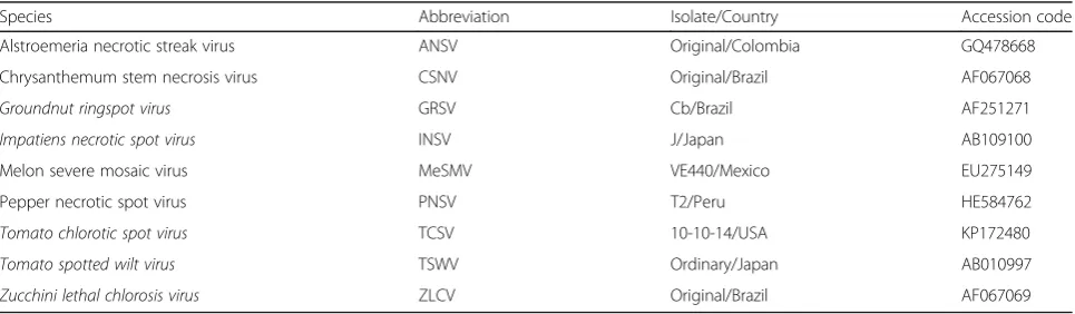
The sequences of the N genes or S RNAs of tospoviruses were obtained from the GenBank of National Center for Biotechnology Information (NCBI) (www.ncbi.nlm.-nih.gov) (Table 1) for design of N gene-specific primer pairs (Table 2). One hundred milligram of plant leaf tis-sues inoculated with ANSV, CSNV-TcCh07A, GRSV-BR, INSV-M and TCSV-BR-03 were ground to fine powder in liquid nitrogen and placed in microcentrifuge tubes for isolation of total RNAs using the Plant Total RNA Mini-prep Kit (GeneMark, GMbiolab Co., Ltd., Taichung, Taiwan) following the manufacturer’s instructions. The complete N gene sequences of tospoviruses were ampli-fied from the aforementioned total RNAs with individual primer pairs (Table 2) using the One-Step RT-PCR Kit (GeneMark). Ten microgram of total RNA, 200 nM indi-vidual primers, 25 U One-Step RT-PCR enzyme mix, 1/5 volume of reaction buffer and 1/5 volume of enhancer buffer (GeneMark) were mixed for amplification. Syn-thesis of cDNAs was conducted at 50 °C for 30 min, and inactivation at 94 °C for 2 min. PCR was performed
by 35 cycles of strand separation at 94 °C for 1 min, annealing at 58 °C for 30 s and synthesis at 72 °C for 1 min; and a final reaction at 72 °C for 7 min. The amplicons were ligated with the TOPO TA cloning vec-tor pCR2.1-TOPO (Invitrogen, Carlsbad, CA), and then
transferred into the DH5α competent cells following
the manufacturer’s instructions. The resultant plasmids named pTOPO-ANSV-N for ANSV, pTOPO-CSNV-N for CSNV-TcCh07A, pTOPO-GRSV-N for GRSV-BR, pTOPO-INSV-N for INSV-M and pTOPO-TCSV-N for TCSV-BR-03 were obtained by the Plasmid Miniprep Purification Kit (GeneMark) following the standard protocols. The nucleotide sequences of all cloned N genes were verified by sequencing using ABI3730 XL DNA Analyzer (Perkin-Elmer Applied Biosysterms, Foster City, CA), performed by Mission Biotech Com-pany (Taipei, Taiwan). The plasmid pTOPO-TSWV-N for TSWV-NY was constructed in the previous study [28].
Design of degenerate primers and virus-specific probes The Lasergene 7 software package (DNASTAR, Madison, WI, USA) was used to analyze sequences. Multiple align-ments of nucleotide sequences of N genes listed in Table 1 were conducted by the MegAlin program. The conserved regions of N genes were used to design degenerate primers, and the virus-specific probes were designed from the variable regions of N genes.
Amplification assay for degenerate primers
PCR was conducted to evaluate the degenerate primers using the constructed plasmids. The reaction mixture of 25μl is comprised of 100 ng of plasmid DNA, 2.5 μl of
10× Taq buffer (50 mM Tris–HCl, pH 8.0, 1 mM EDTA,
1 mM DTT and 50% glycerol) (Protech, Taipei, Taiwan), 1μl of 10 mM dNTPs (Protech), 1μl of 100μM forward primer, 1μl of 100 μM reverse primer and 0.1μl of Pro Plus Taq DNA polymerase (5 U/μl) (Protech). The ampli-fication was carried out by Applied Biosystems GeneAMP PCR System 9700 (Thermo Fisher Scientific Inc., MA,
Table 1Accession codes of N genes or S RNAs used for multiple alignments
Species Abbreviation Isolate/Country Accession code
Alstroemeria necrotic streak virus ANSV Original/Colombia GQ478668
Chrysanthemum stem necrosis virus CSNV Original/Brazil AF067068
Groundnut ringspot virus GRSV Cb/Brazil AF251271
Impatiens necrotic spot virus INSV J/Japan AB109100
Melon severe mosaic virus MeSMV VE440/Mexico EU275149
Pepper necrotic spot virus PNSV T2/Peru HE584762
Tomato chlorotic spot virus TCSV 10-10-14/USA KP172480
Tomato spotted wilt virus TSWV Ordinary/Japan AB010997
Zucchini lethal chlorosis virus ZLCV Original/Brazil AF067069
a
USA) under the setting of a hot start at 95 °C for 5 min; 35 cycles of 95 °C for 30 s, 50 °C for 40 s and 72 °C for 30 s; and a final reaction at 72 °C for 6 min. The ampli-cons were visualized by 2% agarose gel electrophoresis and ethidium bromide (EtBr) staining. For virus detection, the mixture of 25 μl consisting of 200 ng of plant total RNA, 0.2 μl of M-MuLV reverse transcriptase (5 U/μl) (Protech), 2.5 μl of 10× Taq buffer (Protech), 1 μl of 10 mM dNTPs (Protech), 0.1μl of 100μM forward pri-mer, 0.1 μl of 100μM reverse primer and 0.1 μl of Pro Plus Taq DNA polymerase (5 U/μl) (Protech) was used in RT-PCR amplification. Complementary DNA was synthe-sized at 45 °C for 30 min then termination at 95 °C for 5 min. The PCR amplification was set as a hot start at 95 °C for 5 min; 35 cycles of 95 °C for 30 s, 50 °C for 40 s and 72 °C for 30 s; and a final reaction at 72 °C for 6 min. The amplicons were observed by 2% agarose gel electrophoresis and EtBr staining.
Microarray preparation and assay
Twenty micromolar of each probe were spotted on the surface of PVC microchips with an automatic spotting machine (DR. Chip Biotechnology Inc., Miaoli, Taiwan) and immobilized by an ultraviolet crosslinker (UV Light Enterprise Co., Taichung, Taiwan) with 1.2 J for 5 min following the previous report [37]. In addition to the spots for specific tospoviruses, 100 μM of the commer-cial probe Anti-VP1 (Dr. Chip) was spotted for positive control of hybridization. Ten microliter of PCR products
were mixed with 200 μl of commercial DR. Chip Hyb™
buffer (Dr. Chip) containing the 5’ end-biotinylated oligonucleotide (VP1) complementary to the sequence of positive control probe Anti-VP1. The mixture was denatured in boiling water for 5 min then immediately chilled on ice for 3 min. Subsequently, the mixture was transferred into the chip chamber and the chips were incubated at 50 °C in a commercial hybridization oven
(DR. Chip) with vibration for 1 h. After hybridization, the chips were washed by 200 μl of Wash buffer™ (Dr. Chip) in a 1 min incubation for 3 repeats. Then, the chip chamber was inverted and tapped on a paper towel to remove the residual liquid. Thereafter, the mixture of 0.2 μl of DR. Chip Srep-AP™ (Streptavidin-conjugated alkaline phosphatase, Dr. Chip) in 200 μl of DR. Chip Block Reagent™ (Dr. Chip) was transferred into the chip chamber to allow a 30 min incubation at room temperature. The chamber was washed 3 times with 200 μl of wash buffer as described above, followed by rinsing the chips with 200 μl of Detection Buffer™ (Dr. Chip). After discarding the liquid, 4μl of DR. NBT/BCIP™ (nitro blue tetrazolium/5-bromo-4-chloro-3-indolyl phos-phate, Dr. Chip) was mixed with 196μl of detection buf-fer, and the mixture was added to the hybridization chamber. The colorimetric development was allowed to react in the dark for 10 min at room temperature, and then the detection liquid was discarded. The chip was rinsed with deionized water twice, and the developed pat-terns were recorded by DR. Scanning Reader (Dr. Chip).
Establishment of the detection platform of the microarray The PCR product amplified from pTOPO-TSWV-N [28] using the designed primers was serially diluted for testing the detection sensitivity of the microarray. Total RNAs extracted from TSWV-infected pepper samples were used for testing the virus identification by microarray. The pro-tocols of the microarray were described above.
Indirect enzyme-linked immunosorbent assay (ELISA) Indirect ELISA was conducted to detect TSWV in dis-eased plant samples according to the previous report [38]. The crude extracts of plant samples at a 1/20 dilu-tion with coating buffer (0.05 M sodium carbonate, pH 9.6, containing 0.01% sodium azide) were used for assays. The monoclonal antibody against the NSs protein
Table 2The primer pairs used for cloning of N genes of tospoviruses
of TSWV, MAb-TNSs-82D3B4 [39], diluted with conju-gate buffer (PBST containing 2% PVP-40 and 0.2% BSA), was used at a 1/8000 dilution. The alkaline phosphatase (AP)-conjugated goat anti-mouse IgG (Jackson Immu-noResearch Laboratories) was used at a 1/5000 dilution as the secondary antibody. The AP substrate tablets (ρ-Nitrophenyl phosphate disodium salt hexahydrate) (Sigma-Aldrich, St. Louis, MO) dissolved in substrate buffer (9.7% diethanolamine and 0.02% NaN3, pH 9.8) were used for colour development. The absorbance at 405 nm (A405) was determined by Model 680 microplate reader (Bio-Rad) after 10 to 60 min of substrate addition.
Results
Detection test of the designed degenerate primers and virus-specific probes in the microarray
Two conserved regions located at the first 20 nucleotides and middle portion of N genes of the TSWV-serogroup tospoviruses were used to design the forward primer Pr-dTS-f and reverse primer Pr-dTS-r, respectively (Fig. 1a).
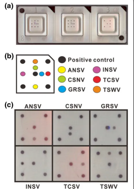
Both primers were synthesized with a 5’-biotinylation (Table 3). The primer pair Pr-dTS-f/Pr-dTS-r was able to amplify a DNA fragment of approximately 0.46 kb from all tested plasmids carrying the N genes of ANSV, CSNV, GRSV, INSV, TCSV or TSWV in PCR (Fig. 1b). The virus-specific probes designed from the N genes of ANSV, CSNV, GRSV, INSV, TCSV and TSWV were synthesized with an addition of 20 thymidine deoxynucleosides (T20) at the 5’terminus for fixing on microchips (Table 3). The spotted positions of individual probes in the microarray are indicated in Fig. 2a and b. The PCR products amplified from different plasmids were incubated with the micro-array for hybridization. Colorization of the specific spots indicated that the virus species can be identified by the microarray (Fig. 2c).
Sensitivity assay of microarray
The PCR product amplified from the plasmid pTOPO-TSWV-N was serially diluted, initiating from 250 to 0.1 ng, to determine the sensitivity of microarray detection.
EtBr staining in agarose gel electrophoresis was performed for comparison. The results showed that the microarray was able to detect a lesser amount of 0.1 ng of the PCR product that is more sensitive than EtBr staining with a detection limitation of 0.2 ng of the PCR product (Fig. 3).
Microarray in virus detection
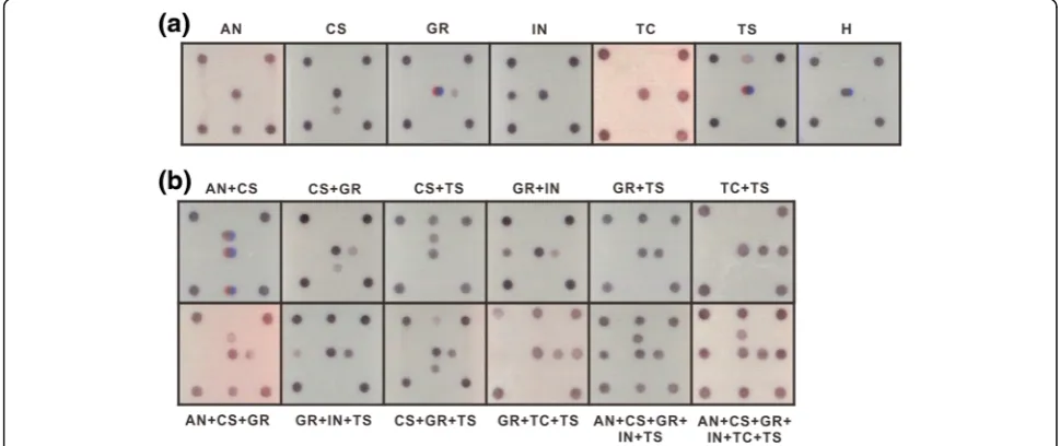
RT-PCR was performed to amplify the DNA fragment of
0.46 kb from the individual total RNAs of C. quinoa
leaves separately inoculated with ANSV, CSNV, GRSV, INSV, TCSV or TSWV as well as the plasmid results shown in Fig. 1b. The amplicons were incubated with probe-spotted microchips mentioned in Fig. 2b, and all tested tospoviruses could be singly detected by the microarray (Fig. 4a). Furthermore, the total RNAs of dif-ferent virus infections were mixed in 2–6 combinations for microarray assays, and individual tospovirus species could be identified (Fig. 4b). Our results demonstrated that the degenerate primer pair is sufficient for detection of the members of the TSWV serogroup and that the microarray is efficient in virus identification.
Application of microarray for virus diagnosis in crop samples
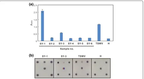
The diseased pepper samples (SY-1 to 6), exhibiting chlorotic spots or ringspots on leaves and fruits, were collected from Sinyi Township, Nantou County, Taiwan in December 2015 for detecting tospovirus infection by indirect ELISA using the monoclonal antibody against the TSWV NSs protein [39]. SY-1 and 3 exhibited an ELISA-positive (Fig. 5a). These samples were also used for assay in the microarray, and the identical result as well as indirect ELISA was obtained (Fig. 5b).
Discussion
An efficient detection method is necessary for inspection of viral diseases in quarantine. The previous studies reported that the tospoviruses ANSV, CSNV, GRSV, INSV, TCSV and TSWV share a serological relationship in their NPs, so that they can be detected, but not be differentiated, by antisera against the NP in serological assays [28, 40]. On the other hand, several consensus sequences can be found among tospoviral genomic sequences. Degenerate primers designed from the consen-sus nucleotide sequences of RdRp and NSm genes have been used to detect tospoviruses at the genus level [30, 31].
Table 3Primers and probes used for the microarray
Primer/Probe Length (bp) Sequence (5’→3’) Specificity
Primer name
Pr-dTS-f 20 Biotin ATGTMTAAGGYHAAGHTYAC TSWV serogroup
Pr-dTS-r 26 Biotin GAAGCWATVAGAGGNADRCTWCCTCC
Probe name
Pb-AN 40 T20ATGGACTTCCTTTGAATGAT ANSV
Pb-CS 40 T20ACGGGCTTAGCTTGAATGAT CSNV
Pb-GR 40 T20ACGGGCTGCCTCTGGCAGAT GRSV
Pb-IN 40 T20ATGGTCTTGCAACCACAGAT INSV
Pb-TS 40 T20ATGGATTACCTCTCGATGAT TSWV
Fig. 2Establishment of the microarray.aThe biochip device.bThe graph represents the biochip elements. The positions of virus-specific probes spotted on the surface of biochips are indicated bycolor spots.
The virus strains with different genotypes will not be excluded when degenerate primers are used in amplifi-cation. By contrast, the N genes of tospoviruses are more diverse, thus it can be the key target for classifica-tion of a tospovirus [6].
Consensus nucleotide sequences can also be found within the N genes of tospoviruses belonging to a phylo-genetical clade. According to this finding, the degenerate primer pair Pr-dTS-f/Pr-dTS-r were designed from the N genes of all nine TSWV-serogroup tospoviruses, and can successfully detect all six tested viruses, ANSV,
CSNV, GRSV, INSV, TCSV and TSWV, in one-step RT-PCR (Fig. 1). Unfortunately, the other three TSWV-serogroup viruses, MeSMV, PNSV and ZLCV, can not be obtained for test in the present study. Sequence compari-son of the N genes of all members of TSWV serogroup was conducted to reveal that INSV shares the lowest nu-cleotide identity (59.3–61.7%) with other tospoviruses (Additional file 1: Table S1) and still can be detected by the degenerate primer pair Pr-dTS-f/Pr-dTS-r. Thus, the detection of MeSMV, PNSV and ZLCV using the degenerate primer pair Pr-dTS-f/Pr-dTS-r can be Fig. 3Sensitivity assay of the microarray. The PCR product amplified from pTOPO-TSWV-N using the primer pair Pr-dTS-f/Pr-dTS-r was diluted as shown for the test.aThe diluted amplicons were analysed by agarose gel electrophoresis with ethidium bromide staining.bThe microarray result. The concentrations of amplicon are indicated as 1: 250 ng; 2: 125 ng; 3: 62.5 ng; 4: 32 ng; 5: 16 ng; 6: 8 ng; 7: 4 ng; 8: 2 ng; 9: 1 ng; 10: 0.5 ng; 11: 0.2 ng; and 12: 0.1 ng
expected even if the virus materials are unavailable. The use of a universal primer pair in simplex RT-PCR significantly increases the efficiency of nucleic acid amplification by comparison with the use of combina-tions of various primer pairs in multiplex RT-PCR. Here, we first integrated the degenerate primers and specific probes into a microarray system to simultan-eously detect and differentiate the TSWV-serogroup tospoviruses that possess twofold higher sensitivity than conventional RT-PCR (Fig. 3). The tospoviruses belong-ing to other serogroups, includbelong-ing Calla lily chlorotic spot virus (CCSV), Capsicum chlorosis virus (CaCV),
Groundnut bud necrosis virus (GBNV), Groundnut chlorotic fan-spot virus (GCFSV), IYSV, Melon yellow spot virus (MYSV), Soybean vein necrosis-associated virus (SVNaV), Tomato necrotic spot-associated virus (TNSaV), Tomato yellow ring virus (TYRV), Tomato
zonate spot virus (TZSV), Watermelon bud necrosis
virus (WBNV) and WSMoV, were also used to test the developed microarray. No unexpected signals were ob-served to demonstrate the excellent specificity of the microarray in TSWV serogroup only (data not shown). Using the same approach, microarrays for detecting tospoviruses in other serogroups will be further developed.
The DNA microarray is basically developed as a com-bination technique of nucleic acid amplification and hybridization for high-throughput assay and has a wide application including use in analyses of gene expression, transcription factor binding and genotyping [41]. It also provides a benefit for simultaneous detection of numer-ous plant RNA viruses [32]. The reliability and time-and-cost-saving are very important issues in develop-ment of detection methods. In this study, we used the PVC biochip-based microarray platform developed by the Dr. Chip Company. Different from the common glass-slide microarray system, the signals on PVC bio-chips can be directly observed by the naked eye and recorded by an optical scanner or camera after coloriza-tion instead of the need of expensive equipment for fluorescent detection. It also provides an advantage of customized probe spotting. The reliability of the PVC biochip has been proven in several applications; e.g., the simultaneous detection and differentiation of seven mastitis-causing pathogens in bovine milk samples [37], Newcastle disease and avian influenza in the poultry
industry [42], and Cymbidium mosaic virus,
Odonto-glossum ringspot virus and CaCV for orchid inspection (Dr. Orchid-3™Kit, Dr. Chip), and the identification of insect species [43] and the sex of owls [44].
The developed microarray was used to inspect virus infection in the fields of Sinyi Township in central Taiwan, and indirect ELISA using MAb-TNSs-82D3B4 was performed for comparison. MAb-TNSs-82D3B4 reacted with TSWV but not reacted with GRSV, INSV and TCSV was mentioned in our previous report [39], and its serological reactivity with ANSV and CSNV was proven by our laboratory later. Thus, in this case, the ELISA-positive result might be resulted from the infec-tions of ANSV, CSNV and/or TSWV (Fig. 5a). The de-veloped microarray helps to clarify the result of indirect ELISA, indicating that two pepper samples, SY-1 and 3, exhibiting ringspots on leaves and fruits, were infected with TSWV only (Fig. 5b). No other TSWV-serogroup tospoviruses, such as ANSV, CSNV, GRSV and INSV, were found in Taiwan yet. Our results also demonstrated the reliability of the microarray in diagnosis of tospoviral diseases.
Tospoviruses are vectored by thrips for oversea distribu-tion. The identification of the thrips category and the in-spection of viruliferous thrips are necessary for preventing an invasion of foreign tospoviruses. The microarray is suf-ficient for multiplex detections. The identifications of tos-poviruses and thrips will be integrated into a biochip to strengthen inspection for quarantine.
Conclusions
In this study, a degenerate primer pair Pr-dTS-f/Pr-dTS-r, designed from the consensus sequences of N genes, was used to amplify a DNA fragment from total RNAs of plant tissues infected by six tested tospoviruses, ANSV, CSNV, GRSV, INSV, TCSV and TSWV. The virus species were further differentiated by hybridization with specific probes spotted on PVC biochips. The microarray platform was used to diagnose TSWV infections in field peppers. Our results showed that the microarray method can be used to simultaneously detect and identify TSWV-serogroup tos-poviruses that provides a great applicable potential in inspection of tospoviruses for quarantine.
Additional file
Additional file 1: Table S1. Nucleotide (above diagonal) and amino acid (below diagonal) identities (%) of the N genes among the members of TSWV serogroup. (DOCX 68 kb)
Abbreviations
ANSV:Alstroemeria necrotic streak virus; CaCV: Capsicum chlorosis virus; CCSV: Calla lily chlorotic spot virus; CSNV: Chrysanthemum stem necrosis virus; ELISA: Enzyme-linked immunosorbent assay; GBNV:Groundnut bud necrosis virus; GCFSV: Groundnut chlorotic fan-spot virus; GRSV:Groundnut ringspot virus; GYSV:Groundnut yellow spot virus; INSV:Impatiens necrotic spot virus; IYSV:Iris yellow spot virus; MAb: Monoclonal antibody; MeSMV: Melon severe mosaic virus; MYSV: Melon yellow spot virus; NP: Nucleocapsid protein; ORF: Open reading frame; PNSV: Pepper necrotic spot virus; RT-PCR: Reverse transcription-polymerase chain reaction; SVNaV: Soybean vein necrosis-associated virus; TCSV:Tomato chlorotic spot virus;
TNSaV: Tomato necrotic spot-associated virus; TSWV:Tomato spotted wilt virus; TYRV: Tomato yellow ring virus; TZSV: Tomato zonate spot virus; WBNV:Watermelon bud necrosis virus; WSMoV:Watermelon silver mottle virus; ZLCV:Zucchini lethal chlorosis virus
Acknowledgements
We thank the virus providers: Dr. R. Provvidenti for TSWV; Dr. Dennis Gonsalves for GRSV; Dr. James W. Moyer for INSV; Dr. Richard Kormelink for ANSV; Dr. Mitsuru Okuda for CSNV; the DSMZ Plant Virus Collection, Germany for TCSV; and the permission of Bureau of Animal and Plant Health Inspection and Quarantine, Council of Agriculture, Executive Yuan for the importation of the viruses. We also thank the great support for the chip production and probe design suggestions from Dr. Kuang-Hui Lu and Yen-Hou Chen (Department of Entomology, National Chung Hsing University) and Cheng-Wei, Lin (BION TECH INC.). Our gratitude also goes to Michael Burton, Asia University.
Authors’contributions
Designed experiments and critically revised the manuscript: LYL, TCC. Performed experiments: LYL, HYY. Collected samples: TCC. Analyzed data: LYL, TCC. Drafted manuscript: THC, TCC. All authors read and approved the final manuscript.
Competing interests
The authors declare that they have no competing interests.
Author details
1Department of Plant Industry, National Pingtung University of Science and Technology, Pingtung 91201, Taiwan.2Department of Biotechnology, Asia University, Wufeng, Taichung 41354, Taiwan.3Department of Plant Medicine, National Pingtung University of Science and Technology, Pingtung 91201, Taiwan.4Department of Medical Research, China Medical University Hospital, China Medical University, Taichung 40402, Taiwan.
Received: 30 September 2016 Accepted: 9 December 2016
References
1. Mumford RA, Barker I, Wood KR. The biology of the tospoviruses. Ann Appl Biol. 1996;128:159–83.
2. Parrella G, Gognalons P, Gebre-Selassiè K, Vovlas C, Marchoux G. An update of the host range oftomato spotted wilt virus. J Plant Pathol. 2003;85:227–64. 3. Pappu HR, Jones RAC, Jain RK. Global status of tospovirus epidemics in
diverse cropping systems: successes achieved and challenges ahead. Virus Res. 2009;141:219–36.
4. Brittlebank CC. Tomato diseases. J Agric Victoria. 1919;17:213–35. 5. Samuel G, Bald JG, Pittman HA. Investigation on“spotted wilt”of tomatoes.
Austral Council Sci Indus Res Bull. 1930;44:1–46.
6. King AMQ, Adams MJ, Carstens EB, Lefkowitz EJ. Virus taxonomy: ninth report of the international committee on taxonomy of viruses. United States of America: Elsevier; 2012. p. 725–41.
7. de Haan P, Kormelink R, Resende RD, Vanpoelwijk F, Peters D, et al.Tomato spotted wilt virus-L RNA encodes a putative RNA-polymerase. J Gen Virol. 1991;72:2207–16.
8. van Knippenberg I, Goldbach R, Peters D. Purified tomato spotted wilt virus particles support both genome replication and transcription in vitro. Virology. 2002;303:278–86.
9. Kormelink R, Storms M, van Lent J, Peters D, Goldbach RW. Expression and subcellular location of the NSm protein oftomato spotted wilt virus(TSWV), a putative viral movement protein. Virology. 1994;200:56–65.
10. Storms MM, Kormelink R, Peters D, van Lent JW, Goldbach RW. The nonstructural NSm protein oftomato spotted wilt virusinduces tubular structures in plant and insect cells. Virology. 1995;214:485–93.
11. Lewandowski DJ, Adkins S. The tubule-forming NSm protein fromtomato spotted wilt viruscomplements cell-to-cell and long-distance movement of tobacco mosaic virushybrids. Virology. 2005;342:26–37.
13. Kikkert M, Verschoor AD, Kormelink R, Peters D, Goldbach RW.Tomato spotted wilt virusglycoproteins exhibit trafficking and localization signals that are functional in mammalian cells. J Virol. 2001;75:1004–12. 14. Sin SH, Mcnulty BC, Kennedy GG, Moyer JW. Viral genetic determinants for
thrips transmission oftomato spotted wilt virus. Proc Natl Acad Sci U S A. 2005;102:5168–73.
15. Takeda A, Sugiyama K, Nagano H, Mori M, Kaido M, Mise K, Tsuda S, Okuno T. Identification of a novel RNA silencing suppressor, NSs protein oftomato spotted wilt virus. FEBS Lett. 2002;532:75–9.
16. Bucher E, Sijen T, de Haan P, Goldbach R, Prins M. Negative-strand tospoviruses and tenuiviruses carry a gene for a suppressor of gene silencing at ananlogous genomic positions. J Virol. 2003;77:1329–36. 17. de Haan P, Wagemakers L, Peters D, Goldbach RW. The S RNA segment of
tomato spotted wilt virushas an ambisense character. J Gen Virol. 1990; 71:1001–7.
18. Goldbach RW, Kuo G. Introduction: proceedings of the international symposium on tospovirus and thrips of floral and vegetable crops. Acta Hortic. 1996;431:21–6.
19. Torres R, Larenas J, Fribourg C, Romero J. Pepper necrotic spot virus, a new tospovirus infecting solanaceous crops in Peru. Arch Virol. 2012;157:609–15. 20. Dong JH, Yin YY, Fang Q, McBeath JH, Zhang ZK. A new tospovirus causing chlorotic ringspot onhippeastrumsp. In China. Virus Genes. 2013;46:567–70. 21. Meng JR, Liu PP, Zou CW, Wang ZQ, Liao YM, Cai JH, Qin BX, Chen BS. First
report of atospovirusin mulberry. Plant Dis. 2013;97:1001. 22. Cheng YH, Zheng YX, Tai CH, Yen JH, Chen YK, Jan FJ. Identification,
characterisation and detection of a new tospovirus on sweet pepper. Ann Appl Biol. 2014;164:107–15.
23. Shimomoto Y, Kobayashi K, Okuda M. Identification and characterization of lisianthus necrotic ringspot virus, a novel distinct tospovirus species causing necrotic disease of lisianthus (eustoma grandiflorum). J Gen Plant Pathol. 2014;80:169–75.
24. Yin Y, Zheng K, Dong J, Fang Q, Wu S, Wang L, Zhang Z. Identification of a new tospovirus causing necrotic ringspot on tomato in China. Virol J. 2014;11:213.
25. Chen TC, Jan FJ. Tomato spotted wilt. In: Tennant P, Fermin G, editors. Virus diseases of tropical and subtropical crops. Oxfordshire: CAB International; 2015. p. 162–76.
26. Ciuffo M, Kurowski C, Vivoda E, Copes B, Masenga V, Falk BW, Turina M. A newtospovirussp. In cucurbit crops in Mexico. Plant Dis. 2009;93:467–74. 27. Hassani-Mehraban A, Botermans M, Verhoeven JT, Meekes E, Saaijer J, Peters
D, Goldbach R, Kormelink R. A distinct tospovirus causing necrotic streak on alstroemeriasp. In Colombia. Arch Virol. 2010;155:423–8.
28. Chen SM, Wang YC, Wu PR, Chen TC. Production of antiserum against the nucleocapsid protein oftomato spotted wilt virusand investigation of its serological relationship with other tospoviruses. Plant Prot Bull. 2014; 56:55–74 (In Chinese).
29. Chen TC, Lu YY, Cheng YH, Li JT, Yeh YC, et al. Serological relationship between melon yellow spot virus andwatermelon silver mottle virusand differential detection of the two viruses in cucurbits. Arch Virol. 2010;155: 1085–95.
30. Chu FH, Chao CH, Chung MH, Chen CC, Yeh SD. Completion of the genome sequence ofwatermelon silver mottle virusand utilization of degenerate primers for detecting tospoviruses in five serogroups. Phytopathology. 2001;91:361–8.
31. Chen TC, Li JT, Lin YP, Yeh YC, Kang YC, Huang LH, Yeh SD. Genomic characterization of calla lily chlorotic spot virus and design of broad-spectrum primers for detection of tospoviruses. Plant Pathol. 2012;61:183–94. 32. Nicolaisen M. An oligonucleotide-based microarray for detection of plant
RNA viruses. J Virol Methods. 2011;173:137–43.
33. Pang SZ, Slightom JL, Gonsalves D. The biological properties of a distinct tospovirus and sequence analysis of its S RNA. Phytopathology. 1993;83:728–33. 34. de Avila AC, de Haan P, Kormelink R, Resende Rde O, Goldbach RW, Peters
D. Classification of tospoviruses based on phylogeny of nucleocapsid gene sequences. J Gen Virol. 1993;74:153–9.
35. Law MD, Moyer JW. A tomato spotted wilt-like virus with a serologically distinct N protein. J Gen Virol. 1990;71:933–8.
36. Takeshita M, Nagai N, Okuda M, Matsuura S, Okuda S, Furuya N, Tsuchiya K. Molecular and biological characterization ofchrysanthemum stem necrosis virus isolates from distinct regions in Japan. Eur J Plant Pathol. 2011;131:9–14. 37. Lee KH, Lee JW, Wang SW, Liu LY, Lee MF, Chuang ST, Shy YM, Chang CL,
Wu MC, Chi CH. (2008). development of a novel biochip for rapid multiplex
detection of seven mastitis-causing pathogens in bovine milk samples. J Vet Diagn Invest. 2008;20:463–71.
38. Chen YH, Dong J, Chien WC, Zheng K, Wu K, Yeh SD, Sun JH, Wang YC, Chen TC. Monoclonal antibodies for differentiating infections of three serological-related tospoviruses prevalent in southwestern China. Virol J. 2016;13:72. doi:10.1186/s12985-016-0525-3.
39. Chen TC, Tsai WT, Kang YC, Wang YC, Yeh SD. Using monoclonal antibodies against the common epitopes of NSs proteins for the prompt detection and differentiation of tospoviruses prevalent in Euro-America and Asia regions. Eur J Plant Pathol. 2016;144:509–24.
40. Wu PR, Chien WC, Okuda M, Takeshita M, Yeh SD, Wang YC, Chen TC. Genetic and serological characterization of chrysanthemum stem necrosis virus, a member of the genustospovirus. Arch Virol. 2015;160:529–36. 41. Bumgarner R. DNA microarrays: Types, applications and their future. Curr
Protoc Mol Biol. 2013; 22: Unit-22.1.. doi: 10.1002/0471142727.mb2201s101 42. Wang LC, Pan CH, Severinghaus LL, Liu LY, Chen CT, Pu CE, Huang D, Lir JT,
Chin SC, Cheng MC, Lee SH, Wang CH. Simultaneous detection and differentiation of Newcastle disease and avian influenza using oilgonucleotide microarrays. Vet Microbiol. 2008;127:217–26.
43. Chen YH, Liu LY, Tsai WH, Haymer DS, Lu KH. Using DNA chips for identification of tephritid pest species. Pest Manag Sci. 2013; doi: 10.1002/ps.3687 44. Wang LC, Severinghaus LL, Chen CT, Liu LY, Pan CH, Huang D, Lee HY, Lir
JT, Chin SC, Pu CE, Wang CH. Sex identification of owls (family strigidae) using oligonucleotide microarrays. J Hered. 2008;99:187–92.
• We accept pre-submission inquiries
• Our selector tool helps you to find the most relevant journal • We provide round the clock customer support
• Convenient online submission • Thorough peer review
• Inclusion in PubMed and all major indexing services • Maximum visibility for your research
Submit your manuscript at www.biomedcentral.com/submit