Brain Tumor Detection by FMRI using
MATLAB
Patel Miteshkumar1, Patel Tarang2, Desai Naincy3
PG Student, Dept. of ELE, SSASIT, Surat, Gujarat, India1
PG Student, Dept. of Biomedical, Ganpat University, Ahmedabad, Gujarat, India2 PG Student, Dept. of ECE, CGPIT, Surat, Gujarat, India3
ABSTRACT: Functional Magnetic Resonance Imaging (fMRI) is one of the best techniques for neuroimaging and have revolutionized the way to understand the brain functions. It measures the changes in the blood oxygen level-dependent (BOLD) signal which is related to the neuronal activity. Complexity of the data, presence of di erent types of noises and the massive amount of data makes the fMRI data analysis a challenging one. It demands e cient signal processing and statistical analysis methods. The inference of the analysis are used by the physicians, neurologists and researchers for better understanding of the brain functions. Here we use Matlab GUI model for tumor detection and using different filter and morphological operation, tumor will detect easily.
KEYWORDS: FMRI, Brain tumor, trilateral filter.
I.INTRODUCTION
Brain tumor is one of the biggest concern for researchers and doctors because patients having this diseases increasing day-by-day. Brain is a most important part of body because most of our activities like speech, storing memory, problem solving, social interaction, emotions etc. is regulated by brain. So, we need to be more careful while doing any treatment or surgery if anyone is having problem like tumor. Treating of tumor is not big concern but detection of tumor at proper place in brain is most challenging part for all researchers and doctors.In the past, most of brain tumor was detected by many imaging techniques. For example, CT (Computed Topography) scan is helpful in diagnosing some types of brain tumors, especially those near or involving bone. They can also show swelling, bleeding, and bone and tissue calcification because of having high radiation X-rays which are not soft tissues of brain. For solving the problem of high radiation, MRI (Magnetic Resonance Imaging) scanner came into action for detecting brain tumor. The main feature of MRI is to scan anatomical structure inside a human organism, including those in the human brain. An MRI’s anatomical imaging views at a high resolution the differences between tissue types with respect to space with help of doing studies with hydrogen water molecule. Now a day, most of tumor patient is treated by MRI scanning because it is cheaper and effective. The modification of MRI is Functional Magnetic Resonance Imaging (FMRI), which uses bold signal for tumor detection.
In this paper, FMRI uses Compose metabolic structure of body. And also it id picture based on level of oxygen. It is used for long distance resolution, and use for diagnostic purposes.Functional Magnetic Resonance Imaging, or fMRI, is working under principle of changing in blood oxygenation and flow that will occur in response to neural activity. When a brain area is more active it consumes more oxygen and to meet this increased demand blood flow increases to the active area. FMRI is used to produce activation maps showing that which parts of the brain are involved in a particular mental process.
As a brain imaging technique FMRI has several significant advantages:
It is non-invasive and doesn’t involve radiation, making it safe for the subject.
It has excellent spatial and good temporal resolution.
II.BASIC PROCEDURE OF FMRI
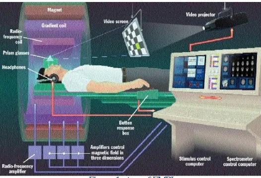
FMRI has same set up that in MRI. Figure 1 shows fundamental set up.In figure, we can see that most of set up is same as doing with MRI but there are some of differences shown. FMRI contains some additional features like headphones, prism glasses, button response box, video screen and video projector. The patient is given with head phone and prism glass to see on video screen. Projector is used to give continuous film on projector. Radio frequency waves is transmitted to patient and at same time doctor will turn on video screen to play film or anything. Button box is given near to patient right hand to change film. When the patient is seeing on video screen, there are some mood changes occur of patient face and based on that activation of some local part of brain. This activation is done because of changes in oxygen level of that particular area. Oxygen will hand over to neurons by hemoglobin in capillary red blood cells. When neuronal activity increases in active area, more oxygen will be required and the local respond will be an increase in blood flow to regions of active brain area. Hemoglobin is diamagnetic when oxygenated but paramagnetic when deoxygenated. This difference will be examined in magnetic properties leads to small differences in the MR signal of blood depending on the degree of oxygenation. Blood oxygenation changes according to the levels of neural activity these differences can be used to detect brain activity. This form of MRI is known as blood oxygenation level dependent (BOLD) imaging.
Figure 1set up of FMRI
The direction of oxygenated blood is change in comply with increased activity. You might expect blood oxygenation to decrease with activation, but in practical it will become more complicated. There is a momentary decrease in blood oxygenation immediately after neural activity increases, known as the “initial dip”. This is pursued by a period where the blood flow increases, not just to a level where oxygen demand is met, but overcompensating for the increased demand. This will have meant that blood oxygenation literally increases following neural activation. The blood flow peaks after around 6 seconds and then falls back to baseline, escorted by a "post-stimulus undershoot". Resulting image will be simplest form for fMRI image. While inventing in the fMRI scanner, the subject watched a screen continuously which oscillated between showing a visual stimulus and being dark every 30 second. Meanwhile the MRI scanner will track the signal all over side the brain. In brain areas responding to the visual stimulus you can expect the signal to went up and down as the stimulus is turned on and off, albeit blurred slightly by the delay in the blood flow response. The 'activity' in a voxel is defined as how closely the time-course of the signal from that voxel matches the expected time-course. Voxels whose signal corresponds tightly are given a high activation score, voxels showing no correlation have a low score and voxels showing the opposite (deactivation) are given a negative score. These can then be translated into activation graph.
The proposed brain tumor detection and localization frame work comprises five steps: 1. image acquisition
4. modified histogram clustering 5. Morphological operations.
After final procedure, tumors will come out with pure white color on pure black backgrounds. First, there will be some background for what is brain tumor and fMRI images at the end of MRI scan. Then, it will come to operations for detecting brain tumor. Brain tumors may be benign or malignant. Primary brain tumors are raised from the brain, and do not excite the surrounding tissues. Primary brain tumors can be malignant and disturb the surrounding tissues. The malignant may or may be not contain cancerous cells. Primary tumors are constituting from cells those are belong to the organ ort issue where they started. The secondary brain tumors are spread to the brain from another place in the body. Brain tumors can affect the normal brain activity. So, accurate detection of tumor is important for human and increase the life expectancy. Brain tumors are classified into Glioblastoma, Gliomas, Medulloblastoma, Ependymomas, CNS Lymphoma, astrocytoma, meningioma and Oligodendroglia. In this procedure, an image will have captured, digitized and processed for performing segmentation and for deriving important information like detection of brain tumor. Because of the complicate organism of brain tissues such as white matter (WM), gray matter (GM) and cerebrospinal fluid (CSF) in the brain images, extracting of meaningful quality is going to be fundamental task. The methods are time consuming.
III. PROPOSED APPROACH FOR DETECTING BRAIN TUMOR
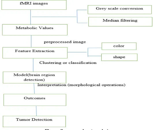
From the fMRI images take the value from graph and stored it as dataset. This dataset is used to classify in between correct data and false data. The proposed diagram for procedure is as follow:
Figure 2 proposed system design
In this approach, segmentation method is used to detect tumor in fMRI images. First, image is divided into segmented images means slice images into different images which is called catchment basins. To find out edge of different images, sober operator is used in this method. Second, the area of the tumor region is sensed by evaluating the total number of pixels in which it serves as the area of each pixel, horizontal and vertical dimensions of image and resolution of the image. The last part is find out location of tumor which is most important step instead of any other step in all processing steps. For that, detected image is subdivided into two parts: The right part of the image is specified as the left hemisphere of the brain and the left part of the image is the right hemisphere of the brain. Then, it need to calculate which part contain more part. If left part contain more pixel values, then image is on left side of hemisphere or else tumor is right side of hemisphere.
morphological operators is to separate the tumor part of the image. Now only the tumor portion of the image is visible, shown as white color. This portion has the highest intensity than other regions of the image. Morphological operators will be applying at last after the segmentation and clustering of image volume information has been done. Now, following sections will be about each part of system design.
In preprocessing or data set uploading section, a dataset about all metabolic values of brain tumor is collected from fMRI images. Mainly in dataset contents are in form of a single database table or a single statistical data matrix, where each column of the table performs as particular variable, and each of row corresponds to a given member of the dataset in question. The data set consist information functioning as height and weight of an object, for each member of the dataset. All data sets are uploaded in preprocessing part. Data pre-processing is crucial step in the data mining process. If there will be much insignificant and bombastic information present or noisy and unreliable data, then the following steps become more difficult. So. We need to be more careful about data we have. Data preparation and filtering steps can take considerable amount of processing time. Data pre-processing includes cleaning, normalization, transformation, feature extraction and selection, etc.
Feature extraction is process for reducing more information that dataset contain. Transforming the input data into the set of features is called feature extraction. If the features extracted are carefully chosen it is expected that the features set will extract the relevant information from the input data. So, in this process color and shape is going to main features to extract. It involves cutting down the amount of resources will be required for describing a large set of data accurately.The result of the proposed procedure will show the final extracted brain tumor from fMRI image.
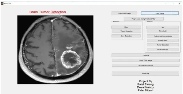
IV. RESULT IN MATLAB
Here, we use MATLAB GUI (Graphical user interface) for the project shown in below figure,
Figure 3 MATLAB GUI of Brain Tumor Detection
Now click on load image button that will help to load the image in MATLAB, the load image will automatically converted to grey scale image and that will show in below figure 4.
After that we use preprocess our image with the help of trilateral filter, here trilateral filter is modification of bilateral filter. Bilateral Filter is used for removing Gaussian noise and impulse noise. Below figure shows the values of variance before filter and after filter and also the improvement in variance.
Figure 5 Segmentation value of Trilateral Filter
The image after preprocessing trilateral filter is as shown below,
Figure 6 Tumor Image after Trilateral filtering
After the trilateral filter we have applied median filtering, here median filtering is used for removal noise, sharpen contrast, highlight contours, detects edge. It is non-linear method and it works by moving through the image pixel by pixels.
After that we used threshold button for thresholding the image of tumor which is shown below,
Figure 8 Thresholding of Tumor
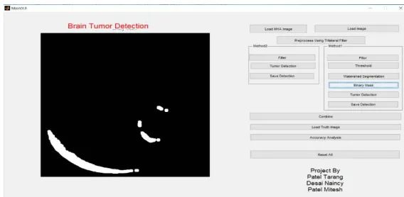
The next step is watershed segmentation, Partitioning an image into its several constituents is called “Segmentation”. Watershed Segmentation gets its name from the manner in which the algorithm segment regions into Catchment basins. Below figure is indicating the segmentation of tumor.
Figure 9 Watersheding segmentation of tumor image
After the segmentation using morphological, dilate and erode process we detected the original tumor part which is shown below,
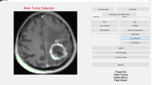
Then the last step is highlight the tumor detection part which is shown in below figure,
Figure 11 Final Tumor Detection
V.CONCLUSION
FMRI is the most advance technique for tumor detection compared to MRI. Using Digital Signal Processing Concepts, we tried to establish tumor in MATLAB by various segments like median filtering, Trilateral filter etc.BOLD signals are most strongly associated with the input to a given area rather than with the output. It is therefore possible that a BOLD signal could be present in a given area even if there is no single unit activity.FMRI has poor temporal resolution.
REFERENCES
[1] Image Processing techniques for brain tumor detection by Vipin Y.Borole, Sunil S. Nimbore and Dr. Seema S. Kawrhekar. [2] Binary Image Analysis.
[3] Median Image Filtering by university of Auckland [4] Image segmentation in color shape by Anisa Chaudhary
[5] Trilateral Filtering for Bio-Medical image by Wilbur C K. Wong, Albert C S. Chung, Simon C H. Yu. [6] The Trilateral Filter for high contrast images and values by Jack Tumblin and Prasun Choudhury.
[7] Detection of Brain Tumor by Mining fMRI Images by Meghana Nagori1, Shivaji Mutkule2, Praful Sonarkar3 Asst. Professor, CSE, Government Engineering College, Aurangabad, India.
[8] International Journal of Innovative Research in Computer and Communication Engineering (An ISO 3297: 2007 Certified Organization)