Original Research Article
A study of incidence, risk factors, clinical profile and management of 50
cases of ectopic pregnancy in a tertiary care teaching hospital
G. Ganitha*, G. Anuradha
INTRODUCTION
Ectopic pregnancy occurs when the fertilized ovum implants outside the uterus.1 Approximately 1-2% of all
pregnancies in developed countries are ectopic.2 In the
developing world, the incidence is much higher and 1 in 10 women admitted with a diagnosis of tubal ectopic pregnancy ultimately die from the condition.3 In the
developing countries, ectopic pregnancy is possibly the second most common cause of maternal death next to postabortal complications in the first three months of
pregnancy.4 Although, overall incidence of ectopic
pregnancy has increased over the past few years, death due to ectopic pregnancy has declined.5,6 The increase in
incidence is because of increase in STD rates, cesarean rates and increasing ART pregnancies. On the other hand, availability of ultrasound and other diagnostic modalities and improvement in health facilities has helped to reduce the maternal morbidity and mortality.6-8 Absence of
identifiable risk factors, varied clinical presentation, and non-availability of ultrasound may cause delay in diagnosis. Delayed diagnosis or late referral resulting in
ABSTRACT
Background: Ectopic pregnancy is a challenging and life-threatening emergency, which can cause significant maternal morbidity and mortality. The present study aims at determining the incidence, risk factors, clinical features, diagnosis, management and outcome of ectopic pregnancies in a tertiary care teaching hospital.
Methods: This was a prospective study of 50 cases of ectopic pregnancies admitted to the Department of Obstetrics and Gynaecology at a tertiary care teaching hospital from August 2012 to September 2013. Relevant data of the 50 patients was tabulated and descriptive analysis was carried out.
Results: The incidence of ectopic pregnancy was 6.3 per 1000 deliveries. Majority of the patients (82%) belonged to 20-30 yrs age group. 70% of the women were multiparous. The commonest site of ectopic pregnancy was fallopian tube (92 %). Past history of pelvic inflammatory disease (40%), abortions (20%), abdominopelvic surgery (14%) and IUCD usage (12%) were among the important risk factors identified. 20% of the patients had no identifiable risk factor. The classical triad of amenorrhea, bleeding per vaginum and abdominal pain was seen in 56 % of the study population. The most important signs which guided the diagnosis of ectopic pregnancy were cervical excitation pain (74%), abdominal tenderness (72%), adnexal mass or fullness (68%) and tenderness in the fornix (68%). Clinical presentation, urinary pregnancy test, culdocentesis and ultrasound were the diagnostic tools used for diagnosis of ectopic pregnancy. The incidence of ruptured ectopic pregnancy was 86%. Majority of the patients underwent salpingectomy (96%). There was no maternal mortality in our study.
Conclusions: Early diagnosis, timely referral, improved access to health care, aggressive management and improvement of blood bank facilities can reduce the maternal morbidity and mortality associated with ectopic pregnancy.
Keywords: Ectopic pregnancy, Pelvic inflammatory disease, Risk factors, Salpingectomy, Tubal pregnancy
Department of Obstetrics and Gynaecology, Tagore Medical College and Hospital (Affiliated to The Tamilnadu Dr MGR Medical University), Rathinamangalam, Chennai 127, Tamilnadu, India
Received: 14 March 2017
Accepted: 18 March 2017
*Correspondence:
Dr.G. Ganitha,
E-mail: drgganitha77@gmail.com
Copyright: © the author(s), publisher and licensee Medip Academy. This is an open-access article distributed under the terms of the Creative Commons Attribution Non-Commercial License, which permits unrestricted non-commercial use, distribution, and reproduction in any medium, provided the original work is properly cited.
ruptured ectopic pregnancy may increase the maternal morbidity and mortality. Early diagnosis can make medical management and conservative surgery feasible. This can have a huge impact on the future fertility of the affected women. This study aims at evaluating the incidence, predisposing risk factors, clinical features, diagnosis and management of ectopic pregnancy in a tertiary care teaching hospital.
METHODS
The present study was carried out in the Department of Obstetrics and Gynaecology at a tertiary care teaching hospital. This was a prospective descriptive study. Study was started after obtaining ethical committee clearance. During the study period from August 2012 to September 2013, 50 cases were diagnosed with ectopic pregnancy. The details of history included age, parity, presenting symptoms, past obstetric history, past history of surgeries or medical disorders, use of contraception and history of infertility. Sexual history was taken in detail to note any high risk for STD/PID. A detailed general physical examination, abdominal and bimanual examination was done. All the patients were subjected to urine pregnancy tests and ultrasound. Culdocentesis was done in few patients. Routine blood and urine investigations were done. All the patients underwent laparotomy or laparoscopy. All 50 patients underwent surgical treatment. Intra operative findings, surgical procedure, blood requirement, post-operative morbidity and outcome were recorded.
Statistical analysis
Information was collected in a structured proforma, tabulated and descriptive analysis was carried out.
RESULTS
During the study period, there were 8994 pregnancies, 7827 deliveries and 50 ectopic pregnancies. Therefore, the incidence of ectopic pregnancy is 1 in 167 pregnancies (0.6%) or 6.3 per 1000 deliveries. The incidence of ectopic pregnancy increased from 4.7 per 1000 deliveries to 6.3 per 1000 deliveries over the past 3 years in the institute.
Table1: Distribution of cases according to age.
Age (years) Percentage
<20 0
20-25 48
26-30 34
31-35 12
>35 6
Table 1 gives the distribution of cases of ectopic pregnancy according to age. The mean age of the patients was 27.1±4.10years. Majority of the patients (48%)
belonged to 22-25 years. 82% of the patients were ≤ 30 years. Table 2 shows the distribution of the cases based on parity. Majority of the patients (70%) were multiparous. 20% of the patients were nulliparous.
Table 2: Distribution of cases according to parity.
Parity Percentage
Nullipara 20
Primipara 10
[image:2.595.312.545.245.374.2]Multipara 70
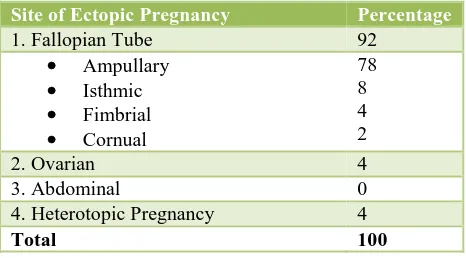
Table 3: Distribution of cases according to site of ectopic pregnancy.
Site of Ectopic Pregnancy Percentage
1. Fallopian Tube 92
Ampullary
Isthmic
Fimbrial
Cornual
78 8 4 2
2. Ovarian 4
3. Abdominal 0
4. Heterotopic Pregnancy 4
Total 100
Table 3 shows the distribution of cases according to the site of ectopic pregnancy. The most common site of ectopic pregnancy was fallopian tube (92%). Ampulla was the commonest site (78%) for ectopic implantation in the fallopian tube. Heterotopic pregnancy is rare where pregnancy is seen in the uterus and tube at the same time. In our study, there were 2 cases of heterotopic pregnancies.
Table 4: Distribution of cases according to risk factors.
Risk Factors Present study (%)
No obvious risk factor 20 H/o pelvic inflammatory disease 40 Previous Ectopic Pregnancy 2 H/o abdominopelvic surgeries 14
Tubectomy/Tubal
surgery 6
LSCS 8
Others (e.g.
Appendicectomy) 0 H/O IUCD usage 12 H/O Oral contraceptive pill
usage 8
H/O Previous abortion 20 H/O infertility 8 H/O Endometriosis 2
examination while 12 patients had two risk factors and 2 patients had multiple risk factors. 10 patients had no identifiable risk factor.
The most common risk factor was pelvic inflammatory disease (40%) followed by H/o previous abortion (20%) and H/o previous abdominopelvic surgery including tubal ligation, LSCS and appendicectomy (14%).
Although any form of contraception decreases the overall risk of pregnancy including ectopic Pregnancy, when contraceptive failure occurs in women using an IUCD or following tubal sterilization, risk of Ectopic Pregnancy is elevated. In our study, 12 % patients were using copper IUCD. H/o infertility due to tubal block or other causes, treatment associated with infertility, endometriosis and H/o previous ectopic pregnancy were other identified risk factors.
Almost 98% patients in our study came with H/O variable period of amenorrhoea. 90% cases complained of abdominal pain. 68% of the patients had bleeding or spotting per vaginum. However, the classical triad of amenorrhoea, abdominal pain and vaginal bleeding was seen in 56% cases. The other symptoms noted in our study were syncope (32%), nausea / vomiting (36%) and urinary symptoms (32%). In our study, 98% of patients had severe pallor. The high incidence of pallor was probably because 90 % of cases were associated with ruptured ectopic pregnancy or tubal abortion. The most important signs which guided in the diagnosis of ectopic pregnancy were cervical excitation pain (74%), abdominal tenderness (72%), adnexal mass or fullness (68%) and tenderness in the fornix (68%).
In the present study, urine pregnancy test was positive in 96% of patients. Culdocentesis was positive in 40% of patients. Ultrasound was able to diagnose 80 % of cases. USG findings suggestive of ectopic pregnancy were extra-uterine gestational sac (14%), haemoperitoneum (60%) and adnexal mass (80%).
In the present study, the incidence of ruptured ectopic pregnancy was 86%. 98% of the patients received one or more units of blood transfusion intra operatively and/or post operatively. All the patients with ectopic pregnancy were managed surgically. 94 % patients underwent laparotomy and 6 % patients had laparoscopic treatment.96% patients underwent unilateral or bilateral salpingectomy or salpingoophrectomy. Milking of tube was performed in 2% of patients. Concurrent dilatation and curettage was performed in the patients who had heterotopic pregnancy (4%). There was no maternal mortality in the present study.
DISCUSSION
In the present study, the incidence of ectopic pregnancy was 0.63%. The incidence of ectopic pregnancy in other
Indian studies conducted during 1996 to 2015 ranged from 0.25% to 1.9%.6-15
We compared the incidence of ectopic pregnancy in the institute from 2011 to 2013 and found an increase in incidence from 4.7 per 1000 deliveries to 6.3 per 1000 deliveries. Similar to our study, there was an increasing trend in the incidence of ectopic pregnancies in the studies conducted by Jophy et al (7.4 per 1000 live births to 15.2 per 1000 live births) and Porwal et al.6,7 Shobeiri
et al conducted a study of 872 women with ectopic pregnancy in Iran during 2000 to 2010. They found that the incidence of ectopic pregnancy increased from 1.5 per 1000 pregnancy in 2000 to 4.8 per 1000 pregnancy in 2010.17
In the present study, 82 % of women were in the age group of 20-30 years. Similar to our study, most studies reported that majority of women diagnosed with ectopic pregnancy belonged to this age group.7,9-16,18,19 This is
probably because sexual activity and fertility of women is highest during this period. In the present study, 70% of the women were multiparous which was comparable with studies by Bhuria et al, Rakhi et al, Yadav et al and Prasanna et al.8,15,18,19
In the present study, the commonest site for ectopic pregnancy was tubal (92%). Among the tubal pregnancies, ampulla was the commonest site (78%). Ampullary pregnancy was seen in 53.84% to 80% of the ectopic pregnancies in other studies.8,9,11,13,18 In our study,
heterotopic pregnancy was seen in 4% of the cases. In our study, 4% of the ectopic pregnancy was ovarian. In other studies, the non-tubal sites for ectopic pregnancy were ovaries, cervix, broad ligament, rudimentary horn of uterus and abdominal cavity. 8,9,11,13,18
In the present study, majority of the women with ectopic pregnancy (40%) had H/O pelvic inflammatory disease. Yadav ST et al, Yadav A et al, Jophy et al and Shivakumar et al also found H/O PID as the major risk factor for ectopic pregnancy.6,8,10,14 Moini et al reported a
strong association between prior PID and ectopic pregnancy.20 Past history of previous abortion with or
without D&C was found to be an important risk factor in most studies including the present study.6,8-12,14,19This is
probably because of tubal damage following post abortal infections.
Although any form of contraception decreases the overall risk of pregnancy including ectopic Pregnancy, when contraceptive failure occurs in women using an IUCD or following tubal sterilization, risk of ectopic Pregnancy is elevated. In our study, we found that IUCD or oral contraceptive pill usage predisposed to ectopic pregnancy. A higher incidence of ectopic pregnancy among IUCD users was noted in most studies.6,8,11
al found that usage of IUCD increased the risk of subsequent ectopic pregnancy four to fivefold.20
Probably, IUCDs predispose to PID or induce inflammatory changes in the endosalpinx leading to subsequent ectopic pregnancy. Therefore, women with poor menstrual hygiene, those at risk of STDs/PID should be suggested alternative (barrier) methods of contraception. In our study, tubal ligation was associated with ectopic pregnancy in 6% of patients. Other studies have reported that the risk of tubal pregnancy following tubal ligation or tubal surgery is 5.4% to 16.21%.6,8-10,14,19
Moini et al reported that women with previous tubal surgery were likely to have ectopic pregnancy two to three times more than controls.20
In the present study, 2% of the study subjects had past history of ectopic pregnancy. Other studies noted that 5.4% to 10.95% of women with ectopic pregnancy had H/o prior ectopic pregnancy.6,8,11,14,18,19 Moini et al have
reported that among all the risk factors of ectopic pregnancy the association between subsequent ectopic pregnancy and previous ectopic pregnancy was the strongest.20 Parashi et al found an increased risk of 7-9
fold in women with previous ectopic pregnancy.21
H/o infertility was found in 8% of women in the present study. Other studies have observed that 10%-23.7% of women with ectopic pregnancy had history of infertility.6,9-12,14,18,19 Tubal pathology, endometriosis,
ovulation induction and ART are the probable reasons for association of infertility with occurrence of ectopic pregnancy. Moini et al found a strong association between infertility and ectopic pregnancy.20 However;
Parashi et al did not find significant association of infertility with occurrence of ectopic pregnancy.21
In the present study, H/o previous LSCS, H/o previous abdominal or pelvic surgeries (excluding tubal ligation) was found in 8% women. Simsek Y et al analysed the risk factors in 35 ectopic pregnancies. They found that 46% women had history of Caesarean section.22 Parashi
et al found that there was a significant relationship between abdominal/pelvic surgery and incidence of ectopic pregnancy.21 In their studies, Wakankar et al and
Yadav A et al reported that 32% and 26.02% of women with ectopic pregnancy respectively had history of LSCS.
8,11A possible explanation for this association is formation
of peritubal adhesions.
Ragab et al conducted a univariate and multivariate analyses of various risk factors for ectopic pregnancy and demographic characteristics. Univariate analyses showed that H/o previous abortion, H/o abdominal surgery, PID, H/o previous D&C and IVF were associated significantly with increased risk of ectopic pregnancy. Multivariate analyses showed that past abdominal surgery, IVF, H/o PID were the only significant risk factors in nulliparous women.23
The present study and other comparative studies show that PID, previous abortions, abdominopelvic surgeries contribute to the risk of subsequent ectopic pregnancy. These risk factors are modifiable. Early diagnosis and adequate treatment of PID, performing D& C under strict aseptic conditions, ensuring adequate haemostasis during surgeries, employing methods to reduce post op adhesions during surgery and adequate antibiotic cover may help in reducing the incidence of ectopic pregnancy.
In the present study, 20% of women had no identifiable risk factor. Other studies have also reported that ectopic pregnancy can occur in women (20%-58.3%) with no identifiable risk factor.6,9,10,14,19 This fact emphasizes that
ectopic pregnancy should be suspected when clinical features are suggestive of ectopic pregnancy even in low risk women. One has to remember that absence of symptoms does not rule out ectopic pregnancy.
Almost 98% patients in our study came with H/o variable period of amenorrhoea. Similar observation was noted by Prasanna et al (96%).19 In other studies, amenorrhoea was
noted in 54.9%- 84.3% patients. 6,9-11,13,18 Abdominal pain
was seen in 90% cases in the present study. Other studies reported that abdominal pain was a frequent and constant symptom in 80%- 95% patients.6,9-11,14,18,19
In the present study, 68% of the patients had bleeding or spotting per vaginum. This was similar to the observations by Yadav ST et al (72.2%), Shivakumar et al (70%), Jophy et al (66.6%) and Wakankar et al (65.4%).6,10,11,14 However, the classical triad of
amenorrhoea, abdominal pain and vaginal bleeding was seen in 56% of the cases in the present study which was comparable to the observation by Wakankar et al (53.84%) and Shetty et al (50%).5,11 Only 22% of the
cases had presented with the classical triad of symptoms in the study by Shukla et al. This shows that unless the obstetrician has high index of suspicion, diagnosis of ectopic pregnancy may be missed or delayed.13
The other symptoms noted in our study were syncope (32%), nausea / vomiting (36%) and urinary symptoms (32%). Syncope was noted in 13%- 31.2% of patients in other studies.6,10,14,15,18 In our study, 98% of patients had
severe pallor. Gaddagi et al and Shivakumar et al noted pallor in 80% of the patients.9,10 In our study, the high
incidence of pallor was probably because 90 % of cases were associated with ruptured ectopic pregnancy or tubal abortion.
Clinical presentation, urinary pregnancy test, culdocentesis and ultrasound were the diagnostic tools used for diagnosis of ectopic pregnancy. In the present study, urine pregnancy test was positive in 96% of patients. This was in concordance with the studies by Gaddagi et al (97.3%), Prasanna et al (94%), Yadav ST et al (100%) and Shukla et al (98.04%).9,13,14,19 In the
present study, culdocentesis was positive in 40% of patients. This was comparable to the study by Gaddagi et al (37.8%).9
In the present study, ultrasound was able to diagnose 80 % of cases. Extra-uterine gestational sac, haemoperitoneum and adnexal mass were noted in 14%, 60% and 80% of cases respectively. In the study by Gaddagi et al, ultrasound revealed heterogenous mass in 40.5% of the cases and fluid in pouch of Douglas in 83.8% of cases.9 Shetty et al reported adnexal mass in
60% of cases, hemoperitoneum in 50 % cases and Gestational sac in adnexa in 8% of cases.5 Nazeem et al
found adnexal mass in 76% of cases, hemoperitoneum in 50 % cases and gestational sac in adnexa in 5.6% of cases.24
In the present study, the incidence of ruptured ectopic pregnancy was 86% as against an incidence of 60.52% - 89.1% as observed in other studies.9, 11-13, 15 This shows
that majority of cases with ectopic pregnancy present as ruptured ectopic pregnancies. This emphasizes the need for early diagnosis. Women with high risk of ectopic pregnancy must be emphasized to consult the obstetrician as early as possible when they miss the periods. Since the incidence of ruptured ectopic pregnancy was high in our study, 98% of the patients were given one or more units of blood transfusion intra operatively and/or post operatively. Blood transfusion was required in 43.5%- 97.3% of patients in other studies.9,14,15 This variability in
requirement for blood transfusion was probably because of the difference in severity of presentation, presence of haemoperitoneum, pre-existing anaemia and availability of blood products.
In the present study, all the patients with ectopic pregnancy were managed surgically. 94 % patients underwent laparotomy and 6 % patients had laparoscopic treatment. In most studies, surgery was the main stay of treatment.7-11 In the present study and in the studies by
Bhuria et al and Shetty et al, 96%, 95.2% and 98% of the patients underwent unilateral or bilateral salpingectomy or salpingoophrectomy respectively.5,15 In the present
study, milking of tube was performed in 2% of patients. Milking of tube was performed in 2% - 8.82 % of patients in other studies.5,8,9,11 Though milking of tube is a tubal
sparing method, the chances of recurrence of ectopic pregnancy in the same side is high. Concurrent Dilatation and curettage was performed in 4% of the patients who had heterotopic pregnancy. Treatment modality for ectopic pregnancy depends on site of pregnancy, ruptured or unruptured pregnancy, availabilty of laparoscopy,
surgical expertise, need to retain fertility and choice of patient.
There was no maternal mortality in our study as reported by many other studies.7-16 This shows that early
diagnosis, timely and prompt management of ectopic pregnancy, availability of adequate blood and blood components improves the outcome of ectopic pregnancies. Delay in seeking healthcare, accessibility to expert health facilities, initial misdiagnosis and delayed referral are important deterrents to prompt management of ectopic pregnancy.25
CONCLUSION
Ectopic pregnancy is one of the commonest gynaecological emergencies with significant maternal morbidity and mortality. The incidence of ectopic pregnancy is on the rise. The incidence of ruptured ectopic pregnancy is high in developing countries due to late diagnosis and delayed referral. In developing countries, PID and postabortal sepsis continue to be the most important risk factor for ectopic pregnancy. Since many patients may not have identifiable risk factors, a high index of suspicion is vital for early diagnosis. Women at high risk for ectopic pregnancy must be counselled about the possibility for future ectopic pregnancy. They should be emphasised to report to their doctor as soon as they miss their periods for early diagnosis. Avoiding unnecessary pregnancies, safe sex practices, using barrier contraceptives, prompt treatment of PID/STDs can bring down the incidence of ectopic pregnancies. Early diagnosis, timely referral, aggressive management, improvement of blood bank facilities can reduce the maternal morbidity and mortality associated with ectopic pregnancy.
Funding: No funding sources Conflict of interest: None declared
Ethical approval: The study was approved by the Institutional Ethics Committee
REFERENCES
1. Farquhar CM: Ectopic pregnancy. Lancet.
2005;366(9485):583-91.
2. Varma R, Gupta J. Tubal ectopic pregnancy. BMJ
Clin Evid. 2009;1406.
3. Leke RJ, Goyaux N, Matsuda T, Thonneau PF.
Ectopic pregnancy in Africa: a population-based study. Obstet Gynecol. 2004;103:692-97.
4. Thonneau P, Hijazi Y, Goyaux N, Calvez T, Keita N.
Ectopic pregnancy in Conakry, Guinea.Bull World Health Organ. 2002;80:365-70.
5. Shetty VH, Gowda S, Lakshmidevi M. Role of ultra
6. Jophy R, Thomas A, Mhaskar A. Ectopic pregnancy -5 year experience. J Obstet Gynecol Ind. 2002;52:55-8.
7. Gupta R, Porwal S, Swarnkar M, Sharma N,
Maheshwari P. Incidence, trends and risk factors for ectopic pregnancies in a tertiary care hospital of Rajasthan. JPBMS. 2012;16(07):1-3.
8. Yadav A, Prakash A, Sharma C, Pegu B, Saha
MK.Trends of ectopic pregnancies in Andaman and Nicobar Islands.Int JReprod Contracept Obstet Gynecol. 2017;6:15-9.
9. Gaddagi RA, Chandrashekhar AP. A Clinical Study
of Ectopic Pregnancy. J Clin Diagn Res. 2012;6(5):867-9.
10. Shivakumar HC, Umashankar KM, Ramaraju HE.
Analysis of forty cases of ectopic pregnancies in tertiary care hospital in south India. Indian Journal of Basic and Applied Medical Research; 2013: 3(1):235-241.
11. Wakankar R, Kedar K. Ectopic Pregnancy- A rising
Trend. Int J Sci Stud. 2015;3(5):18-22.
12. Mufti S, Rather S, Mufti S, Rangrez RA, Wasiqa,
Khalida. Ectopic pregnancy: an analysis of 114 cases. JK Practitioner. 2012;17(4):20-3.
13. Shukla DB, Jagtap SV, Kale PP, Thakkar HN.Study
of ectopic pregnancy in a tertiary care centre.Int J Reprod Contracept Obstet Gynecol. 2017;6:975-9.
14. Yadav ST, Kaur S, Yadav SS. Ectopic pregnancy an
obstetric emergency: retrospective study from medical college Ambala, Haryana, India.Int J Reprod Contracept Obstet Gynecol. 2016;5:2210-4.
15. Bhuria V, Nanda S, Chauhan M, Malhotra V. A
retrospective analysis of ectopic pregnancy at a tertiary care centre: one year study. Int J Reprod Contracept Obstet Gynecol. 2016;5:2224-7.
16. Nair L, Peter N, Rose A. International Journal of
Biomedical Research 2015;6(05):331-3.
17. Shobeiri F, Tehranian N, Nazari M. Trend of ectopic
pregnancy and its main determinants in Hamadan province, Iran (2000-2010). BMC research notes. 2014;7(1):733.
18. Rakhi, Mital PL, Hooja N, Agarwal A, Makkar P,
Andleeb F. Ectopic pregnancy: a devastating catastrophe.Sch J App Med Sci. 2014;2(3A):903-7.
19. Prasanna B, Jhansi CB, Swathi K, Shaik MV. A
study on risk factors and clinical presentation of ectopic pregnancy in women attending a tertiary care centre. IAIM. 2016;3(1):90-6.
20. Moini A, Hosseini R, Jahangiri N, Shiva M,
Akhoond MR. Risk factors for ectopic pregnancy: A case–control study. J Res Med Sci. 2014;19:844-9.
21. Parashi S, Moukhah S, Ashrafi M. Main risk factors
for ectopic pregnancy: a case–control study in a sample of Iranian women. Int J Fertil Steril. 2014;8:147-54.
22. Simsek Y, Oguzhan A M. Analysis of ectopic
pregnancies admitted to emergency department. Turk J Emerg Med. 2015;15(4):151–154.
23. Ragab A, Mesbah Y, El-Bahlol I, Fawzy M,
Alsammani MA. Predictors of ectopic pregnancy in nulliparous women: A case-control study. Middle East Fertility Society Journal.2016;21(1):27-30.
24. Naseem I, Bari V, Nadeem N. Multiple parameters in
the diagnosis of ectopic pregnancy. J Pak Med Assoc. 2005;55(2):74-6.
25. Awoleke JO, Adanikin AI, Awoleke AO. Ruptured
tubal pregnancy: predictors of delays in seeking and obtaining care in a Nigerian population. Int J Women Health. 2015;7:141-7.
Cite this article as: Ganitha G, Anuradha G.A study of incidence, risk factors, clinical profile and