Original Research Article
Evaluation of cervical erosion in symptomatic women
Sindhu C. Guptha*
INTRODUCTION
Cervical erosion when occurs there is a zone of columnar epithelium on the vaginal portion of the cervix replacing the normal stratified squamous epithelium that is usually found outside the external os.1,2 Cervical erosion has also been known as cervical ectropion and cervical ectopy. Cervical ectropion is a normal physiological state. After menarche, the columnar epithelium within the endocervical canal grows and everts exposing the glandular epithelium on the ectocervix.1-3 The exposed glandular epithelium is gradually replaced by squamous
epithelium in a process called metaplasia.1 This area of metaplastic change is called transformation zone. It is where most squamous cell cervical cancer arises.1,2 The prevalence of cervical ectropion in women of reproductive age is between 17% and 50%.4
The presence of ectropion is influenced by estrogens. Hence it is often found in women after the menarche, during pregnancy or with use of the combined oral contraceptive pill. The prevalence increases with parity and use of the combined oral contraceptive pill. Ectropion is rare in postmenopausal women as a result of declining estrogen levels, the cervix shrinks and inverts, ABSTRACT
Background: The objective of this study was evaluation of symptomatic women with cervical erosion.
Methods: Prospective clinical study done in the department of obstetrics and gynecology, VIMS, Ballari for one-year period from June 2016 to May 2017. All patients with symptoms of white discharge, post coital bleeding and menstrual irregularities were examined by per speculum examination and all those who had cervical erosion (112 Patients) were included in the study. Pap smear and colposcopy were done in all subjects. Colposcopic guided biopsy was done in women with abnormal findings on colposcopy. Histological prediction of colposcopic findings was done according to the Reid’s modified colposcopic index. The result of the biopsy was correlated with the predicted histology of the Reid’s modified colposcopic index and statistical analysis done to calculate diagnostic efficacy of colposcopy in the evaluation of symptomatic women with cervical erosion.
Results: Out of the 112 patients, 5.35% (6/112) had abnormal Pap smear. All patients underwent colposcopy and colposcopic guided biopsy, 11.6% (13/112) had abnormal colposcopic findings. Histopathology confirmed CIN I in 6.25% (7/112) and CIN II in 1.78% (2/112) patients. The sensitivity, specificity, PPV, NPV and accuracy of PAP smear was found to be 22.22%, 96.11%, 33.33%, 93.39% and 90.17%. The above values for colposcopy were found to be 88.89%, 95.15%, 61.54%, 98.99% and 94.64%.
Conclusions: Colposcopy should be done in all symptomatic patients with cervical erosion as it is a good diagnostic tool for premalignant conditions of the cervix and correlates well with histopathological findings.
Keywords: Cervical erosion, Colposcopy, Histopathology, Pap smear
Department of Obstetrics and Gynecology, Bangalore Medical College and Research Institute, Bangalore, Karnataka, India
Received: 26 February 2020
Accepted: 30 March 2020
*Correspondence:
Dr.Sindhu C. Guptha,
E-mail: sindhuguptha66@gmail.com
Copyright: © the author(s), publisher and licensee Medip Academy. This is an open-access article distributed under the terms of the Creative Commons Attribution Non-Commercial License, which permits unrestricted non-commercial use, distribution, and reproduction in any medium, provided the original work is properly cited.
drawing the squamous epithelium covering the ectocervix into the endocervical canal.1-3
It is common in women of lower socioeconomic groups, poor general hygiene, early marriage and multiple pregnancies. These are also the risk groups for cervical dysplasia which occurs as a consequence of metaplasia.1 The presence of the metaplastic columnar epithelium can manifest a myriad of symptoms including:
• Increased vaginal discharge • Leucorrhea
• Postcoital bleeding • Pelvic pain
• Recurrent cervicitis • Dyspareunia.1,3
There is evidence that invasive cancer of the cervix develops from carcinoma in situ or dysplasia. The rate of invasive cancers can be significantly reduced by screening of the cervix by cytology and colposcopy.5 Cervical cancer is an ideal target for screening and treatment because of the long pre-clinical stage of cervical cancer during which precursor lesions can be treated conservatively and successfully.6
The principal methods used in the early detection of cancer of the uterine cervix are Pap smear cytological examination and colposcopy. Screening programmes either organized or opportunistic have led to large decline in the incidence of cervical cancer in developed countries. In contrast, the incidence and prevalence of cervical cancer remains large in high-risk developing countries because of ineffective or no screening.7 It is more realistic and effective to target screening in high-risk women using a highly sensitive test, with emphasis on high coverage of the targeted population.8
Despite the existence of national guidelines, the screening coverage in India is appalling and is mainly attributed to inequality between infrastructure, resources and outsized population. As a result, very often diagnosis of cervical cancer is based on opportunistic screening or after the onset of symptoms.8 The diagnostic algorithm of various organized screening programmes consists of cytology and colposcopy (which help in detecting the abnormality) and which can be established by histology (grade of lesion).6
The present study was, therefore undertaken to evaluate symptomatic women with cervical erosion by Pap smear and colposcopy and to evaluate the role of colposcopy as a screening and diagnostic tool for cervical cancer and other cervical lesions.
Objectives
• Evaluation of symptomatic women with cervical erosion by Pap smear and colposcopy
• Correlation of the colposcopic findings with cytology and histopathology.
METHODS
A prospective observational study done over a period of 1 year, from June 2016 to May 2017 in the department of obstetrics and gynecology at a tertiary care hospital in Ballari.
The study participants were patients attending the department of obstetrics and gynecology OPD during the study period and who fulfilled the inclusion criteria and who gave valid consent.
Inclusion criteria
• All patients with the complaints like white discharge per vaginum, heavy menstrual bleed, intermenstrual bleed, post coital bleed, pelvic pain or dyspareunia and having cervical erosion on per speculum examination.
Exclusion criteria
• Pregnancy
• Women ≤6 weeks after delivery • Patients on oral contraceptive pills
• Known case of CIN or treated case of CIN or cervical cancer
• Women with frank growth on the cervix
• Women with bleeding at the time of examination.
Patients meeting the study criterion and who gave consent were recruited in the study.
Written informed consent was obtained and patients were explained regarding the procedure and the complications that may arise due to the procedure.
Table 1: Modified B.G. Prasad classification.9
Socio economic class 1961 (Original) X correction factor Current (modified)
I ≥100×63 6300 Upper
II 50-99×63 3150-6299 Upper middle
III 30-49×63 1890-3149 Lower middle
IV 15-29×63 945-1889 Upper lower
Demographic data like age, married life and parity of the patient were noted. Relevant medical, menstrual and obstetric history was taken.
To ascertain the socioeconomic status of the individual modified B.G. Prasad classification (Table 1) was used. It
is a simple and widely used scale by researchers to determine the socioeconomic status of study subjects in health studies in India. It is based on the per capita income of an individual and can be applied to both urban and rural populations. It is constantly updated to take inflation and depreciation of rupee into account.9
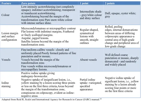
Table 2: Modified Reid’s colposcopic index (RCI).
Feature Zero points 1 point 2 points
Colour
Low-intensity acetowhitening (not completely opaque); indistinct acetowhitening; transparent or translucent acetowhitening.
Acetowhitening beyond the margin of the transformation zone Pure snow-white colour with intense surface shine. Flat lesions with indistinct margins, Feathered or finely scalloped margins.
Angular, jagged lesions.
Satellite lesions beyond the margin of the transformation zone. change and peripheral area of low-grade change
Vessels
Fine/uniform-calibre vessels- closely and uniformly placed Poorly formed patterns of fine punctation and/or mosaic.
Vessels beyond the margin of the transformation zone.
Fine vessels within microcondylomatous or micropapillary lesions.
Positive iodine uptake giving mahogany-browncolor.
Negative uptake of insignificant lesion, i.e., yellow staining by a lesion scoring three points or less on the first three criteria,Areas beyond the margin of the transformation zone, conspicuous on colposcopy, evident as iodine-negative areas. staining by a lesion already scoring four points or more on the first three criteria
Adapted from Reid R, Scalzi and International Agency for Research in Cancer (IARC) manual.10
Table 3: Histopathological correlation with colposcopy grading.11
Colposcopic grading RCI score Histopathology (HPE)
Grade 1 0-2 Normal/inflammatory/subclinical/viral infection/CIN I
Grade 2 3-5 CIN II
Grade 3 6-8 CIN III - carcinoma in situ/microinvasive/frank cancer
A Pap smear was taken and simultaneously colposcopy was done, if no cervico-vaginal infections were present. Cervico-vaginal infections were given antibiotic course of 7 days and then Pap smear and colposcopy were done. The colposcopy instrument which was used is COLpro222DX pro-MIS with magnification 6X-40X. All the patients underwent both the acetic acid and the Schiller’s test (Lugol’s Iodine) before they were subjected to colposcopy directed biopsy. Biopsy was
taken from the acetowhite areas and the iodine negative areas. 4 quadrant biopsy taken when colposcopy findings were normal.
histopathological report was obtained. Histopathology was considered to be gold standard.
Depending on their degree of dysplasia and considering the age and the parity of the patients, they were given appropriate treatment.
Accuracy of colposcopic examination and Pap smear was calculated by standard statistical methods. Sensitivity, specificity, positive predictive valve (PPV), negative predictive valve (NPV), positive and negative likelihood ratio of colposcopic examination and Pap smear were calculated.
The Table 3 shows the correlation with RCI score using colposcopy and HPE.
RESULTS
Total number of cases were 112. Among which the complaints they presented with;
• WDPV (white discharge per vagina): 76 patients • HMB (heavy menstrual bleeding): 21 patients • IMB (intermensrual menstrual bleeding): 9 patients • PCB (post coital bleeding): 6 patients.
Figure 1: Distribution of cases by the presenting complaints.
Table 4: Distribution of cases according to age.
Age No. of cases Percentage
20-29 13 11.61%
30-39 43 38.39%
40-49 46 41.08%
50-59 10 8.92%
Total 112 100%
The Table 4 shows the distribution of cases according to age. The maximum number of patients were of the age group 40-49 years contributing to 41.08% (46/112) cases and least number of cases belonged to age group of 50-59 years (8.92%).
As shown in Table 5, maximum number of patients belonged to class IV of modified BG Prasad classification 45.54% (51/112).
Table 5: Distribution of cases according to their socioeconomic status (according to modified BG
Prasad classification using Table 1).
Socioeconomic status (modified B.G. Prasad)
No. of
cases Percentage
Class II 3 2.67%
Class III 21 18.75%
Class IV 51 45.54%
Class V 37 33.04%
Total 112 100%
Table 6: Distribution of cases according to duration of marriage.
Married life (years) No. of cases Percentage
≤10 12 10.71%
11-20 49 43.75%
21-30 46 41.08%
>30 5 4.46%
Total 112 100%
As shown in the Table 6, the present study among 112 patients,12 (10.71%) were married for less than 10 years, 49 (43.75%) had a married life of 11-20 years duration, 46 patients (41.08%) were married for 21-30 years and 5 patients (4.46%) were married for more than 30 years.
Table 7: Distribution of cases according to parity.
Parity No. of cases Percentage
1 4 3.57%
2 28 25%
3 56 50%
4 22 19.65%
5 2 1.78%
Total 112 100 %
Figure 2: Distribution of cases as per presence of koilocytes.
WDPV=76
HMB=21
IMB=9
PCB=6
Koilocytes Present
(17)
Koilocytes Absent
(95)
As shown in the Table 7, the present study majority of patients were multiparous. Only 4 patients (3.57%) were primipara. Majority were para 3 contributing to 50% (56/112) cases.
The presence of koilocytes in histopathology indicates active infection with human papilloma virus (HPV). As shown in the Figure 2, the present study koilocytes were present in 17 cases among total of 112 cases (15.17%).
Table 8: Pap smear report.
Pap smear No. of
cases Percentage
Normal smear 5 4.47%
Inflammatory smear 96 85.71%
ASCUS (atypical squamous
Pap smear was done for all 112 patients among which the findings were as shown in the Table 8.
A total 5 patients had a normal Pap smear, 96 patients had inflammatory smear, 5 patients had ASCUS, 4 patients had LSIL, 2 patients had HSIL as per Bethesda system of reporting Pap smear.
Table 9: Colposcopic findings with acetic acid (5%).
Acetowhite areas No. of cases
Absent 3
Present 109
Total 112
As shown in the Table 9, the present study, among 112 cases acetowhite areas on the cervix on application of 5% acetic acid was seen in 109 cases and in remaining 3 cases there were no acetowhite areas.
Table 10: Colposcopic findings with Lugol’s iodine.
Lugols iodine No. of cases
Normal (Mahogany brown stained) 24
Unstained areas 76
Yellow brown areas 12
Total 112
As shown in the Table 10, the present study, among 112 cases on application of Lugols iodine, 24 patients had mahogany brown staining of cervix which is normal.76
patients had unstained areas on the cervix and 12 patients had yellow brown areas.
Using Reids colposcopic index as explained in Table 2 for 112 cases, RCI was calculated and the value for each case is categorised as shown in Table 11 and Figure 3.
Table 11: Distribution of cases by Reid’s colposcopic index (RCI).
The Table 12 shows the Histopathology report of cervical biopsy specimens of all 112 cases. In the present study 101 cases had chronic cervicitis, 7 cases had mild dysplasia, 2 cases had moderate dysplasia and remaining 2 cases had a normal biopsy report.
Table 12: Histopathological findings.
Histopathological findings No. of
cases Percentage
Normal 2 1.79%
Chronic cervicitis 101 90.17%
Mild dysplasia 7 6.25%
Moderate dysplasia 2 1.79%
Total 112 100%
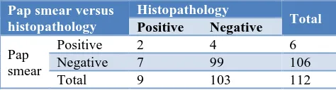
The Table 13 shows correlation between Pap smear findings and histopathology. Histopathological findings were considered to be gold standard.
Table 13: Correlation of Pap smears findings with HPE report.
The Table 14 shows correlation between colposcopic findings and histopathology. Histopathological findings were considered to be gold standard.
The Table 15 shows the statistical analysis values of colposcopy and Pap smear considering histopathology report to be gold standard. The accuracy of colposcopy was 94.6% and that of Pap smear 90.17%.
Table 15: Comparing the statistical analysis of Pap smear and colposcopy with respect to HPE.
Statistics Pap smear Colposcopy
Sensitivity 22.22 88.89
Specificity 96.11 95.15
Positive predictive value 33.33 61.54 Negative predictive value 93.39 98.99 Positive likelihood ratio 5.72 18.31 Negative likelihood ratio 0.80 0.12
Accuracy 90.17 94.64
The Figure 3 shows colposcopic appearance of the cervix in a case of cervical erosion on application of normal saline.
Figure 3: Colposcopic appearance of the cervix in a case of cervical erosion on application of
normal saline.
Figure 4: Colposcopic appearance of the cervix in a case of cervical erosion on application of acetic
acid (5%).
The Figure 5 shows colposcopic appearance of the cervix on application of Lugols iodine. Unstained areas are seen on the cervix.
The acetowhite areas as seen in Figure 4. It shows colposcopic appearance of the cervix on application of 5% acetic acid and unstained areas as seen in Figure 5 were subjected to biopsy.
Figure 5: Colposcopic appearance of the cervix in a case of cervical erosion on application of
Lugols iodine.
DISCUSSION
Routine cervical cytology is an effective screening tool for CIN and cervical cancer. However, negative cytology does not always rule out CIN. In this study Pap smear diagnosed abnormality in 6 women (5.35%) whereas colposcopy diagnosed abnormalities in 13 (11.6%) women and these 7 would have missed if colposcopy was not done simultaneously. Referral for colposcopy may still reveal CIN even if a recent smear result is negative. • In the present study among the 112 patients, 5.35%
(6/112) had abnormal Pap smear, 11.6% (13/112) had abnormal colposcopic findings. Histopathology confirmed CIN I in 6.25% (7/112) and CIN II in 1.78% (2/112) patients.
• The sensitivity, specificity, PPV, NPV and accuracy of Pap smear was found to be 22.22%, 96.11%, 33.33%, 93.39% and 90.17%. The above values for colposcopy were found to be 88.89%, 95.15%, 61.54%, 98.99% and 94.64%. The accuracy of colposcopy was found to be more than the accuracy of Pap smear.
• In study by Patil et al, colposcopy was done in all patients (n=120) along with cytological examination and the findings co-related in 102 patients (85%) and did not correlate in 15 patients (12.5%).4
• In a study by Gupta V et al, colposcopy was done following cytological examination in 177 (59%) patients. Findings correlated in 160 (90.39%).2 Colposcopic guided biopsy was obtained in 104 (34.66%) patients. The correlation between colposcopic findings and HPE results was seen in 90
Cervical erosion
Acetowhite area
Unstained areas
(88.23%) patients which is also comparable with this study.12
• In a study by Kumari M et al on colposcopy in cervical erosion patients, 68% had normal findings and 30% had abnormal colposcopy.13
• In the study by Bangal VB et al out of 100 patients of cervical erosion, 67% had normal colposcopic findings and 30% had abnormal findings.14
• Jyothi et al in their study of 200 high risk patients in the age group of 35 to 60 years reported abnormal
Since the cancer cervix has long duration of premalignant conditions in which it can be diagnosed and treated, evaluation of premalignant conditions of the cervix correlates well with histopathological findings. Hence it should be done in all symptomatic patients with cervical erosion as it is an office procedure, with good diagnostic accuracy and it permits direct visualization of the lesion. In conclusion colposcopy using RCI has a satisfactory diagnostic efficacy and the good correlation between colposcopic impression and histopathology makes it a valid tool in the diagnosis and management of benign and precancerous lesions. Hence colposcopy should be offered as a diagnostic method in all patients with unhealthy cervix.
ACKNOWLEDGMENTS
Authors would like to thank all patients who participated in the study. Authors also, thankful to teachers, seniors, colleagues and friends for their help and guidance and parents who have been authors constant support, inspiration and guide.
Funding: No funding sources Conflict of interest: None declared
Ethical approval: The study was approved by the Institutional Ethics Committee
REFERENCES
1. Matiluko A. Cervical ectropion Part 1: Appraisal of a common clinical finding. Trends in Urology, Gynaecol Sexual Health. 2009;14(3):10-2.
2. Sellors JW, Sankaranarayanan R. Colposcopy and treatment on cervical intraepithelial neoplasia: A Beginners’ Manual, IARC Press, Lyon, France. 2003:8-12.
3. Hua X, Zeng T, Zhang R. Using platelet-rich plasma for the treatment of symptomatic cervical ectopy. Int J Gynaecol Obstet. 2012;119:26-9.
4. Patil P, Sharma P. Colposcopic evaluation of cervical erosion in symptomatic women. Int J Reprod Contracept Obstet Gynecol. 2017;6:2207-11.
5. Nayani ZS, Hendre PC. Comparison and correlation of pap smear with colposcopy and histopathology in evaluation of cervix. J Evol Med Dent Sci. 2015;4(53):9236-47.
6. Chaudhary RD, Inamdar SA, Hariharan C. Correlation of diagnostic efficacy of unhealthy cervix by cytology, colposcopy and histopathology in women of rural areas. Int J Reprod Contracept Obstet Gynecol. 2014;3:213-8. 7. Sankaranarayanan R, Budukh AM, Rajkumar R.
Effective screening programmes for cervical cancer in low-and middle-income developing countries. Bulletin World Health Organization. 2001;79:954-62.
8. Aswathy S, Quereshi A, Kurian B, Leelamoni K. Cervical cancer screening: current knowledge and practice among women in a rural population of Kerala, India. Indian J Med Res. 2012;136:205-10.
9. Vasudevan J, Mishra AK, Singh Z. An update on B. G. cervix on naked eye examination. J Evol Med Dent Sci. 2014;3(4):902-6.
11. Guptha N, Chandra M. Colposcopy made easy-A Hands on manual for Practicing Doctors and Postgraduates, 1st edition, Jayapee Brothers Medical Publishers ltd,; 2009:223.
12. Gupta V, Tandon A, Nanda A, Sharma A, Bansal N. Colposcopic evaluation of cervical lesions: a prospective study. Int J Clin Trials. 2014;1:110-3. 13. Kumari M, Murari K, Kumari M. Role of colposcopy in
management of cervical erosion in rural population of Eastern Bihar. Inter J Cur Res. 2016;8(11):41268-70. 14. Bangal VB, Patil NA, Gavhane SP, Shinde KK,
Colposcopy guided management of cervical erosions in rural population. Sch J App Med Sci. 2014;2(1C):261-5.
15. Jyoti R, Gupta P, Rao R, Sood PL, Parasher N. Correlation between colposcopy, cytology, and histopathology in high risk patients for cervical cancer in perimenopausal women in Himachal Pradesh, India. J South Asian Federat Menop Soc. 2013;1(1):21-3. 16. Ramesh G, Sudha R, Jayashree AK, Padmini J.
Colposcopic evaluation of the unhealthy cervix. J Clin Diagnost Res. 2012;6(6):1026-8.