Genes Required for the Engulfment
of
Cell Corpses During Programmed
Cell Death in Caenorhabditis elegans
Ronald E. Ellis, Dean M. Jacobson and H.
Robert
Horvitz
Howard Hughes Medical Institute, Department of Biology, Massachusetts Institute of Technology, Cambridge, Massachusetts 02139
Manuscript received December 26, 1990 Accepted for publication May 1 1, 199 1
ABSTRACT
After programmed cell death, a cell corpse is engulfed and quickly degraded by a neighboring cell. For degradation to occur, engulfing cells must recognize, phagocytose and digest the corpses of dying cells. Previously, three genes were known to be involved in eliminating cell corpses in the nematode
Caenorhabditis elegans: ced-1, ced-2 and nuc-I. We have identified five new genes that play a role in this process: ced-5, ced-6, ced-7, ced-8 and ced-10. Electron microscopic studies reveal that mutations in each of these genes prevent engulfment, indicating that these genes are needed either for the recognition of corpses by other cells or for the initiation of phagocytosis. Based upon our study of double mutants, these genes can be divided into two sets. Animals with mutations in only one of these sets of genes have relatively few unengulfed cell corpses. By contrast, animals with mutations in both sets of genes have many unengulfed corpses. These observations suggest that these two sets of genes are involved in distinct and partially redundant processes that act in the engulfment of cell corpses.
C
ELL death plays an important role in the normal development of most animals (SAUNDERS 1966;COWAN et at. 1984; TRUMAN 1984; ELLIS, YUAN and
HORVITZ 1992). Usually the corpses of dead cells a r e quickly eliminated from the body, because they a r e engulfed and degraded by other cells (e.g. STOCKER, EDWARDS a n d TRUMAN 1978; WYLLIE, KERR a n d CURRIE 1980; LOCKSHIN 198 1). Because dying cells, but not living cells, are engulfed, there must be signals that mark cell corpses for engulfment. To understand how cell corpses are recognized by other cells, and how the process of phagocytosis is initiated, we have begun a genetic analysis of engulfment in the nema- tode Caenorhabditis elegans.
Programmed cell death plays a major role in the development of a C. elegans hermaphrodite-of the
1090 cells formed, 13 1 undergo cell death (SULSTON a n d HORVITZ 1977; SULSTON et at. 1983). These programmed cell deaths require the products of the genes ced-3 and ced-4, which act within cells that die to cause their deaths (ELLIS and HORVITZ 1986; YUAN a n d HORVITZ 1990). T h e dying cells a r e quickly en- gulfed and degraded by neighboring cells, and their corpses remain visible for only a few minutes (SULSTON
a n d HORVITZ 1977; ROBERTSON and THOMSON 1982;
SULSTON et al. 1983). The process of engulfment can begin even before a cell that will die has been com- pletely separated from its sister by cell division. As engulfment proceeds, the dying cell splits into mem- brane-bound fragments, and the nuclear membrane degenerates. T h e last recognizable features of the dead cell a r e whorls of membrane contained in vacu- oles within the engulfing cell. After dead cells have been engulfed, a nuclease controlled by the gene nuc-
Genetics 129: 79-94 (September, 1991)
1 digests the DNA of the dead cell (SUISTON 1976;
HEDGECOCK, SULSTON and THOMSON 1983), and the
remaining cellular debris is degraded.
Mutations in the genes ced-1 and ced-2 prevent dead cells from being engulfed by neighboring cells, and in these mutants cell corpses remain visible for many
hours (HEDGECOCK, SULSTON and THOMSON 1983).
T h a t cells die in the absence of engulfment indicates that the engulfment process is not the cause of most programmed cell deaths. The single previously known
ced-2 allele has a maternal effect. Since earlier mutant hunts could not efficiently detect maternal-effect mu- tations that prevent the degradation of cell corpses, we developed a screen to identify more such muta- tions. I n this manuscript, we describe the isolation and characterization of 24 new mutations that affect the engulfment of cell corpses generated by programmed cell death in C. elegans.
MATERIALS AND METHODS
Strain maintenance and nomenclature: Techniques for culturing C. elegans are described by BRENNER (1974). Un- less otherwise indicated, strains were grown at 20". C. elegans genetic nomenclature is described by HORVITZ et ul.
(1 979).
Strains used: The wild-type parent of most of the strains we used is the C. elegans Bristol strain N2 (BRENNER 1974). However, some strains contain the ced-l(n1506) mutation, which was isolated in a strain derived from TR679 (COLLINS, SAARI and ANDERSON 1987; FINNEY 1987). We also used strains containing the following mutations: LGZ sem- 4(n1378) (M. STERN, personal communication), ces-l(n703)
(ELLIS and HORVITZ 1991), unc-75(e950), ced-l(el735) (HED- GECOCK, SULSTON and THOMSON 1983), unc-59(e261); LGlZ
clr- Z(el745) (E. HEDGECOCK, personal communication);
80 R. E. Ellis, D. M. Jacobson and H. R. Horvitz
(RIDDLE, SWANSON and ALBERT 1981), sma-j(e491), par- j(it62) (KEMPHUES et al. 1988), sma-4(e729), sma-2(e502), unc-32(e189), glp-l(q231) (AUSTIN and KIMBLE 1987), unc- 16(e109), unc-69(e587), unc-50(e306), dPy-l8(e364); LGZV dpy-9(e12), ced-2(el752) (HEDGECOCK, SULSTON and THOM- SON 1983), lin-l(e1275) (HORVITZ and SULSTON 1980), dpy- 13(e184), unc-5(e53), unc-24(e138), mec-j(e1338) (CHALFIE and SULSTON 1981), let-61(~65) (ROGALSKI, MOERMAN and BAILLIE 1982), let-63(~170) (ROGALSKI, MOERMAN and BAIL-
LIE 1982), let-69(~684) (ROGALSKI and BAILLIE 1985), let- 72(s685) (ROGALSKI and BAILLIE 1985), let-96(~1112) (CLARK et al. 1988), let-307(~1171) (CLARK et al. 1988), let-j08(sl705) (CLARK et al. 1988), let-651(~1185) (CLARK et al. 1988), him- 8(e1489) (HODGKIN, HORVITZ and BRENNER 1979), dpy- ZO(e1282) (HORVITZ and SULSTON 1980), unc-22(~7) (MOER- MAN and BAILLIE 1979), unc-31(el69), unc-3O(e191), ced- j(n717) (ELLIS and HORVITZ 1986); LGV unc-42(e270), egl- 41(n1077) (DESAI and HORVITZ 1989), him-5(e1467, e1490)
(HODGKIN, HORVITZ and BRENNER 1979); LG X unc-
10(e102), xol-l(y9) (MILLER et al. 1988), dpy-6(e14), unc- 27(e155), unc-9(elOl). Mutations without a reference are described by BRENNER (1 974).
In addition, we used strains containing the following chromosomal aberrations: nDfl7 ZZZ (FINNEY 1987), sDf2 N (MOERMAN and BAILLIE 1981), sDf64 N (D. BAILLIE, per- sonal communication), nTl(N;V) (FERGUSON and HORVITZ 1985), uDfl X (SAVAGE et al. 1989), and yDf5 X (MILLER et al. 1988). Some nematode strains used in this work were provided by the Caenorhabditis Genetics Center.
Isolation of new ced alleles: We mutagenized sem-4 her- maphrodites with ethyl methanesulfonate (EMS) as de- scribed by BRENNER (1 974), and then transferred individual L4 larvae or young adults to 35 mm plates, which were incubated at either 20 O or 25 O f The sem-4 mutation prevents worms from laying eggs due to the absence of sex muscles,
so that the mother fills with eggs and larvae, forming a bag- of-worms (M. STERN, personal communication). T o screen for new ced mutants, we transferred 100 to 200 F2 bags-of- worms from a single Po to a microscope slide, prepared for observation by Nomarski optics as described by SULSTON and HORVITZ (1 977). The worms were mounted in a drop of M9 salt solution (SULSTON and BRENNER 1974) containing 30 mM NaNs (AVERY and HORVITZ 1987) to prevent them from moving. Using Nomarski microscopy (SULSTON and HORVITZ 1977), we screened for bags-of-worms containing eggs or larvae with undegraded cell corpses. Cell corpses are round and highly refractile, and can be quickly identified in embryos or young larvae (HEDCECOCK, SULSTON and THOMSON 1983). Most of the cells that die during embryo- genesis do so several hours before morphogenesis is com- plete and are quickly degraded (SULSTON et al. 1983), so we scored only fully developed embryos or young larvae, which normally contain no cell corpses. Mutants were recovered from the slide after the cover slip had been gently pushed to one side.
We screened 13,488 F2 bags-of-worms by this procedure. These worms were selected randomly from the progeny of about 6,420 F, animals. For maternal-effect mutations, only Fs homozygotes will contain visibly mutant Fs progeny, so
this screen represents 13,488/2 = 6,744 haploid genomes scored out of the total of 12,840 haploid genomes present among the F,. T o correct for the fact that some chromo- somes from the F, animals would have been homozygous in more than one of the F2 bags-of-worms we scored, we estimated the probability of our scoring one particular hap- loid genome out of the 12,840 present among the F1, given that we screened 6,744 haploid genomes: 1
-
(12,839/ 12,840)6744 = 0.41. Thus, we actually screened about 0.41(12, 840) = 5,264 haploid genomes for recessive mu-tations that show a maternal effect. Zygotic mutations can be recognized in both F2 homozygous and heterozygous bags-of-worms, so this screen represents about (1 3,488)(3)/ (2) = 20,232 haploid genomes scored for zygotic mutations. Correcting for the number of haploid genomes scored more than once indicates that we screened about 10,150 haploid genomes for recessive, zygotic mutations.
In addition, we have characterized three other ced muta- tions: the mutation n1506 was found by G. GARRIGA in a mut-2 strain in which several C. elegans transposons are active (COLLINS, S A A R I ~ ~ ~ ANDERSON 1987; FINNEY 1987), n1812 is a spontaneous mutation discovered by C. DESAI in an egl- 41(e1077) him-5(e1467) strain, and the mutation n1813 was found by M. STERN in a strain of sup(nl789) worms isolated from a 7-ray mutagenesis of clr-l(el745) animals.
Genetic mapping: ced-1 was mapped to linkage group Z near the gene unc-75 by HEDGECOCK, SULSTON and THOM- SON (1983). We showed that ced-1 lies between unc-75 and unc-59 by three-factor mapping: 8/10 Unc-75 non-Unc-59 and 3/29 Unc-59 non-Unc-75 recombinant chromosomes from unc-75 ced-l(n1506) unc-59/+
+
+
heterozygotes car- ried ced-l(n1506) (see Figure 3).The map position of ced-2 was determined by HEDGE- COCK, SULSTON and THOMSON (1 983).
The mutation ced-5(n1812) was mapped to linkage group
ZV by linkage to unc-5 (data not shown). The following cross demonstrates that ced-5 is located between unc-5 and dpy-
20: from a ced-5(n1812)/unc-5 dpy-20 hermaphrodite 2/3 Unc non-Dpy and 2/9 Dpy non-Unc recombinant progeny segregated ced-5. A four-factor cross demonstrates that ced- 5 must lie very near the gene mec-3: from a ced-5(n1812)/ unc-24 mec-3 dpy-20 hermaphrodite, 21/21 Unc non-Mec non-Dpy, 017 Dpy Mec non-Unc, O/l Unc Mec non-Dpy, and 7/7 Dpy non-Mec non-Unc recombinants segregated ced-5. Recent work in our laboratory has shown that ced-5 lies to the right of mec-3 and to the left of him-8 (S. GUS and R. ELLIS, unpublished results). Deficiency mapping places ced-5 in the region uncovered by sDj2, but not uncov- ered by sDf64: 28/28 ced-5(n1812)/sDf2 animals had unen- gulfed cell corpses, whereas 12/12 ced-5(n1812)/sDf64 ani- mals were wild type. The presence of the deficiencies in these strains was confirmed by using 1 % nicotine to test for deletion of the nearby gene unc-22 (MOERMAN and BAILLIE
1979).
T h e mutation ced-6(n1813) was mapped to linkage group
Z l Z by linkage to unc-32 (data not shown). The following cross confirms this linkage, and demonstrates that ced-6 lies to the left of unc-32: from a ced-6(n1813)/dpy-l7 unc-32 hermaphrodite 118 Dpy non-Unc and 919 Unc non-Dpy recombinant progeny segregated ced-6. A four-factor cross shows that ced-6 lies between lon-1 and sma-3, near the gene daf-4: from a lon-l(el85) ced-6(n1813) sma-3/daf-4 herma- phrodite 011 6 Sma Daf non-Lon and 3/3 Sma non-Daf non- Lon recombinant chromosomes carried the ced-6 mutation. The genes par-3 and sma-4 are probably located to the right of ced-6: from a ced-6(nl813)/lon-l(e185)par-3 hermaphrod- ite 1/2 Lon non-Par recombinant animals segregated ced-6, and from a ced-6(n1813)/lon-l sma-4 hermaphrodite 2/3 Sma non-Lon recombinant animals segregated ced-6. Fi- nally, in lon-1 ced-6(n1813)/nDf17 animals cell corpses are engulfed normally, so ced-6 is probably not removed by the deletion nDfl7.
right of glp-1 and to the left of unc-69: from a sma-2 ced- 7(n1892) unc-69/glp-l hermaphrodite 516 Sma non-Glp non-Unc and 112 Unc Glp non-Sma recombinant chromo- somes carried the ced-7 mutation. Finally, a five-factor cross demonstrated that ced-7 lies to the left of unc-16: from a ced-7(n1892)/dpy-l7 lon-l(e1820) unc-16 unc-50 herma- phrodite 618 Dpy non-Unc and 0110 Unc-16 Unc-50 non- Dpy non-Lon recombinant progeny segregated ced-7. The mutation ced-7(n1893) is also linked to unc-32: from a ced-
7(n1893)/unc-32 dpy-18 hermaphrodite 013 Unc non-Dpy and 717 Dpy non-Unc recombinant progeny segregated ced- 7 .
The mutation ced-8(nl891) was mapped to the X chro- mosome because it showed sex-linkage (data not shown). T h e following cross confirms this linkage: from ced- 8(n1891)/dpy-6 unc-9 hermaphrodites 017 Dpy non-Unc and 919 Unc non-Dpy recombinant progeny segregated ced-8. Although we do not know on what side of dpy-6 the gene ced-8 is located, it must be very close: from a ced-8(n1891)/ unc-10 xol-1 dpy-6 hermaphrodite 21/21 Unc non-Dpy re- combinant progeny segregated ced-8, and from a ced- 8(n1891)/dpy-6 unc-27 hermaphrodite 818 Unc non-Dpy and 0/9 Dpy non-Unc recombinant progeny segregated ced- 8. The mutation ced-8(n1999) is also linked to dpy-6: from a ced-8(n 1999)/dpy-6 unc-9 hermaphrodite 141 14 Unc non- Dpy and 013 Dpy non-Unc recombinant progeny segregated ced-8. Finally, unc-42/+; ced-8(nl89l)/uDfl animals segre- gated only 6/21 Ced eggs, but unc-42/+; ced-8(n1891)/yDfS animals segregated 19/19 Ced eggs, showing that ced-8 is uncovered by yDf5 but not by u D f l .
The mutation ced-lO(nl993) was mapped to linkage group IV by linkage to unc-5 (data not shown). The following cross confirms this linkage, and shows that ced-10 lies to the left of unc-24: from a ced-lO(n1993)/unc-24 mec-3 dpy-20 her- maphrodite 0/12 Unc non-Dpy and 515 Dpy non-Unc re- combinant progeny segregated ced-10, and two Unc Dpy chromosomes with an unselected recombination event also carried the ced-10 mutation. Furthermore, ced-10 lies be- tween lin-l and dpy-13: from a ced-lO(n1993) him-8llin-l dpy-13 heterozygote, 6/12 Lin non-Dpy recombinant prog- eny segregated ced-10.
Complementation tests. In general, we assigned each new ced mutation to a complementation group by crossing males mutant for a known ced gene with sem-4 hermaphrodites homozygous for a new ced mutation. Because sem-4 animals cannot lay eggs (M. STERN, personal communication), cross- progeny hermaphrodites laid all the FP eggs present on the test plates. We viewed several of these F2 eggs using No- marski microscopy to see if the older embryos had unen- gulfed cell corpses. If the new ced mutation fails to comple- ment the ced gene being tested, then all the eggs will have unengulfed corpses. Otherwise, many of the F2 eggs will appear wild-type. Furthermore, if the ced mutations comple- ment and both show a maternal effect, then all of the FP eggs will appear wild type. In these crosses, all of the mutations tested showed segregation typical of recessive mutations.
In a few complementation tests, sem-4 was not used as a marker. The mutation n1893 was assigned to the ced-7 gene by showing that all of the eggs laid by n1893/ced-7(n1892) heterozygotes had unengulfed corpses, and the mutation n1999 was assigned to the ced-8 gene by showing that all of the eggs laid by n1999/ced-8(n1891) heterozygotes had unengulfed corpses. Besides the ced mutations isolated in the sem-4 mutagenesis, we also characterized three ced mu- tations isolated by colleagues. Based on map position, the mutations ced-5(n1812) and ced-6(n1813) defined new ced genes. T h e third such mutation, n1506, failed to comple- ment ced-l(el735): all cross-progeny of ces-l(n703sd); ced-
l(el735); him-5 males mated with ced-l(n1506) hermaphrod- ites had unengulfed corpses.
We also tested mutations in each new gene for comple- mentation with mutations in nearby genes {data not shown). The mutation ced-5(n1812) complements mutations in the following genes: mec-3,1et-61,1et-63,1et-69,1et-72,1et-96, let- 307,1et-308 and let-651. The mutation ced-6(n1813) comple- ments mutations in lon-1, par-3, and daf-4. The mutation ced-7(n1892) complements mutations in unc-16 and g&-1. Finally, the mutation ced-8(n1891) complements mutations in xol-1, dpy-6 and unc-27.
Strain constructions: To build double mutants using the various ced genes, we relied on two different approaches. In some cases we made the appropriate double heterozygotes, and then screened their offspring for animals with an in- creased number of undegraded corpses. We used this tech- nique only when we knew or suspected that two ced genes would combine to have a much stronger effect on the engulfment of cell corpses than either single mutant alone. If we could not screen for an enhanced phenotype, we used marker mutations, either in cis or in trans to one of the ced genes, to aid in building the double mutants. In all cases where the phenotype of a double mutant was not different from that of the single mutants, we used complementation tests to confirm the genotype of the putative double mutant strains. The fact that the ced-1 and ced-8 genes do not show maternal effects aided in these strain constructions by allow- ing us to identify animals homozygous for either ced-1 or ced-8 one generation before a Ced phenotype is expressed by mutants for any of the other genes. As an example, to construct the ced-I; ced-7 double mutant we used the unc- 32 gene as a cis marker for the presence of ced-7. From ced- I/+; unc-32 ced-7/+
+
heterozygotes we isolated Ced prog- eny, which must be homozygous for ced-1 because the ced-7 gene shows a maternal effect. In the next generation we isolated an Unc animal, which was putatively ced-1; unc-32 ced-7, and verified its genotype by complementation tests. We used similar techniques to construct triple mutants.Cell corpse assays: T o study the number and distribution of cell corpses in the pharynges of ced mutants, we anesthe- tized the worms by mounting them in a drop of 30 mM NaNs, and then examined the animals using Nomarski mi- croscopy, as described above. Except where stated other- wise, we scored only young L1 animals with four cells in the gonad, the number present from hatching until the middle of the first larval stage (KIMBLE and HIRSH 1979). For each animal scored, we noted the exact position in the pharynx of each corpse. From these data we derived the average number of corpses in the pharynx for each strain, and also the frequency at which NSM sister corpses were present. For mutations with an average number of corpses in the pharynx between 0.4 and 1.2, we also determined how many corpses were present in four separate regions of the phar- ynx: the procorpus, the metacorpus, the isthmus, and the terminal bulb (ALBERTSON and THOMSON 1976), as well as how often the NSM sister corpses were preserved (see Figure 6). The 95% confidence limits for the average number of corpses were determined by the t-test, performed with the StatVim ZZ program (Abacus Concepts, Inc., Berkeley, Cal- ifornia).
In our cell corpses assays shown in Tables 5 and 6, the following strains contained additional mutations besides the two ced mutations indicated:
82 R. E. Ellis, D. M. Jacobson and H. R. Horvitz
dpy-9 ced-2(el752) ced-lO(nl993) him-8 lon-l(e185) ced-6(n1813); ced-Z(nl812) ced-5(n1812); ced-S(n1891) unc-27
lon-l(el85) ced-6(n1813) unc-32 ced-7(n1892) ced-6(n1813); ced-S(n1891) unc-27
lon-l(e185) ced-6(n1813); ced-8(n2093) lon-l(e185) ced-6(n1813); ced-lO(nl993) unc-32 ced-7(n1892); ced-S(n1891) unc-32 ced-7(n1892); ced-g(n2093) unc-32 ced-7(n1892); ced-lO(n1993) ced-lO(nl993); ced-S(nl891) unc-27
In the assays described in Table 6 we used these strains:
ced-l(el735); unc-32 ced-7(n1892); ced-5(n1812) ced-l(el735); unc-32 ced-7(n1892); ced-S(n1891) unc-32 ced- 7(n 1892); ced-5(n 18 12); ced-S(n 189 I)
lon-l(e185) ced-6(n1813); dpy-9 ced-2(e1752) ced-5(n1812)
lon-l(e185) ced-6(n1813) unc-32 ced-7(n1892); ced- unc-30
B(n2093).
Electron microscopy: We used Nomarski optics to select worms of an appropriate age that contained several unde- graded cell corpses. These worms were recovered from the slide, immobilized by chilling on ice, and decapitated with a razor blade to improve subsequent fixations. The worms were fixed in 2% glutaraldehyde and 2% paraformaldehyde, 0.1 M Na cacodylate (pH 7.4) at 20" for one hour (KARNOV- SKY 1965), then rinsed twice in 0.1 M Na cacodylate (pH 7.4), and placed in 1 % osmium tetroxide, 0.1 M Na caco- dylate (pH 7.4) at 4" for 1 hr. The worms were stained en bloc for 2 hr at 4 " in 2% uranyl acetate, and subsequently dehydrated through an ethanol series and embedded in Epon 812. We collected pale gold sections, stained them with uranyl acetate and lead citrate (ROBERTSON and THOM-
SON 1982), and examined them with a JEOL JEM-1200EXII transmission electron microscope.
Laser microsurgery: Laser microsurgery (SULSTON and WHITE 1980) was performed using an optical system similar to that used by STERNBERG and HORVITZ (1981). Specifi- cally, we used a VSL-DYE (mirror) dye laser excited by a VSL-337 nitrogen laser (Laserscience, Inc., Cambridge), as described by AVERY and HORVITZ ( 1 987).
Most living cells can absorb several pulses of a laser microbeam before they are killed, and irradiated cells very rarely burst even when dying. For example, when we irra- diated eight HSN neurons (WHITE et al. 1986) with five pulses each from our laser microbeam, none of the cells exploded and two of the eight cells survived. Under identical conditions, the random cell corpses we tested in the head and pharynx usually exploded when we fired a single pulse of a laser beam at the cell surface.
RESULTS
ced-2 shows a maternal-rescue effect: H . ELLIS
(personal communication) observed that the single previously known ced-2 allele, e 1 7 5 2 , shows a mater- nal-rescue effect: homozygous ced-2 progeny of a homozygous ced-2 mother have unengulfed cell corpses, but homozygous ced-2 progeny of a ced-2/+
mother appear wild type, as do all ced-2/+ animals themselves (Figure 1). Mutations in the gene ced-1 do not show any maternal effects. These results suggested
a
I +
J"'
ced-21 ced-2
ced
embryos+I+
(f
X ced-2lced-2I
Q
Ccd embryos
t
ced-21 + nonCed embryos
FIGURE l.-ced-2 mutations show a maternal-rescue effect. (a)
ced-2 homozygotes from a ced-2/+ mother appear wild type. (b) ced- 2 /+ progeny from a ced-2 mother also appear wild type. In the zygote a ced-2(+) allele contributed by the father is sufficient to rescue the mutant phenotype, so ced-2 mutations do not show a strict maternal effect.
that other genes like ced-2 might not have been iden- tified in previous screens for ced mutants, because these screens were of FP progeny from mutagenized animals, and maternal-effect mutations can be recog- nized only in the following generation.
Isolation of new ced mutations: Because living animals with undegraded cell corpses can be recog- nized using Nomarski optics but appear normal in general morphology and behavior (HEDGECOCK, SULS-
TON and THOMSON 1983), we used Nomarski micros- copy to screen for new ced mutants. To allow the identification of both maternal-effect and zygotic mu- tations, we examined the Fs offspring of individual F2 animals after EMS mutagenesis. We mutagenized sem-
4 animals, which are unable to lay eggs because they lack sex muscles (M. STERN, personal communication); these animals eventually fill with eggs and newly hatched larvae and die, forming bags-of-worms (Fig- ure 2). If an F2 animal is homozygous for a maternal- effect mutation, then all of the FB progeny within that
/
EMSsern-4
F1
F3 animals inside F2 bag-of-worms
FIGURE 2.-The screen for new mutations affecting the degra- dation of cell corpses. After mutagenizing sem-4 animals with EMS,
we used Nomarski microscopy to examine individual FS animals, held within the body of their mother, for undegraded cell corpses. All of the FP animals are generated either by
+/+
FI mothers, or by rare ced/+ F , animals carrying a mutation in one of the cedgenes. If a new mutation shows a maternal effect, the cedlced FP
;~nimals will themselves appear wild type, but their progeny con- tained within the bag-of-worms will all have undegraded cell corpses. If a new ced mutation is zygotic, ced/+ FY bags-of-worms will contain about 25% FS animals with undegraded cell corpses, and cedlced Fs bags-of-worms will contain 100% F5 animals with
undegraded cell corpses.
of killing new mutants. (4) Mutant strains can be easily mated and analyzed genetically, because the sem-4
mutation blocks egg-laying by preventing sex muscle development; by contrast, many other mutations that cause animals to become bags-of-worms block devel- opment of the vulva, which is required for mating (FERCUSON and HORVITZ 1985).
We screened the FS progeny within 13,488 F2 bags- of-worms after EMS mutagenesis of sem-4 animals. This screen represents about 5,000 haploid genomes examined for mutations that show a recessive mater- nal-effect phenotype. Such mutations must be homo- zygous in F2 animals to be detected. This screen also represents about 10,000 haploid genomes examined for recessive zygotic mutations, which can be detected in progeny from either homozygous or heterozygous FP animals (see MATERIALS AND METHODS). From this screen we isolated 21 mutations that prevent the degradation of cell corpses. A few additional strains might have carried mutations with very weak effects on the degradation of cell corpses, but the phenotypes of these strains were difficult to score and the strains were therefore discarded. We also studied three ced
TABLE 1
New ced mutations
Gene Alleles ced- I n l 5 0 6 , n 1 9 9 5 , n 2 0 0 0 , n 2 0 8 9 ,
ced-2 n 1994
ced-5 n l R 1 2 , n 2 0 0 2 ced-6 nlR13, n2095
ced- 7 7x1892, n1893, n1996, n1997. n 2 0 9 1 , n 2 0 9 2
n1998, n2001, n2094, n2096
ced-R n1891, n1999, n2090, n2093 ced- I0 n1993
The 24 ced mutations we have characterized are a l l recessive. Complementation tests (see MATERIALS A N D METHODS) show that these mutations define seven genes-the previously known genes
ced-1 and ced-2 (HEDGECOCK, SULSTON and THOMSON 1983). and the new genes ced-5, ced-6, ced-7, ced-R and ced-10.
mutations isolated by other members of our labora- tory (see MATERIALS A N D METHODS).
We have characterized these 24 mutations. All are recessive, and the homozygotes are viable. These mu- tations define seven complementation groups, which include the previously known genes ced-1 and ced-2,
and five new genes, ced-5, ced-6, ced-7, ced-8 and ced-
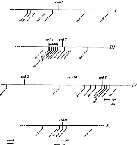
10 (Table 1, MATERIALS AND METHODS). T h e map locations of these genes are shown in Figure 3.
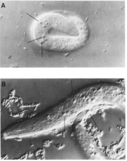
Phenotypes of the new mutants: Each of the 24 new mutations we studied prevents many cell corpses from being degraded (e.g., Figure 4). In each of the mutants the appearance of the corpses, as visualized using Nomarski optics, is the same as in the ced-1 and
ced-2 mutants isolated by HEDGECOCK, SULSTON and THOMSON (1 983). These corpses resemble those seen in wild-type animals during the stage of programmed cell death immediately before degradation (SULSTON and HORVITZ 1977). Occasionally, older corpses in these mutants contain vacuoles, lose the shiny appear- ance of fresh corpses, or even seem to be slowly disintegrating.
For each ced mutation, we examined the progeny of ced/+ heterozygotes, and determined the propor- tion that contain undegraded cell corpses (Table 2). About one fourth of the progeny of each ced-1 or ced- 8 heterozygote appear mutant, indicating that these mutations are recessive and that the wild-type gene is required in the zygote. By contrast, all alleles of the genes ced-2, ced-5, ced-6, ced-7 and ced-I0 show a maternal-rescue effect-almost all of the offspring from these heterozygotes appear wild type.
The new ced mutations prevent engulfment: T h e new ced mutations might prevent either the engulf- ment of cell corpses or the degradation of cell corpses after they are engulfed. To distinguish between these alternatives, we used electron microscopy to examine cell corpses in ced-5, ced-6, ced-7, ced-8 and ced-I0
84 R. E. Ellis, D. M. Jacobson and H. R. Horvitz
ced-1
ced-6 ced-7
"-
1-
I
---
111
ced-2
ced-IO
ced-5
I
I
I
---
IV
ced-8
FIGURE 3.-A genetic map showing portions of linkage groups I , III, IV and X . The positions of the ced genes involved in the engulfment of cell corpses are based upon data presented in MATERIALS AND METHODS.
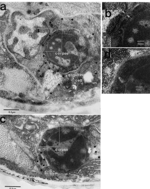
die in the ventral nervous system during larval devel- opment (ROBERTSON and THOMSON 1982), we fixed late L1 and early L2 larvae and examined dying cells descended from the ventral cord precursor cells P9 through P12. Some of the corpses we examined were in early stages of cell death, whereas others had re- mained undegraded for several hours. None of the dead cells we observed had been engulfed in the ced- 5 ( n 1 8 1 2 ) , c e d d ( n l 8 1 3 ) , c e d - 7 ( n 1 8 9 2 ) and ced- lO(n1993) mutants, whereas all of the corpses found in wild-type animals were being engulfed normally ( n
= 8 , 4, 4, 3 and 6 corpses, respectively; see Figure 5
for representative photographs).
Undegraded corpses are visible in ced-8 embryos viewed with Nomarski optics, but not in L2 larvae, so
it is possible that ced-8 does not affect postembryonic cell deaths (see below). We therefore examined both embryos and larvae of ced-g(n1891) mutants by elec- tron microscopy. Of five undegraded embryonic corpses visible in the heads of newly hatched ced-8
animals, none had been engulfed. By contrast, the two corpses we examined in the ventral cords of ced- 8 larvae were being engulfed normally. These obser- vations indicate that mutations in ced-8 affect the engulfment of cell corpses during embryogenesis. We therefore conclude that all of our newly discovered
ced genes, like ced-1 and ced-2 (HEDGECOCK, SULSTON and THOMSON 1983), affect the process of engulfment rather than a later step of degradation.
Cell Corpse Engulfment Genes 85
TABLE 2
Many ced mutations show maternal-rescue effects
FIGURE 4.-Two Nomarski pllotornicr(~gr~lphs of a representa- tive mutant defective in the degradation of cell corpses. (A) a ced- 5(n1812) embryo, and (B) a ced-5(nl812) LI larvae. Many unde- graded cell corpses are visible as bright, round refractile objects. Some of these corpses are indicated with lines.
these seven ced genes appear fully penetrant; in each animal some undegraded corpses are visible in the head during late embryogenesis. By contrast, 8 of the 10 wild-type embryos we examined contained no cell corpses in this region, and the remaining two embryos each had only a single corpse. However, the expres- sivity of each ced mutation appeared low and varied from animal to animal. Although 71 cells die during development of the head (SULSTON et al. 1983), pre- liminary observations revealed an average of around 30 corpses in the heads of late embryos of the strong- est mutant strains, and an average of 2 to 3 such corpses in embryos of the weakest mutant strains (e.g.,
see Table 9). T o quantify the effects of the ced muta- tions on the engulfment of cell corpses, and also to look for cell-specific effects of mutations in the ced
genes, we focused on cell deaths in one organ of the animal-the pharynx (Figure 6).
The pharynx has several advantages for these stud- ies: (1) Only 22 cells die during pharyngeal develop- ment, which is a small and easily countable number.
(2)
Almost all cell deaths in the pharynx occur within a two hour period during mid-embryogenesis (SULS- TON et al. 1983). By counting the number of corpses present in L1 larvae shortly after hatching (about nine hours after the pharyngeal cells die), we can study the effects of the different ced mutations on a variety of cell corpses after essentially the same interval of time has passed for each corpse. (3) T h e pharynx is sur- rounded by a basement membrane, and has a wellZygotic nlwxtions Matern;d-resctre nwtations
Percent Ced Percent Ced
eggs from
red/+ par-
eggs from
red/+ par- Gene Allele ents (n) Gene Allele rnls ( n )
ced-1 n2089 n 1506 el 735 n691 n2092 n2091 n2000 n l 9 5 1 n1995
ced-8 n2093 n2090 n189I n1999
25 (60) 26 (46) 26 (42) 24 (25) 15 (102) 29 ( 5 6 )
33 (36) 29 (28) 15 (34)
17 (97) 26 (91) 20 (30) 20 (45)
ced-2 el 752 n I994
ced-5 n l 8 1 2 n2002
ced-6 n2095 nlR13
ctd-7 n 1892
n1997 n1998 n2096 n200 I n2094 n I996 n 1893
ced-I0 n1993 2 (41)
To determine which mutations show a maternal effect, we used Nomarski microscopy to count the number of eggs (produced by self-fertilization from ced/+ hermaphrodites) that had unengulfed cell corpses. All eggs produced by self-fertiliwtion from cedlced
hermaphrodites appear mutant (data not shown). We include data for the mutations ced-I(t1735) and ced-2(el752), previously charac- terized by HEDGECOCK, SULSTON and THOMSON ( I 983); ced-l(n69 I) ,
a spontaneous mutation (H. ELLIS, personal communication): and
ced-l(nlY5l)a mutation we isolated in a separate E M S mutagenesis experiment.
defined structure (Figure 6; ALBERTSON and THOM-
SON 1976), making it easy to observe the distribution of cell corpses. (4) Although it is difficult to determine the exact identity of most embryonic corpses, because of their characteristic positions we can reliably identify the corpses formed by the deaths of two specific cells in the pharynx, the sisters of the two NSM neurons (ELLIS and HORVITZ 199 1).
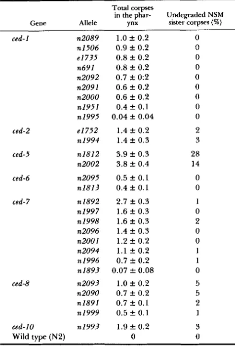
To measure the expressivity of each ced mutation we developed two assays using the pharynx. First, we determined the average number of cell corpses found in the pharynges of young larvae. Second, we studied the effects of these mutations on a specific pair of dying cells, by determining the frequencies at which the NSM sister corpses remain undegraded. Table 3 shows the results of these two assays.
T h e most striking feature of Table 3 is that all of the ced mutations are very low in expressivity. These mutants contain 0-4 corpses per pharynx at hatching, far fewer than the total of 22 corpses formed as the pharynx develops. Furthermore, the NSM sister corpses are engulfed most or all of the time in these
86 R. E. Ellis, D. M. Jacobson and H. R. Horvitz
FIGURE 5.-Electron micrographs of cell corpses in wild-type and ced-10 animals. (a) An engulfed corpse in a wild-type larva at about the time of the LI molt. The corpse is PI 1 .aap (SULSTON and HORVITZ 1977). which is engulfed by the hypodermis (ROBERTSON and THOMSON
TABLE 3
Expressivities of the ced mutants
Total corpses
in the phar- Undegraded NSM Gene Allele vnx sister corpses (W)
ced-1 n2089
n 1506 e l 735 n691 n2092 n2091 n2000 n1951 n 1995
1.0
*
0.2 0.9 f 0.20.8 f 0.2 0.8 f 0 . 2 0.7 f 0:2 0.6 f 0.2 0.6 f 0.2 0.4 ? 0.1 0.04 f 0.04
ced-2 e1752 1.4 f 0.2 2
n1994 1.4 f 0.3 3
ced-5 n1812 3.9 f 0.3 28
n2002 3.8 f 0.4 14
ced-6 n2095 0.5 f 0.1 0
n1813 0.4 f 0.1 0
ced-7 n1892
n1997 n 1998 n2096 n2001 n2094 n 1996 n1893
2.7 f 0.3 1.6 f 0.3 1.6 f 0.3 1.4 f 0.3 1.2 f 0.2 1 . 1 f 0.2 0.7 f 0.2 0.07 f 0.08
ced-8 n2093 1.0 f 0.2 5
n2090 0.7 f 0.2 5
n1891 0.7 f 0.1 2 nI999 0.5 f 0.1 1 ced- 10 n1993 1.9 f 0.2 3
Wild type (N2) 0 0
The numbers of corpses present in the pharynges of L1 larvae the total number of corpses observed (22 cells die during formation of the pharynx), and also the percentage of NSM sister corpses remaining undegraded (two of the cells that die during pharyngeal development are the sisters of the NSM neurons). The data shown for ced-2(nI994), ced-5(n2002) and the ced-7 alleles n1997, n1998, n2096 and n1893 represent the average of 50-60 animals; all other data represent the average of 100 animals. The 95% confidence limits were calculated with the t-test, using the S t u t V h I1 program (Abacus Concepts, Inc., Berkeley, California).
were assayed as described in MATERIALS A N D METHODS. We pI-eSent
eliminates the function of its particular ced gene, or
(2)
there is redundancy in the system for engulfing dead cells, so that more than one gene must be inac- tivated to prevent all engulfments from occurring, or (3) some corpses might disappear without being en- gulfed. We consider these possibilities below.Are any of the ced mutations null alleles? The effects of all of the ced mutations we describe are recessive, which suggests that each mutation might
cause a reduction or loss of gene function. Table 3 reveals that there are significant differences in ex- pressivity among these mutations, and these differ- ences provide hints about which mutations might cause a complete loss of gene function. For example, the ced-1 alleles form a series, with weak alleles such as n1951 and n 1 9 9 5 , and a number of strong alleles. These strong alleles include ced-l(n1506), a potential transposon insertion (our unpublished observations), and ced-l(n691), a spontaneous mutation. Further- more, we isolated five ced-1 mutations from a screen of about 10,000 haploid genomes, a frequency equiv- alent to the value of 1/2,000 expected for loss-of- function mutations in typical C . eleguns genes (BREN- NER 1974; MENEELY and HERMAN 1979; GREENWALD and HORVITZ 1980). These observations suggest that the strong ced-1 alleles might result in a complete loss
of ced-1 function. Because many cell corpses are en- gulfed in strong ced-1 mutants, the null phenotype of
ced-1 probably is of low expressivity.
We isolated eight ced-7 mutations from a screen of about 5,000 haploid genomes, so ced-7 alleles occur very frequently. However, this series of alleles does not end with a group of strong mutations. Rather,
ced-7(n1892) stands alone as the strongest allele. Thus the ced-7 gene is very sensitive to mutation, but we do not know which mutations, if any, eliminate ced-7
function. We recovered four ced-8 alleles from a screen of about 10,000 haploid genomes, which is also close to the frequency expected for loss-of-function mutations. Furthermore, ced-8 homozygotes and ced-
8(nl89l)/yDf5 animals resemble one another-both
live and contain undegraded cell corpses-so it is pos- sible that some ced-8 alleles completely eliminate gene function. Similarly, ced-5(n1812)/sDj2 animals live and resemble ced-5 mutants, so this ced-5 mutation also might be a null allele. Our data do not indicate if any existing mutations in ced-2, ced-6 and ced-10 result in partial or complete losses of gene function.
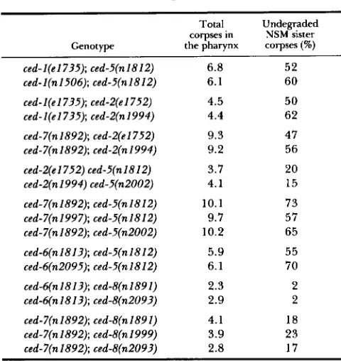
The ced genes fall into two functional sets: T o determine if some of the ced genes that act in the engulfment of cell corpses are functionally redundant, which would account for the low expressivities seen in the single mutants, we built various double mutant combinations. We then measured the expressivity of the Ced phenotype of each of the double mutants, using the assays described above. Our results are summarized in Table 4. We also constructed addi- tional double mutants to test for allele-specific effects. These data, shown in Table 5 , indicate that the inter-
engulfing pseudopod is visible in some regions, whereas in other regions it is squeezed between the corpse and a neighboring cell and no cytoplasm can be seen. One such compressed region is enlarged in (b), which shows four parallel membranes, one from the neighboring cell and one from the corpse (indicated with black arrowheads), and two from the engulfing cell (indicated with white arrows). (c) An unengulfed corpse in a ced-10 larva at about the time of the L1 molt. The corpse is PI 1 .aap, and the ventral cuticle is visible at the bottom of the micrograph. The membrane of the corpse is indicated with small arrowheads, and the membrane of the hypodermal cell is marked with black arrows where it faces neighboring cells. Note that the hypodermal cell has not produced an engulfing pseudopod. (d) An enlargement of (c), showing the membrane of the dead cell next to that of a neighbor, and the absence of membranes from an engulfing pseudopod.
88 R. E. Ellis, D. M. Jacobson and
€3.
R. HorvitzprOCOrpUS mehCOrpUS isthmus terminal bulb
n
n
nn
FIGURE 6.-The approximate posi-Left side: tions of the cell corpses formed during
pharyngeal development. The locations
drawn based upon (1) the anatomy of the pharynx (ALBERTSON and THOMSON 1976), (2) the cell lineage of the pharynx (SULSTON et al. 1983) and (3) our obser- vations of ced mutants (data not shown). NSM sister corpses Because of this variation, the I2 sister I2 sister corpses corpses cannot be distinguished by loca-
dorsal 0.
ventral
0 of these corpses vary somewhat, and are
0
Right side: ~
-9
3
tion from those of adjacent dying cells.The identification of the NSM sister corpses is described by ELLIS and HORV- dorsal
ventral
0:
ITZ (1 99 1).
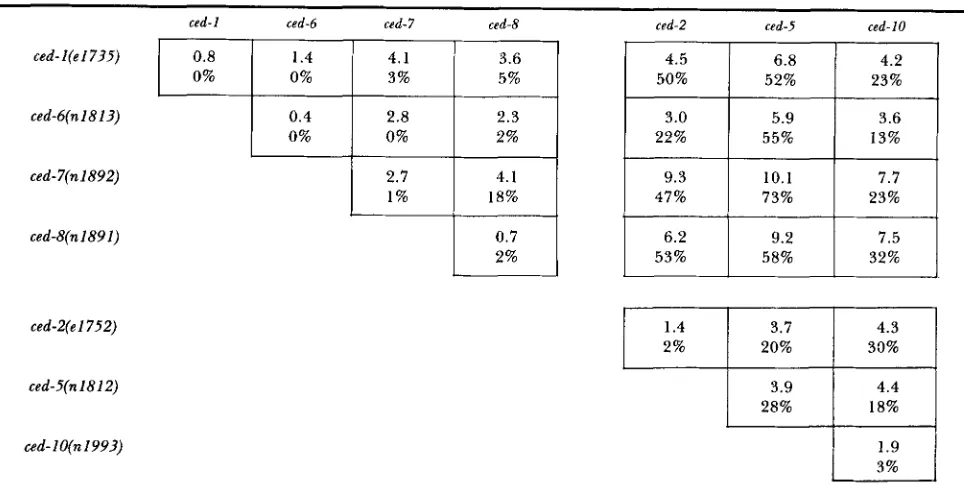
TABLE 4
Expressivities of ced double mutants
ced-l(e17?5)
ced-6(n 18 13)
ced-7(n1892)
ced-g(n1891)
ced-2(el752)
ced-5(n1812)
ced- 1 O(n 199?)
ced-1 ced-6 ced-7 ced-8 ced-2 csd-5 ced-10
0.8
23% 52%
50% 5%
3%
4.2 6.8
0% 0%
4.5 3.6
4.1 1.4
0.4 2.3 2.8 3.0 5.9 3.6
0%
32% 58% 53% 2%
7.5 9.2
6.2 0.7
23% 73%
47% 18%
1 %
7.7 10.1
9.3 4.1
2.7
13% 55%
22% 2%
0%
1.4
20% 2%
4.3 3.7
3% 1.9 18% 28%
4.4 3.9
33%
The upper value in each box is the average number of corpses in the pharynges of young L1 larvae, and the lower value represents the percentage of NSM sister corpses remaining undegraded in these animals; the values for each single mutant represent the average of at least 100 animals, and are taken from Table 3. The assays are described in MATERIALS A N D METHODS.
actions between different ced genes do not depend on the particular alleles we used to construct the strains. The results of both assays show that the expressivities of certain double mutants are much greater than those of any single ced mutant. For example, ced-7; ced-5
worms have an average of 10 corpses in the pharynx, and we have observed individuals of this genotype with
up
to 17 pharyngeal corpses. By contrast, ced-7and ced-5 single mutants have an average of three to four pharyngeal corpses, with a maximum of seven observed in some individuals. Furthermore, the NSM
sister corpses remain unengulfed 73% of the time in the ced-7; ced-5 double mutant, but only 28% of the
time in ced-5 animals and 1 % of the time in ced-7
animals.
Analysis of the double mutants suggests that two distinct sets of genes might operate in the engulfment of dead cells. Double mutants among ced-2, ced-5 and
ced-10 show the same expressivity as ced-5 single mu- tants, whereas double mutants between ced-2, ced-5 or
ced-10, and ced-1, ced-6, ced-7 or ced-8 generally display
TABLE 5
Expressivities of double mutant combinations are not allelespecific
Total
corpses in NSM sister Undegraded Genotype the pharynx corpses (%)
ced-l(e1735); ced-5(n1812) 6.8 52
ced-l(n1506); ced-5(n1812) 6.1 60 ced-l(el735); ced-Z(e1752) 4.5 50
ced-l(e1735); ced-Z(n1994) 4.4 62
ced-7(n1892); ced-Z(el75.2) 9.3 47
ced-7(n1892); ced-Z(n1994) 9.2 56
ced-Z(el752) ced-5(n1812) 3.7 20
ced-Z(n1994) ced-5(n2002) 4.1 15
ced-7(n1892); ced-5(n1812) 10.1 73
ced-7(n1997); ced-5(n1812) 9.7 57
ced-7(n1892); ced-5(n2002) 10.2 65
ced-6(n1813); ced-5(n1812) 5.9 55
ced-6(n2095); ced-5(n1812) 6.1 70
ced-6(n1813); ced-b'(n1891) 2.3 2
ced-6(n1813); ced-8(n2093) 2.9 2
ced-7(n1892); ced-8(n1891) 4.1 18
ced-7(n1892); ced-8(n1999) 3.9 23
ced-7(n1892); ced-tI(n2093) 2.8 17
Each set represents double mutants containing different alleles of the same two ced genes. The assays used are described in
MATERIALS AND METHODS. Each value represents the average of 30
animals. The strains used in the studies presented in Table 4 are listed first in each set shown in this table.
ced-7 and ced-8 act in a second process. Some of the double mutant combinations within the ced-I, ced-6, ced-7, ced-8 set are stronger than any of the single mutants within this set, which could be either because some of these mutations are not null alleles, or because these four genes do not act in a single process.
Because genotypes with the strongest effects on engulfment do not prevent all 22 corpses formed during development of the pharynx from disappear- ing, we examined the triple mutants shown in Table
6 to see if any showed an even more severe phenotype. None of these triple mutants is more severe than the strongest double mutants. Instead, each triple mutant appears similar to the strongest double mutant com- bination that it contains. T h e fact that triple mutants are not more severe than double mutants supports the idea that there are only two processes of engulfment defined by the seven ced genes.
Some cell corpses probably disappear without en- gulfment: T w o observations suggest that cell corpses might occasionally disappear without being engulfed. First, preliminary experiments with a laser microbeam indicate that corpses are more fragile than living cells, suggesting that some unengulfed corpses might burst (see MATERIALS AND METHODS). All that is left of a corpse after destruction with a laser beam is a small amount of debris, similar to material we have some- times seen where we expect cell corpses in the various
ced mutants (our unpublished observations). Such de-
bris was not counted in our assays for the number of cell corpses. Second, HEDGECOCK, SULSTON and
THOMSON (1 983) noticed that some cell corpses sepa- rate from the developing embryo and drift into the egg fluid. Based on these two observations, we con- clude that some corpses probably disappear in the absence of engulfment, which could explain why we never find the corpses of all dead cells in the severe double and triple mutants we have assayed.
The ced genes act on the same corpses, but with different relative effects: Analysis of double mutant combinations suggests that the two engulfment proc- esses do not act on entirely distinct groups of cells. Mutations in each of the ced genes show synergistic effects on the engulfment of the NSM sister corpses in some double mutant combinations (Table
4).
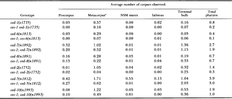
Therefore all of these genes must play a role in the engulfment of the NSM sister corpses. We also studied the roles of the different ced genes in the engulfment of two other pharyngeal corpses, those formed by the sisters of the two I2 neurons. T h e mutation ces- I(n703) prevents both the I2 sisters and the NSM sisters from dying but does not affect other cell deaths (ELLIS and HORVITZ 1991), so we compared ces-I; ceddouble mutants with the corresponding ced single mutants (Table
7).
When the I2 sisters survive, there are fewer corpses in the metacorpus of all of the cedmutants (although the decrease for ced-8 is small and might not be significant), which indicates that most or
all of the ced genes affect the engulfment of the I2 sister corpses. As a control, we also measured the number of corpses found in the procorpus, the isth- mus, the terminal bulb, and the anterior end of the isthmus, where the NSM sisters die (Figure 6). In general, the control values were unaffected by the presence of a ces-1 mutation.
Although the ced genes do not act on distinct groups of corpses, mutations in the ced genes appear to have different relative effects on different corpses. For example, seven cells, including the sisters of the two I2 neurons, die in the metacorpus of the pharynx. In
ces-1; ced-2(e1752) animals, in which the I2 sisters survive, there is an average of only 0.04 corpses in the metacorpus, whereas ced-2(el752) animals have an average of 1.05 corpses in this region. Thus, in the metacorpus, ced-2(el752) affects mainly the engulf- ment of the I2 sisters (Table
7).
By contrast, ced- 7(n1892) appears to affect other corpses in the meta- corpus in addition to the I2 sisters, because in these mutants a ces-1 mutation decreases by only half the number of corpses in this location.90 R. E. Ellis, D. M. Jacobson and H. R. Horvitz
TABLE 6
Expressivities of triple mutants
Total corpses
Genotype in the pharynx Undegraded NSM sister corpses ( W )
ced-l(e1735); ced-7(n1892); ced-5(n1812) 11.1 67
ced-7(n1892); ced-5(n1812) 10.1 73
ced-l(el735); ced-5(n1812) 6.8 52
ced-l(el735); ced-7(n1892) 4.1 3
ced-l(e1735); ced-7(n1892); ced-8(n1891) 3.9 12
ced-7(n1892); ced-8(n1891) 4.1 18
ced-I(e1735); ced-g(n1891) 3.6 5
ced-l(el735); ced-7(n1892) 4.1 3
ced-7(n1892); ced-5(n1812); ced-8(n1891) 8.2 78
ced-7(n1892); ced-5(n1812) 10.1 73
ced-5(n1812); ced-g(n1891) 9.2 58
ced-7(n1892); ced-g(n1891) 4.1 18
ced-6(n1813); ced-2(e1752) ced-5(n1812) 5.5 60
ced-6(n1813); ced-Z(nl812) 5.9 55
ced-6(n1813); ced-2(e1752) 3.0 22
ced-2(e 1 7 5 2 ) ced-5(n 18 12) 3.7 20
ced-6(n1813); ced-7(nl892); ced-8(n2093) 4.5 10
ced-7(n1892); ced-8(n2093) 2.8 17
ced-6(n1813); ced-8(n2093) 2.9 2
ced-6(n1813); ced-7(nl892) 2.8 0
The expressivities of each triple mutant and of its constituent double mutants are listed. Each value represents the average of 30 animals. The assays used are described in MATERIALS AND METHODS.
TABLE 7
Distributions of pharyngeal corpses in ces-I(n703); ced double mutants
Average number of corpses observed:
Terminal Total
Genotype Procorpus Metacorpus" NSM sisters Isthmus bulb pharynx
ced- 1 (e 1 735) 0.03 0.57 0.00 0.02 0.16 0.8
ces-1 ced-l(el735) 0.00 0.16 0.00 0.00 0.07 0.2
ced-6(n1813) 0.03 0.29 0.00 0.00 0.03 0.4
ces- I ; ces-h(n I81 3) 0.00 0.07 0.00 0.01 0.06 0.1
ced- 7(n 1892) 0.32 1.02 0.01 0.01 1.36 2.7
ces-I; ced-7(n1892) 0.20 0.52 0.0 1 0.0 1 1.15 1.9
ced-8(n1891) 0.16 0.28 0.03 0.0 1 0.19 0.7
ces-I; ced-8(n1891) 0.1 3 0.22 0.0 1 0.04 0.33 0.7
ced-2(el752) 0.0 1 1.05 0.04 0.02 0.32 1.4
ces-1; ced-2(el752) 0.02 0.04 0.00 0.00 0.25 0.3
ced-5(n 18 12) 0.42 1.71 0.55 0.13 1.04 3.9
ces-1; ced-5(n1812) 0.27 0.62 0.0 1 0.09 2.03 3.0
ced- I O(n 1993) 0.08 1.22 0.05 0.03 0.53 1.9
ces-I: ced-lO(n1993) 0.10 0.49 0.0 1 0.00 0.36 1
.o
The metacorpus contains the I2 sisters.
Cell corpse assays are described in MATERIALS AND METHODS, the anatomy of the pharynx is described by ALBERTSON and THOMSON
(1 976). and a diagram showing the distribution of dying cells in the pharynx is shown in Figure 6. Note that the I2 sister corpses are located in the metacorpus. The total numbers of dying cells in each region are: procorpus (6); metacorpus, which contains the two I2 sisters (7); NSM sisters (2); isthmus (1); and terminal bulb (6). The final column shows the average number of corpses found in the whole pharynx, as presented in Table 3. We scored 100 animals of each genotype for each mutation, except for ced-B(n1891), for which we scored 150 animals.
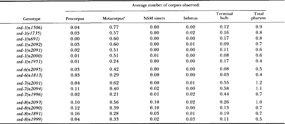
the four ced-8 alleles (Table 8). Our results show, for are gene-specific and not allele-specific. There are example, that of the mutations listed, those in ced-8 also overall variations in expressivity among the ced
have the greatest relative effects on the NSM sister genes. For example, all alleles of ced-2 are weaker corpses, and those in ced-7 have the greatest effects than either allele of ced-2, and both alleles of ced-2 are on corpses in the terminal bulb. In general, the effects much weaker than either allele of ced-5 (Table 3).
Cell Corpse Engulfment Genes
TABLE 8
The spatial distributions of corpses in ced mutants are gene-specific
Average number of corpses observed: Genotype
ced-l(n1506) ced-l(el735) ced-l(n691) ced-l(n2092) ced-l(n2091) ced-l(n2000) ced-I(n1951)
Terminal Total
Procorpus Metacorpus" NSM sisters Isthmus bulb pharynx
0.04 0.77 0.00 0.00 0.12 0.9
0.03 0.57 0.00 0.02 0.16 0.8
0.00 0.60 0.00 0.00 0.17 0.8
0.03 0.60 0.00 0.01 0.09 0.7
0.02 0.5 1 0.00 0.00 0.1 1 0.6
0.01 0.5 1 0.01 0.00 0.08 0.6
0.01 0.24 0.00 0.00 0.17 0.4
ced-6(n2095) 0.03 0.42 0.00 0.00 0.08 0.5
ced-6(n1813) 0.03 0.29 0.00 0.00 0.03 0.4
ced-7(n2001) 0.04 0.62 0.00 0.0 1 0.55 1.2
ced-7(n2094) 0.1 1 0.40 0.02 0.00 0.58 1.1
ced-7(n 1996) 0.02 0.21 0.0 1 0.02 0.44 0.7
ced-8(n2093) 0.10 0.56 0.10 0.02 0.26 1
.o
ced-8(n2090) 0.12 0.39 0.10 0.00 0.13 0.7
ced-8(n1891) 0.16 0.28 0.03 0.0 1 0.19 0.7
ced-8(n1999) 0.04 0.33 0.02 0.03 0.1 1 0.5
* T h e metacorpus contains the I2 sisters.
T h e assays are described in Table 7. We scored 100 animals of each genotype for each mutation, except for ced-8(n1891), for which we
scored 150 animals.
velopment: We determined the role the ced genes play in cell death at different stages of development. Mutations in ced-1, ced-2, ced-5, ced-6 and ced-10 affect cell corpses formed in the head and pharynx during embryogenesis, and also corpses formed in the ventral nerve cord during larval development (Table 9). By contrast, mutations in the genes ced-7 and ced-8 affect corpses mainly during embryogenesis, since there are many undegraded corpses in ced-7 and ced-8 embryos, but not in the ventral nerve cord during larval devel- opment. Furthermore, the embryonic corpses in ced-
7
and ced-8 animals disappear during larval develop- ment, so that these mutants eventually contain far fewer corpses than even ced-l(n1995) mutants, al- though in other respects ced-I(n1995) is the weakestced mutation we have characterized (Table 3). It seems possible either that ced-7 and ced-8 play a role specific to embryogenesis, or else that mutations in these genes slow down but do not prevent engulfment, and so are most easily observed during the relatively short em- bryonic period. In our ultrastructural studies (see above), both of the larval ced-8 corpses we examined were being engulfed, but the four ced-7 corpses were unengulfed, which indicates that mutations in ced-7
can affect larval corpses, although these corpses even- tually disappear.
DISCUSSION
After cells die by programmed cell death, their corpses are quickly engulfed by other cells and then degraded (WYLLIE, KERR and CURRIE 1980; HEDGE- COCK, SULSTON and THOMSON 1983). In a few cases,
TABLE 9
Number of corpses in embryos and L3/L4 larvae L3/L4 larvae
Embryos Embryonic deaths Larval deaths
Genotype Head Head Pharynx Tail" posterior tral cord ven-
ced-I(n1995) ced-l(el735)
ced-2(el752)
ced-5(n1812)
ced-6(n1813)
ced-1 O(n 1993)
ced-7(n1997) ced-7(n1892)
ced-b'(n1891)
11.3 22.2
9.0
27.4
22.6
18.3
28.1 26.8
6.3
5.5 >10
-9
>10
>10
-7
0.2 0.3
0.2
0.5 1.2 1.2
0.5 2.2 5.8
1.5 1.5 3.7
3.2 4.0 3.8
0 1.8 3.0
1.5 1.8 4.3
0.2 0 0.0
0.5 0 0.2
0.2 0 0.0
T h e tail contains both embryonic and larval deaths.
~~~
We used Nomarski microscopy to determine the average number of corpses in the heads of three embryos of each genotype, and also to determine the average number of corpses found in different body regions of six larvae of each genotype during the L3/L4 molt. T h e wild-type pattern of cell deaths was determined by SULSTON and HORVITZ ( 1 977) and by SULSTON et al. (1983). T h e 7 1 cells that die during formation of the head (excluding the pharynx) and the 22 cells that die during formation of the pharynx all die embryonically. T h e tail contains 13 cells that die embryonically, and two that die during larval development. In the posterior ventral cord, one descendant of P9, one descendant of P10, two descend- ants of PI 1 and three descendants of P12 die during larval devel- opment. >IO, greater than 10 (in these animals the number of corpses also appeared roughly less than or equal to the embryonic value); -, approximately equal to.