ABSTRACT
ROCK, KYLIE DANIELLE. Sex-Specific Effects of Firemaster
®550 on Placenta, the
Developing Brain, and Behavior. (Under the direction of Dr. Heather Patisaul).
Chemical flame retardants (FRs) are commonly applied to consumer products including foam-based furniture and infant products, textiles, and electronics. Widespread human exposure and evidence of endocrine disruption have raised concerns regarding the possible toxicity of these chemicals,
particularly on the developing brain. While certain mixtures, such as the polybrominated diphenyl ethers (PBDEs), have been phased-out, due to known toxic effects, use of alternative FR mixtures have rapidly increased. Firemaster® 550 (FM 550), which is composed of brominated and organophosphate (OP)
compounds, has become one of the most commonly used FRs in baby products and residential furniture, yet we still know very little about its toxicity. We identified evidence of endocrine disruption and sex-specific behavioral effects in rats exposed to FM 550 during development. Furthermore, we have shown that components of FM 550 can be placentally and lactationally transferred to the developing offspring. Throughout gestation, the placenta is a critical coordinator of fetal growth and development, including neurodevelopment. Certain chemicals, including FRs, have been found to accumulate in placental tissue. Given the important role the placenta plays in fetal programming, the studies reported in this dissertation were conducted to test the hypothesis that placental dysfunction may be a critical but underappreciated mechanism by which environmental chemicals, such as FRs, induce changes in brain and behavior.
Wistar rat dams were exposed to a range of human relevant doses during the perinatal window (0, 100, 300, and 1000 g/day; GD 9 – PND 21) for offspring behavioral assessments or during the
We hypothesized that sex-biased placental accumulation would result in sex-specific changes in placental function and ultimately lead to sex differences in neurodevelopmental outcomes. Using untargeted (RNA-seq and metabolomics) and targeted (qRT-PCR and neurotransmitter analysis) approaches we conducted a thorough molecular investigation of these placentas. Multiple pathways relevant to brain development were altered by FM 550 exposure, such as endocrine, inflammatory, and neurotransmitter signaling pathways. Some changes were sex-specific, including changes in serotonin (5-HT) that were observed in both the placenta and fetal forebrain of female offspring. These findings indicate that FM 550 has the potential to impact brain development by altering normal placental functions and highlights 5-HT signaling between the placenta and brain as a potentially causal mechanism.
Due to widespread use of the OPs (TPHP and ITPs), in complex mixtures like FM 550 or
Sex-Specific Effects of Firemaster
®550 on Placenta, the Developing Brain, and Behavior.
by
Kylie Danielle Rock
A dissertation submitted to the Graduate Faculty of
North Carolina State University
in partial fulfillment of the
requirements for the degree of
Doctor of Philosophy
Toxicology
Raleigh, North Carolina
2019
APPROVED BY:
_______________________________
_______________________________
Dr. Heather Patisaul Dr. Seth Kullman
Committee Chair
_______________________________
_______________________________
Dr. John Meitzen
Dr. John Godwin
ii
DEDICATION
iii
BIOGRAPHY
Kylie Danielle Rock was born October 26th, 1990 in Plattsburgh, New York. She graduated from
Elizabethtown-Lewis Central School in 2008. During her undergraduate career, at St. Lawrence University in Canton, New York, she spent three and a half years assisting with research projects in the labs of Dr. Marilyn Mayer and Dr. Alexander Schreiber. She graduated with a Bachelor of Science in Biology in 2012. She went on to work as a technician in the lab of Dr. Heather Stapleton at Duke
iv
ACKNOWLEDGMENTS
v
TABLE OF CONTENTS
List of Tables ... viii
List of Figures ... ix
Chapter 1 - Environmental Mechanisms of Neurodevelopmental Toxicity ... 1
Literature Review ... 1
Abstract ... 2
Keywords ... 2
Introduction ... 2
Defining Developmental Neurotoxicity ... 3
Mechanisms of Developmental Neurotoxicity ... 3
Oxidative Stress ... 3
Altered Neurotransmitter Systems ... 5
Neuroendocrine Disruption ... 6
Immune System Disruption ... 7
Indirect Toxicity ... 8
Conclusions ... 8
References ... 9
Chapter 2 - Sex Specific Placental Accumulation and Behavioral Effects of Developmental Firemaster 550 Exposure in Wistar Rats ... 15
Abstract ... 16
Introduction ... 16
Methods ... 17
Animals ... 17
Experiment 1: Placental Accumulation of FM 550 Components ... 18
Dosing Prep ... 18
Animal Husbandry and Exposure ... 18
Tissue Collection ... 18
Placental Analysis ... 18
Experiment 2: Behavioral Consequences of Developmental FM 550 Exposure ... 18
Animal Husbandry and Exposure ... 18
Behavior Testing ... 19
Statistical Analysis ... 19
Results ... 20
Accumulation of FM 550 Components in Placenta ... 20
Juvenile Behavior ... 20
Adult Behavior ... 21
Discussion ... 22
References ... 26
Appendix I Supplementary Methods ... 97
Chapter 3 - Molecular Effects of Developmental FM 550 Exposure in Wistar Rat Placenta and Fetal Forebrain... ... 29
Abstract ... 30
Introduction ... 30
vi
Animals ... 32
Dosing Prep ... 32
Animal Husbandry and Exposure ... 32
Tissue Collection ... 32
RNA Sequencing ... 33
RNA-seq Data Processing ... 33
Quantitative Real-Time PCR ... 34
GC-TOF-MS Metabolomics ... 34
GC-TOF-MS Data Analysis ... 34
Neurotransmitters ... 34
Statistical Analyses... 35
Results ... 35
Impact of Prenatal FM 550 Exposure on Placental Gene Expression ... 35
Impact of Prenatal FM 550 Exposure on Placental Neurotransmitters and Metabolites ... 37
Impact of Prenatal FM 550 Exposure on Fetal Forebrain Gene Expression ... 39
Impact of Prenatal FM 550 Exposure on Fetal Forebrain Neurotransmitters ... 40
Discussion ... 43
Conclusions ... 45
References ... 46
Appendix II Supplementary Methods, Tables, and Figures ... 104
Chapter 4 - Effects of Prenatal Exposure to a Mixture of Organophosphate Flame Retardants on Placental Gene Expression and Neurotransmitter Levels ... 50
Abstract ... 51
Introduction ... 52
Materials & Methods ... 55
Animals ... 55
Dosing Prep ... 56
Animal Husbandry and Exposure ... 57
Tissue Collection ... 58
Serum Processing and Analysis ... 59
Placenta Processing and Analysis ... 60
RNA-Sequencing... 60
RNA-Seq Data Processing ... 61
Quantitative Real-Time PCR ... 61
Neurotransmitter Analysis ... 62
5-HT Immunohistochemistry ... 64
Confocal Microscopy and Image Analysis ... 65
Statistical Analysis ... 65
Results ... 66
Accumulation of OPFR Components in Placenta ... 66
Impact of Prenatal OPFR Exposure on Placental Gene Expression – RNA-seq ... 68
Impact of Prenatal OPFR Exposure on Placental Gene Expression – qRT-PCR ... 69
Impact of Prenatal OPFR Exposure on Placental Neurotransmitters ... 72
Impact of Prenatal OPFR Exposure on Development of 5-HTergic Projections ... 75
Discussion ... 76
Conclusions ... 81
References ... 83
vii
Chapter 5 - Conclusions ... 89
Novel Findings ... 90
Dissertation Summary ... 91
Limitations ... 94
Broader Impacts ... 95
Appendices………. ... 97
Appendix I– Chapter 2 Supplementary Methods ... 97
Appendix II– Chapter 3 Supplementary Methods, Tables, and Figures ... 104
Appendix III – Chapter 4 Supplementary Figures and Table ... 183
viii
LIST OF TABLES
Chapter 1
Table 1. Representative neurotoxic chemicals and how they can impact brain development through multiple mechanisms ... 4 Chapter 3
Table 1. Summary of untargeted metabolomics analysis in whole placenta ... 39 Supplemental Table 1. Sample size of groups evaluated for thyroid hormone levels ... 106 Supplemental Table 2. Average thyroid hormone concentrations (ng/mL) in sampled rat
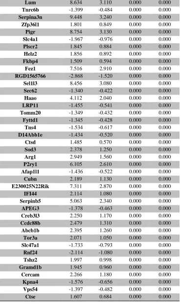
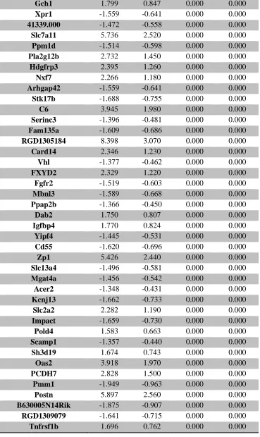
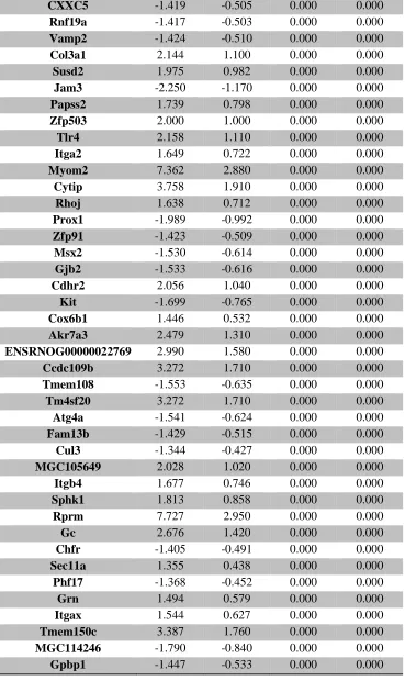
serum ... 107 Supplemental Table 3. RNA-seq analysis of differentially expressed placental genes ... 108 Supplemental Table 4. RNA-seq analysis of differentially expressed forebrain genes ... 173
Chapter 4
Table 1. Summary of chemicals in OPFR mixture and accumulation in maternal serum and
ix
LIST OF FIGURES
Chapter 2
Figure 1. Chemical structure of the four FM 550 components ... 17 Figure 2. Accumulation of TBB, TBPH, and TPHP in placental tissue following gestational
exposure to FM 550 ... 21 Figure 3. Effects of perinatal FM 550 exposure on juvenile behavior in the light dark box (L/D) ... 22 Figure 4. Effects of perinatal FM 550 exposure on juvenile behavior in the open field (OF) ... 23 Figure 5. Effects of perinatal FM 550 exposure on adult behavior in the light dark box
(L/D) ... 24 Figure 6. Effects of perinatal FM 550 exposure on adult behavior in the elevated plus maze
(EPM) ... 24 Figure 7. Effects of perinatal FM 550 exposure on adult behavior in activity wheels ... 25 Supplemental Figure 1. Quantifying and qualifying m/z for analytes and standards ... 100
Chapter 3
Figure 1. Top 10 canonical pathways identified by IPA analysis, in placenta and fetal forebrain, and changes in placental gene expression verified by qRT-PCR ... 36 Figure 2. Effects of prenatal FM 550 exposure on the relative abundance of genes related to
inflammatory signaling in placental tissue ... 37 Figure 3 Effects of prenatal FM 550 exposure on the relative abundance of genes related to
endocrine signaling in placental tissue ... 38 Figure 4. Effects of prenatal FM 550 exposure on neurotransmitter and neurotransmitter
metabolite levels in whole placenta ... 40 Figure 5. Effects of prenatal FM 550 exposure on the relative abundance of genes related to
5-HT signaling and metabolism in placental tissue ... 41 Figure 6. Effects of prenatal FM 550 exposure on neurotransmitter and neurotransmitter
metabolite levels in fetal forebrain ... 42 Figure 7. Effects of prenatal FM 550 exposure on the relative abundance of genes related to
5-HT signaling and metabolism in fetal forebrain ... 43 Supplemental Figure 1. Unsupervised principal component analysis (PCA) of placental
x Supplemental Figure 2. Unsupervised principal component analysis (PCA) of fetal forebrain
RNA-seq data ... 179 Supplemental Figure 3. Effects of prenatal exposure on the relative abundance of genes
related to FXR/RXR signaling and endocrine signaling ... 180 Supplemental Figure 4. Average levels of total thyroxine (TT4) and total triiodothyronine
(TT3) from the serum of dams during gestation (GD 18) or lactation (PND 21) and in serum from male and female pups (PND 21) ... 181 Chapter 4
Figure 1. Representative images of placental zones for micropunch collection ... 59 Figure 2. Levels of individual chemicals and total OPFR burden in whole placenta separated by
sex ... 68 Figure 3 Top 15 canonical pathways identified by IPA analysis using annotations for all tissue
types and annotations for placenta/nervous system tissue ... 69 Figure 4. Effects of prenatal OPFR exposure on placental genes responsible for protection
against oxidative stress and exposure to corticosterone ... 71 Figure 5. Effects of prenatal OPFR exposure on the relative abundance of genes involved in
the transport, synthesis, and metabolism of 5-HT and levels of tryptophan, 5-HT and 5-HIAA ... 73 Figure 6. Effects of prenatal OPFR exposure on the expression of a gene that metabolizes
kynurenine as well as the relative abundance of kynurenine and its metabolites ... 74 Figure 7. Effects of gestational OPFR exposure on the extension of 5-HT and thalamocortical
immunolabeled axons in the GD 14 brain ... 75 Figure 8. Summary of observed OPFR effects on metabolites and genes involved in the 5-HT
and kynurenine metabolic pathways ... 80 Supplemental Figure 1. Unsupervised principal component analysis (PCA) of placental
RNA-seq data ... 184 Supplemental Figure 2. Cluster dendrogram and unsupervised principal component
analysis (PCA) of female and male placentas ... 185 Chapter 5
Figure 1. Summary of dissertation findings: Disruption of serotonin signaling between the placenta and fetal brain as a novel mechanism of FR induced changes in brain and
1
CHAPTER 1
Environmental Mechanisms of Neurodevelopmental Toxicity
15
CHAPTER 2
Sex Specific Placental Accumulation and Behavioral Effects of Developmental Firemaster 550 Exposure in Wistar Rats
29
CHAPTER 3
Molecular Effects of Developmental FM 550 Exposure in Wistar Rat Placenta and Fetal Forebrain
50
CHAPTER 4
Effects of Prenatal Exposure to a Mixture of Organophosphate Flame Retardants on Placental Gene Expression and Serotonergic Innervation in the Developing Rat Brain
Kylie D. Rock1, Genevieve St Armour1, Brian Horman1, Allison Phillips3, Matthew Ruis3, Allison K.
Stewart4, Dereje Jima2,5, Heather M. Stapleton3, Heather B. Patisaul1,2 1Department of Biological Sciences, North Carolina State University, Raleigh, NC, USA
2Center for Human Health and the Environment, North Carolina State University, Raleigh, NC, USA 3Nicholas School of the Environment, Duke University, Durham, NC 27708, USA
4Molecular Education, Technology, and Research Innovation Center, North Carolina State University,
Raleigh, NC, USA
51 Abstract
Concerns regarding the potential neurotoxicity of chemical flame retardants (FRs) are on the rise, in part due to the increased use of organophosphate flame retardants (OPFRs) that are structurally similar to known neurotoxicants. Human exposure to OPFRs is widespread, as they are commonly used in complex mixtures as FRs, like the commercial mixture Firemaster® 550 (FM 550), and as plasticizers.
We have previously shown that developmental FM 550 exposure can lead to sex-specific behavioral effects in rats and highlighted the placenta as a potential target tissue of toxicity. Throughout gestation, the placenta is a critical coordinator of fetal growth and development, including neurodevelopment. We have shown in rats and humans that FRs accumulate in placental tissue; in some cases, to a greater degree in male-associated placentas than female-associated placentas. These placentas also showed evidence of sex-specific functional changes, including a reduction in serotonin (5-HT) turnover in female-associated placentas that was also observed in the fetal forebrain of female offspring. The placenta serves as a primary source of 5-HT needed for fetal forebrain development from GD 10 – 15. Given the important role the placenta plays in fetal programming, disruption of placental 5-HT may be a critical but
52 Introduction
Chemical flame retardants (FRs) have long been suspected of contributing to rising rates of neurodevelopmental disorders, with the now phased out polybrominated diphenyl ethers (PBDEs) specifically linked to deficits in memory and cognitive function [1-4]. Replacement brominated flame retardants (BFRs) have also been shown to have toxic effects, including effects on brain and behavior [5-9], and are environmentally problematic because of their persistence, thermostability, lipophilicity, and resistance to biotic and abiotic degradation. In response to these concerns the production and application of organophosphate flame retardants (OPFRs), as an alternative FR class, has seen a notable increase in recent years, with global consumption jumping from 100,000 tons in 1992 to 680,000 tons in 2015 [10-12]. While these alternatives show less bioaccumulation than the PBDEs [13], likely due to their rapid metabolism [13-15], their potential toxicity has not been studied to the same extent as the BFRs.
Neurotoxicity is a significant concern because structurally related compounds with an organophosphorous backbone have been shown in a variety of model systems to affect brain development and are suspected developmental neurotoxicants in humans [7, 16-20]. Firemaster® 550 (FM 550) is a commercial FR
mixture, comprised of both BFRs and OPFRs, that has rapidly become one of the most commonly used FRs for baby products and residential furniture [21, 22]. We have previously reported evidence of behavioral effects in rats following developmental exposure, and identified multiple pathways by which gestational FM 550 exposure may impact the placenta and developing forebrain [6, 23]. The present studies were conducted to assess the degree to which the OPFR components of FM 550 can impact neurodevelopmental outcomes by altering placental function and disrupting neurotransmitter signaling between the placenta and developing brain.
53 made up of organophosphate components, including triphenyl phosphate (TPHP also known as TPP) and a mixture of isopropylated triarylphosphate isomers (ITPs) [27] (Table 1). TPHP is a high production volume chemical (10 – 50 million lbs/year), and has been used as a plasticizer in products like polyvinyl chloride (PVC) and circuit boards for decades [28]. As a result, TPHP has become one of the most abundant and frequently detected OPFRs in indoor dust samples in the U.S. as well as globally, including Egypt, Japan, Germany, and Portugal [21, 29-32]. Evidence of human exposure from biomonitoring studies have shown nearly ubiquitous detection of diphenyl phosphate (DPHP), the primary metabolite of TPHP, in urine samples [33-39]. Fewer studies have looked at the environmental prevalence of the ITPs, as this isomeric mixture was not fully characterized until 2017 by Phillips et al. [27]. There is a growing body of literature, however, showing that exposure in the U.S. is very common. One study detected the ITPs on 100% of the silicone wristbands worn by human participants [40] while another study found detectable levels of mono-isopropylphenyl phenyl phosphate (mono-ipPPP), which is a confirmed metabolite of the ITPs, in 98% of urine samples [35]. Accumulating evidence of widespread exposure and concerns regarding potential neurotoxicity highlights the importance of investigating the
neurodevelopmental effects of replacement FRs like TPHP and the ITPs.
54 been shown to be a target of the organophosphate pesticide chlorpyrifos [46-49]. Therefore, we
hypothesized that disruption of 5-HT signaling may be a critical mechanism by which TPHP and the ITPs can impact brain development and behavior.
Throughout gestation, the placenta is a critical coordinator of fetal growth and development, including neurodevelopment. It is a highly active endocrine organ that is responsible for the transfer and synthesis of hormones, monoamines, and other signaling factors required for proper brain development [50-53]. For example, the fetal side of the placenta serves as a critical and primary source of 5-HT for the developing fetal forebrain from GD 10 – 15, a time frame that precedes the organization and function of the brain’s serotonergic network [51, 52]. Placental function is dynamic, adapting to the needs of the fetus and changes in the intrauterine environment, and sex-specific, with placental sex (XX vs XY) determining how and to what degree the placenta responds [50, 53, 54]. Such adaptability is believed to play a role in sex-specific fetal programming, including changes in brain development that may contribute to sex differences in risk of neurodevelopmental disorders. One well-studied example is the
programmatic effects of prenatal stress, which has been shown to result in male-specific placental inflammation and offspring hyperactivity [55]. Notably, maternal inflammation has been shown to disrupt forebrain development via increased placental 5-HT output to the fetal brain [56]. In our previous work in Wistar rats, we found that FM 550 accumulates in placental tissue and can alter various aspects of placental function, including endocrine, inflammatory, and neurotransmitter signaling [6]. Furthermore, we found evidence that 5-HT signaling between the placenta and fetal forebrain may be disrupted as a result of FM 550 exposure [23]. 5-HT plays an important role in fetal brain development, and our
previously published data shows changes in behaviors associated with 5-HT signaling well into adulthood following developmental FM 550 exposure [6]. Therefore, we hypothesized that disruption of placental output, particularly 5-HT output, could be an important mechanism by which the organophosphate components of FM 550 influence sex-specific neurodevelopmental programming.
55 contributes to the adverse phenotypes. The present studies focused on the OPFRs, with an emphasis on disruption of 5-HT production and signaling between the placenta and fetal forebrain. To probe these and other modes of action we employed a combination of hypothesis-generating (RNA-sequencing) and targeted (quantitative real-time PCR and targeted NT analysis) tools to examine Wistar rat placentas following gestational exposure to 500, 1000, and 2000 g OPFRs/kg/day (TPHP and ITPs, Table 1) as well as the global 5-HT agonist 5-methoxytryptamine (5 mg/kg/day) as a positive control for fetal 5-HT disruption. In addition, we assessed development of the 5-HTergic system in fetal Wistar rat brains in order to see if there were any neurodevelopmental phenotypes associated with changes in placental function.
Materials and Methods
Animals
Animal care, maintenance, and experimental protocols met the standards of the Animal Welfare Act and the U.S. Department of Health and Human Services “Guide for the Care and use of Laboratory Animals” and were approved by the North Carolina State University (NCSU) Institutional Animal Care and Use Committee (IACUC). A supervising veterinarian approved and monitored all procedures throughout the duration of the project. For each aim, Wistar rats were obtained from Charles River (Raleigh, NC) and bred in house as indicated in humidity-and-temperature controlled rooms, each with 12 h:12 h light, dark cycles at 25°C and 45-60% average humidity in the AAALAC approved Biological Resource Facility at NCSU. As in our prior studies [57, 58], and in accordance with recommended practices for EDC research [59-61], all animals were housed in conditions specifically designed to minimize unintended EDC exposure including use of glass water bottles with metal sippers, soy-free diet, woodchip bedding, and thoroughly washed polysulfone caging. The ARRIVE (Animal Research:
56 Refinement and Reduction of Animals in Research) initiative to improve the standard of reporting of research using animals.
Dosing Prep
All animals were orally dosed using a concentrated ITP-mixture ethanol-based solution (~100 mg/ml) prepared in the Stapleton lab. The commercial mixture was weighed using a Mettler Toledo A21 Comparator microbalance and dissolved in 100% ethanol to make the concentrated stock. This
concentrated stock contained the same mixture and percent composition of TPHP and ITPs used in a prior study conducted in the Stapleton lab [27]. The chemical components included TPHP (Cas No. 26040-51-7), 2-isopropylphenyl diphenyl phosphate (2IPPDPP; Cas No. 28108-99-8; 93925-52; 64532-94-1), 3-isopropylphenyl diphenyl phosphate (3IPPDPP), 4-3-isopropylphenyl diphenyl phosphate (4IPPDPP), 2,4-diisopropylphenyl diphenyl phosphate (24DIPPDPP), bis(2-isopropylphenyl) phenyl phosphate
(B2IPPPP; Cas No. 69500-29-4), bis(3-isopropylphenyl) phenyl phosphate (B3IPPPP),
57
Animal Husbandry and Exposure
58
Tissue Collection
Sacrifice on GD14 was performed four hours after final dosing (12:00h ± 60 minutes) to control for time post-exposure and time of collection. All dams and fetuses were weighed and sacrificed by CO2
asphyxiation and rapid decapitation. Fetal heads were flash frozen or drop-fixed in 4% paraformaldehyde. A single paw was collected from each fetus to determine sex via PCR as previously described [69, 70]. Fetal age was assigned using Witischi, Thelier, Carnegie and criteria as guides. Placentas were collected, dissected, and flash frozen on powdered dry ice. Placental dissection consisted of separating the decidua, myometrium, and mesometrium (referred to as the maternal side of the placenta) from the
trophospongium and labyrinth zone (referred to as the fetal side of the placenta). The majority of the placentas were dissected, however when litters were large (greater than 12) a single placenta was placed in 10% neutral buffered formalin for gross histopathological assessment. Placentas, four from the control group (two of each sex), four from the 2 mg/kg OPFR group (two of each sex), and five from the 8 mg/kg OPFR group (three males and two females) were sent to the Histology Laboratory and the associated Anatomic Pathology Core at the NCSU College of Veterinary Medicine for pathological analysis (no gross pathology was identified) and confirmation of gestational age.
59
Serum Processing and Analysis
Serum samples were processed according to [71]. Samples (approximately 1 mL) were allowed to thaw and then were spiked with the internal standard 13C-TPHP (Wellington Laboratories). Deionized water (1 mL) and formic acid (0.5 mL) were added to deproteinate the serum. Samples were vortexed and subsequently sonicated for 15 minutes. Serum extracts were purified in two rounds of solid phase
extraction (SPE). The first SPE purification step used Waters Oasis PRiME HLB extraction cartridges (500 mg, 6 cc). Briefly, cartridges were conditioned with 5 mL of dichloromethane, 5 mL of methanol, and 5 mL of deionized water. After samples were loaded, the cartridges were washed with 5 mL deionized water and vacuumed to dryness. Analytes were eluted from the cartridges using 10 mL of 1:1 dichlormethane/ethyl acetate. Extracts were then concentrated to near dryness under a gentle stream of nitrogen and reconstituted in 1 mL of hexane. The second SPE purification step used Waters Sep-Pak Ca Certified Silica cartridges (1 g, 6 cc). Briefly, cartridges were conditioned with 10 mL of hexane and sample extracts were loaded. Analytes were eluted from the cartridges using 10 mL of 1:1 hexane/ethyl acetate. Samples were concentrated under a gentle stream of nitrogen, reconstituted in hexane, and spiked with the recovery standard dTPHP (Sigma Aldrich). Analytes were quantified using previously described gas chromatography/mass spectrometry methods and six-point calibration curves [27]. Average recovery of 13C-TPHP was 97 ± 11%.
60
Placenta Processing and Analysis
Dissected placentas from litters younger than GD 14 (GD12 – GD13.5) were sent to the Stapleton lab to quantify OPFR levels in the fetal and maternal sides of the placenta (n = 4 per sex per group, control and 2,000 µg OPFRs only). Approximately 100 mg of maternal placental tissue and approximately 200 mg of fetal placental tissue were ground in 3g of sodium sulfate and spiked with internal stand 13C-TPHP (Wellington Laboratories). The tissues were extracted by sonication in 1:1 hexane:dichloromethane and purified using Florisil SPE cartridges (SUPELCO Supelclean ENVI 6mL 0.5g). Briefly, cartridges were conditioned with 5 mL of methanol and 3 mL of hexane. The sample was loaded onto the cartridge and eluted with 8 mL of hexane (fraction one) followed by 10 mL of ethyl acetate (fraction two). Fraction two was blown down to near dryness under a gentle stream of nitrogen, reconstituted in 0.5 mL of hexane and spiked with recovery standard dTPHP (Sigma Aldrich). Analytes were quantified using the same methods for serum analysis.
RNA-Sequencing
As an initial hypothesis-generating approach, placental punches from the fetal side (5 per sex per group, from vehicle and 8mg/kg OPFRs) underwent RNA sequencing (RNA-seq). The experimental design and analysis were developed in consultation with the NCSU Genomic Sciences Laboratory (GSL). RNA extraction was performed with the Qiagen RNEasy Miniprep kit according to the manufacturer protocol (Qiagen, Cat. 74134). RNA quality was determined using an Agilent 2100 Bioanalyzer and all samples, for both tissue types, were found to have an RNA integrity number (RIN) = 10. Sequencing libraries were prepared as described previously [72], using NEBNext Ultra Directional RNA Library Prep kit and NEBNext Poly(A) mRNA Magnetic Isolation Module (catalogs E7420 and E7490; New England Biolabs), to be compatible with Illumina sequencing. Isolation, heat fragmentation, and priming was
61 performed using a 125-bp single-end protocol on a single lane of an Illumina HiSeq2500 sequencer. Approximately, 33 million uniquely mapped reads were generated per placental library.
RNA-Seq Data Processing
Data analysis for placenta RNA-seq was performed by the Bioinformatics Core of the NCSU Center for Human Health and Environment. Briefly, quality control of read data was evaluated with FastQC. Alignment was performed using STAR short read aligner [73] to Rattus norvegicus (rn6) reference genome. The number of reads mapped to genome features was determined using htseq-count script from HTSeq [74] python package and count data was imported to R statistical computing
environment [75] to normalize count data, estimate dispersion, and fit a negative binomial model for each gene, using an extended model matrix considering the main effects of both sex and treatment as well as the interaction term.
Initially, genes that had no counts in more than 2/3 of the replicate samples were excluded from the analysis. Data normalization based on dispersion and differential analysis were conducted using DESeq2 [76] package. We fitted a generalized linear model (~Gender + Dose) between the expression count, Gender (M, F) and Dose (Control, High). Finally, differential expression analysis was preformed between Female vs Male and high vs control dose. The overall mean of the normalized counts, the log2(fold change), the p-value, and the adjusted p-value (padj), adjusted for multiple testing using the Benjamini-Hochberg False Discovery Rate, were calculated.
Quantitative Real-Time PCR
62 of FM 550 [23]. More specifically, we assessed changes in the expression of genes involved in the transport (solute carrier family 6 member 4: Slc6a4), synthesis (dopa decarboxylase: Ddc, tryptophan hydroxylase 1: Tph1), and metabolism of 5-HT (monoamine oxidase a: Maoa). Because 5-HT synthesis is dependent on the availability of tryptophan we also quantified the expression of enzymes related to tryptophan metabolism (tryptophan 2,3- dioxygenase: Tdo2, indoleamine 2,3-dioxygenase 1 & 2: Ido1 & Ido2) and kynurenine metabolism (kynurenine 3-monooxygenase: Kmo). Finally, we followed up on one-specific gene of interest identified by our RNA-seq data, superoxide dismutase 2 (Sod2). RNA extractions were performed as described above for RNA-sequencing. RNA concentrations were normalized to 20 ng/μl prior to cDNA synthesis. cDNA synthesis was performed using high capacity RNA-to-cDNA kits (Applied Biosystems, Cat. 4387406) according to the manufacturer instructions. Incubation for reverse transciptase reactions was 60 min at 37 °C, 5 min at 95 °C, and the cDNA stored at -20 °C until use. A QuantStudio™ 6 Flex Real-Time PCR System and Taqman probes were used for quantitative real-time PCR (qRT-PCR) as previously detailed [72]. Triplicate reactions were run as well as negative controls (no template present) for each Taqman assay. A house keeping gene (18s rRNA) was used to normalize CT values for differences in starting concentrations of cDNA. Relative changes in expression were determined using the Livak ΔΔ CT method [77].
Neurotransmitter Analysis
Relative abundance of kynurenine pathway metabolites were measured in the fetal portion of placentas (2 per sex per dose group, from vehicle and 8mg/kg OPFRs), including tryptophan (Trp), kynurenine (Kyn), kynurenic acid (Kyna), quinolinic acid (Quin), xantherinic acid (Xan), Picolinic Acid (Pic), Serotonin (Ser) and 5-hydroxyindole acetic acid (HIAA). Placenta samples were transferred to 2.0 mL Eppendorf tubes and 100 mL of acidified mobile phase and 500 uL of cold methanol were added prior to homogenization using an Omni tissue homogenizer with disposable tips. After ca. 10 s of
63 taken to dryness on a SpeedVac. The samples were then resuspended in mobile phase A based on extract weight (30 mg/mL) and centrifuged for 15 min (4°C, 3000 xg). The supernatant was transferred directly to an LCMS autosampler vial for analysis. Chromatographic separation was achieved using a Waters Acquity BEH C18 column (2.1 x 100 mm) and a linear gradient of 1-100% acetonitrile with 0.1% formic acid. High resolution mass spectrometry data was acquired using a Thermo Orbitrap ID-X mass
spectrometer in positive mode with a mass range of m/z 70-1000 and a dwell time of 0.6 sec based on the chromatographic peak width of 6 sec allowing 10 scans across each peak for accuracy in
quantification. MS1 data was collected with a resolution of 120,000 and an AGC target of
2.0e5. MS2 data was collected using a prioritized ddMS2 method. The first order scan priority was guided
by a targeted list of metabolites in the kynurenine pathway (AGC target 1.0e5, resolution 30K, and fixed HCD collision energy of 30). The second order scan priority provided MS2 data for other metabolites in
the extracts for global metabolite profiling (AGC target 1.0e5, resolution 30K, and stepped HCD collision energy of 10, 35, 50).
64
5-HT Immunohistochemistry
Fixed fetal heads were cryosectioned (Leica CM1900) into 3 serial sets of 20 µm sagittal sections, mounted on Superfrost Plus slides (Fisher, Pittsburgh, PA), and stored at -80 oC.
Immunolabeling of 5-HT, Netg1a and HuC/D was consistent with what we have done previously in floating sections using antibodies validated previously by us or others and listed in the Antibody Registry (http://antibodyregistry.org/) [78]. All slides were brought to room temperature and allowed to dry for 15-minutes. Then slides were placed in room temperature methanol for 10-minutes, followed by three 10-minute rinses in KPBS. A PAP pen was used to create a hydrophobic barrier around the perimeter of each slide for incubation. Slices were preincubated overnight in 2% donkey serum (Colorado Serum Co., Denver, CO) and 0.3% Triton X-100 in 500 μl KPBS (LKPBS) per slide in a humidity controlled
chamber at 4°C. The LKPBS was gently poured off, then the slides were incubated in a humidity controlled chamber at 4°C in a 500 μl cocktail of primary antibodies directed against 5-HT (rabbit anti-5-HT, 1:2000; Immunostar, Hudson, WI), Netg1a (goat anti-Netrin-G1a, 1:1000; R&D Systems,
Minneapolis, MN), and HuC/D (mouse anti-HuC/HuD 16A11, 1:500, Invitrogen, Carlsbad, CA) in LKPBS. HuC/D is a neuronal marker and was used to confirm all 5-HT cell bodies were neuronal [78, 79]. Netg1a positive axonal projections are sensitive to 5-HT disruption and thus a critical endpoint, but also served as a critical anatomical landmark. All slides underwent six 10-minute washes in KPBS, then incubated for 1 hour at 4°C in a cocktail of secondary antibodies (Alexa-Fluor donkey anti-rabbit 488, Alexa-Fluor donkey anti-goat 555, Alexa-Fluor donkey anti-mouse 647; Invitrogen) each diluted to 1:200 in LKPBS. The slides were rinsed for 10-minutes in KPBS, counterstained with Hoechst 33258
65
Confocal Microscopy and Image Analysis
A developing rat atlas was used in order to verify neuroanatomical landmarks to ensure that only GD 14 fetal heads were analyzed. 5-HT, Netg1a, and HuC/D triple-immunofluorescent label was visualized in a midline section of the fetal brain in all exposure groups: control (6♀, 7♂), 2 mg/kg OPFR (5♀, 5♂), 4 mg/kg OPFR (4♀, 5♂), 8 mg/kg OPFR (6♀, 6♂), and 5-MT (8♀, 8♂). Anatomical
identification of each section was made by using a cluster of dorsal thalamic neurons that were Netg1a-immunoreactive [52], which takes on a distinct fan shape close to the midline, whose projections originate in the pre-thalamus and extend dorsally towards the thalamus and hippocampus. Using the developing rat atlas as a guide figure 66 E14 sagittal 2 was our reference for cell body images and figure 67 sagittal slice 3 was our reference for 5-HTergic and Netg1a positive projections. Immunolableing of all triple-labeled sections was visualized using a Leica DM6 B epifluorescent microscope. Leica Application Suite X software a measurement tool was used in order to determine the length of 5-HTergic projections, from the base of the Netg1a-immunoreactive cluster. Length was also determined for the Netg1a-immunoreactive population and to determine the spread of 5-HTergic cell bodies.
Statistical Analysis
female-66 associated placentas from the same litter. Because equal variance was not found for all endpoints, gene expression was analyzed using non-parametric approaches. Placental gene expression (qRT-PCR) data was first analyzed using a 2-tailed exact Mann-Whitney U test with males and females binned together to compare exposure groups to controls. The data were then examined within sex and a 2-tailed exact Mann-Whitney U test used to identify sex differences between male and female controls, and exposure-related effects. Fetal forebrain projection and cell body measurements in the control groups were first analyzed by an unpaired 2-tailed t-test to test for a sex difference. Possible exposure effects were probed by one-way ANOVA followed by Dunnett’s multiple comparisons test. Female 5-HTergic projections appeared to have a dose response even though the ANOVA did not reveal a significant effect of dose. Thus, a Brown-Forsyth test was conducted to confirm equality of group variances, and then a test for linear trend was conducted as the post-hoc analysis.
Results
Accumulation of OPFR components in placenta
A greater number of OPFR components were detected in placental tissue (7/9) than in dam serum (3/9) (Table 1). Two components were detected in both dam serum and placental tissue, 4IPPDPP and B2IPPPP, with levels consistently higher in the placenta. In dam serum 24DIPPDPP was also detected and, at an average concentration of 14.8 ± 3.4 ng/ml, was the most abundant of the three. TPHP,
2IPPDPP, 4IPPDPP, B2IPPPP, B3IPPPP, B4IPPPP, and T3IPPP were observed in both the maternal and fetal sides of the placenta. In control placentas, levels were either below the MDL or very low in
comparison to placentas exposed to 8 mg/kg OPFR, with the exception of TPHP on the fetal side of the placenta. The greatest amount of placental accumulation was observed for 4IPPDPP, with levels reaching up to 10.5 ± 1.8 ng/g ww on the maternal side of male-associated placentas. Sex differences in
67 No significant effect of sex was observed for OPFR levels on the maternal side of the placenta; however a significant effect of sex was observed for OPFR levels on the fetal side (F (1, 48) = 8.2, p ≤ 0.006). Post-hoc analysis revealed only suggestive difference for TPHP with male associated placentas containing more than female (p = 0.07). Analysis of the whole placenta also revealed a significant effect of sex (F (1, 48) = 4.8, p ≤ 0.03) on OPFR levels, however post-hoc analysis did not reveal a significant effect of sex for any of the individual OPFR components (Figure 2A). A significant difference in OPFR levels was also found for the maternal versus the fetal side (F (1, 112) = 4.4, p ≤ 0.04). When examining the individual components, post-hoc analysis only showed a significant difference in accumulation of TPHP (p ≤ 0.006) with levels higher on the maternal side. We also looked at the total burden of OPFRs in exposed placentas and although there was not a significant difference between male and
68 associated placentas from the same litter, total burden was suggestively higher in male-associated
placentas (Figure 2B).
Impact of prenatal OPFR exposure on placental gene expression – RNA-seq
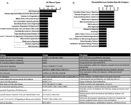
RNA-seq of the placental transcriptome was performed and analyzed as a hypothesis-generating approach to identify possible modes of action. Unfortunately, one female – associated placenta sample from the 8 mg/kg OPFR dose group had to be removed from the analysis for technical reasons. RNA-seq results were first analyzed by sex, but lack of separation in an unsupervised PCA prohibited this approach (Supplemental Figure 1) and revealed reasonable separation for only the female-associated placenta samples (Supplemental Figure 2). Therefore, pathways analysis was only conducted on female-associated placenta data. Differential expression of 79 genes, at a P adjusted value of 0.05, was found in female-associated placental tissue from dams exposed to 8 mg/kg OPFR. Ingenuity Pathway Analysis (IPA, QIAGEN) software was used to identify potential pathways perturbed by OPFR exposure. Several different iterations were employed using the full gene list with a P adjusted value of 0.1 as a cutoff; no cutoff value was implemented for fold-change. The first iteration included annotations from all tissue types (Figure 3A), while the second iteration included annotations from only placenta and nervous system tissue types (Figure 3B) because we were specifically interested in how changes in placental function may impact the brain. A number of the same pathways were identified in both analyses and are enriched with genes that play roles in energy production as well as cellular growth and development (Figure 3C).
69
Impact of prenatal OPFR exposure on placental gene expression – qRT-PCR
Follow-up analyses on genes of interest by qRT-PCR, to validate RNA-seq findings, used a larger sample size (n = 8 – 11 per sex per group) and considered sex as a biological variable. It has been well established that there are sex-differences between male and female-associated placentas, including differences in gene expression [54]. Therefore, in order to assess baseline sex differences the data was first normalized to the control values for a single sex, in this case males. When analyzed for baseline sex differences only one of the genes showed a significant difference in expression between control males and
70 females. Relative abundance of Tph1 was found to be significantly higher in control male-associated placentas than female-associated placentas (U = 4, p ≤ 0.01; Figure 5E).
Expression of Sod2 was found to be significantly altered by OPFR exposure (K-W = 31.5, p ≤ 0.0001), with both the 4 mg/kg OPFR and 8 mg/kg OPFR dose groups showing a significant reduction compared to controls (p ≤ 0.001; p ≤ 0.0001; Figure 4B). This relationship was also found when female and male-associated placentas were separated for analysis (K-W = 16.8, p ≤ 0.0008; K-W = 14.4, p ≤ 0.003). A significant reduction in the relative abundance of Sod2 was found at both the 4 mg/kg OPFR and 8 mg/kg OPFR dose for female (p ≤ 0.04; p ≤ 0.002) and at the highest dose (8 mg/kg OPFR) for male-associated placentas (p ≤ 0.006; Figure 4C). While a significant reduction in expression was not observed for 4 mg/kg OPFR male-associated placentas, a suggestive decrease was observed (p = 0.06). Expression of Sod2 was also impacted by treatment with 5-MT (U = 36, p ≤ 0.001), in both female and male-associated placentas (U = 12, p ≤ 0.02; U = 6, p ≤ 0.02; Figure 4C).
71 While expression of Tph1 and Maoa were unchanged, relative abundance of Ddc was
significantly altered by exposure to OPFRs (K-W = 9.9, p ≤ 0.02) with the 4 mg/kg OPFR dose group showing a decrease in relative abundance compared to controls (p ≤ 0.05; Figure 5H). When stratified by sex expression of Ddc was significantly impacted by OPFR exposure (K-W = 18.9, p ≤0.0003), the 4 mg/kg OPFR and 8 mg/kg OPFR dose groups showed a significant reduction in the relative abundance of Ddc (p ≤ 0.03; p ≤ 0.02; Figure 5 I). Expression of Ddc also appeared to be lower in the 5-MT group, but did not reach statistical significance (U = 70, p = 0.08) in the combined analysis (Figure 5I). Although not impacted by OPFR exposure, expression of Tph1 was significantly altered by 5-MT treatment (U = 58, p ≤ 0.05; Figure 5D). When stratified by sex treatment with 5-MT lead to a significant reduction in the
72 expression of Tph1 in male-associated placentas (U = 1, p ≤ 0.008; Figure 5E). Relative abundance of the 5-HT transporter, Slc6a4, was significantly impacted by OPFR exposure (K-W = 15.9, p ≤ 0.001; Figure 5A)with expression in both the 2 mg/kg OPFR and 4 mg/kg OPFR groups significantly lower than in unexposed controls (0 mg/kg OPFR) (p ≤ 0.05; p ≤ 0.0004; Figure 5A). When separated by sex this relationship held true for both female and male-associated placentas (K-W = 7.7, p ≤ 0.05; K-W = 9.4, p ≤ 0.03). Female-associated placentas showed a significant decrease in Slc6a4 at the lowest dose (p ≤ 0.03), while male-associated placentas showed a significantly lower relative abundance at both the 2 mg/kg OPFR and 4 mg/kg OPFR dose compared to controls (p ≤ 0.04; p ≤ 0.01; Figure 5B). Kmo, which is responsible for metabolizing kynurenine, was not altered by OPFR exposure but was significantly impacted by treatment with 5-MT (U = 41, p ≤ 0.002; Figure 6B), which only turned out to be true for male-associated placentas when analyzed by sex (U = 6, p ≤ 0.02; Figure 6C).
Impact of prenatal OPFR exposure on placental neurotransmitters
73 Figure 5. Effects of prenatal OPFR exposure on the relative abundance of genes involved in the transport, synthesis, and metabolism of 5-HT and levels of tryptophan, 5-HT and 5-HIAA. (A and B) Slc6a4, the 5-HT transporter, was lower in the 2mg/kg and 4mg/kg OPFR dose when sexes were combined for analysis, and in male –associated placentas when separated by sex. In female-associated placentas levels were only lower in the 4mg/kg OPFR dose group. (D and E) Expression of Tph1, enzyme that converts tryptophan to 5-hydroxytryptophan (5-HTP) (C), was significantly lower in the 5-MT group in both the combined analysis and in male-associated placentas when separated by sex. (E) Tph1 also showed a significant sex difference in the unexposed controls, with male-associated placentas having greater expression than female-associated placentas (cp ≤ 0.05). (H) Expression of Ddc, the enzyme that converts 5-HTP to 5-HT (G), was lower
in the 4mg/kg OPFR group in the combined analysis, while a suggestive decrease was observed in the 5-MT animals. (I) When separated by sex, lower expression of Ddc was observed in the 4mg/kg and 8mg/kg females. (L and M) Expression
of Maoa, the enzyme that converts 5-HT to 5-HIAA (K), was unaffected. (F, J, and N) Relative abundance of tryptophan,
74 Figure 6. Effects of prenatal OPFR exposure on the expression of a gene that metabolizes kynurenine as well as the
75
Impact of prenatal OPFR exposure on development of 5-HTergic projections
Length was determined for 5-HTergic projections, a cluster of Netg1a-immunoreactive neurons, and the spread of 5-HTergic cell bodies in GD 14 fetal brains. No significant sex differences were detected for any of the analyses when female and male controls were compared. OPFR exposure did not significantly alter the length of Netg1a projections or the spread of 5-HTergic cell bodies. In female brains, 5-HTergic projections were not significantly impacted by OPFR exposure, however a significant linear trend was detected (F (1, 17) = 4.5, p ≤ 0.05, r2 = 0.97; Figure 7A). Length of 5-HTergic
projections was significantly altered by OPFR exposure in male offspring (F (3, 19) = 3.6, p ≤ 0.03), with both the 4 mg/kg OPFR and 8 mg/kg OPFR dose groups showing a significant increase in length of projections (p ≤ 0.007; p ≤ 0.02; Figure 7A). A suggestive increase in length of 5-HTergic projections was observed in male brains exposed to 5-MT (p = 0.08; Figure 7A).
76 Discussion
The present study supports and expands upon our previous findings that gestational exposure to FM 550 alters placental function, and identifies tryptophan metabolism and the neurotransmitter 5-HT as particularly vulnerable to the OPFRs. Accumulation of OPFRs in exposed placentas was not sex-specific, which differs from our prior study that identified sex specific TPHP placental accumulation following gestational exposure to FM 550 [6]. However, when looking at the total placental burden of OPFR components, male-associated placentas appear to be more heavily exposed, which may play a role in the male-biased exposure-related phenotypes observed in the placenta and developing brain. Altered placental pathways indicate that gestational OPFR exposure may disrupt aspects of placental energy production, cellular development, and protein synthesis all of which could negatively impact the health and development of the placenta, fetus, and fetal brain. Most significantly, exposure-related increases in placental tryptophan, 5-HT, 5-HIAA, kynurenine, and several other kynurenine metabolites were observed and showed some sex bias, with higher levels in male-associated placentas. The observed increase in neuroactive metabolites in the placenta was associated with a concomitant increase in the length of 5-HTergic projections in the fetal forebrain. These phenotypes were more pronounced in male offspring. Collectively, our data suggest that there may be a causal and sex-specific link between OPFR-induced placental disruption and changes in the development of the 5-HTergic system.
77 form heterodimers with retinoid x receptor (RXR) and regulate the transcription of genes involved in endocrine and metabolic regulation as well as fetal development [81, 82]. We have previously shown that gestational exposure to FM 550 can lead to the activation of both the farsenoid x receptor (FXR)/RXR and liver x receptor (LXR)/RXR pathways [23]. Therefore, RXR and the nuclear receptors it forms complexes with, especially RAR, FXR, and LXR, may be particularly susceptible to OPFRs.
Mitochondrial dysfunction was also identified as an important pathway perturbed by OPFR exposure. Mitochondria play a significant role in energy production, metabolism, and response to
oxidative stress [83]. Oxidative stress has been identified as a potential risk factor in the pathophysiology of placenta-related disorders, including preeclampsia (PE) and intrauterine growth restriction (IUGR). Over the course of pregnancy mRNA expression of various antioxidant enzymes, such as Sod2, increase in their expression in order to protect the placenta from oxidative stress in an increasingly rich oxygenated environment [84]. Within the mitochondrial dysfunction pathway, genes perturbed by OPFR exposure included ATP synthase membrane subunit c locus 1 (Atp5mc1), parkinsonism associated deglycase (Park7), NADH:ubiquinone oxidoreductase subunit A13 (Ndufa13), and Sod2. Superoxide dismutases, including Sod2 located in the mitochondria, are antioxidant enzymes that aid in protecting against oxidative stress [85]. While the RNA-seq data set showed an increase in the expression of Sod2, by 1.5 fold, qRT-PCR actually showed a significant decrease in both female and male associated placentas. Although our qRT-PCR findings did not confirm our RNA-seq data, alterations in the expression of Sod2 may indicate OPFR induced oxidative stress in our exposed placentas. Overall, these findings suggest that placentas exposed to OPFRs may be susceptible to the effects of oxidative stress which may play a role in functional changes similar to phenotypes observed with PE and IUGR.
78 decrease in expression has been observed in several models of prenatal stress [88-90]. The programmatic effects of prenatal stress on neurodevelopment likely stem from increased exposure to maternal
glucocorticoids in utero. Therefore, overexposure to corticosterone may be a potential mechanism by which OPFR exposure alters aspects of brain development and behavior, and warrants further investigation.
We consistently found evidence that the synthesis and metabolism of placental 5-HT was altered as a result of OPFR exposure. Neurotransmitter analysis revealed an increase in the relative abundance of tryptophan (the precursor to 5-HT), 5-HT, and its primary metabolite, 5-HIAA, in placentas exposed to the highest dose (8 mg/kg OPFR). All of these outcomes appeared to be more pronounced in male-associated placentas than female-male-associated placentas. It is unclear whether these changes in 5-HT are indicative of changes in maternal blood levels or placentally- derived 5-HT. The 5-HT transporter, Slc6a4, is highly expressed in both human and rodent placentas [91] and has been localized to the villous trophoblasts where it regulates the amount of 5-HT taken up into the placenta from maternal circulation [91]. While other groups have demonstrated that 5-HT in maternal circulation is not transferred to the fetus [52], expression of Slc6a4 at the maternal interface suggests a functional role for this vasoactive, mitogenic neurotransmitter in the placenta. We observed a significant decrease in the expression of Slc6a4 in exposed female and male-associated placentas. Although it is not yet fully understood how placentally derived 5-HT is transferred to the fetus, it is possible that changes in Slc6a4 function in the placenta could affect the storage and transfer of 5-HT to the fetus.
It has recently been established that the fetal side of the placenta is the sole source of 5-HT for the developing forebrain from GD 10 – 15, at which time 5-HTergic projections innervate the forebrain and provide a local source of HT [51, 52]. Therefore, changes in placental tryptophan metabolism and 5-HT synthesis during this critical gestational window could lead to significant alterations in brain
male-79 associated placentas, we did observe a significant decrease in the expression of Ddc in female-associated placentas. Ddc is responsible for converting the intermediate, 5-hydroxytryptophan, to 5-HT. The observed decrease in expression may be the placentas response to high levels of 5-HT. These phenotypes were not recapitulated with the 5-HT agonist, 5-MT, however male-associated placentas exposed to 5-MT did show a significant decrease in the expression of Tph1. It is important to note that although we did not see major effects of OPFR exposure on the expression of these enzymes we did not look at changes in enzyme activity. Follow-up studies focusing specifically on placental 5-HT synthesis and metabolism should also assess activity levels of these enzymes. Furthermore, prenatal stress has been shown to increase the fraction of free tryptophan in maternal blood, which was associated with increased levels of tryptophan, 5-HT, and 5-HIAA in the fetal brain [92]. It is possible that the observed increases in placental tryptophan are of maternal blood origin. In future studies, it may be important to assess effects of OPFR exposure on the maternal hypothalamic-pituitary-adrenal axis, as several of the phenotypes we have observed in in this study, including elevated levels of tryptophan, also appear in models of prenatal stress.
80 enzyme Kmo is believed to drive the neurotoxic shift in the brain, leading to the production of QA and its neurotoxic precursor 3-hydroxykynurenine (3-HK) [93]. We did not see an increase in the relative abundance of QA or 3-HK in OPFR exposed placentas, and expression of Kmo only showed significant changes in response to 5-MT exposure. However, increased abundance of kynurenine in the placenta could result in overexposure of the fetal brain to kynurenine and its neuroactive metabolites. A recent study in mice showed that kynurenine is able to cross the placenta to a greater degree than kynurenine acid, and leads to a subsequent increase in 3-HK in the fetal brain [94]. Therefore, the observed increase in kynurenine in OPFR exposed placentas may alter fetal brain exposure to neurotoxic metabolites. A summary of changes in 5-HT and kynurenine metabolic pathways in exposed placentas can be found below in Figure 8.
81 In our previous study, gestational exposure to FM 550 resulted in a significant decrease in 5-HT turnover, determined by looking at the ratio of 5-HIAA:5-HT, indicating that there may be reduced 5-HT signaling in the fetal brain. 5-HT plays a very important role in brain development including
synaptogenesis, cell proliferation and migration, myelination, and differentiation [95, 96]. Development and targeting of 5-HTergic axons occurs during the window in which placentally derived 5-HT is being supplied to the fetal forebrain. A recent study in mice showed that changes in placental output of 5-HT was associated with blunting of 5-HT axonal outgrowth in the fetal forebrain [56]. Therefore, 5-HT signaling between the placenta and fetal forebrain may be important for the establishment of the 5-HTergic system itself. In this study we chose to look at development of the 5-HT system (length of projections and spread of cell bodies) as well as the development of thalamocoritcal axons (TCAs), which are known to be serotonin sensitive. While we did not see any significant effect of OPFR exposure on the spread of 5-HT cell bodies or length of TCAs, we did see a significant effect on the length of 5-HTergic projections (a phenotype that was at least in part recapitulated by 5-MT). The 5-HTergic system has been implicated in a variety of neurological disorders [97], including autism spectrum disorder where
hyperserotonemia is suspected to be an endophenotype [98]. Hyperinnervation of the developing forebrain by 5-HTergic projections may be a critical mechanism by which OPFR exposure can impact brain physiology and behavior. While we have seen changes in serotonin-dependent behaviors, including anxiety and hyperactivity, in Wistar rat offspring exposed to FM 550 we have not yet established whether or not this OPFR mixture can impact behavior.
Conclusions
82 knowledge gap and indicate that altered placental function, resulting from OPFR exposure may be a route by which this chemical mixture can disrupt brain development and alter later in life behaviors. More specifically, disruption of neuroactive metabolites, including the NT 5-HT, in OPFR exposed placentas was found in conjunction with overgrowth of 5-HTergic projections in the fetal forebrain. While there is some overlap between this study and changes in placental function as a result of FM 550 exposure, including increased levels of placental 5-HT, there are some notable differences. For example, with FM 550 exposure we saw a lot of changes in gene expression related to endocrine and inflammatory signaling that were not observed in this study. This provides some preliminary evidence on FR class (brominated versus organophosphate) and mechanism of action, with the BFRs contributing to endocrine disruption and inflammation and the OPFRs perturbing neurotransmitter signaling in the placenta. While this study, and our previous work on FM 550, utilized complex mixtures, independent examination of individual components could further our mechanistic understanding. Greater resolution on changes in 5-HT signaling between the placenta and fetal brain would also be beneficial, and may be obtainable in future studies by measuring levels of tryptophan and 5-HT in the placenta as well as maternal and fetal blood. Our results provide novel evidence that gestational exposure to OPFRs can impact physiological endpoints in the rat placenta and development of the 5-HTergic system in the fetal forebrain.
Funding
This work was supported by NIEHS 1F31ES02900-01 to KDR, NIEHS R56ES022957 to HBP NIEHS T32ES021432 and P30ES025128 to NCSU and NIEHS R01ES016099 to HMS.
Acknowledgments
83 References