_____________________________________________________________________________________________________
*Corresponding author: E-mail: [email protected];
(Past name: British Journal of Medicine and Medical Research, Past ISSN: 2231-0614, NLM ID: 101570965)
Regenerative Endodontics – Looking Inward
Shivani Mallishery
1*and Tanvi Shah
11Nair Hospital Dental College, Mumbai, India.
Authors’ contributions
This work was carried out in collaboration between both authors. Author SM designed the study and wrote the protocol. Author TS managed the literature searches. The first draft of the manuscript was written and revised by both the authors. Both authors read and approved the final manuscript.
Article Information
DOI: 10.9734/JAMMR/2020/v32i730454
Editor(s):
(1) Dr. Chan-Min Liu, Xuzhou Normal University, China.
Reviewers:
(1)Patrícia Diogo, University of Coimbra, Portugal. (2)D. N. Nirupama, India. (3)Jorge Paredes Vieyra, Universidad Autonoma de Baja California Tijuana BC, Mexico. Complete Peer review History:http://www.sdiarticle4.com/review-history/57036
Received 17 March 2020 Accepted 24 May 2020 Published 30 May 2020
ABSTRACT
Regenerative endodontics has become a revolutionizing tissue engineering concept in the treatment of immature permanent teeth for over two decades. It has been described as a ‘paradigm shift’ in the treatment of immature teeth, since it fosters continued root maturation. An immature necrotic permanent tooth is usually a result of trauma or infection due to which the tooth becomes non-vital before completing root development. In such cases, the root walls are left thin and weak with an open apex. Traditional apexification procedures may resolve pathology but have not been able to prove tooth survival due to absence of continued root development and risk of root fracture. A successful regenerative endodontic procedure (REP) results in resolution of signs and symptoms of pathology, radiographic signs of healing, proof of continued root development as well as presence of pulp vitality due to the regeneration of pulp tissue in the root canal. Various stem cells, growth factors, scaffolds and suitable environment form the tetrad of elements necessary to induce regeneration of dental pulp. While there has been some success in isolating dental pulp cells with in-vitro experiments, it has been proven to be rather difficult to implement the
same in a practical perspective ex vivo. Although there has been clinical success related to REP,
histologically they seem to undergo guided endodontic repair rather than true regeneration of physiologic pulp tissue. This review provides an overview of components of tissue engineering, clinical protocol and predictable outcomes for REP and recent advances in regenerative dentistry.
Keywords: Regenerative endodontics; tissue engineering; stem cells; clinical outcomes; immature teeth; open apex.
ABBREVIATIONS
BMP : Bone Morphogenic Protein BMSSC : Bone Marrow Stem Cells
DFPC : Dental Follicle Progenitor Cells DPSC : Dental Pulp Stem Cells
PDLSC : Periodontal Ligament Stem Cells
PRP : Platelet Rich Plasma
PRF : Platelet Rich Fibrin
REP : Regenerative Endodontic Procedures
SCAP : Stem Cells from Apical Papilla
SHED : Stem Cells from Human Exfoliated Deciduous Teeth
1. INTRODUCTION
One of the greatest challenges in endodontics is the management of an immature permanent tooth with a non-vital pulp. Incorporation of traditional endodontic treatment like apexification in a non-vital immature permanent tooth results in cessation of root development which renders the formation of thin and weak roots that are highly prone to root fractures [1-2]. Hence, alternative approaches that allow quantitative and/or qualitative increase in length and thickness of the root should be pursued.
According to American Association of
Endodontists Glossary of Endodontic Terms,
regenerative endodontic procedures are
biologically based procedures designed to
physiologically replace damaged tooth
structures, including dentin and root structures, as well as cells of the pulp-dentin complex [3-4]. The primary function of pulp is to produce and maintain the vitality of dentin. Therefore, regenerative pulp should be capable of forming dentin to replace and repair the lost tissues. The term regenerative endodontics procedures (REP) refers to all the events that aim to regenerate and repair the pulp-dentin complex.
The first evidence of regeneration of dental tissues was in 1932 by G. L. Feldman, who
showed evidence of regeneration of dental pulp under certain optimal biological conditions
[5]. In 1971, a pioneer study in regenerative
endodontics conducted by Nygaard-Ostby
concluded that bleeding induced within a vital or necrotic canal led to resolution of signs and symptoms of necrotic cases and in certain cases, apical closure [6]. In the year 2000, Gronthoset al. identified and isolated odontogenic progenitor
cells in an adult dental pulp, which proved to be a breakthrough in the regeneration of dental tissues [7].
Tissue engineering is the field of functional restoration of tissue structure and physiology for impaired or damaged tissues because of cancer,
diseases and trauma [8]. Regenerative
endodontics, a type of tissue engineering, has two common terminologies associated with it- revascularization and revitalization.
Revasculari-zation refers to the re-establishment of
vascularity in the pulp space post-injury to the original vascularity of the pulp of a traumatized immature tooth. Revitalization, on the other hand, describes non-specific vital tissues rather than just blood vessels [9]. For immature teeth with
non-vital pulp, such revascularization/
revitalization treatment induces physiological root formation (apexogenesis), which thus results in
tissue regeneration. By restoring root
development and reinforcing dentinal walls, the strength of the root, and hence, long term retention of the tooth increases. Such treatment modality can prove to be an efficient alternative to conventional apexification procedures [10].
The aim of the present review is to perform critical analysis and summarize the available evidence on components of tissue engineering, clinical protocol, predictable outcomes and limitations of REP. To perform this review, a web-based search on PubMed was done to find relevant literature on REP published in the last 10 years. Both abstracts and free full text articles of dental journals were reviewed. A combination of keywords were used as search terms. These
include ‘regenerative endodontics’, ‘tissue
engineering’, ‘outcomes’, ‘dental stem cells’, ‘challenges’, ‘dental scaffolds’, ‘growth factors’.
2. REGENERATIVE CAPACITY OF
DENTAL TISSUES
The tooth is a complex organ that is formed by highly organized mineralized tissues encasing the dental pulp. Different mineralized tissues
have different regenerative capabilities.
Ameloblasts, derived from ectoderm, produce
enamel after being stimulated by the
ectomesen-chyme lead to the formation of dentin and cementum respectively. These cells, unlike ameloblasts, have limited regenerative capacity. In response to stimuli causing mild injury at the pulp-dentin interface, progenitor cells are derived from pulp which produce tertiary dentin [12]. The dentin so produced helps in separating the damaged tooth structure from pulp, thus maintaining pulp vitality. Another example of regenerative capacity of dentin is dentin-bridge formation upon application of calcium hydroxide as a pulp capping agent [13]. Similar to dentin, cementum is also laid down throughout life in the form of cellular cementum at the root apex to compensate for passive eruption of the tooth [14]. Alveolar bone is derived from osteoblasts, which exhibit rapid turnover in response to
mechanical stimulus [15]. Guided Tissue
Regeneration (GTR) has been successfully
incorporated into clinical practice to
allow selective regeneration of functional
periodontal ligament by using a barrier
membrane [16].
3. ELEMENTS OF TISSUE ENGINEERING
The components of tissue engineering include stem cells, scaffolds and growth factors. The
process involves incorporation of ex vivo
expanded stem cells and growth factors incorporated within a 3-dimensional natural or synthetic polymer that provide an environment for cell proliferation and differentiation [17]. Although these three components are important, they cannot yield successful results without the
fourth major component - a conducive
environment. Tissue engineering approach
involved in regenerative dentistry includes either in vivo implantation of an in-vitro cell culture
with/without polymers or direct in vivo
implantation of isolated cells and scaffolds [18].
3.1 Stem Cells
Stem cell biology is one of the fundamental components of regenerative medicine. The origin of any tissue can be traced back to its stem cells. Stem cells exhibit two properties- self renewal and plasticity. Self-renewal explains the property of these cells to divide thus producing more of themselves, while the potential of these cells to differentiate into different mature cell types is explained by the term plasticity [19].
These cells can be classified based on their origin as be embryonic stem cells (ES) or
post-natal stem cells and based on their plasticity as pluripotent (capacity of maturing into cells belonging to any of the three germ layers) or multipotent (capacity to differentiate only into cells of the tissues from which they are derived) [20]. Although ES cells are more valuable in tissue engineering due to their pluripotency and greater plasticity, legal and ethical issues associated with their sourcing is a concern. Hence, multipotent post-natal stem cells are widely studied. Further, these cells could either be autogenous, allogeneic or xenogeneic.
A stem cell niche, identified in several connective tissues, is the microenvironment in which the stem cells reside and represents as little as 1% of total population. The identification of such niches is best performed after stimulation of injury to the tissues [20]. However, the source of these cells is still unclear. Most stem cells found in the orofacial region are mesenchymal stem cells. Stem cell population applied in REP include.
3.1.1 Dental pulp stem cells (DPSC)
These cells, isolated from human dental pulp, are capable of regenerating the odontoblasts with mineralized tubules and fibrous tissues with blood vessels; very similar to the pulp-dentin complex of a normal human tooth and have the unique ability to form mineralized tissues both in-vitro and in-vivo [7]. DPSCs share a similarity in
gene expression with the precursors of
osteoblasts, ie, Bone Marrow Stem Cells (BMSSCs): they exhibit the capacity of self-renewal following in-vivo transplantation and can develop into diverse phenotypes like adipocytes and neural precursors [21]. However, in contrast to BMSSCs, these cells have 30% higher proliferation rate and higher growth
potential [22].
3.1.2 Stem cells from apical papilla (scap)
apical periodontitis and are highly significant in REP [20].
3.1.3 Periodontal ligament stem cells (pdlsc)
These are mesenchymal stem-cells isolated not just from human permanent teeth but also from deciduous and supernumerary teeth [24], which can differentiate into periodontal ligaments, cementum, alveolar bone, blood vessels and peripheral nerves. However, obtaining these cells could be difficult since they are collected from atraumatic extraction of healthy teeth, which is practiced only in the case of impacted teeth or for orthodontic purpose. It is now stated that PDLSC can also be isolated and expanded from inflamed PDL tissues, such as granulation tissue of periodontitis affected intra-bony pockets (i-PDLSC) [25]. However, i-PDLSCs have lesser osteogenic and cementum regeneration capacity compared to cells derived from healthy teeth [26]. Further, PDLSCs obtained from older adults have lesser regenerative capacity compared to younger donors [27].
3.1.4 Dental follicle progenitor cells (dfpcs)
DFPCs are isolated from the ectomesenchymal dental follicle or dental sac, which is responsible for the formation of periodontium [28-29]. Like other stem cells, they demonstrate adipogenic, neurogenic and osteogenic differentiation [30]. However, these cells are more proliferative than DPCS and SCAP [31] and display fibroblast-like morphology [28]. In vitro, they are capable of differentiating into PDL-like structures or calcified nodules with bone or cementum-like attributes. These calcified nodules resemble calcifications seen in calcifying epithelial odontogenic tumors or cemento-ossifying fibroma [32]. Further, after in-vivo implantation, STRO-1 positive dental follicle stem cells can form cementum and immortalized dental follicle stem cells are capable of forming new PDL [33].
3.1.5 Stem cells from human exfoliated deciduous teeth (shed)
These cells were isolated and expanded ex vivo by Miura et al. from the remnants of living, normal pulp tissue in exfoliated deciduous crown [34]. SHED develop at the 6th week of embryonic development and share common molecular characteristics with neural crest cells. Unlike other dental stem cells, SHED are easily accessible since they are sourced from naturally
occurring tooth exfoliation phenomena.
Compared to their adult counterparts (DPSCs), SHED have higher proliferative capacity and are less mature, hence exhibit greater potential of multi potential differentiation [35]. Due to their higher neurogenic potential, SHED have been extensively studied for the treatment of neural tissue injury or degenerative diseases, like Parkinson’s Disease [36]. Researchers believe these cells can be stored (SHED banking) and can be used as a successful stem cell therapy for the treatment of various medical conditions [37].
3.2 Growth Factors/Morphogens
Growth factors are extracellularly secreted signals that bind to specific receptors on cells and play a major role in regulation of endogenous cells or stem cell recruitment, migration, proliferation and differentiation. The growth factors may be released by blood clot, Platelet Rich Plasma (PRP), Platelet Rich Fibrin
(PRF) or from dentin matrix upon
demineralization (caries, acid etching etc). Growth factors used in regenerative endodontics are platelet-derived growth factor (PDGF), transforming growth factor (TGF-β), Bone Morphogenic Protein (BMP), vascular endothelial growth factor (VEGF), basic fibroblast growth factor (bFGF), Colony stimulating factor (CSF), platelet derived growth factor (PDGF), epidermal growth factor (EFG) and insulin-like growth factor (IGF) [38]. These can be used for-
● Proliferation and differentiation of stem cells- PDGF, BMP, CSF, EGF, FGF, IGF ● Modulation of humoral and cellular immune
response- Interleukin 1-13 ● Angiogenesis- VEGF
● Wound healing and tissue regeneration- TGF alpha and beta.
Amongst these growth factors, BMP and TGF are the most crucial in regeneration of dental tissues in adults. They can either be used in vivo, where they are directly applied to exposed pulp or ex vivo, where they are first isolated with dental pulp stem cells which stimulate the formation of odontoblasts and finally transplanted to regenerate dentin [39].
3.3 Scaffold
and differentiation. Ideal properties of a scaffold include scaffold porosity to facilitate diffusion, biocompatibility and biodegradability, effective transportation of oxygen and nutrients, ability to support cell growth and differentiation, non-toxicity and adequate physical and mechanical strength [40]. Scaffolds can either be natural or synthetic.
3.3.1 Natural scaffolds
Natural scaffolds can be autologous like PRP, PRF and blood clot or derived from natural substances like collagen or glycosaminoglycans. PRP and PRF are first- and second-generation platelet concentrates respectively that stimulate the proliferation of stem cells. Although PRP has a rich source of growth factors, PRF has higher concentration of cytokines and stimulates faster healing. Blood clot has fewer cytokines and growth factors than PRP or PRF [40]. Intentional periapical filing to induce blood clot formation or venous blood drawn from patients can cause discomfort to the patient. Hence, collagen and glycosaminoglycan are used, which provide excellent tensile strength to the tissues, allow easy placement of stem cells and growth factors and control over resorption rate by altering its density. However, it is observed that collagen may adversely affect pulp tissue regeneration since pulp cells in collagen undergo marked contraction [41].
3.3.2 Synthetic scaffolds
Polymers such as polyglycolic acid (PGA), polylactic acid (PLA), polylactic co-glycolic acid
(PLGA), polycaprolactone (PCL) are
commercially available synthetic scaffolds. They allow precise control of physiochemical features like degradation rate, microstructure, strength, porosity and undergo degeneration by hydrolysis [42]. Scaffolds containing inorganic compounds like hydroxyapatite and tricalcium phosphate can be used to enhance osteoconductivity. The major disadvantage associated with synthetic scaffolds is inflammation at the site of implantation [42].
3.4 Environment
Tissue engineering, or regeneration of tissues requires a tetrad of elements. A conducive environment is crucial for any stem cell to proliferate or differentiate in vivo or in vitro. These include- A well-sealed restoration in vivo to prevent contamination of pulp [43], disinfection of the root canal system using intracanal irrigants like Sodium hypochlorite or chlorhexidine and
antibiotic pastes [2] and large-scale in vitro regeneration of tissues requires bioreactors that mimic the internal environment of the body and provide appropriate physiological stress to
enhance the mechanical properties of
regenerated tissues [44].
4. PROCEDURE
REP is the mainly considered for a permanent necrotic tooth with an open apex in a compliant patient with no allergy to medications and antibiotics used in the procedure. Moreover, since the pulp canal space is involved in the procedure, it should be ensured that this canal space is not required for other restorative purpose [45].
Based on Cvek’s classification of root
development, immature necrotic permanent teeth suitable for REP include Stage 1 (less than one-half of root formation with open apex), stage 2 (one-half of root formation) and stage 3 (two-third of root development with open apex), due to short root, thin canals and wide-open apex. However, teeth at stage 4 of root development (nearly completed root with an open apex) can be treated by both REP and apexification, since the root has developed enough to withstand apexification [46].
Because of the encouraging results of
regenerative procedures in young immature permanent teeth it has been tried for use in adult teeth with closed apices.
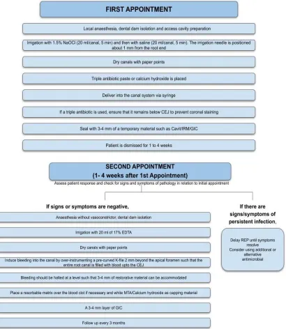
4.1 Traditional REP for Permanent Teeth with an Open Apex
Traditional REP is performed by inducing bleeding into the canal by over-instrumentation in immature permanent teeth.
4.2 REP Using Platelet Rich Plasma for Permanent Teeth with an Open Apex
Liu et al. showed that PRP stimulates cell proliferation and differentiation of the dentin-pulp complex, which suggested that PRP could be used as a scaffold for pulp capping [47].
4.3 Rep for Adult Mature Permanent Teeth
in adults. These include cell transplantation of ex vivo cultivated stem/progenitor cells and cell homing by molecules that recruit the patient’s resident cells [49].
In a series of case reports, Paryani K et al. and Saoud et al. used modified regenerative
endodontic procedures to enhance the
probability of pulp revascularization in mature
necrotic teeth. The resolution of apical
radiolucency and regression of clinical signs and symptoms along with ingrowth of new vital tissue into the chemo-mechanically debrided canals were observed at recall appointments [50-51].
Fig. 2. Regenerative endodontic procedure using Platelet Rich Plasma (PRP) for a permanent tooth with an open apex [48]
The size of the apical foramen has been much stressed upon when attempting regeneration in a permanent tooth with closed apex. Cells such as fibroblasts, osteoblasts, cementoblasts and endothelial cells migrate through the apical foramen into the canal to produce PDL, bone, cementum, blood vessels in the canal space.
determine if the size of the apical foramen actually influences the outcome of REP in teeth with closed apex.
5. OUTCOME OF REGENERATIVE
ENDODONTIC PROCEDURES
For any clinical procedure, the challenge starts when we try to define what a successful outcome is. When results for revascularization procedures are considered, evidence of root formation and reestablishment of pulpal function is vital to measure the success of treatment [3].
Reports demonstrate that regenerative
endodontics is a viable treatment option which has been described as a ‘paradigm shift’ that allows for continued root development, a return of vitality and health in formerly necrotic immature teeth [53]. Successful outcome of REP could mean different things in patient, clinician and researcher-centered perspective [54].
5.1 Patient-based Outcomes
Criteria of treatment success for clinicians and researchers may not always meet the needs and desires of the patients. Outcomes need to focus more on patient satisfaction as they are the primary contributors for their own health and we need to respect patient autonomy. The following are patient-centered outcomes of REP.
5.1.1 Resolution of symptoms
When standardized treatment protocols for REPs and apexification procedures (MTA and Calcium hydroxide) were compared, REP and MTA plug apexification procedures were equally effective in resolving signs and symptoms of disease and survival over 18 months in 100% and 95% of all patients, respectively, whereas apexification procedures using calcium hydroxide were significantly less effective (77%) [10]. However, Alobiad et al. found REPs to promote healing in 79% of patients treated, whereas apexification procedures promoted healing in 100% of the patients [55].
5.1.2 Survival of tooth
From a patient’s perspective, an ideal treatment should result in increased functional life of the tooth. This is especially important when survival of an immature permanent tooth is considered, since early loss of a permanent tooth will not only cause malocclusion, but also impair craniofacial
development. As mentioned earlier, REP has shown greater survival rate than MTA [10].
5.1.3 Esthetics
An important patient-centered outcome is preservation or restoration of esthetics. However, coronal staining may occur when TAP or MTA is used as an intracanal medicament. This staining is believed to be caused by minocycline, a constituent of TAP [56-58]. However, this coronal staining can be prevented by either using a dentinal adhesive to block dentinal tubules or by substituting minocycline with cefuroxime or Arestin [59].
5.2 Clinician-based Outcomes
Clinicians base the success of treatment based on clinical and radiographic exam such as no pain, soft tissue swelling or sinus tract (between first and second appointments), resolution of apical radiolucency (6-12 months after treatment) and positive response to pulp sensitivity tests.
5.2.1 Radiographic signs
One of the primary clinician-centered outcomes is radiographic signs of resolution of apical lesion and continued root development. Increased width of root walls is generally observed before apparent increase in root length and often occurs 12-24 months after treatment. In comparison to apexification, a significant increase in root canal length and width was seen in teeth treated with REP as compared to the teeth treated for MTA or Calcium Hydroxide Apexification [60-61]. Several studies have concluded that although REPs
predictably promote healing of apical
periodontitis in more than 90% of the cases, radiographic root development is far less predictable [23].
5.2.2 Vitality response
Positive response to pulp sensitivity test is considered to be a sign of pulp vitality [62]. Re-establishment of pain perception indicates the presence of vital, vascularized tissues with normal physiological response. Clinicians have noted positive response to cold or electric pulp sensitivity testing in 60% of published cases [63].
5.3 Scientist/researcher-based Outcomes
research, to promote substantial future advances in regenerative endodontics.
5.3.1 Animal studies
Several animal studies were conducted to
determine the histological outcome of
regenerative endodontic procedures. The effect of different combinations of stem cells, growth
factors and scaffolds has been studied
extensively on dog and ferret models [64-67]. All these studies showed formation of hard tissues at the apex and apical closure, however none of the studies showed true regeneration of the pulp-dentin complex. It has been observed that
the newly formed hard tissue appears
cellular, atubular, hence resembling cementum [68].
5.3.2 Histology
According to a study conducted by Torabinejad et al. and Nosrat et al. on human immature permanent teeth using PRP, the tissues that grow into the root canals after regenerative endodontic procedures resemble periodontium, that is, fibrotic PDL, collagen fibres and cementum-like hard tissues [67,69]. However, no evidence of odontoblasts could be seen histologically. Nosrat et al. compared the
histological sections of human immature
permanent teeth using a novel hydroxyapatite scaffold and blood clot. All the specimens histologically showed dentin, dentin associated newly formed mineralized tissue, newly formed connective tissues and the PDL. However, none of the specimens showed pulp-like tissues [70].
6. FUTURE OF REGENERATIVE
ENDODONTICS
Regenerative procedures require more research as they have a potential to replace conventional root canal therapy in the future, provided a predictable, feasible and economical protocol can be developed for the same [10,71-73]. Since its onset over 40 years ago, several research advancements have been made in the field. These mainly include components of cell-based regenerative therapy and tissue engineering. Potential areas of research in the development of regenerative endodontics include.
6.1 3-D Bioprinting
Synthetic 3D scaffolds can be generated using a bioink, which is composed of blending printable
alginate hydrogel with soluble and insoluble fractions of the dentin matrix [74]. It involves precise placement of cells into a soft scaffold as directed by a computer-aided design. The
soluble dentin molecules of the matrix
significantly enhance odontogenic differentiation of the mesenchymal stem cells encapsulated in these bioprinted hydrogels. Such bioprinting allows for precise and reproducible positioning of cellularized scaffolds, which can be useful to study cell interactions and the effect of cell organization on cell growth and function [74].
6.1.1 Injectable scaffold
This is a type of 3-D scaffold, composed of injectable hydrogel, which is placed in the root canal to provide a substrate for organized cell proliferation. To promote bone regeneration, calcium phosphate cement scaffolds have been used along with osteoinductive growth factors [75].
6.2 Gene Therapy
Gene therapy involves introducing specific genes into target cells, either to compensate for abnormal genetic material or to stimulate the natural biological process for regeneration of a desired tissue. These genes can be delivered using viral (adenovirus, lentivirus, herpes simplex virus) or non-viral (plasmids, peptides, DNA-ligand complexes, liposomes) vectors, either ex vivo or in vitro [76,3]. The viral vectors are modified such that they do not cause disease but are capable to stimulate an infection. Gene
therapy can be applied in regenerative
endodontics by delivering mineralizing genes into the vital pulp cells of necrotic or symptomatic teeth [77]. The ex vivo therapy is preferred for stimulation of reparative dentin [78]. However, except for a research done by Rutherford in 2001 using c-DNA encoded mouse BMP-7 [79], currently the knowledge based on this therapy is purely theoretical. Widespread clinical application of such therapy is yet to be developed considering the potential health hazard posed by the vectors in this therapy [80].
6.3 Vascular and Nervous Regeneration
is vital for proliferation and differentiation of stem cells [8]. Angiogenesis can be accelerated in an engineered tissue by slowly releasing growth factors like VEGF from the scaffold. Utilizing gene therapy along with VEGF and BMP can be potentially used in regeneration of pulp-dentin complex [81].
6.4 Bioengineered Tooth
It is said that an organ can be bioengineered by reproducing the developmental processes during organogenesis [82]. Like tooth, several organs develop from interactions between epithelial and mesenchymal cells. Hence a bioengineered tooth can be developed in vitro by replicating the steps of development of a tooth germ. Ikeda et al. and Oshima et al. demonstrated in an adult mouse, that a fully functional bioengineered tooth can be achieved through the transplantation of a bioengineered tooth germ. This tooth, which was erupted and occluded, had the correct tooth and alveolar structure, hardness of mineralized tissues for mastication, and response to noxious stimulations in cooperation with tissues of oral and maxillofacial region [83-84]. Therefore, these bioengineered teeth can be used as a replacement for missing or lost teeth.
7. LIMITATIONS
7.1 Patient Co-operation
REP is usually indicated for relatively young patients. Such patients are often nervous, frightened, or impatient. Further, when PRP is
utilized for such procedures, venipuncture is required. Hence, REP could be challenging to perform if patient cooperation is compromised.
7.2 Barriers in Stem Cell Transplantation
Stem cell transplantation is a common practice in medicine, especially in the treatment of
haematopoeitic disorders. However, extensive ex
vivo culturing of mesenchymal stem cells is required, while ensuring proper protocols of isolation and expansion are followed. Lack of such affordable facilities and dental stem cell banking system, and the risks of immune rejection restricts the use of these cell-based therapies in clinical practice [85,86].
7.3 Tooth Discolouration
As mentioned earlier, coronal staining may occur when antibiotics pastes containing minocycline
and bismuth oxide are used as an intracanal medicament [56-58]. Additionally, these materials show greater color change when they come in contact with blood [87].
7.4 Unpredictable Outcome
The nature of tissue formed in the canal after REP remains unpredictable. According to studies conducted by Nosrat et al.and Torabinejad et al., the regenerated tissues resemble periodontium, rather than the pulp. However, Shimizu et al. showed the presence of pulp-like connective tissue after revascularization in more than one-half of the canal [88]. Further, clinical trials involving long-term prognosis and impact of REP beyond 18 months are limited, and hence, unknown [89].
7.5 Lack of expertise
Treatment of teeth with immature roots is challenging due to thin dentin walls, hence root fracture can easily occur during mechanical debridement. Lack of both endodontic expertise and facilities involved in the handling of stem cells for clinical application of cell-based REP makes it a challenging technique currently.
8. CONCLUSION
Regenerative endodontic procedures provide a successful alternative for the treatment of necrotic teeth with open apex, by permitting continued root development. Several studies have demonstrated root development with high rates of resolution of clinical and radiographic signs and symptoms upon treatment using REP. However, current clinical protocols of REP foster repair rather than regeneration. Moreover, the resemblance of the regenerated tissue to a healthy pulp-dentin complex is debatable.
CONSENT
It is not applicable.
ETHICAL APPROVAL
It is not applicable.
COMPETING INTERESTS
Authors have declared that no competing interests exist.
REFERENCES
1. Cvek M. Prognosis of luxated non-vital
maxillary incisors treated with calcium hydroxide and filled with gutta-percha. A
retrospective clinical study. Dental
Traumatology. 1992;8(2):45-55. Available:https://doi.org/10.1111/j.1600-9657.1992.tb00228.x
2. Huang GJ. Apexification: The beginning of
its end. International Endodontic Journal. 2009;42(10):855-66.
Available:https://doi.org/10.1111/j.1365-2591.2009.01577.x
3. Murray PE, Garcia-Godoy F, Hargreaves
KM. Regenerative endodontics: A review of current status and a call for action. Journal of Endodontics. 2007;33(4):377-90.
Available:https://doi.org/10.1016/j.joen.200 6.09.013
4. Available:https://www.aae.org/specialty/clin
ical-resources/glossary-endodontic-terms/. [Accessed on 14/5/2020]
5. Bansal R, Jain A. Current overview on
dental stem cells applications in
regenerative dentistry. Journal of Natural Science, Biology, and Medicine. 2015;6(1): 29.
DOI: 10.4103/0976-9668.149074
6. Nygaard-Östby B, Hjortdal O. Tissue
formation in the root canal following pulp
removal. European Journal of Oral
Sciences. 1971;79:333-349.
DOI: 10.1111/j.1600-0722.1971.tb02019.x
7. Gronthos S, Mankani M, Brahim J, Robey
PG, Shi S. Postnatal human dental pulp stem cells (DPSCs) in vitro and in vivo. Proceedings of the National Academy of Sciences. 2000;97(25):13625-30.
Available:https://doi.org/10.1073/pnas.240 309797
8. Nakashima M, Akamine A. The application
of tissue engineering to regeneration of
pulp and dentin in endodontics. Journal of Endodontics. 2005;31(10):711-8.
Available:https://doi.org/10.1097/01.don.00 00164138.49923.e5
9. Lin LM, Kahler B. A review of regenerative
endodontics: current protocols and future directions. Journal of Istanbul University Faculty of Dentistry. 2017;51(3 Suppl 1): S41.
DOI: 10.17096/jiufd.53911
10. Jeeruphan T, Jantarat J, Yanpiset K,
Suwannapan L, Khewsawai P, Hargreaves KM. Mahidol study 1: Comparison of radiographic and survival outcomes of
immature teeth treated with either
regenerative endodontic or apexification methods: A retrospective study. Journal of endodontics. 2012;38:1330-1336.
Available:https://doi.org/10.1016/j.joen.201 2.06.028
11. Thesleff I, Tummers M. Tooth
organogenesis and regeneration. In Stem
Book [Internet]. Harvard Stem Cell
Institute; 2009. [PMID: 20614625]
12. Smith AJ, Patel M, Graham L, Sloan AJ, Cooper PR. Dentine regeneration: Key roles for stem cells and molecular signalling. Oral Biosci Med. 2005;2(2/3): 127-32.
13. Swarup SJ, Rao A, Boaz K, Srikant N,
Shenoy R. Pulpal response to nano hydroxyapatite, mineral trioxide aggregate and calcium hydroxide when used as a direct pulp capping agent: An in vivo study. Journal of Clinical Pediatric Dentistry. 2014;38(3):201-6.
Available:https://doi.org/10.17796/jcpd.38. 3.83121661121g6773
14. Gottlieb B. Continuous deposition of
cementum. The Journal of the American
Dental Association. 1943;30(11):842-
7.
Available:https://doi.org/10.14219/jada.arc hive.1943.0172
15. King GJ, Keeling SD, Wronski TJ.
Histomor-phometric study of alveolar bone turnover in orthodontic tooth movement. Bone. 1991;12(6):401-9.
Available:https://doi.org/10.1016/8756-3282(91)90029-I
16. Takata T, Wang HL, Miyauchi M.
Available:https://doi.org/10.1034/j.1600-0765.2001.360508.x
17. Neel EA, Chrzanowski W, Salih VM, Kim
HW, Knowles JC. Tissue engineering in dentistry. Journal of dentistry. 2014;42(8): 915-28.
[PMCID: PMC5090995 PMID: 27857762] 18. Huang GT. Dental pulp and dentin tissue
engineeringandregeneration-advancement
and challenge. Frontiers in bioscience (Elite edition). 2011;3:788-800.
DOI: 10.2741/e286 [PMCID: PMC3289134] [NIHMSID: NIHMS331027] [PMID: 21196351]
19. Zipori D. The stem state: Plasticity is
essential, whereas self-renewal and
hierarchy are optional. Stem cells. 2005; 23(6):719-26.
Available:https://doi.org/10.1634/stemcells. 2005-0030
20. Diogenes A, Henry MA, Teixeira FB,
Hargreaves KM. An update on clinical
regenerative endodontics. Endodontic
Topics. 2013;28(1):2-3.
Available:https://doi.org/10.1111/etp.12040 21. Gronthos S, Brahim J, Li W, Fisher LW, Cherman N, Boyde A, DenBesten P, Robey PG, Shi S. Stem cell properties of human dental pulp stem cells. Journal of dental research. 2002;81(8):531-5. Available:https://doi.org/10.1177/15440591 0208100806
22. Shi S, Bartold PM, Miura M, Seo BM,
Robey PG, Gronthos S. The efficacy of mesenchymal stem cells to regenerate and repair dental structures. Orthodontics & craniofacial research. 2005;8(3):191-9. Available:https://doi.org/10.1111/j.1601-6343.2005.00331.x
23. Sonoyama W, Liu Y, Fang D, Yamaza T,
Seo BM, et al. mesenchymal stem cell-mediated functional tooth regeneration in swine. Plos One. 2006;1(1):e79.
DOI: 10.1371/journal.pone. 0000079
24. Seo BM, Miura M, Gronthos S, et al.
Investigation of multipotent postnatal stem cells from human periodontal ligament. The Lancet. 2004;364:149-155.
Available:https://doi.org/10.1016/S0140-6736(04)16627-0
25. Park JC, Kim JM, Jung IH, Kim JC, Choi SH, Cho KS, Kim CS. Isolation and characterization of human periodontal ligament (PDL) stem cells (PDLSCs) from
the inflamed PDL tissue: In vitro and in
vivo evaluations. Journal of Clinical
Periodontology. 2011;38(8):721-31. Available:https://doi.org/10.1111/j.1600-051X.2011.01716.x
26. Zhu W, Liang M. Periodontal ligament
stem cells: current status, concerns, and future prospects. Stem Cells International. 2015;2015.
Available:https://doi.org/10.1155/2015/972 313
27. Zheng W, Wang S, Ma D, Tang L, Duan Y,
Jin Y. Loss of proliferation and
differentiation capacity of aged human periodontal ligament stem cells and rejuvenation by exposure to the young extrinsic environment. Tissue Engineering Part A. 2009;15(9):2363-71.
Available:https://doi.org/10.1089/ten.tea.20 08.0562
28. Handa K, Saito M, Yamauchi M, Kiyono T,
Sato S, Teranaka T, Narayanan AS. Cementum matrix formation in vivo by cultured dental follicle cells. Bone. 2002; 31(5):606-11.
Available:https://doi.org/10.1016/S8756-3282(02)00868-2
29. Morsczeck C, Götz W, Schierholz J,
Zeilhofer F, Kühn U, Möhl C, Sippel C, Hoffmann KH. Isolation of precursor cells (PCs) from human dental follicle of wisdom teeth. Matrix Biology. 2005;24(2):155-65. Available:https://doi.org/10.1016/j.matbio.2 004.12.004
30. Morsczeck C, Völlner F, Saugspier M,
Brandl C, Reichert TE, Driemel O, Schmalz G. Comparison of human dental follicle cells (DFCs) and stem cells from human exfoliated deciduous teeth (SHED) after neural differentiation in vitro. Clinical Oral Investigations. 2010;14(4):433-40. Available:https://doi.org/10.1007/s00784-009-0310-4
31. Saito MT, Silvério KG, Casati MZ, Sallum EA, Nociti Jr FH. Tooth-derived stem cells: Update and perspectives. World Journal of Stem Cells. 2015;7(2):399.
DOI: 10.4252/wjsc.v7.i2.399
32. Maiorano E, Renne G, Tradati N, Viale G.
Cytogical features of calcifying epithelial odontogenic tumor (Pindborg tumor) with abundant cementum-like material. Virchows Archiv. 2003;442(2):107-10.
Available:https://doi.org/10.1007/s00428-002-0722-x
33. Chalisserry EP, Nam SY, Park SH, Anil S.
Journal of tissue engineering. 2017;8:2041731417702531.
Available:https://doi.org/10.1177/20417314 17702531
34. Miura M, Gronthos S, Zhao M, Lu B, Fisher
LW, Robey PG, Shi S. SHED: Stem cells from human exfoliated deciduous teeth. Proceedings of the National Academy of Sciences. 2003;100(10):5807-12.
Available:https://doi.org/10.1073/pnas.093 7635100
35. Wang X, Sha XJ, Li GH, Yang FS, Ji K, Wen LY et al. Comparative characteri-zation of stem cells from human exfoliated deciduous teeth and dental pulp stem cells. Archives of oral biology. 2012; 57(9):1231-40.
Available:https://doi.org/10.1016/j.archoral bio.2012.02.014
36. Wang J, Wang X, Sun Z, Wang X, Yang H,
Shi S et al. Stem cells from human-exfoliated deciduous teeth can differentiate into dopaminergic neuron-like cells. Stem Cells and Development. 2010;19(9):1375-83.
Available:https://doi.org/10.1089/scd. 2009.0258
37. Arora V, Arora P, Munshi AK. Banking
stem cells from human exfoliated
deciduous teeth (SHED): Saving for the
future. Journal of Clinical Pediatric
Dentistry. 2009;33(4):289-94.
Available:https://doi.org/10.17796/jcpd.33. 4.y887672r0j703654
38. Saber SE. Tissue engineering in
endodontics. Journal of Oral Science. 2009;51(4):495-507.
Available:https://doi.org/10.2334/josnusd.5 1. 495
39. Nakashima M. Bone morphogenetic
proteins in dentin regeneration for potential use in endodontic therapy. Cytokine & Growth Factor Reviews. 2005;16(3):369-76.
Available:https://doi.org/10.1016/j.cytogfr.2 005.02.011
40. Gathani KM, Raghavendra SS. Scaffolds
in regenerative endodontics: A review. Dental Research Journal. 2016;13(5): 379.
41. Huang GT, Sonoyama W, Chen J, Park
SH. In vitro characterization of human dental pulp cells: various isolation methods and culturing environments. Cell and Tissue Research. 2006;324(2):225. Available:https://doi.org/10.1007/s00441-005-0117-9
42. Sharma S, Mittal N. A comparative
evaluation of natural and artificial scaffolds in regenerative endodontics: A clinical study. Saudi Endodontic Journal. 2016; 6(1):9.
DOI: 10.4103/1658-5984. 171995
43. Nör JE. Buonocore memorial lecture:
Tooth regeneration in operative dentistry. Operative dentistry. 2006;31(6):633-42. Available:https://doi.org/10.2341/06-000
44. Malhotra N, Mala K. Regenerative
endodontics as a tissue engineering approach: Past, current and future. Australian Endodontic Journal. 2012;38: 137-48.
Available:https://doi.org/10.1111/j.1747-4477.2012.00355.x
45. American Association of Endodontists.
AAE clinical considerations for a
regenerative procedure. [Accessed on 20/4/2020]
Available:https://f3f142zs0k2w1kg84k5p9i1
o-wpengine.netdnassl.com/specialty/wpcont ent/uploads/sites/2/2018/06/Consideration sForRegEndo_AsOfApril2018.pdf
46. Kim SG, Malek M, Sigurdsson A, Lin LM, Kahler B. Regenerative endodontics: A
comprehensive review. International
Endodontic Journal. 2018;51(12):1367-88. Available:https://doi.org/10.1111/iej.12954 47. Liu ZN, Jiang T, Wang YX. Platelet-riched
plasma promotes potential mineralizing capacity of human dental pulp cells in vivo. Beijing da xue xue bao. Yi xue ban= Journal of Peking University. Health Sciences. 2011;43(2):276-9.
48. Jadhav GR, Shah N, Logani A.
Platelet-rich plasma supplemented revasculari-zation of an immature tooth associated with a periapical lesion in a 40-year-old man. Case Reports in Dentistry. 2014; 2014.
Available:https://doi.org/10.1155/2014/479 584
49. He L, Kim SG, Gong Q, Zhong J, Wang S,
Zhou X, Ye L, Ling J, Mao JJ.
Regenerative endodontics for adult
patients. Journal of Endodontics. 2017; 43(9):S57-64.
Available:https://doi.org/10.1016/j.joen.201 7.06.012
50. Paryani K, Kim SG. Regenerative
Available:https://doi.org/10.1016/j.joen.201 3.04.029
51. Saoud TM, Sigurdsson A, Rosenberg PA,
Lin LM, Ricucci D. Treatment of a large cystlike inflammatory periapical lesion associated with mature necrotic teeth using regenerative endodontic therapy. Journal of Endodontics. 2014;40(12):2081-6.
Available:https://doi.org/10.1016/j.joen.201 4.07.027
52. Fang Y, Wang X, Zhu J, Su C, Yang Y, Meng L. Influence of apical diameter on the outcome of regenerative endodontic treatment in teeth with pulp necrosis: A review. Journal of Endodontics. 2018; 44(3):414-31.
53. Huang GT. A paradigm shift in endodontic
management of immature teeth:
conservation of stem cells for
regeneration. J Dent. 2008;36:379–386. Available:https://doi.org/10.1016/j.jdent.20 08.03.002
54. Diogenes A, Ruparel NB, Shiloah Y,
Hargreaves KM. Regenerative endodontics: A way forward. The Journal of the
American Dental Association. 2016;
147(5):372-80.
Available:https://doi.org/10.1016/j.adaj.201 6.01.009
55. Alobaid AS, Cortes LM, Lo J, Nguyen TT, Albert J, Abu-Melha AS, Lin LM, Gibbs JL. Radiographic and clinical outcomes of the treatment of immature permanent teeth by revascularization or apexification: A pilot retrospective cohort study. Journal of Endodontics. 2014;40(8):1063-70.
Available:https://doi.org/10.1016/j.joen.201 4.02.016
56. Reynolds K, Johnson JD, Cohenca N. Pulp
revascularization of necrotic bilateral
bicuspids using a modified novel
technique to eliminate potential coronal discolouration: A case report. Int Endod J. 2009;42(1):84-92.
Available:https://doi.org/10.1111/j.1365-2591.2008.01467.x
57. Kim JH, Kim Y, Shin SJ, Park JW, Jung IY.
Tooth discoloration of immature permanent incisor associated with triple antibiotic therapy: A case report. J Endod. 2010; 36(6):1086-1091.
Available:https://doi.org/10.1016/j.joen.201 0.03.031
58. Petrino JA, Boda KK, Shambarger S,
Bowles WR, McClanahan SB. Challenges
in regenerative endodontics: A case series. J Endod. 2010;36(3):536-541.
Available:https://doi.org/10.1016/j.joen.200 9.10.006
59. Krastl G, Allgayer N, Lenherr P, Filippi A, Taneja P, Weiger R. Tooth discoloration induced by endodontic materials: A literature review. Dental Traumatology. 2013;29(1):2-7.
Available:https://doi.org/10.1111/j.1600-9657.2012.01141.x
60. Bose R, Nummikoski P, Hargreaves K. A
retrospective evaluation of radiographic
outcomes in immature teeth with
necrotic root canal systems treated with regenerative endodontic procedures. J Endod. 2009;35:1343–1349.
Available:https://doi.org/10.1016/j.joen.200 9. 06.021
61. Nagy MM, Tawfik HE, Hashem AA,
Abu-Seida AM. Regenerative potential of immature permanent teeth with necrotic pulps after different regenerative protocols. Journal of Endodontics. 2014;40(2):192-8. Available:https://doi.org/10.1016/j.joen.201 3.10.027
62. Weisleder R, Yamauchi S, Caplan DJ,
Trope M, Teixeira FB. The validity of pulp testing: A clinical study. JADA 2009; 140(8):1013-1017.
Available:https://doi.org/10.14219/jada.arc hive.2009.0312
63. Torabinejad M, Turman M. Revitalization of
tooth with necrotic pulp and open apex by using Platelet rich plasma: A case report. J Endod. 2011;37(2):265–268.
[PubMed: 21238815]
Available:https://doi.org/10.1016/j.joen.201 0.11.004
64. Torabinejad M, Corr R, Buhrley M, Wright K, Shabahang S. An animal model to study
regenerative endodontics. Journal of
endodontics. 2011;37(2):197-202. Available:https://doi.org/10.1016/j.joen.201 0.10.011
65. Thibodeau B, Teixeira F, Yamauchi M,
Caplan DJ, Trope M. Pulp revasculari-zation of immature dog teeth with apical periodontitis. Journal of endodontics. 2007; 33(6):680-9.
Available:https://doi.org/10.1016/j.joen.200 7.03.001
66. Zhu X, Zhang C, Huang GT, Cheung GS,
Available:https://doi.org/10.1016/j.joen.201 2.09.001
67. Torabinejad M, Faras H, Corr R, Wright
KR, Shabahang S. Histologic examinations
of teeth treated with 2 scaffolds: A pilot animal investigation. Journal of
Endodontics. 2014;40(4):515-20.
Available:https://doi.org/10.1016/j.joen.201 2.03.006
68. Yoo YJ, Lee W, Cho YA, Park JC, Shon
WJ, Baek SH. Effect of conditioned
medium from preameloblasts on
regenerative cellular differentiation of the
immature teeth with necrotic pulp
and apical periodontitis. Journal of
endodontics. 2014;40(9):1355-61.
Available:https://doi.org/10.1016/j.joen.201 4.02.009
69. Nosrat A, Kolahdouzan A, Hosseini F,
Mehrizi EA, Verma P, Torabinejad M. Histologic outcomes of uninfected human immature teeth treated with regenerative endodontics: 2 case reports. Journal of Endodontics. 2015;41(10):1725-9.
Available:https://doi.org/10.1016/j.joen.201 5.05.004
70. Nosrat A, Kolahdouzan A, Khatibi AH,
Verma P, Jamshidi D, Nevins AJ, Torabinejad M. Clinical, radiographic and
histologic outcome of regenerative
endodontic treatment in human teeth using a novel collagen-hydroxyapatite scaffold. Journal of Endodontics. 2019;45(2):136-43.
Available:https://doi.org/10.1016/j.joen.201 8.10.012
71. Gupta P, Gada S, Shetty H. Regenerative endodontics: An evidence based review. J Cont Med A Dent. 2015;3(1):12-9.
Available:http://dx.doi.org/10.18049/jcmad/ 312
72. Witherspoon DE, Small JC, Regan JD,
Nunn M. Retrospective analysis of open apex teeth obturated with mineral trioxide aggregate. J Endod. 2008;34:11711176. Available:https://doi.org/10.1016/j.joen.200 8.07.005
73. Andreasen JO, Farik B, Munksgaard EC.
Long-term calcium hydroxide as a root canal dressing may increase risk of root fracture. Dent Traumatol. 2002;18:134-137.
Available:https://doi.org/10.1034/j.1600-9657.2002.00097.x
74. Athirasala A, Tahayeri A, Thrivikraman G, França CM, Monteiro N, Tran V, Ferracane
J, Bertassoni LE. A dentin-derived
hydrogel bioink for 3D bioprinting of cell laden scaffolds for regenerative dentistry. Biofabrication. 2018;10(2):024101. Available:https://doi.org/10.1088/1758-5090/aa9b4e
75. Kim HJ, Kim UJ, Leisk GG, Bayan C,
Georgakoudi I, Kaplan DL. Bone
regeneration on macroporous aqueous-derived silk 3-D scaffolds. Macromolecular Bioscience. 2007;7(5):643-55.
Available:https://doi.org/10.1002/mabi.200 700030
76. HS Shilpashree, Shriprasad Sarapur.
Gene therapy in dentistry: A review. New York State Dental Journal. 2013;79(5):60.
77. Lovelace TW, Henry MA, Hargreaves KM,
Diogenes A. Evaluation of the delivery of mesenchymal stem cells into the root canal space of necrotic immature teeth after clinical regenerative endodontic procedure. Journal of Endodontics. 2011;37(2):133- 8.
78. Nakashima M, Iohara K, Zheng L. Gene
therapy for dentin regeneration with bone morphogenetic proteins. Current gene therapy. 2006;6(5):551-60.
Available:https://doi.org/10.2174/15665230 6778520665
79. Rutherford RB. BMP-7 gene transfer to
inflamed ferret dental pulps. European Journal of Oral Sciences. 2001;109(6):422-4.
Available:https://doi.org/10.1034/j.1600-0722.2001.00150.x
80. Bansal R, Bansal R. Regenerative
endodontics: A state of the art. Indian Journal of Dental Research. 2011;22(1): 122.
Available:http://www.ijdr.in/text.asp?2011/2 2/1/122/79977
81. Rutherford RB. BMP-7 gene transfer to
inflamed ferret dental pulps. European Journal of Oral Sciences. 2001;109(6):422-4.
Available:https://doi.org/10.1034/j.1600-0722.2001.00150.x
82. Duailibi SE, Duailibi MT, Vacanti JP, Yelick
PC. Prospects for tooth regeneration.
Periodontology 2000. 2006;41(1):177-
87.
Available:https://doi.org/10.1111/j.1600-0757.2006.00165.x
83. Ikeda E, Morita R, Nakao K, Ishida K,
Nakamura T, Takano-Yamamoto T,
Ogawa M, Mizuno M, Kasugai S, Tsuji T.
Fully functional bioengineered tooth
therapy. Proceedings of the National Academy of Sciences. 2009;106(32): 13475-80.
Available:https://doi.org/10.1073/pnas.090 2944106
84. Oshima M, Mizuno M, Imamura A, Ogawa
M, Yasukawa M, Yamazaki H, Morita R, Ikeda E, Nakao K, Takano-Yamamoto T, Kasugai S. Functional tooth regeneration using a bioengineered tooth unit as a mature organ replacement regenerative therapy. PloS One. 2011;6(7).
Available:https://doi.org/10.1007/s10266-014-0168-z
85. Mao JJ, Kim SG, Zhou J, et al.
Regenerative endodontics: Barriers and strategies for clinical translation. Dental Clinics. 2012;56:639-649.
Available:https://doi.org/10.1016/j.cden.20 12.05. 005
86. Huang GT, Al-Habib M, Gauthier P.
Challenges of stem cell-based pulp and dentin regeneration: A clinical perspective. Endodontic Topics. 2013;28(1):51-60.
Available:https://doi.org/10.1111/etp. 12035
87. Lenherr P, Allgayer N, Weiger R, et al. Tooth discoloration induced by endodontic materials: A laboratory study. Int Endod J. 2012;45:942–9
Available:https://doi.org/10.1111/j.1365-2591.2012.02053.x
88. Shimizu E, Jong G, Partridge N,
Rosenberg PA, Lin LM. Histologic
observation of a human immature
permanent tooth with irreversible
pulpitis after revascularization/regeneration procedure. J Endod. 2012;38(9):1293-1297.
DOI: 10.1016/j.joen.2012.06.017
89. Tong HJ, Rajan S, Bhujel N, Kang J,
Duggal M, Nazzal H. Regenerative
endodontic therapy in the management of nonvital immature permanent teeth: A systematic review-outcome evaluation and meta-analysis. J Endod. 2017;43(9):1453-1464.
DOI: 10.1016/j.joen.2017.04.018
_________________________________________________________________________________ © 2020 Mallishery and Shah; This is an Open Access article distributed under the terms of the Creative Commons Attribution License (http://creativecommons.org/licenses/by/4.0), which permits unrestricted use, distribution, and reproduction in any medium, provided the original work is properly cited.
Peer-review history:
![Fig. 2. Regenerative endodontic procedure using Platelet Rich Plasma (PRP) for a permanent tooth with an open apex [48]](https://thumb-us.123doks.com/thumbv2/123dok_us/1753331.1225010/7.612.101.514.114.596/regenerative-endodontic-procedure-using-platelet-rich-plasma-permanent.webp)