ABSTRACT
CHICHLOWSKI, MACIEJ. Effect of probiotic consortium on level and mechanism of intestine function. (Under the direction of Warren James Croom, Jr.).
A probiotic consortium or direct-fed microbial (DFM) is a live-microbial food supplement that improves health and performance, in broilers and other animals. They may have potential as an alternative to supplemental feed antibiotics use within the poultry industry to prevent enteric disease. Mechanisms by which DFM operate, collectively called “competitive exclusion”, includes spatial exclusion,
micro-environmental alterations, production of antimicrobial substances and epithelial barrier integrity. The majority of recent research suggests the likelihood of a small but additive series of beneficial changes from the animal’s exposure to DFM. The interactions between intestinal microbiota, the gut epithelium, and the immune system are important in the competitive exclusion process.
Data presented in this study suggest that DFM increase metabolic efficiency via changes in intestinal physiology and metabolism, especially when compared to the traditional feed supplement prophylactic, salinomycin. Also, the effects of DFM on ileal glucose and proline absorption and their relationship to gastrointestinal energy
spatial relationships between the gastrointestinal bacteria and gastrointestinal epithelium, described in this study, confirm previous assumptions about the ability of DFM to
physically exclude the colonization of bacterial pathogens, preventing enteric disease. In summary, it is likely that the beneficial effects of DFM are the result of the summation of a complex, multi-variate series of alterations in gut microbial and whole body
EFFECT OF PROBIOTIC CONSORTIUM ON LEVEL AND MECHANISM OF INTESTINE FUNCTION
by
MACIEJ CHICHLOWSKI
A dissertation submitted to the Graduate Faculty of North Carolina State University
In partial fulfillment of the Requirements for the Degree of
Doctor of Philosophy
PHYSIOLOGY
Raleigh, North Carolina 2006
APPROVED BY:
James Croom, Ph.D.
Chair of Advisory Committee
Matthew Koci, Ph.D.
BIOGRAPHY
Maciej Chichlowski was born in Poznan, Poland in 1978. Since the early childhood he was interested in the life sciences and decided to pursue a career in Physiology. He received his B.S. Degree in Animal Science at University of Agriculture in Poznan, and a Diploma in Food Chain Management at Dronten Professional University of Agriculture in
ACKNOWLEDGMENTS
There are many people who influenced my research, work, as well as personal life during these past few years. It is not possible to mention all of them here, but I am grateful for all their help and support. First of all, I would like to thank my advisor, Dr. Jim Croom, who provided mentorship and guidance so necessary in graduate research. I can truly admit, that working with Dr. Croom helped me to gain a certain perspective on science, life, and the world. I learned from him that hypothesis-driven analysis is a key to a successful research. Also, he taught me how to think “outside the box” in many areas. Further, I would like to thank Dr. Matt Koci, who introduced me to molecular biology field. Our discussions gave me inspiration for many new research ideas. I would like to show gratitude to rest of my Committee, Dr. Odle and Dr. Christensen. I am also very thankful to everyone in
Dr.Croom’s Nutrition Physiology laboratory, the technician Linda Daniel, and lab mates Carson and Rocky. Without your help I would not be able to perform all of those experiments.
I want to show appreciation to many friends who greeted me so warmly in North Carolina; Mary, Hans, Gail and Ed, I would never survive without your help. Also, many thanks to my fellow graduate students. Finally, much appreciation to my parents for their support and patience.
TABLE OF CONTENTS
LIST OF TABLES... viii
LIST OF FIGURES ...ix
CHAPTER 1: LITERATURE REVIEW ...1
Abstract ...2
Introduction...3
What are probiotics and how can they impact poultry production systems?...5
What are the factors associated with colonization? ...6
What is known about the metabolism of the major probiotic organisms within the GI tract? ...9
What mechanisms of actions have been postulated for how probiotics enhance poultry health and productivity? ...12
What are components of competitive exclusion? ...13
Do probiotic have an effect on maintenance of epithelial barrier integrity? ...17
What is the effect of probiotic on maintenance and enhancement of intestinal immune function? ...20
Conclusions...26
Figures...27
References...30
Abstract ...45
Introduction...46
Materials and Methods...48
Experimental Design...48
Sample Collection...49
Whole Body Oxygen Consumption...50
Ileal and Cecal Oxygen Consumption...50
Real Time RT-PCR...51
Results...52
Discussion ...54
Tables and Figures ...60
References...67
CHAPTER 3: DIRECT-FED MICROBIAL AND SALINOMYCIN SUPPLEMENTATION AND GASTROINTESTINAL FUNCTION IN CHICKEN BROILERS ...71
Abstract ...72
Introduction...74
Materials and Methods...77
Experimental Design...77
Sample Collection and Analyses...78
Results...80
Discussion ...83
References...98
CHAPTER 4: MICRO-ARCHITECTURE AND SPATIAL RELATIONSHIP BETWEEN BACTERIA AND ILEAL, CECAL AND COLONIC EPITHELIUM IN CHICKS FED A DIRECT-FED MICROBIAL AND SALINOMYCIN ...103
Abstract ...104
Introduction...106
Materials and Methods...108
Experimental Design...108
Sample Collection and Analyses...109
Results...110
Histomorphometrics...110
Scanning Electron Microscopy...111
Discussion ...113
Tables and Figures ...120
References...130
LIST OF TABLES
Table Page
CHAPTER 2
1. Body Weight (BW) and feed intake of 18 d old broiler chickens ...60
2. Dry matter (DM) of serosa and mucosa in ileum, jejunum and intact colon and cecum in 3 wk old broiler chickens...61
3. Adjusted weight of intestinal segments in 3 wk old broiler chickens...62
4. Whole- body oxygen consumption and CO2 production in 3 wk old broiler chickens...63
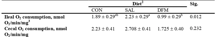
5. Ileal and Cecal Oxygen consumption in 3 wk old broiler chickens ...64
CHAPTER 3 1. Total duodenal, jejunal, ileal, cecal, and colonal protein and DNA contents in chicken at 21 d of age ...89
2. Jejunal fluid VFA concentrations [mM] and molar percentages at 21 d of age ...90
3. Ileal fluid VFA concentrations [mM] and molar percentages at 21 d of age ...91
4. Cecal fluid VFA concentrations [mM] and molar percentages at 21 d of age ...92
5. Lactic acid concentration in 21 d old chicken ...93
6. Ileal glucose and proline transport and efficiency of 21 d old chicken ...94
7. Analysis of estimated total ileal glucose, proline and oxygen flux ...95
8. Ileal oxygen consumption in 21 d old chicken ...96
LIST OF FIGURES
Figure Page
CHAPTER 1
1. SEM micrograph of chicken ileal mucosa at d 21 after hatch...27 2. SEM micrograph of chicken ileal mucosa at d 21 after hatch...28 3. SEM micrograph of chicken ileal mucosa at d 21 after hatch...29
CHAPTER 2
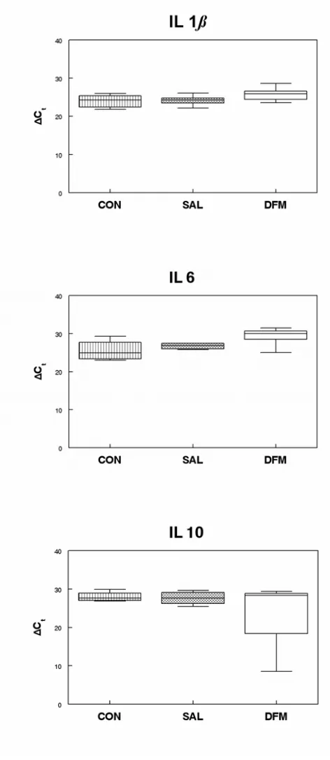
1. Ileal and cecal O2 consumption in 3 wk old broiler chickens...65 2. Whisker plot of cytokine production in the broiler chicken ileum...66
CHAPTER 3
1. Apparent energetic efficiency (APEE) of total ileal glucose and proline uptake in response to DFM or SAL treatment in chicken broilers on d 21...97
CHAPTER 4
8. SEM micrograph of the colonic surface of the 21 d old chicken broiler...128 9. SEM micrograph of the ileal surface of the 21 d old broiler chicken...129
CHAPTER 1: LITERATURE REVIEW
ABSTRACT
The poultry industry is facing a forthcoming ban of antibiotic feed additives, and
there is a growing interest in finding valuable alternatives to the prevention of disease and
growth enhancement supplements. The effects of probiotic or direct fed microbials (DFM)
on gut health and performance in poultry as well as other species are presented. The
interactions between intestinal microbiota, the gut epithelium, and the immune system are
important in the competitive exclusion process. The mechanisms by which probiotics
operate include spatial exclusion, micro-environmental alterations, production of
antimicrobial substances and epithelial barrier integrity. The majority of recent research in
this field suggests the likelihood of a small but additive series of beneficial changes from the
animal’s exposure to probiotics. Further investigations are required to fully characterize the
effects and sustained outcomes of probiotic and DFM treatments in poultry.
INTRODUCTION
The metabolic activity and energy requirements of the intestinal microbiota is
comparable to that which takes place in the liver, the most metabolically active organ
(Isolauri et al., 2004). In vertebrates, there are more microbial cells within the
gastrointestinal (GI) tract than within the body-proper (Hove et al., 1999; Mai, 2004). The
microorganisms most commonly observed are bacteria and yeast.
There are two populations of microorganisms that are found within the GI tract of
poultry. The first, the autochthonous bacteria, colonize the gut by inoculation resulting from
normal feeding activities of the bird (Gusils et al., 1999). The second, allocthonous bacteria,
are exogenous in nature and are introduced into the GI tract through the feed or drinking
water as direct fed microbials (DFM) or probiotics (Bird et al., 2002; Fooks and Gibson,
2002; Patterson and Burkholder, 2003). Modern nutritionists use the terms probiotics and
DFM interchangeably, but currently the term probiotic is most often used. So, for the
purposes of this review, probiotic or probiotics will be used to denote the use of both
probiotics and direct fed microbials.
A large body of data in the literature now indicates that allocthonous bacteria
introduced via probiotics can prevent infection and colonization of the GI tract by
opportunistic pathogens (Lin, 2003; Netherwood et al., 1999; Ouwehand and Vesterlund,
2003). Introduction of such probiotics is believed to prevent or attenuate clinical and sub
clinical enteric pathogens in poultry, thereby, resulting in enhanced growth and performance.
amongst poultry flocks (Patterson and Burkholder, 2003). This interest is because of
growing concerns about antibiotic resistance, and bans or potential bans on antibiotic usage
in poultry and other animal productions systems in Europe and the US (Klose et al., 2006;
Patterson and Burkholder, 2003).
Unfortunately, the mode of action of probiotics is poorly understood. Many papers
have been published on individual physiological actions of specific consortia of probiotic
organisms, but to date, however, poultry scientists have failed to develop a comprehensive
and integrated model of how probiotics work within the body of the bird. Such information
is essential for the development of more efficacious probiotics. Once these mechanisms are
elucidated, it may be possible to use modern molecular biological techniques to develop
more efficacious and useful probiotics.
This review will briefly describe traditional definitions of probiotics and summarize
what is currently understood about their colonization in the GI tract, their metabolism and
their mechanisms of action in altering host animal health and performance as well as their
What are probiotics and how can they impact poultry production systems?
Probiotics are “live microbial feed supplements, which beneficially affect the host
animal by improving its intestinal microbial balance” (Fuller, 1989) or
“a live microbial feed that is beneficial to health” (Salminen et al., 1998). They may contain
only one, or several (a consortium) different bacterial species. The mechanisms of action of
different bacterial strains in a probiotic consortium may differ (Davis and Anderson, 2002;
Mai, 2004). Additionally, different subtypes within the same species may have different
biological effects; isolates within the same species can be unique, and may have differing
areas of adherence, specific immunological effects, and other biological actions (Isolauri et
al., 2004). Hence, probiotics containing similar species of bacteria may, in fact, differ in
efficacy.
Although many articles in the current scientific and popular literature refer to the
“beneficial effects” of probiotics, these articles are often vague as to exactly what benefits
are conferred upon the animal by the probiotic (Fooks and Gibson, 2002; Netherwood et al.,
1999; Patterson and Burkholder, 2003). In poultry production systems, benefits and efficacy
can be easily defined. Any feed supplement or therapy that enhances poultry health and
performanceas measured by enhanced animal health, growth and/or feed efficiency can be
defined as beneficial or efficacious. In the case of probiotics, the preponderance of literature
suggests that these production endpoints are beneficially impacted by the ability of consortia
What are the factors associated with colonization?
Successful probiotic colonization depends on the survival and stability of the
probiotic strain, specificity of the strain relative to host, dose and frequency of
administration, health and nutritional status of the host, effect of age, stress and genetics of
the host. (Bomba et al., 2002). In general, probiotic bacteria are anaerobes or facultative
anaerobes (Isolauri et al., 2004). In poultry, probiotic organism colonization, as measured by
colony forming units (CFUs), increases as you go from the beak distally to the colon
(Bouzaine et al., 2005).
The crop, proventriculus and gizzard have very low anaerobic bacterial numbers due
to the presence of the O2 consumed with the feed as well as the low environmental pH that is
associated with the secretion of HCl within the proventriculus (Mason et al., 2005). The
small intestine has large bacterial numbers consisting of facultative anaerobes such as
Lactobacilli, Streptococci, and Enterobacteria as well as anaerobes such as Bifidobacterium
spp., Bacteroides spp., and Clostridia spp. at levels of ~ 104 to108 CFU/ml (Salanitro et al.,
1978). The most heavily colonized regions of the GI tract are the colon and cecum with
colonization of 1010 to 1013 CFU/ml (Simon et al., 2004).
Autocthonous and allocthonous (probiotic) bacteria are present in three different
major microenvironments within the GI tract, namely in the intestines, cecum and colon
(Rastall, 2004). These first areas of the gut where gut microbes are found are in the digesta,
which is created by the consumption of a rich milieu of feed nutrients and water. The digesta
is an ideal environmental niche for many bacteria to flourish. Probiotic bacteria can be found
attached to individual feed particles such as starch granules (Figure 1A and 1B). Other
the digesta. The second area of the gut where microbes are found is within the mucous
blanket that covers the epithelial lining of the GI tract including the intestinal villi (Figure 1C
and 1D). The mucous not only serves as an environment within which these microbes exist,
but also serves as a source of nutrients for bacteria (Gaskins, 2003). Finally, bacteria can
also exist on the surface of epithelial cells or they can actually be attached to epithelial cells
(Figures 2A and 2B). Figure 4 depicts lactobacillus occupying the surface area above the
opening to a goblet cell on the ileal villus of a chick at d 21. Figure 5 depicts a cluster of
segmented fusiform bacteria embedded into the cytoplasm of enterocytes of the ileum
(Heczko et al., 2000; Klaasen et al., 1992). The significance of the occurrence of bacteria in
relation to the GI architecture and its biological activity has not been fully established
(Yamauchi and Snel, 2000).
The ability of many strains of probiotic bacteria to physically adhere to portions of
the various GI microenvironments speaks to their ability to enhance bird enteric health
(Jacobsen et al., 1999). Attachment is considered a very first step in the colonization of the
host mucosal surfaces. That permits them to resist peristalsis and removal from the gut.
However, adherent probiotic bacteria usually do not colonize the intestinal mucosa for long
periods, and they are normally eliminated after a few days when the host stops ingesting
them (Marteau et al., 2004).
It has been suggested that Lactobacilli can colonize the nonsecretory gastric
epithelium by attaching to epithelial cells and can continuously inoculate gastric contents and
the lower regions of the intestinal tract (Rojas and Conway, 1996). It has also been
cells than do hydrophilic strains (Wadstrom et al., 1987). The highest adhesion values were
obtained at pH 7 (Gusils et al., 1999).
Lactobacilli, whether shed from epithelial surfaces or multiplying in ingested food, permeate all regions of the digestive tract in poultry (Gusils et al., 1999). However, very few
studies have investigated adhesion and colonization, because of the complexity of the
intestinal mucosa and the extensive interaction between cell types within the GI tract
(Sarem-Damerdji et al., 1995). Generally, Lactobacilli adhere to epithelial surfaces by interactions
occurring between specific molecules on the bacterial cells and on the GI surface of the host
(Gusils et al., 1999).
Since it is very difficult to study bacterial adhesion in vivo, most experiments use in
vitro models. Microbial Adhesion to Solvents (MATS) is a technique that have been used to investigate bacterial cell affinities for polar and non-polar solvents (Kankaanpaa et al., 2004).
Non-polar solvents have been used to estimate the hydrophobic properties, while polar
solvents have been used to help estimate Lewis acid/base properties (Briandet et al., 1999).
The low affinities of Lactobacilli for non-polar solvents indicate that these bacteria possess a
hydrophilic rather than hydrophobic cellular surface (Kankaanpaa et al., 2004). When those
microorganisms were cultured with free polyunsaturated fatty acids (PUFA), hydrophobicity
was diminished.
Huang and Adams (2003) used human intestinal epithelial cell line to study probiotic
bacterial adhesion. This cell line is a model for investigating such mechanisms, because it
spontaneously differentiates under standard in vitro culture conditions, and the differentiated
cells then express characteristics of mature enterocytes. This study utilized Lactobacillus
testing adhesion of propionibacteria strains. In that experiment large numbers of
L. acidophilus were observed that were adhering to the surface of the human enterocyte cell
line by scanning electron microscopy (SEM); while very few B. lactis were observed on the
surface. Additionally, polyunsaturated fatty acids (PUFA) were reported to alter bacterial
adhesion sites on Caco-2 cells (Bomba et al., 2002). That study suggests that dietary PUFA
affects the attachment sites for the GI microbiota, possibly by modifying the composition of
fatty acids in the intestinal wall. The stimulatory effect of PUFA upon adhesion of
Lactobacilli may be useful for enhancing the effectiveness of probiotics in inhibiting digestive tract pathogen colonization (Bomba et al., 2002).
It has also been observed that not all strains of Lactobacillus adhere to enterocytes,
indicating that this property is strain specific (Servin and Coconnier, 2003). Also, a
dose-dependent inhibition of adherence of Enterotoxigenic E.coli, Enteropathogenic E.coli
(EPEC), and S. typhimurium to Caco-2 cells by strains of Bifidobacteria and Lactobacillus
has been reported (Servin and Coconnier, 2003). Lactobacillus animalis has been
demonstrated to inhibit growth of Salmonella strains, produce antimicrobial substances, and
to inhibit in vitro adhesion (Gusils et al., 1999).
What is known about the metabolism of the major probiotic organisms within the GI tract?
As mentioned above, probiotic organisms can be divided into two general groups
based on their tolerance to O2, i.e. anaerobes and facultative anaerobes. The facultative
They reduce the redox potential in the gut and render the environment suitable for obligate
anaerobes (Fooks and Gibson, 2002).
Obligate anaerobic bacteria are those species capable of anaerobic fermentation only
(Wagner and Cerniglia, 2005). Anaerobic fermentation is the metabolic processes performed
by microorganisms that transform substrates, mainly carbohydrates and proteins, to provide
metabolites. These metabolites act as electron acceptors via substrate level phosphorylation
as opposed to O2, which is the major ultimate electron acceptor in aerobic respiration (Fooks
and Gibson, 2002; Jozefiak et al., 2004). Anaerobic fermentation results in the production of
microbial metabolic end-products such as lactate, succinate, acetate, propionate, and butyrate
(short chain volatile fatty acids; VFA), H2, CO2, CH4 as well as bacterial biomass (Cummings
and Macfarlane, 1997). Most of the VFA formed by intestinal bacteria are absorbed and
metabolized by the bird, thereby contributing to the host energy requirements (Cummings
and Macfarlane, 1997). Some bacterial metabolites that result from the breakdown and
fermentation of proteins, such as ammonia, phenols and amines are toxic (Macfarlane and
Cummings, 1999).
Bifidobacterium
Bifidobacteria are believed to be a major component of the microbial barrier to pathogenic bacterial infection (Gibson and Roberfroid, 1995). They are gram-positive,
non-spore forming rod or club-shaped bacteria, with distinct cellular bifurcations. Bifidobacteria
make a significant contribution to carbohydrate fermentation in the colon. Hexoses are
Stouthammer, 1968). The principal end products of fermentation are acetate and lactate,
which are produced in a 3:2 ratio (Fooks and Gibson, 2002).
Bifidobacteria produce a wide-range of antimicrobial agents that are effective against both gram-positive and gram–negative organisms. These agents might include antimicrobial
peptides, such as defensins, cathelicidins, and lyzozyme (Dommett et al., 2005). In addition
to producing antimicrobial agents, Bifidobacteria competitively exclude pathogens by
competing for epithelial and mucosal binding sites and nutrients.
Bifidobacteria have been reported to alter fecal bacterial enzyme activities, reduce antibiotic induced side effects, inhibit mammary and liver tumors, and they have been
reported to, in conjunction with oligofructose, reduce 1,2-dimethylhydrazine, reduce induced
colonic carcinogenesis in mice (Marteau, 2000). Oral supplementation of Bifidobacterium
lactis to elderly subjects increased the production of total, helper CD4+, and activated CD25+ T lymphocytes and natural killer cells (NK); and it increased the phagocytic activity of
mononuclear and polymorphonuclear phagocytes and the tumoricidal activity of NK (Gill et
al., 2001).
Lactobacilli
Lactobacilli are gram-positive, non-spore forming rods, usually non-motile, that are catalase negative, and do not reduce nitrate (Fooks and Gibson, 2002). They are generally
accepted as safe to ingest, and have together with Bifidobacterium, therefore been granted
GRAS status (Salminen et al., 1998). Lactobacilli are involved in both homo- and
pyruvates molecules which are then converted into lactate. Two moles of ATP are generated
per mole of glucose with this type of fermentation. Heterolactic fermentation acts via the
pentose phosphate pathway, to produce lactate, CO2 and ethanol, generating one mol of ATP
per mol of glucose. Phosphoketolase is the key enzyme involved in this process (Fooks and
Gibson, 2002). Lactobacilli, besides producing lactic acid, which is deleterious to many
micro-organisms, are also capable of producing antibacterial proteins and bacteriocins
(Marteau et al., 2004). Of these, some bacteriocins display a wide antibacterial spectrum
against gram-positive bacteria.
What mechanisms of actions have been postulated for how probiotics enhance poultry health and productivity?
Numerous health benefits have been ascribed to probiotics when used in poultry and
other species of animals, including man. Although the mechanisms of action associated with
the beneficial effects of probiotics are still unclear and likely multifaceted (Bird et al., 2002;
Fooks and Gibson, 2002), the single most frequently used term in describing their effects on
enteric health is “competitive exclusion” (Edens et al., 1997). This term was originally used
to describe the physical blocking of opportunistic pathogen colonization by the colonization
of organisms in the probiotic consortia (Klose et al., 2006) (Figure 3A-3D). Others and we
speculate that beside competitive exclusion there exist a number of mechanisms the increase
both enteric and whole-bird health (Bauer et al., 2006; Chichlowski et al., 2006a; Chichlowski et al., 2006b; Hugo et al., 2006). Virtually all actions of probiotics, both
states in the GI tract and other body tissues. Actions of probiotics include alterations in the
microbial microenvironments, alteration of the host animal’s metabolism, modifications of
the host’s immune system, improvement in feed digestion and absorption, and the production
of antimicrobial compounds (Chichlowski et al., 2006a; Cummings and Macfarlane, 1997;
Galdeano and Perdigon, 2006; Ichikawa et al., 1999; Mead, 1989). Potential benefits from
the ingestion of probiotics include: 1) the production of β-galactosidase which improves
tolerance to lactose (Savaiano et al., 1984); 2) treatment and reduction of intestinal
infections (Colombel et al., 1987); 3) the suppression of cancer (Kinouchi et al., 1998;
Reddy, 1998); 4) a decrease in the incidence of coronary heart disease (Schaafsma et al.,
1998), and others.
What are components of competitive exclusion? Physical interference
One of the first mechanisms that was proposed as a beneficial action of probiotic
bacteria was their ability to physically colonize environmental niches within the intestinal
tract, especially in the lower intestinal tract, which is favored by enteric pathogens
(Mercenier et al., 2003). Colonization by probiotic organisms may physically exclude
colonization by pathogens. For example, colonic crypts and intestinal villi crypts are the
favorite sites colonized by Salmonella spp. (Garriga et al., 1998; Mare et al., 2006; Servin
and Coconnier, 2003). Additionally, work in our laboratory suggests that probiotics may also
Lactobacilli, whether attached to epithelial surfaces or multiplying in ingested food, permeate all regions of the digestive tract in poultry (Gusils et al., 1999). Very few studies
have investigated adhesion and colonization, because of the complexity of the intestinal
mucosa and the extensive interactions that occur between cell types (Sarem-Damerdji et al.,
1995). It has been proposed that Lactobacilli adhere to the epithelial surfaces by interactions
occurring between specific molecules on the bacterial cells and with molecules on the GI
surface of the host (Gusils et al., 1999). Henriksson et al. (1991) propose that Lactobacilli
adhere to the stomach epithelial cells through proteinaceus components located on the
bacterial surface. Recently, it was suggested that lactic acid bacteria display various surface
determinants, and that these are involved in their interaction with intestinal epithelial cells.
Those determinants include passive forces, electrostatic interactions, hydrophobic forces,
steric forces, lipoteichoic acids, and specific structures such as external appendages covered
by lectins (Servin and Coconnier, 2003).
Competition for binding sites
Probiotics exclude the colonization of pathogens by preventing their adhesion to the
epithelium (Kohler et al., 2003). The ability of a probiotic strain to adhere to mucus and
epithelial cell surfaces is one of the main selection criteria for a candidate probiotic
(Kankaanpaa et al., 2004). Gastrointestinal microbial colonization is related to the
bacterium’s intestinal ability to adhere and is initiated from the initial ingestion of microbes
at the day of age. Attachment is considered to be the very first step in the colonization of
removal by intestinal peristalsis and is a prerequisite for adhesion to the enterocyte surface
(Erickson et al., 1992).
The exact mechanism by which probiotic bacteria prevent the attachment and
colonization of pathogens can vary from organism to organism. Lactobacillus plantarum
inhibits pathogen adhesion without competing for binding sites. This probiotic bacterium
induces the transcription and excretion of the mucins MUC2 and MUC3 from goblet cells
and thereby inhibits the adherence of EPEC to the intestinal surface (Mack et al., 1999).
Another example is a Lactobacillus spp. which directly inhibit the attachment of Salmonella,
E.Coli and other food borne pathogens (Duggan et al., 2002). Lactobacilli have also been
reported to suppress the growth of Shigella flexneri, Salmonella typhimurium, Clostridium
difficile and other pathogens (Forestier et al., 2001; Pochapin, 2000). The exact mechanisms of this inhibition are unknown.
Micro-environmental alterations
Probiotics can alter the microenvironments of the GI tract via a very complex and
interdependent mechanism that is based on digesta substrate utilization (Cummings and
Macfarlane, 1997; Delzenne and Williams, 2002). These alterations often result in changes
in the physical environment of the gut in such a manner that opportunistic pathogens cannot
compete as well as changes in GI epithelial function and metabolism. Probiotic organisms
compete with pathogens for nutrients thus preventing them from acquiring energy to
effectively compete in the gut environment (Fooks and Gibson, 2002). The collateral effect
nutrients in the gut digesta (Mead, 1989). These weak organic acids lower the pH of the gut
environment below that essential for the survival of such pathogenic bacteria as E. coli and
Salmonella (Isolauri et al., 2004).
Volatile fatty acids serve as energy yielding substrates to the host animal, in addition
to inhibiting the growth of pathogenic bacteria. Volatile fatty acids can affect colonic
epithelial cell transport, colonocyte metabolism, growth and differentiation, hepatic control
of lipid and carbohydrates, and they provide energy to the muscle, kidney, heart and brain
(Gibson, 1999). Volatile fatty acids are rapidly absorbed from the small intestinal tract and
colon, stimulate electrolyte and water absorption within the intestinal tract and have a major
effect on the growth of epithelial cells. The colonic epithelium derives 60-70% of its energy
from bacterial fermentation (Cummings and Macfarlane, 1997).
Production of antimicrobial substances
Probiotic organisms are known to produce a class of small, antimicrobial molecules
that are collectively known as bacteriocins (Marteau et al., 2004). These bacteriocins can
kill pathogenic bacteria or impede their colonization (Marteau et al., 2004). They are
proteins, or protein complexes, produced by certain strains of bacteria, which can have
antagonistic actions against species related to the producer bacterium. Other, non-bacteriocin
compounds which inhibit the growth of pathogens are also produced by probiotic bacteria
(Meghrous et al., 1990). The polyamine derivate piperidine, which is also produced by
intestinal microflora as the result of amino acid degradation, has been shown to inhibit the
binding and internalization of Salmonella and Shigella to intestinal epithelial cells in vitro
Do probiotics have an effect on maintenance of epithelial barrier integrity?
One of the major secondary functions of the GI tract is to act as a protective barrier,
which shields the body from organisms and substances that do not serve as nutrients
(Bar-Shira and Friedman, 2006). There are two major mechanisms which epithelial barrier
maintains integrity. The mucous barrier or “blanket” (Figure 1D), which is composed of a
complex mixture of proteins and carbohydrates and the tight junctions or zona occludens
(tight junctions)via which the intestinal epithelial cells attach to one another to form an
unbroken, contiguous biological barrier. Mechanisms of probiotic action are closely related
to both of them.
Mucous layer
A relatively thick layer of mucus that is secreted by goblet cells in the gut wall covers
the epithelial cells in the small intestine. This mucus consists of mucin, many small
associated proteins, glycoproteins, lipids, and glycolipids (Gusils et al., 2003). It also
contains soluble receptors that recognize specific adhesion proteins (Metcalfe et al., 1991).
Both pathogens and probiotic bacteria adhere to specific glycoconjugates on the microvilli in
the small intestinal tract.
Access to the Epithelium
Non-nutrient substances and microorganisms can enter the body through the GI tract
in one of several ways; including transepithelial transit, paracellular transit and through the
effectively in preventing the entrance of antigens and pathogens that could prove harmful to
the bird into the blood stream. Indeed, the function of “M” cells themselves may be
important in acclimatizing the animal’s immune system to non life-threatening antigens
(Pohlmeyer et al., 2005). Furthermore, Tyrer et al. (2006) suggested that
pathogen-associated molecular pattern interactions with pattern recognition receptors are key factors in
“M” cell recognition of intestinal antigens for mucosal immune priming.
Tight Junctions
The development of intestinal barrier function is determined by the assembly of tight
junction and adherent junction proteins (Kohler et al., 2003). That is also the most
vulnerable point of intestinal bacteria penetration, however, there is a very scarce data
regarding effects of probiotic on tight junction structure. Tight junctions can be viewed by
electron microscopy as a series of discrete contacts between plasma membranes of adjoining
cells containing a multiprotein complex that affiliates with the underlying actin cytoskeleton
(Nusrat et al., 2001). The tight junction and so-called adherens junction are known as the
apical junction complex. The adherens junction in the intestine has been documented to be a target for infectious diseases (Barton et al., 2001). Furthermore, infectious agents induce
inflammatory response associated with an influx of leukocytes and the release of cytokines
such as interferon-gamma (IFN-γ), and tumor necrosis factor-alpha (TNF-α, not identified in
birds yet), both of which are known to modify tight junction structure and paracellular
PUFA and probiotics
It is possible that consumption of specific PUFA may enhance the intestinal barrier
(Bomba et al., 2002). Claudins and occludin have been identified as tight junctions specific
integral membrane proteins (Furuse et al., 1998), which are regulated by immune mediators
and whose expression restricts paracellular transport of macromolecules. Occludin is
approximately 65kDa and is attached to the cytoskeleton by another group of proteins
including cingulin and 7H6 (Jiang et al., 1998). Occludin, which plays a major role in tight
junctions, was up regulated by γ-linolenic acid (GLA; 18:3, ω-6) and by eicosapentaenoic
acid (EPA; 20:5,
ω
-3) but was down regulated by arachidonic acid (AA; 20:4, ω-4) andlinoleic acid (LA; 18:2, ω-6; (Jiang et al., 1998).
The extra cellular domain of occludin binds to another occludin molecule on an
adjacent cell and thus forms a tight junction (Furuse et al., 1996). Pathogens aim to destroy
the integrity of the epithelial barrier to gain easy access to the gut interstitium, which allows
further access to the blood stream and systemic spreading of the organism (Kohler et al.,
2003). Probiotics are known to enhance the epithelial barrier. Madsen et al. (2001) have
shown that a commercial mixture of various Bifidobacterium and Lactobacilli strains can
enhance the epithelial barrier in IL-10 knock-out mice.
Nutrient transport
The trophic effects of probiotics include increases in the specific and total activities
of the brush-border membrane enzymes in the jejunal mucosa of growing rats (Marteau et
corresponding increase of the sodium D-glucose cotransporter-1, SGLT-1 (Buts et al.,
2002). It has also been also reported that the oral administration of Lactobacillus casei
increased the crypt cell production rate of the jejunum, ileum, cecum and distal colon in rats
(Ichikawa et al., 1999). Additionally, Chichlowski et al. (2006b) have reported that
probiotic consortium increases passive absorption of glucose in the chicken ileum.
What is the effect of probiotic on maintenance and enhancement of intestinal immune function?
Introduction to mucosal immunity
The intestinal immune system has several unique elements to maintain the
equilibrium with bacterial flora and the rest of the organism (Mowat, 2003). There is a
constant interaction among different cell types, including members of the innate and the
adaptive immune systems, and bacteria in the gut lumen, epithelium or lamina propria (Eberl,
2005). Furthermore, the epithelial lining is an important permeability barrier between the
external environment and the internal body environment (Nusrat et al., 2001). The dendritic
cells sample bacterial antigen, and migrate to the T-cell zones within the epithelium
(Macpherson and Uhr, 2004). Presentation of microbial antigen to immune response cells
can be to both CD4+ helper cells and to CD8+ cytotoxic/suppressor cells. Then, adaptive
immune response can be induced, that can lead to effector T-cells and immunoglobulin A
(IgA) production (Niess et al., 2005). Probiotics may reach the inductive mucosal immune
system through several routes including the specialized M cells near the Peyers patches and
contains at least two types of immunocompetent cells, the intraepithelial lymphocytes and the
intestinal epithelial cells (Donnet-Hughes et al., 2001). Also, epithelial cells express many
important immune molecules and a wide range of cytokines, which modulate their interaction
with T lymphocytes (Strober, 1998). The number of Peyer’s patches (lymphoid aggregates)
and Ig A producing cells increase in the presence of the intestinal microbiota (Isolauri et al.,
2004) which constructs an immunological barrier of the gut mucosa. It has been suggested
that surface Ig A attached to the mucosal membrane may limit or inhibit the adherence of
enteropathogens that are invading the GI tract (Perdigon et al., 1990). Furthermore, the
intestinal microbiota provide an important stimulus for the maturation of the immune system
(Isolauri et al., 2004).
Th1 and Th2 responses
The cytokines released by intestinal immune cells in the presence of bacteria, may
indicate either a Th1 immunogenic response or a Th2 humoral response (Gonnella et al.,
1998). Th1 cell-driven responses are exemplified by a delayed-type hypersensitivity
reaction, together with regulation of phagocytes. More specifically, Th1 cells secrete
interferon-γ (IFN-γ), tumor necrosis factor (TNF; not identified in chickens yet),
IL (interleukin)-2, IL-12 and are central to the development of cellular immunity against
intracellular pathogens. Th2 cells promote Ig E production and eosinophil activity
(Donnet-Hughes et al., 2001). Th2 cells produce IL-4, IL-5 and IL-13 and promote allergic
inflammation (Avery et al., 2004; Das, 2002). This is especially true for IL-4 and IL-5,
during and after antigen presentation. These naïve Th cells produce all of these cytokines
and are considered the precursor of the Th1 and Th2.
Establishment and maintenance of intestinal tolerance is mostly dependent on
suppressive cytokines such as IL-10 and transforming growth factor-β (TGF-β) produced by
regulatory T cells and T helper cells characteristic of the intestinal immune system (Izcue et
al., 2006; Pessi et al., 2000). Probiotics seem to shift the Th1/Th2 response balance toward
Th1 (Das, 2002), they also augment the production of TGF-β, IL-10 and Ig A (Gaskins,
1997). For example, the expression of the key proinflammatory cytokine (Th2), IL-8 was
inhibited by a probiotic consortium (Vidal et al., 2002). Furthermore, Bifidobacteria isolated
from infants suffering from allergic diseases induced the secretion of TNF-α, IL-1β, IL-6 and
IL-12 by macrophages in vitro, while Bifidobacteria from healthy infants stimulated the
secretion of IL-10 (He et al., 2002). Also, non-pathogenic enteric microbes, like
Bifidobacteria, exert an immunosuppressive effect on intestinal epithelial cells by inhibition of the transcription factor NF-κB pathway (Neish et al., 2000). The tolerogenic effects of
the gut microbiota may partially be mediated by generation of regulatory T cells (Singh et al.,
2001), however, for immune regulation; specific adherence properties may be required
(O'Hara et al., 2006).
Cytokines produced by epithelial cells transmit information on the relative state of
intestinal health to intra-epithelial T lymphocytes and immune cells in the underlying lamina
propria (Gaskins, 2003). Cytokines play an important role in these mucosal humoral and
cell-mediated responses (Peters et al., 2005). CD4+ T helper cells exert their effect through
the secretion of pro-inflammatory (IL-1β, IL-6) or immunomodulatory (IL-10) cytokines
In one study, Lactobacillus casei fed mice have shown an increase in IL-10, while a
significant induction of IL-2 and IL-12 was observed in mice fed L. acidophilus (Perdigon et
al., 2002). These effects were dose dependent. It has also been reported that a commercial
probiotic consortium, containing strains of Lactobacilli and Bifidobacterium could enhance
the epithelial barrier in IL-10 knock-out mice which serve as a model for Inflammatory
Bowel Disease (Kohler et al., 2003). Madsen and co-workers (Madsen et al., 2001) have also
demonstrated that the production of proinflammatory cytokine secretion is down-regulated
by attenuation of the NF-kB pathway.
Signaling pathways
Probiotics may have the ability to directly influence the inflammatory response
elicited by pathogens by downregulating a specific signaling pathways (Kohler et al., 2003).
There are several pathways proposed for activation of immune response by gut microflora or
when cells are infected by a variety of pathogens, including MAP kinase and NF-κB
pathways. Also, several enteric organisms have been shown to inhibit these pathways, and
thus the subsequent induction of inflammatory mediators (Neish et al., 2000). Active or
acute inflammation of the intestinal epithelium triggered by the bacterial antigens is
associated with the migration of polymorphonuclear leukocytes from the microvasculature
into the lumen (Nusrat et al., 2001). The primary polymorphonuclear leukocyte in poultry is
heterophil, the avian equivalent to mammalian neutrophil (Kogut et al., 2006). These cells
are involved in the phagocytosis and killing of invading microbes. Heterophils activate
Adenosine, in particular, seems to play a central role in mediating
neutrophil-epithelial crosstalk in most species. Adenosine is generated from neutrophil-derived 5’AMP
through protein kinase A (PKA) and it stimulates trans-epithelial chloride flux, up-regulates
CD73 (which possesses the enzymatic activity of 5’nucleotidase), and induces IL-6 secretion
by epithelial cells, which in turn activate neutrophils to modulate inflammation (Nusrat et al.,
2001). However, it has been demonstrated that Salmonella typhimurium activates a protein
kinase C (PKC)-dependent signal transduction pathway, which is independent of NF-κB, and
influences transepithelial neutrophil movement (Kohler et al., 2003). The PKC is a cellular
receptor for the second lipid messenger diacylglycerol (DAG) and is therefore a crucial
element in signal transduction pathways. In addition, the cyclic-AMP (cAMP) production
activated by microflora, can also have a profound effect on pro and anti-inflammatory
cytokines production, which increases IL-10 production by monocytes, increases IL-8
production by mast cells, and increases IL-6 production by astrocytes (Singh et al., 2006).
Further, an inflammatory state can modify tight junctions and paracellular permeability
across the intestinal epithelium (Shen and Turner, 2006).
Clearly, all DFM do not share the same immunomodulating properties, and can even
have opposite effects on some parameters (Marteau et al., 2004). Mercenier et al. (2003)
reported large variation in the ability of different Lactobacillus strains to induce pro- and
anti-inflammatory cytokines. Colonization of the small intestine with commensal organisms
may result in altered sensitivity to various luminal stimuli. These considerations may be
particularly important during animal growth, given the energetic costs likely associated with
It has been shown that probiotic bacteria may cause the intestinal epithelial cells to
limit immune activation by inhibiting the ubiquitination and degradation of the IκB signaling
pathway (Neish et al., 2000). Ubiquitination is a common covalent modification of cellular
proteins that serves a number of regulatory functions, often targeting modified proteins for
the control of cellular degradation. The results lead to a significant reduction in the amount
of IL-8 that is secreted from the intestinal epithelial cells. Also, the lipoteichoic acid from
Lactobacillus johnsonii and
L. acidophilus has been shown to downregulate the inflammatory response in epithelial cells to lipopolysaccharide and enteric bacteria.
Toll-like receptors
The host’s innate immune response distinguishes signals from pathogens and
commensals via Toll-Like Receptors (TLR) (Marteau et al., 2004). TLRs are the highly
conserved receptors existing in most animal cells and are important initiators of innate
immunity (Doyle and O'Neill, 2006). The TLRs mediate mechanisms of intestinal epithelial
tolerance versus intolerance (Cario and Podolsky, 2005). The immune cells express multiple
TLRs in order to recognize the specific microbial environment involved, and trigger an
appropriate adaptive response as well as to induce antimicrobial effector pathways, which
eventually leads to efficient elimination of host-threatening pathogens (Kobayashi et al.,
2002). Immunomodulation may be due to an increased transport of antigens across the
mucosal barrier (via increased intestinal permeability) or to an up-regulation of antigen
CONCLUSIONS
It is likely that the beneficial effects of probiotics are the result of the summation of a
complex, multi-variate series of alterations in gut microbial and whole body metabolism.
Those alterations might include whole body and tissue oxygen consumption, absorption of
nutrients, production of cytokines, as well as configurations and intestine histomorphometry.
Data described above demonstrate also that some probiotic species can communicate with the
epithelium and immune system, modulating tissue physiology and response to host’s
infection. Overall, probiotic products are putative alternative to growth promoting
antibiotics. However, more research is necessary to define mechanisms of action of
FIGURES
Figure 1. SEM micrograph of chicken ileal mucosa at d 21 after hatch. Tissues were fixed in 1%OsO4 and
Figure 2. SEM micrograph of chicken ileal mucosa at d 21 after hatch. Tissues were fixed in 1%OsO4 and examined using JEOL 5900LV microscope at 20kV. A: SEM micrograph of
Figure 3. SEM micrograph of chicken ileal mucosa at d 21 after hatch. Tissues were fixed in 1%OsO4 and
REFERENCES:
Avery, S., L. Rothwell, W.D. Degen, V.E. Schijns, J. Young, J. Kaufman, and P. Kaiser.
2004. Characterization of the first nonmammalian T2 cytokine gene cluster: the
cluster contains functional single-copy genes for IL-3, IL-4, IL-13, and GM-CSF, a
gene for IL-5 that appears to be a pseudogene, and a gene encoding another
cytokinelike transcript, KK34. J Interferon Cytokine 24(10):600-10.
Bar-Shira, E., and A. Friedman. 2006. Development and adaptations of innate immunity in
the gastrointestinal tract of the newly hatched chick. Dev. Comp. Immunol.
30:930-941.
Barton, E.S., J.C. Forrest, J.L. Connolly, J.D. Chappell, Y. Liu, F.J. Schnell, A. Nusrat, C.A.
Parkos, and T.S. Dermody. 2001. Junction Adhesion Molecule Is a Receptor for
Reovirus. Cell 104:441-451.
Bauer, E., B.A. Williams, H. Smidt, M.W. Verstegen, and R. Mosenthin. 2006. Influence of
the gastrointestinal microbiota on development of the immune system in young
animals. Curr Issues Intest Microbiol. 7(2):35-51.
Bird, A., W. Croom, and B. McBride. 2002. Dietary management of the intestinal microbiota
using probiotics and prebiotics in human and animals. Proceedings of the Australian
Poultry Science Symposium 14:not given.
Bomba, A., R. Nemcova, S. Gancarcikova, R. Herich, P. Guba, and D. Mudronova. 2002.
Improvement of the probiotic effect of micro-organisms by their combination with
maltodextrins, fructo-oligosaccharides and polyunsaturated fatty acids. Br J Nutr.
Bouzaine, T., R.D. Dauphin, P. Thonart, M.C. Urdaci, and M. Hamdi. 2005. Adherence and
colonization properties of Lactobacillus rhamnosus TB1, a broiler chicken isolate.
Lett. Appl. Microbiol. 40:391-396.
Briandet, R., T. Meylheuc, C. Maher, and M.N. Bellon-Fontaine. 1999. Listeria
monocytogenes Scott A: cell surface charge, hydrophobicity, and electron donor and
acceptor characteristics under different environmental growth conditions. Appl
Environ Microbiol 65(12):5328-5333.
Buts, J., N. De Keyser, C. Stilmant, E. Sokal, and S. Marandi. 2002. Saccharomyces
boulardii enhances N-terminal peptide hydrolysis in suckling rat small intestine by
endoluminal release of a zinc-binding metalloprotease. Pediatr. Res. 51:528-534.
Cario, E., and D.K. Podolsky. 2005. Intestinal epithelial Tollerance versus inTollerance of
commensals. Molecular Immunology 42:887-893.
Chichlowski, M., J. Croom, M. Koci, G. Davis, and L. Daniel. 2006a. Primalac, direct fed
microbial supplement, decreases energy expenditures in young broilers (abstract).
Poult Sci.(Suppl 1):152.
Chichlowski, M., W.J. Croom Jr, M.A. Froetschel, M.D. Koci, B.M. McBride, R. Qiu, and
L.R. Daniel. 2006b. Effect of Primalac, direct fed microbial, on ileal absorption,
energy expenditure and intestinal microbial fermentation (abstract). Poult Sci. (Suppl
1):33.
Colombel, J.F., A. Cortot, C. Neut, and C. Romond. 1987. Yoghurt with Bifidobacterium
longum reduces erythromycin-induced gastrointestinal effects. Lancet. 2:43.
Das, U.N. 2002. Essential fatty acids as possible enhancers of the beneficial actions of
probiotics. Nutrition 18:786-789.
Davis, G.S., and K.E. Anderson. 2002. The Effects of Feeding the Direct-Fed Microbial,
PrimaLac, on Growth Parameters and Egg Production in Single Comb White Leghorn
Hens. Poult. Sci. 81:755-759.
de Vries, W., and A.H. Stouthammer. 1968. Fermentation of glucose, lactose, mannitol and
xyloseby bifidobacteria. J. Bacteriol. 96:472-478.
Delzenne, N.M., and C.M. Williams. 2002. Prebiotics and lipid metabolism. Curr Opin
Lipidol. 13(1):61-7.
Dommett, R., M. Zilbauer, J.T. George, and M. Bajaj-Elliott. 2005. Innate immune defence
in the human gastrointestinal tract. Molecular Immunology 42:903-912.
Donnet-Hughes, A., E. Schiffrin, and M. Turini. 2001. The intestinal mucosa as a target for
dietary polyunsaturated fatty acids. Lipids 36:1043-1052.
Doyle, S.L., and L.A.J. O'Neill. 2006. Toll-like receptors: From the discovery of NF[kappa]B
to new insights into transcriptional regulations in innate immunity. Biochemical
Pharmacology In Press, Corrected Proof.
Duggan, C., J. Gannon, and W.A. Walker. 2002. Protective nutrients and functional foods for
the gastrointestinal tract. Am. J. Clin. Nutr. 75:789-808.
Eberl, G. 2005. Inducible lymphoid tissues in the adult gut: recapitulation of a fetal
developmental pathway? Nat Rev Immunol 5:413-420.
Edens, F.W., C.R. Parkhurst, I.A. Casas, and W.J. Dobrogosz. 1997. Principles of ex ovo
competitive exclusion and in ovo administration of Lactobacillus reuteri. Poult Sci.
Erickson, A.K., J.A. Willgohs, S.Y. McFarland, D.A. Benfield, and D.H. Francis. 1992.
Identification of two porcine brush border glycoproteins that bind the K88ac adhesin
of Escherichia coli and correlation of these glycoproteins with the adhesive
phenotype. Infect Immun 60(3):983-988.
Fooks, L., and G. Gibson. 2002. Probiotics as modulators of the gut flora. Br. J. Nutr.
88:S39-S49.
Forestier, C., C. De Champs, C. Vatoux, and B. Joly. 2001. Probiotic activities of
Lactobacillus casei rhamnosus: in vitro adherence to intestinal cells and antimicrobial
properties. Res Microbiol. 152:167.
Fuller, R. 1989. Probiotics in man and animals. J. Appl. Bact. 66:365-378.
Furuse, M., K. Fujimoto, N. Sato, T. Hirase, and S. Tsukita. 1996. Overexpression of
occludin, a tight junction-associated integral membrane protein, induces the
formation of intracellular multilamellar bodies bearing tight junction-like structures. J
Cell Sci 109:429-435.
Furuse, M., K. Fujita, T. Hiiragi, K. Fujimoto, and T. S. 1998. Claudin-1 and -2: novel
integral membrane proteins localizing at tight junctions with no sequence similarity to
occludin. J Cell Biol 141:1539-1550.
Galdeano, C.M., and G. Perdigon. 2006. The Probiotic Bacterium Lactobacillus casei
Induces Activation of the Gut Mucosal Immune System through Innate Immunity.
Clin. Vaccine Immunol. 13:219-226.
Garriga, M., M. Paschual, J. Monfort, and M. Hugas. 1998. Selection of lactobacilli for
Gaskins, H. 2003. The commensal microbiota and developments of mucosal defense in the
mammalian intestine. 9th International Symposium on Digestive Physiology in Pigs
1:57-71.
Gaskins, H.R. 1997. Immunological aspects of host/microbiotica interactions at the intestinal
epithelium, p. 537-587, In R. L. Mackie, et al., eds. Gastrointestinal
Microbiology:Volume 2, Gastrointestinal Microbes and Host Interactions, Vol. 2.
Chapman & Hall, New York.
Gibson, G., and M. Roberfroid. 1995. Dietary Modulation of the human colonic
microbiota:introducing the concept of prebiotics. J. Nutr. 125:1401-1412.
Gibson, G.R. 1999. Dietary modulation of the human gut microflora using the prebiotics
oligofructose and inulin. J. Nutr. 129:1438S-1441S.
Gill, H.S., K.J. Rutherfurd, M.L. Cross, and P.K. Gopal. 2001. Enhancement of immunity in
the elderly by dietary supplementation with the probiotic Bifidobacterium lactis
HN019. Am J Clin Nutr 74(6):833-9.
Gonnella, P.A., Y. Chen, J. Inobe, Y. Komagata, M. Quartulli, and H.L. Weiner. 1998. In
situ immune response in gut-associated lymphoid tissue (GALT) following oral
antigen in TCR-transgenic mice. J Immunol. 160:4708-4718.
Gusils, C., S. Gonzalez, and G. Oliver. 1999. Some probiotic properties of chicken
lactobacilli. Can. J. Microbiol. 45:981-987.
Gusils, C., O. Oppezzo, R. Pizarro, and S. Gonzalez. 2003. Adhesion of probiotic lactobacilli
He, F., H. Morita, H. Hashimoto, M. Hosoda, J. Kurisaki, A.C. Ouwehand, E. Isolauri, Y.
Benno, and S. Salminen. 2002. Intestinal Bifidobacterium species induce varying
cytokine production. J. Allergy Clin. Immunol. 109:1035-1036.
Heczko, U., A. Abe, and B.B. Finlay. 2000. Segmented filamentous bacteria prevent
colonization of enteropathogenic Escherichia coli O103 in rabbits. J Infect Dis.
181:1027-33.
Henriksson, A., R. Szewzyk, and P.L. Conway. 1991. Characteristics of the adhesive
determinants of Lactobacillus fermentum 104. Appl Environ Microbiol
57(2):499-502.
Hove, H., H. Norgaard, and P.B. Mortensen. 1999. Lactic acid bacteria and the human
gastrointestinal tract. Eur. J. Clin. Nutr. 53:339-350.
Huang, Y., and M.C. Adams. 2003. An in vitro model for investigating intestinal adhesion of
potential dairy propionibacteria probiotic strains using cell line C2BBe1. Lett Appl
Microbiol. 36:213-216.
Hugo, A.A., G.L. De Antoni, and P.F. Perez. 2006. Lactobacillus delbrueckii subsp lactis
strain CIDCA 133 inhibits nitrate reductase activity of Escherichia coli. International
Journal of Food Microbiology 111:191-196.
Ichikawa, H., T. Kuroiwa, A. Inagaki, R. Shineha, T. Nishihira, S. Satomi, and T. Sakata.
1999. Probiotic Bacteria Stimulate Gut Epithelial Cell Proliferation in Rat. Dig. Dis.
Sci. 44:2119-2123.
Isolauri, E., S. Salminen, and A.C. Ouwehand. 2004. Probiotics. Best Pract Res Clin
Izcue, A., J.L. Coombes, and F. Powrie. 2006. Regulatory T cells suppress systemic and
mucosal immune activation to control intestinal inflammation. Immunol Rev.
212:256-71.
Jacobsen, C.N., V. Rosenfeldt Nielsen, A.E. Hayford, P.L. Moller, K.F. Michaelsen, A.
Paerregaard, B. Sandstrom, M. Tvede, and M. Jakobsen. 1999. Screening of probiotic
activities of forty-seven strains of Lactobacillus spp. by in vitro techniques and
evaluation of the colonization ability of five selected strains in humans. Appl.
Environ. Microbiol. 65:4949-4956.
Jiang, W.G., R.P. Bryce, D.F. Horrobin, and R.E. Mansel. 1998. Regulation of tight junction
permeability and occludin expression by polyunsaturated fatty acids. Biochem
Biophys Res Commun. 244(2):414-420.
Jozefiak, D., A. Rutkowski, and S. Martin. 2004. Carbohydrate fermentation in the avian
ceca: a review. Anim. Feed Sci. Technol. 113:1-15.
Kankaanpaa, P., B. Yang, H. Kallio, E. Isolauri, and S. Salminen. 2004. Effects of
polyunsaturated fatty acids in growth medium on lipid composition and on
physicochemical surface properties of lactobacilli. Appl Environ Microbiol
70(1):129-136.
Kinouchi, T., K. Kataoka, S.R. Bing, H. Nakayama, M. Uejima, K. Shimono, T. Kuwahara,
S. Akimoto, I. Hiraoka, and O. Y. 1998. Culture supernatants of Lactobacillus
acidophilus and Bifidobacterium adolescentis repress ileal ulcer formation in rats
treated with a nonsteroidal antiinflammatory drug by suppressing unbalanced growth
Klaasen, H.L., J.P. Koopman, F.G. Poelma, and A.C. Beynen. 1992. Intestinal, segmented,
filamentous bacteria. FEMS Microbiol Rev. 8:165-180.
Klose, V., M. Mohnl, R. Plail, G. Schatzmayr, and A.P. Loibner. 2006. Development of a
competitive exclusion product for poultry meeting the regulatory requirements for
registration in the European Union. Mol. Nutr. Food. Res. 50:563-571.
Kobayashi, K., L.D. Hernandez, J.E. Galan, J. Janeway, Charles A., R. Medzhitov, and R.A.
Flavell. 2002. IRAK-M Is a Negative Regulator of Toll-like Receptor Signaling. Cell
110:191-202.
Kogut, M.H., C. Swaggerty, H. He, I. Pevzner, and P. Kaiser. 2006. Toll-like receptor
agonists stimulate differential functional activation and cytokine and chemokine gene
expression in heterophils isolated from chickens with differential innate responses.
Microbes and Infection 8:1866-1874.
Kogut, M.H., M. Iqbal, H. He, V. Philbin, P. Kaiser, and A. Smith. 2005. Expression and
function of Toll-like receptors in chicken heterophils. Developmental & Comparative
Immunology 29:791-807.
Kohler, H., B. McCormick, and W. Walker. 2003. Bacterial-enterocyte crosstalk: Cellular
mechanisms in health and disease. J. Pediatr. Gastroenterol. Nutr. 36:175-185.
Kohler, H., S.P. Rodrigues, A.T. Maurelli, and B.A. McCormick. 2002. Inhibition of
Salmonella typhimurium enteropathogenicity by piperidine, a metabolite of the
polyamine cadaverine. J. Infect. Dis. 186:1122-1130.
Lan, Y., M.W.A. Verstegen, S. Tamminga, and B.A. Williams. 2005. The role of the
61:95-Lin, D.C. 2003. Probiotics As Functional Foods. Nutr Clin Pract 18:497-506.
Macfarlane, G.T., and J.H. Cummings. 1999. Probiotics and prebiotics: can regulating the
activities of intestinal bacteria benefit health? Br. Med. J. 318:999-1003.
Mack, D., S. Michail, S. Wei, L. McDougall, and M. Hollingsworth. 1999. Probiotics inhibit
enteropathogenic E. coli adherence in vitro by inducing intestinal mucin gene
expression. American Journal of Physiology 276:G941-950.
Macpherson, A., and N. Harris. 2004. Interactions between commensal intestinal bacteria and
the immune system. Nature Reviews: Immunology 4:478-485.
Macpherson, A.J., and T. Uhr. 2004. Induction of Protective IgA by Intestinal Dendritic Cells
Carrying Commensal Bacteria. Science 303:1662-1665.
Madsen, K., A. Cornish, P. Soper, C. McKaigney, H. Jijon, C. Yachimec, J. Doyle, L. Jewell,
and C. DeSimone. 2001. Probiotic bacteria enhance murine and human intestinal
epithelial barrier function. Gastroenterology 121:580-591.
Mai, V. 2004. Dietary Modification of the Intestinal Microbiota. Nutr. Rev. 62:235-242.
Mare, L., G.M. Wolfaardt, and L.M.T. Dicks. 2006. Adhesion of Lactobacillus plantarum
423 and Lactobacillus salivarius 241 to the intestinal tract of piglets, as recorded with
fluorescent in situ hybridization (FISH), and production of plantaricin 423 by cells
colonized to the ileum. J. Appl. Microbiol. 100:838-845.
Marteau, P. 2000. Role of the intestinal flora in gastrointestinal diseases. Lancet 356
Supplement:s28.
Marteau, P., P. Seksik, P. Lepage, and J. Dore. 2004. Cellular and physiological effects of
Mason, C.K., M.A. Collins, and K. Thompson. 2005. Modified electroporation protocol for
Lactobacilli isolated from the chicken crop facilitates transformation and the use of a
genetic tool. J. Microbiol. Methods 60:353-363.
Mead, G.C. 1989. Microbes of the avian cecum: Types present and substrates utilized. The
Journal of Experimental Zoology 3:48-54.
Meghrous, J., P. Euloge, A.M. Junelles, J. Ballongue, and H. Petitdemange. 1990. Screening
of Bifidobacterium strains for bacteriocin production. Biotechnol. Lett. 12:575-580.
Mercenier, A., S. Pavan, and B. Pot. 2003. Probiotics as biotherapeutic agents: present
knowledge and future prospects. Curr Pharm Des. 9(2):175-91.
Metcalfe, J.W., K.A. Krogfelt, H.C. Krivan, P.S. Cohen, and D.C. Laux. 1991.
Characterization and identification of a porcine small intestine mucus receptor for the
K88ab fimbrial adhesin. Infect Immun 59:91-96.
Mowat, A.M. 2003. Anatomical basis of tolerance and immunity to intestinal antigens. Nat
Rev Immunol 3:331-341.
Neish, A., A. Gewirtz, H. Zeng, A.N. Young, M. Hobert, V. Karmali, A. Rao, and J. Madara.
2000. Prokaryotic regulation of epithelial responses by inhibition of IkB-a
ubiquitination. Science 289:1560-1563.
Netherwood, T., H.J. Gilbert, D.S. Parker, and A.G. O'Donnell. 1999. Probiotics Shown To
Change Bacterial Community Structure in the Avian Gastrointestinal Tract. Appl.
Environ. Microbiol. 65:5134-5138.
Niess, J.H., S. Brand, X. Gu, L. Landsman, S. Jung, B.A. McCormick, J.M. Vyas, M. Boes,
Dendritic Cell Access to the Intestinal Lumen and Bacterial Clearance. Science
307:254-258.
Nusrat, A., S. Sitaraman, and A. Neish. 2001. Interaction of bacteria and bacterial toxins with
intestinal epithelial cells. Curr. Gastroenterol. Rep. 3:392-398.
O'Hara, A.M., P. O'Regan, A. Fanning, C. O'Mahony, J. MacSharry, A. Lyons, J.
Bienenstock, L. O'Mahony, and F. Shanahan. 2006. Functional modulation of human
intestinal epithelial cell responses by Bifidobacterium infantis and Lactobacillus
salivarius. Immunology 118:202-215.
Ouwehand, A.C., and S. Vesterlund. 2003. Health aspects of probiotics. IDrugs 6:573-580.
Patterson, J.A., and K. Burkholder. 2003. Application of prebiotics and probiotics in poultry
production. Poult. Sci. 82:627-631.
Perdigon, G., C. Galdeano, J. Valdez, and M. Medici. 2002. Interaction of lactic acid bacteria
with the gut immune system. Eur J Clin Nutr. 56:S21-S26.
Perdigon, G., S. Alvarez, M.E. Nader de Macias, M.E. Roux, and A. Pesce de Ruiz Holgado.
1990. The oral administration of lactic acid bacteria increase the mucosal intestinal
immunity in response to enteropathogens. J. Food Prot. 53:404-410.
Pessi, T., Y. Sutas, M. Hurme, and E. Isolauri. 2000. Interleukin-10 generation in atopic
children following oral Lactobacillus rhamnosus GG. Clin. Exp. Allergy
30:1804-1808.
Peters, I.R., C.R. Helps, E.L. Calvert, E.J. Hall, and M.J. Day. 2005. Cytokine mRNA
quantification in histologically normal canine duodenal mucosa by real-time
Pochapin, M. 2000. The effect of probiotics on Clostridium difficile diarrhea. Am J
Gastroenterol. 95:S11-33.
Pohlmeyer, I., J. Jorns, U. Schumacher, E.J.M. Van Damme, W.J. Peumans, U. Pfuller, and
U. Neumann. 2005. Lectin Histochemical Investigations of the Distal Gut of Chicks
with Special Emphasis on the Follicle-associated Epithelium. J. Vet. Med. Series A
52:138-146.
Rastall, R. 2004. Bacteria in the gut: Friends and foes and how to alter the balance. J. Nutr.
134:2022S-2026S.
Reddy, B.S. 1998. Prevention of colon cancer by pre- and probiotics: evidence from
laboratory studies. Br J Nutr. 80:S219-S223.
Rojas, M., and P.L. Conway. 1996. Colonization by lactobacilli of piglet small intestinal
mucus. J Appl Bacteriol. 81:474-480.
Romagnani, S. 1997. The Th1/Th2 paradigm. Immunol Today. 18(6):263-266.
Salanitro, J.P., I.G. Blake, P.A. Muirehead, M. Maglio, and J.R. Goodman. 1978. Bacteria
isolated from the duodenum, ileum, and cecum of young chicks. Appl Environ
Microbiol. 35(4):782-90.
Salminen, E., A.C. Ouwehand, and E. Isolauri. 1998. Clinical application of probiotic
bacteria. Int. Dairy J. 8:563-572.
Sarem-Damerdji, L., F. Sarem, L. Marchal, and J.P. Nicolas. 1995. In vitro colonization
ability of human colon mucosa by exogenous Lactobacillus strains. FEMS Microbiol