Review
1
Modeling of disordered protein structures using
2
Monte Carlo simulations and knowledge-based
3
statistical force fields
4
Maciej Pawel Ciemny 1,2, Aleksandra Elzbieta Badaczewska-Dawid 1, Monika Pikuzinska 1,
5
Andrzej Kolinski 1, Sebastian Kmiecik 1,*
6
1 Faculty of Chemistry, Biological and Chemical Research Center, University of Warsaw, Pasteura 1,
7
02-093 Warsaw, Poland;
8
2 Faculty of Physics, University of Warsaw, Pasteura 5, 02-093, Poland;
9
[email protected] (M.P.C.); [email protected] (A.E.B-D.);
10
[email protected] (M.P.); [email protected] (A.K.); [email protected] (S.K.);
11
* Correspondence: [email protected] (S.K.); Tel.: +48-22-55-26-365 (S.K.)
12
13
Abstract: The description of protein disordered states is important for understanding protein
14
folding mechanisms and their functions. In this short review, we briefly describe a simulation
15
approach to modeling disordered protein interactions and unfolded states of globular proteins. It is
16
based on the CABS coarse-grained protein model that uses a Monte Carlo (MC) sampling scheme
17
and a knowledge-based statistical force field. We review several case studies showing that
18
description of protein disordered states resulting from CABS simulations is consistent with
19
experimental data. The case studies comprise investigations of protein-peptide binding and protein
20
folding processes. The CABS model has been recently made available as the simulation engine of
21
multiscale modeling tools enabling studies of protein-peptide docking and protein flexibility. Those
22
tools offer customization of the modeling process, driving the conformational search using distance
23
restraints, reconstruction of selected models to all-atom resolution and studies of large protein
24
systems in a reasonable computational time. Therefore, CABS can be combined in integrative
25
modeling pipelines incorporating experimental data and other modeling tools of various resolution.
26
Keywords: coarse-grained; CABS model; MC simulations; statistical force fields; disordered protein;
27
protein structure;
28
29
30
Graphical Abstract
31
32
1. Introduction
33
There is a growing body of evidence that some proteins act in multiple structural states [1]. It
34
has been demonstrated that the ability of these proteins to switch between distinct structural states
35
may be crucial for their function and regulation [1]. Additionally, a number of key biological
36
functions have been proven to be performed by disordered or partially unstructured proteins [2].
37
Some proteins fold and obtain their structure only upon binding to their partners, while others form
38
so called “fuzzy complexes” in which both proteins retain a certain degree of disorder [3]. These
39
discoveries modified the core biochemistry principle of “structure determines function”. As for now,
40
a consensus has been reached that protein function may be a result of an interplay between protein
41
structure and its dynamics [4,5].
42
Internal protein motions may be studied both experimentally and with computational methods
43
[6,7]. For example, nuclear magnetic resonance (NMR) spectroscopy is one of the richest sources of
44
information on protein structure and dynamics, especially when accompanied with assisting
45
methods that enhance resolution or provide an additional insight into the dynamics of structures [8].
46
This approach, however, results in an averaged image of the structural ensemble.
47
A variety of computational techniques have been developed to assist these challenging
48
experimental studies [7,9]. In the last decades, molecular modeling was dominated by
structure-49
based models or Go-like models (approaches that are biased toward known folded conformations
50
[10,11]). These indeed lead to significant speedup of simulations but may result for example in an
51
unrealistic picture of protein folding, which in reality may also depend on non-native interactions
52
[12–14].
53
Recent works show that methods combining experimental data and computational approaches
54
may produce the most promising pictures of protein equilibrium dynamics [15,16]. However, the
55
development of these methods poses a number of challenges – both in terms of the validity of the
56
approach and its computationally efficient implementation.
Molecular Dynamics (MD) has been so far the most widespread computational method for the
58
investigation of protein motions [17]. However, standard all-atom MD implementations are limited
59
to sub-microsecond timescales and may suffer from limited sampling despite recent significant
60
advances in code optimization and hardware [18]. To overcome this problem various MD extensions
61
which enhance sampling have been proposed. These extensions include for example
replica-62
exchange MD, metadynamics, Markov state models and simulated annealing algorithms [6,19–22].
63
A number of non-MD sampling methods have also been developed to provide a comprehensive
64
image of protein dynamics using limited computational resources. Of these, Monte Carlo (MC) is
65
perhaps the most commonly used and generally applicable sampling method [11]. MC randomly
66
generates conformations and uses an energy-based acceptance criterion that promotes
pseudo-67
trajectory convergence to an energetic minimum. On the expense of losing a direct image of the
68
timescales or kinetics of the ensemble, MC manages to overcome some of the major limitations of MD
69
[23].
70
Aside from the sampling method, a further extension of effective timescales is possible by using
71
a simplified representation of protein structures to reduce the number of a system’s degrees of
72
freedom. The accuracy of the available coarse-grained (CG) models may vary from detailed, almost
73
atomistic representations (Primo [24], Rosetta [25]), medium resolution models (in which a single
74
amino acid is represented by 3 to 5 beads: UNRES [26], CABS [27], AWSEM [28], MARTINI [29],
75
PaLaCe [30]), Scorpion [31]) to significantly simplified models like SURPASS [32]. Applications and
76
implementations of these and other CG models are described in detail in a recent review [11].
77
In addition to the representation and sampling method, the choice of the force field to perform
78
the simulation determines the success of modeling. Traditionally, force fields are divided into two
79
main groups: physics-based, which involve (usually pairwise) interaction terms [33], and those
80
employing a statistical approach; however, most of the successful approaches are usually a mixture
81
of the two. A statistical force field is constructed using the probability of a chosen observable (or a set
82
of observables) in a given ensemble of structures [34]. Early attempts focused on straightforward
83
pairwise contacts [35]; however, with further development, more complex observables were
84
analyzed. This resulted in a generation of more sophisticated knowledge-based force fields or scores,
85
such CABS [27], Rosetta [36], DOPE [37], GOAP [38], QUARK [39], Bcl::Score [40] or BACH [34].
86
Newly developed approaches go a step further and improve the results by combining these methods
87
with experimental data [41,42]. An example of such approach is RosettaEPR [43], which includes
88
distance data from site-directed spin labeling electron paramagnetic resonance experiments. It is
89
generally agreed that statistical force fields frequently allow more accurate scoring than
physics-90
based potentials [11]. The combination of knowledge-based force fields or scores with effective
91
sampling schemes seems to be a promising approach to a number of problems [11], such as protein
92
structure prediction [41,42,44,45], investigation of protein interactions [46] or studies of protein
93
dynamics [47–49].
94
This review briefly describes one of these approaches: an MC-based and knowledge-based
95
interaction scheme for modeling protein-peptide interactions and unfolded states of globular
96
proteins using the CABS coarse-grained protein model. Firstly, the main features of the CABS method
97
will be described, with a focus on their applicability for modeling disordered or unfolded proteins or
98
their fragments. Subsequently, representative case studies will be discussed to provide detailed
99
insights into the modeling results obtained for systems characterized by a varying level of disorder.
100
2. CABS dynamics and interaction model
101
Since its development, the CABS model (C-alpha, C-beta and Side chain model) has been
102
applied to a variety of modeling problems, such as protein folding mechanisms [48–55], protein
103
structure prediction [56–59], protein-peptide docking including large-scale conformational flexibility
104
[60–66] and simulations of near-native fluctuations of globular proteins [67–71]. When combined with
105
careful bioinformatics selection of the generated models, CABS proved to be one of the two most
106
accurate structure prediction tools evaluated in the CASP (Critical Assessment of protein Structure
107
Prediction) experiment [58]. Since CABS outputs protein systems in C-alpha representation, the
obtained models need to be reconstructed to all-atom representation for practical applications. In
109
various multiscale modeling tools discussed below, CABS has been integrated with the
MODELLER-110
based reconstruction procedure [72]. Other reconstruction scenarios are also possible to ensure the
111
best possible quality of local protein structure. This can be realized by combination of different tools
112
for protein backbone reconstruction from the C-alpha trace and side chain reconstruction, like BBQ
113
[73] or SCWRL [74] for example, and optionally further refinement [75].
114
In this review, we discuss the applicability of the CABS CG model and its knowledge-based
115
statistical force field [27] to the modeling of disordered or unfolded protein states. In the CABS model
116
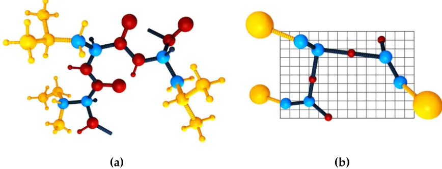
the polypeptide chain representation is reduced to up to four unified atoms per residue (see Figure
117
1). These interaction centers represent lattice-confined C-alpha atoms, C-beta atoms, the united side
118
chain pseudo-atom and, additionally, pseudo-atoms representing geometrical centers of peptide
119
bonds needed to define the hydrogen pseudo-bond. An example of a polypeptide chain in CABS
120
representation is presented in Figure 1, b. Even though the restriction of the C-alpha trace to the
121
underlying low spacing (0.61 Å [27]) cubic lattice may appear to be a drastic simplification, it is not.
122
Allowing small fluctuations of the C-alpha – C-alpha distance enables hundreds of possible
123
orientations of this pseudo bond, and thereby the resulting model chains do not show any noticeable
124
directional biases. Furthermore, the averaged resolution of the C-alpha traces is acceptable and below
125
0.5 Å [27]. Additionally, the lattice representation enables pre-calculation of local moves and
126
corresponding changes of interactions, leading to a few times faster simulations in comparison with
127
otherwise equivalent continuous space CG models [11].
128
(a)
(b)
Figure 1. A three-residue protein fragment in: all-atom (a) and CABS model (b) representation. The
129
spheres represent atoms: blue, C-alpha and C-beta atoms (the same in both representations); yellow,
130
side chain atoms (one atom in CABS); red, atoms involved in the peptide bond (one
pseudo-131
atom in CABS placed in the geometric center of the peptide bond. A single slice (layer) of the lattice
132
that confines the C-alpha trace in the CABS model is also presented.
133
The CABS model uses a knowledge-based statistical force field that consists of generic,
sequence-134
independent interaction terms that favor protein-like conformations, and sequence-dependent
135
interaction terms that determine some structural details [11,27,76]. The generic force field terms are
136
derived from general features of polypeptide chains that result in protein-like behavior of the model
137
chains. They account for properties of protein chains such as local stiffness, their biases toward
138
secondary structures and packing compactness. The residue-residue interaction terms are derived
139
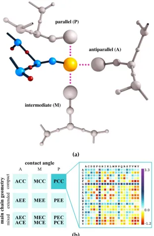
from contact geometry statistics (illustrated in Figure 2, a). The resulting force field takes a form of a
140
precomputed matrix of contact pseudo-energies, presented schematically in Figure 2, b. Additionally,
141
to allow successful modeling of membrane proteins the CABS force field can be extended by
142
introducing effective dielectric constant terms [77].
143
The main difference between CABS and other statistical force fields used in CG models of similar
144
resolution [11] is the context and orientation dependence of side chain interaction pseudo-energy that
145
encodes characteristic patterns observed in globular proteins. For instance, the oppositely charged
side chains in single globules mostly contact in an almost parallel fashion (usually on the surface of
147
a globule), while the antiparallel contacts (usually in the buried regions of the protein globule) are
148
very rare. Therefore, in the context dependent force field these antiparallel contacts of oppositely
149
charged residues are treated as repulsive. This way, the CABS force field implicitly incorporates
150
information on the complicated interaction patterns with the solvent (via contact statistics) and its
151
entropic contribution to system thermodynamics [11,27].
152
(a)
(b)
Figure 2. Key elements of a residue-residue interaction term in the CABS model force field. Panel (a)
153
shows three examples of contact geometries in CABS representation – parallel (P), antiparallel (A)
154
and intermediate (M) – used to derive contact statistics from experimental data. Panel (b) shows an
155
example matrix of contact energies which depend on the geometry of the contacting pair, main chain
156
geometry (compact (C) or extended (E)) for both amino acids (left part of the panel), and also on the
157
amino acid identities (right part of the panel, the amino acids are represented using the one-letter
158
code). The PCC matrix is presented which shows interaction energies between residues being in
159
parallel orientation (P), where one residue belongs to a compact type of structure (C) and the second
160
one as well (C).
Using the mean-force force field derived from folded proteins to simulations of less-structured
162
systems raises justified questions about the validity of this approach in studies of the non-native
163
regions of conformational space. The folding events observed in simulations performed using the
164
CABS force field are consistent with both the experimental data and all-atom MD simulations
165
[48,50,78,79]. Thus, it is hypothesized that unstructured (unfolded, partially unfolded or intrinsically
166
disordered) proteins to a significant extent share similar stabilizing interaction patterns with the
167
patterns observed for their well-structured counterparts [80,81].
168
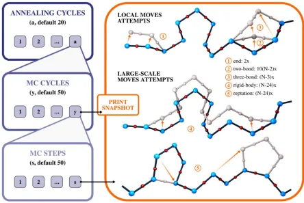
The CABS method uses the MC asymmetric Metropolis sampling scheme that governs a set of
169
local motions as well as multi-residue, small distance moves of the C-alpha atoms (see Figure 3). The
170
method uses a replica exchange algorithm with simulated annealing to enhance the sampling of
171
conformational states. The simulation is organized as a set of nested loops, in which the s number of
172
MC steps are organized into the y number of MC cycles, and these in the a number of annealing
173
cycles. Each of the MC steps consists of a per-set number of attempts to perform each of the five
174
standard precomputed moves. The available motions and the details of implementation of the
175
sampling scheme are presented in Figure 3.
176
177
178
Figure 3. Sampling scheme of the CABS model. Blue panels show implementation details of Monte
179
Carlo (MC) iterations (loops). The orange panel shows all motions that may be performed in a single
180
MC step. The simulation is organized as a set of nested loops, in which the s number of MC steps is
181
organized into the y number of cycles, and these in a annealing cycles (number of a, y or s cycles can
182
be controlled by the user in CABS-flex and CABS-dock standalone packages [70]). In the orange panel,
183
numbers 1 to 5 denote the available moves, presented together with the number of attempts to
184
perform a move in each of the MC steps. The resulting trajectory is comprised of simulation snapshots
185
saved at the end of each MC cycle.
186
The combination of the key features of CABS – its representation, force field and the scale of the
187
movements used in the MC scheme – makes it suitable for the investigation of protein
pseudo-188
dynamics. As mentioned above, the fine-grained lattice improves sampling efficiency, achieving
189
effective timescales of milliseconds. As compared with MD simulations this is a considerably broader
190
time range. The chosen micro-motions allow (via accumulation over simulation steps) cooperative,
191
large-large scale motions. The ensemble of structures produced by the CABS method resembles a
dynamic ensemble averaged over the effective timescale. Due to the nature of the method the picture
193
of local dynamics is distorted (on the level of local moves); however, it may be argued that mesoscale
194
pseudo-dynamics (such as conformational transitions or loop movements) recovers the realistic
195
picture of protein motions averaged over time.
196
The long-time dynamics picture is more accurate, but the timescale is not a priori defined. It
197
should be also noted that the time scaling of CABS dynamics at various temperatures can be different,
198
due to hidden entropic contributions in the force field, accounting for implicit solvent effects and
199
multi-body interactions encoded in the statistical force field. Nevertheless, the effective timescale of
200
MC dynamics can be approximately identified by comparison with MD trajectories from sufficiently
201
long simulations. This comparison was thoroughly discussed previously, and the results were
202
compared to MD results [67] and NMR ensembles [69].
203
The CABS model is presently used as a simulation engine of a few multiscale modeling tools
204
that merge CABS with models reconstruction to all-atom resolution. Those include the CABS-dock
205
method for flexible protein-peptide docking (available as a web server [60] at
206
http://biocomp.chem.uw.edu.pl/CABSdock and a standalone application at
207
https://bitbucket.org/lcbio/cabsdock/). In comparison to other protein-peptide docking tools,
208
reviewed recently [82], CABS-dock offers a unique opportunity for modeling large-scale
209
rearrangements of protein receptor structure during on-the-fly docking of fully flexible peptides.
210
CABS-flex, another CABS-based tool, enables fast simulations of protein flexibility (available as a web
211
server [71] at http://biocomp.chem.uw.edu.pl/CABSflex and a standalone application [70] at
212
https://bitbucket.org/lcbio/cabsflex/). CABS-flex has been also incorporated as the module in the
213
Aggrescan3D method for prediction of protein aggregation properties (available as a web server [83]
214
at http://biocomp.chem.uw.edu.pl/A3D and a standalone application at
215
https://bitbucket.org/lcbio/aggrescan3d). By using CABS-flex predictions, Aggrescan3D enables
216
predicting the impact of protein conformational fluctuations on aggregation properties. Finally, the
217
CABS model is used in the CABS-fold method for protein structure prediction: in the de novo fashion
218
(from an amino acid sequence only), guided by user-provided templates or user-provided distance
219
restraints (available as a web server [56] at http://biocomp.chem.uw.edu.pl/CABSfold/).
220
3. CABS applications to simulation of disordered or unfolded proteins
221
In this section, we review CABS applications to simulations of protein-peptide binding (section
222
3.1) and folding of globular proteins (section 3.2). We briefly discuss modeling results for the binding
223
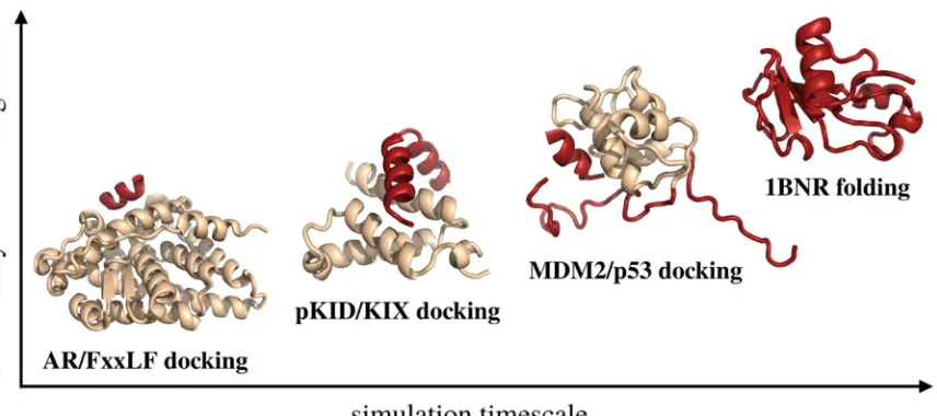
of three protein-peptide systems and protein folding of one protein system. Figure 4 shows native
224
conformations of these systems, determined by X-ray crystallography or NMR. In the figure, they are
225
arranged according to the size of a fully flexible fragment of the modeled system, effective timescales
226
required for a meaningful simulation of their motions and thus the modeling difficulty: (1) modeling
227
of FxxLF motif peptide docking to an androgen receptor (AR), (2) investigation of binding and folding
228
of an unstructured pKID protein to KIX protein, (3) modeling of p53-derived peptide docking to the
229
MDM2 protein receptor with partially unstructured regions and (4) simulation of the de novo folding
230
of barnase. The simulations were performed using the CABS-dock method for protein-peptide
231
docking [60] and CABS-flex methodology [70,71] that enable running de novo folding simulations.
233
Figure 4. Presentation of the modeling cases discussed in this work. The modeled systems are
234
arranged according to the size of the fully flexible fragment of the modeled system and the effective
235
timescales required to observe their motions. The regions of the systems that were modeled as fully
236
flexible are marked with red, while the regions in which backbone fluctuations were limited to 1 Å
237
RMSD with beige.
238
3.1 Protein-peptide binding
239
The CABS-dock method has been extensively tested using the PeptiDB benchmark set of
protein-240
peptide complexes [60,63,84]. One of the benchmark cases is the androgen receptor ligand binding
241
domain (AR) in complex with a peptide with the FxxLF motif [85] (PDB code: 1T7R). To further
242
analyze the interaction details of this complex, we performed blind global docking (using no
243
knowledge about the binding site and peptide conformation) using CABS-dock [60]. As the input we
244
used information on peptide sequence (incorporating the FxxLF motif: SSRFESLFAGEKESR),
245
peptide secondary structure information assigned by the DSSP method [86] and the structure of the
246
AR protein receptor. In this docking study, the peptide structure has been simulated as fully flexible,
247
while fluctuations of the protein receptor have been limited to small backbone movements around
248
the input structure (around 1 Å). The docking simulation started from random peptide conformations
249
placed in random positions around the receptor structure. During simulation, the peptide remained
250
unstructured until it was bound to the receptor binding site (Figure 5, a). The docking simulations
251
provided a set of high quality models – the best model was characterized by a peptide-RMSD
(root-252
mean-square deviation) value of 1.97 Å – and contact maps in strong agreement with the
253
experimental data. As expected from the experimentally obtained structures and sequence analysis
254
[85] the FxxLF interaction motif residues were most frequently involved in stabilizing hydrophobic
255
interactions with the receptor. These high frequency contacts are clearly visible in Figure 5, a.
257
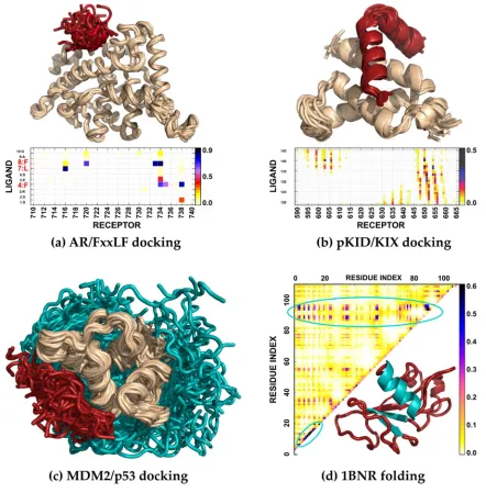
Figure 5. Case studies of modeling disordered or unfolded structures of proteins with CABS-based
258
tools. In the figures, red or cyan marks structure fragments simulated as fully flexible (cyan was used
259
to mark regions of interest discussed in the text), while beige marks regions whose motions were
260
confined to small backbone movements (around 1 Å from the input structure). (a) Modeling of the
261
dynamics of a flexible peptide representing the FxxLF motif in the proximity of the binding site of AR
262
protein together with an averaged contact map showing frequency of residue-residue contacts during
263
the docking simulation. (b) Modeling of coupled folding and binding of the disordered pKID to the
264
KIX domain [61]; the map presents the frequency of contacts of near-native conformations obtained
265
in the simulation. (c) Modeling of p53 peptide binding to the MDM2 receptor [62], which includes
266
fully flexible regions of the protein receptor (shown in cyan) interacting with a fully flexible peptide
267
(shown in red). (d) Modeling of barnase folding [50] in the de novo fashion (using no knowledge about
268
the structure); the map is a residue-residue contact map showing relative contact frequencies in
269
denaturing conditions; the protein fragments that form the folding nucleation site are colored in cyan
270
in the presented folded structure of barnase.
271
The study of the pKID/KIX system [61] involved performing a folding simulation of an
272
intrinsically disordered protein (pKID) and its binding to a well-structured KIX receptor (Figure 5,
273
b). According to the experimental studies, the pKID structure is disordered in its unbound form with
274
a slight propensity toward a helix. In the complex with the KIX protein, pKID adopts a characteristic
conformation of two perpendicular helices that wrap around the receptor. However, most simulation
276
results for the coupled folding and binding of this system published prior to the CABS-based study
277
used models which biased pKID toward its native conformation (see the discussion in [61]). Using
278
our method for studying this system enabled fully flexible treatment of the pKID protein and resulted
279
in the formulation of a well-supported by the NMR data hypothetical explanation of the binding
280
mechanism involving two encounter complexes. The obtained models presented high fractions of
281
native contacts and allowed identification of residues essential for the binding and stabilization of
282
the complex.
283
In the simulation of MDM2/p53 binding [62], the most challenging task was to adequately model
284
the flexibility of the relatively long, unstructured regions of the protein receptor in addition to the
285
fully flexible peptide [62,87] (Figure 5, c). To provide a detailed insight into MDM2/p53 binding, we
286
performed CABS-dock simulations and captured system behavior in agreement with the
287
experimental data [62]. During the simulation, the flexible N- and C- terminal MDM2 fragments
288
remained significantly disordered. The best resulting model was characterized by a peptide-RMSD
289
value of 2.76 Å and 54% of the native contacts while the top ranked model by 3.74 Å and 60%,
290
respectively. During simulations, we observed ensembles of models in which the peptide adopted
291
different conformations loosely bound to the binding site and models in which the N-terminal highly
292
flexible MDM2 fragment was interacting with the binding site. These findings are in agreement with
293
the experimental data suggesting that p53-MDM2 binding is affected by significant rearrangements
294
of the N-terminal MDM2 fragment (see discussion in [62]).
295
3.2 Folding and flexibility of globular proteins
296
The CABS model has been applied to de novo simulations of protein folding (using no
297
knowledge about the protein structure) for several model systems that have been extensively studied
298
by experiment and simulation tools. Those studies include barnase [49,50], chymotrypsin inhibitor
299
[49,50], B1 domain of protein G [48,49], B domain of protein A [51] and others [49,52]. The CABS
300
modeling protocol was also extended to enable studies of the chaperonin effect on the folding
301
mechanism [53]. The obtained pictures, which covered protein dynamics from highly denatured
302
states to ensembles close to the folded states, agreed well with available experimental data.
303
For example, simulation of barnase folding resulted in the adequate reproduction of the folding
304
pathway in strong agreement with NMR data for denatured states and phi-value analysis [50]. The
305
performed simulations show that barnase folding starts with developing a folding nucleation site
306
that consists of protein fragments corresponding to two strands of a beta sheet and one of the helices
307
in the folded structure (presented in Figure 5, d). In addition, the characteristic patterns of
308
hydrophobic interactions that are crucial for the initiation and sustenance of folding are in agreement
309
with the experimental data ( see discussion in [50], the contact map resulting from these simulations
310
is presented in Figure 5, d).
311
4. Conclusions
312
The presented case studies review the applications of the CABS model in simulations of
313
disordered or unfolded protein states. As discussed, the method succeeded in capturing the
314
experimentally determined features of the investigated systems, such as binding site localization, key
315
contacts, peptide hot-spot areas, distinctive conformational states of the system, transient encounter
316
complexes and intermediate states in protein folding [48,50,61,62]. Additionally, CABS enables an
317
investigation of fluctuations of globular proteins around the native (input) structure [67–71].
318
It is, however, noteworthy that statistical force fields suffer from inherent limitations, depending
319
on the chosen method of derivation. The transferability of these methods may be limited as they are
320
applicable always to a certain subset of proteins. Therefore, the performance of knowledge-based
321
approaches may be poor for rare or atypical structures, for which appropriate statistics of contact
322
patterns could not be collected – for example beta-helical proteins. It should also be noted that
323
interactions with solvent are averaged and treated implicitly, which may lead to significant
discrepancies if the method is applied to non-standard solvent conditions (such as extreme pH
325
values).
326
One of the most challenging tasks in modeling protein systems is the effective incorporation of
327
sparse experimental data to drive the modeling procedure. In the CABS model, the experimental data
328
may be readily introduced into the simulation as geometry distance restraints and weighted
329
according to their certainty. A thorough discussion of this possibility is presented in the
330
documentation of CABS-based tools for the fast modeling of protein flexibility and protein-peptide
331
docking [64,70,71]. On a similar basis, CABS simulations can be guided by computational predictions
332
from other sources or integrated with other modeling tools of various resolution. Therefore, the CABS
333
model can be incorporated into integrative modeling pipelines that would benefit from its effective
334
sampling scheme. The recently published standalone application and web server tools are available
335
for integration with external pipelines (access links are presented in the last paragraph of Section 2).
336
337
Author Contributions: S.K. and A.K. conceptualized this review. It was written by all the authors.
338
Funding: This research was funded by NCN Poland, grant number MAESTRO2014/14/A/ST6/00088.
339
Conflicts of Interest: The authors declare no conflict of interest.
340
Abbreviations
341
CABS Cα, Cβ, Side chain model
MC Monte Carlo
NMR nuclear magnetic resonance
MD molecular dynamics
CG coarse-grained
AR DSSP RMSD
androgen receptor
dictionary of protein secondary structure root-mean-square deviation of atomic positions PDB
CASP
Protein Data Bank
Critical Assessment of protein Structure Prediction
References
342
1. Dishman, A. F.; Volkman, B. F. Unfolding the Mysteries of Protein Metamorphosis. ACS Chem.
343
Biol. 2018, 13, 1438–1446, doi:10.1021/acschembio.8b00276.
344
2. Uversky, V. N. Dancing protein clouds: The strange biology and chaotic physics of
345
intrinsically disordered proteins. J. Biol. Chem. 2016, 291, 6681–8, doi:10.1074/jbc.R115.685859.
346
3. Wright, P. E.; Dyson, H. J. Intrinsically disordered proteins in cellular signalling and
347
regulation. Nat. Rev. Mol. Cell Biol. 2015, 16, 18–29, doi:10.1038/nrm3920.
348
4. Henzler-Wildman, K.; Kern, D. Dynamic personalities of proteins. Nature 2007, 450, 964–72,
349
doi:10.1038/nature06522.
350
5. Vendruscolo, M.; Dobson, C. M. Dynamic visions of enzymatic reactions. Science (80-. ). 2006,
351
313, 1586–7, doi:10.1126/science.1132851.
352
6. Wei, G.; Xi, W.; Nussinov, R.; Ma, B. Protein Ensembles: How Does Nature Harness
353
Thermodynamic Fluctuations for Life? the Diverse Functional Roles of Conformational
354
Ensembles in the Cell. Chem. Rev. 2016, 116, 6516–51, doi:10.1021/acs.chemrev.5b00562.
355
7. Best, R. B. Computational and theoretical advances in studies of intrinsically disordered
356
proteins. Curr. Opin. Struct. Biol. 2017, 42, 147–154, doi:10.1016/j.sbi.2017.01.006.
357
8. Kay, L. E. NMR studies of protein structure and dynamics. J. Magn. Reson. 2011, 213, 477–491,
358
doi:10.1016/J.JMR.2011.09.009.
9. Robustelli, P.; Piana, S.; Shaw, D. E. Developing a molecular dynamics force field for both
360
folded and disordered protein states. Proc. Natl. Acad. Sci. 2018, 115, E4758–E4766,
361
doi:10.1073/pnas.1800690115.
362
10. Bowman, G. R.; Voelz, V. A.; Pande, V. S. Taming the complexity of protein folding. Curr.
363
Opin. Struct. Biol. 2011, 21, 4–11, doi:10.1016/j.sbi.2010.10.006.
364
11. Kmiecik, S.; Gront, D.; Kolinski, M.; Wieteska, L.; Dawid, A. E.; Kolinski, A. Coarse-Grained
365
Protein Models and Their Applications. Chem. Rev. 2016, 116, 7898–7936,
366
doi:10.1021/acs.chemrev.6b00163.
367
12. Zhang, Z.; Chan, H. S. Competition between native topology and nonnative interactions in
368
simple and complex folding kinetics of natural and designed proteins. Proc. Natl. Acad. Sci.
369
2010, 107, 2920–5, doi:10.1073/pnas.0911844107.
370
13. Shan, B.; Eliezer, D.; Raleigh, D. The unfolded state of the C-terminal domain of the ribosomal
371
protein L9 contains both native and non-native structure. Biochemistry 2009, 48, 4707–19,
372
doi:10.1021/bi802299j.
373
14. Rothwarf, D. M.; Scheraga, H. A. Role of non-native aromatic and hydrophobic interactions
374
in the folding of hen egg white lysozyme. Biochemistry 1996, 35, 13797–807,
375
doi:10.1021/bi9608119.
376
15. Cavalli, A.; Montalvao, R. W.; Vendruscolo, M. Using chemical shifts to determine structural
377
changes in proteins upon complex formation. J. Phys. Chem. B 2011, 115, 9491–4,
378
doi:10.1021/jp202647q.
379
16. Fu, B.; Kukic, P.; Camilloni, C.; Vendruscolo, M. MD Simulations of Intrinsically Disordered
380
Proteins with Replica-Averaged Chemical Shift Restraints. Biophys. J. 2014, 106, 481a,
381
doi:10.1016/j.bpj.2013.11.2714.
382
17. Greener, J. G.; Filippis, I.; Sternberg, M. J. E. Predicting Protein Dynamics and Allostery Using
383
Multi-Protein Atomic Distance Constraints. Structure 2017, 25, 546–558,
384
doi:10.1016/j.str.2017.01.008.
385
18. Klepeis, J. L.; Lindorff-Larsen, K.; Dror, R. O.; Shaw, D. E. Long-timescale molecular dynamics
386
simulations of protein structure and function. Curr. Opin. Struct. Biol. 2009, 19, 120–7,
387
doi:10.1016/j.sbi.2009.03.004.
388
19. Bernardi, R. C.; Melo, M. C. R.; Schulten, K. Enhanced sampling techniques in molecular
389
dynamics simulations of biological systems. Biochim. Biophys. Acta - Gen. Subj. 2015, 1850, 872–
390
877, doi:10.1016/j.bbagen.2014.10.019.
391
20. Shukla, D.; Hernández, C. X.; Weber, J. K.; Pande, V. S. Markov state models provide insights
392
into dynamic modulation of protein function. Acc. Chem. Res. 2015, 48, 414–22,
393
doi:10.1021/ar5002999.
394
21. Kolinski, A. Toward more efficient simulations of slow processes in large biomolecular
395
systems: Comment on “Ligand diffusion in proteins via enhanced sampling in molecular
396
dynamics” by Jakub Rydzewski and Wieslaw Nowak. Phys. Life Rev. 2017, 22–23, 75–76,
397
doi:10.1016/j.plrev.2017.07.003.
398
22. Rydzewski, J.; Nowak, W. Ligand diffusion in proteins via enhanced sampling in molecular
399
dynamics. Phys. Life Rev. 2017, 22–23, 82–84, doi:10.1016/j.plrev.2017.03.003.
400
23. Maximova, T.; Moffatt, R.; Ma, B.; Nussinov, R.; Shehu, A. Principles and Overview of
401
Sampling Methods for Modeling Macromolecular Structure and Dynamics. PLoS Comput. Biol.
2016, 12, e1004619, doi:10.1371/journal.pcbi.1004619.
403
24. Hatherley, R.; Brown, D. K.; Glenister, M.; Bishop, Ö. T. PRIMO: An interactive homology
404
modeling pipeline. PLoS One 2016, 11, e0166698, doi:10.1371/journal.pone.0166698.
405
25. Das, R.; Baker, D. Macromolecular Modeling with Rosetta. Annu. Rev. Biochem. 2008, 77, 363–
406
382, doi:10.1146/annurev.biochem.77.062906.171838.
407
26. Czaplewski, C.; Karczyńska, A.; Sieradzan, A. K.; Liwo, A. UNRES server for physics-based
408
coarse-grained simulations and prediction of protein structure, dynamics and
409
thermodynamics. Nucleic Acids Res. 2018, 46, W304–W309, doi:10.1093/nar/gky328.
410
27. Kolinski, A. Protein modeling and structure prediction with a reduced representation. Acta
411
Biochim. Pol. 2004, 51, 349–371, doi:035001349.
412
28. Davtyan, A.; Schafer, N. P.; Zheng, W.; Clementi, C.; Wolynes, P. G.; Papoian, G. A.
AWSEM-413
MD: Protein structure prediction using coarse-grained physical potentials and
414
bioinformatically based local structure biasing. J. Phys. Chem. B 2012, 116, 8494–503,
415
doi:10.1021/jp212541y.
416
29. Marrink, S. J.; Tieleman, D. P. Perspective on the Martini model. Chem. Soc. Rev. 2013, 42, 6801,
417
doi:10.1039/c3cs60093a.
418
30. Pasi, M.; Lavery, R.; Ceres, N. PaLaCe: A coarse-grain protein model for studying mechanical
419
properties. J. Chem. Theory Comput. 2013, 9, 785–93, doi:10.1021/ct3007925.
420
31. Basdevant, N.; Borgis, D.; Ha-Duong, T. Modeling protein-protein recognition in solution
421
using the coarse-grained force field SCORPION. J. Chem. Theory Comput. 2013, 9, 803–13,
422
doi:10.1021/ct300943w.
423
32. Dawid, A. E.; Gront, D.; Kolinski, A. SURPASS Low-Resolution Coarse-Grained Protein
424
Modeling. J. Chem. Theory Comput. 2017, 13, 5766–5779, doi:10.1021/acs.jctc.7b00642.
425
33. Lopes, P. E. M.; Guvench, O.; MacKerell, A. D. Current Status of Protein Force Fields for
426
Molecular Dynamics Simulations. In; Humana Press, New York, NY, 2015; pp. 47–71.
427
34. Cossio, P.; Granata, D.; Laio, A.; Seno, F.; Trovato, A. A simple and efficient statistical potential
428
for scoring ensembles of protein structures. Sci. Rep. 2012, 2, 351, doi:10.1038/srep00351.
429
35. Tanaka, S.; Scheraga, H. A. Medium- and Long-Range Interaction Parameters between Amino
430
Acids for Predicting Three-Dimensional Structures of Proteins. Macromolecules 1976, 9, 945–
431
950, doi:10.1021/ma60054a013.
432
36. Tsai, J.; Bonneau, R.; Morozov, A. V.; Kuhlman, B.; Rohl, C. A.; Baker, D. An improved protein
433
decoy set for testing energy functions for protein structure prediction. Proteins Struct. Funct.
434
Genet. 2003, 53, 76–87, doi:10.1002/prot.10454.
435
37. Shen, M.; Sali, A. Statistical potential for assessment and prediction of protein structures.
436
Protein Sci. 2006, 15, 2507–2524, doi:10.1110/ps.062416606.
437
38. Zhou, H.; Skolnick, J. GOAP: A Generalized Orientation-Dependent, All-Atom Statistical
438
Potential for Protein Structure Prediction. Biophys. J. 2011, 101, 2043–2052,
439
doi:10.1016/J.BPJ.2011.09.012.
440
39. Xu, D.; Zhang, Y. Ab initio protein structure assembly using continuous structure fragments
441
and optimized knowledge-based force field. Proteins Struct. Funct. Bioinforma. 2012, 80, 1715–
442
1735, doi:10.1002/prot.24065.
443
40. Woetzel, N.; Karakaş, M.; Staritzbichler, R.; Müller, R.; Weiner, B. E.; Meiler, J. BCL::Score—
444
Knowledge Based Energy Potentials for Ranking Protein Models Represented by Idealized
Secondary Structure Elements. PLoS One 2012, 7, e49242, doi:10.1371/journal.pone.0049242.
446
41. Ovchinnikov, S.; Park, H.; Kim, D. E.; Liu, Y.; Wang, R. Y.-R.; Baker, D. Structure prediction
447
using sparse simulated NOE restraints with Rosetta in CASP11. Proteins Struct. Funct.
448
Bioinforma. 2016, 84, 181–188, doi:10.1002/prot.25006.
449
42. Ovchinnikov, S.; Kim, D. E.; Wang, R. Y.-R.; Liu, Y.; DiMaio, F.; Baker, D. Improved de novo
450
structure prediction in CASP11 by incorporating coevolution information into Rosetta.
451
Proteins Struct. Funct. Bioinforma. 2016, 84, 67–75, doi:10.1002/prot.24974.
452
43. Hirst, S. J.; Alexander, N.; Mchaourab, H. S.; Meiler, J. RosettaEPR: An integrated tool for
453
protein structure determination from sparse EPR data. J. Struct. Biol. 2011, 173, 506–514,
454
doi:10.1016/J.JSB.2010.10.013.
455
44. Yang, J.; Zhang, W.; He, B.; Walker, S. E.; Zhang, H.; Govindarajoo, B.; Virtanen, J.; Xue, Z.;
456
Shen, H. Bin; Zhang, Y. Template-based protein structure prediction in CASP11 and retrospect
457
of I-TASSER in the last decade. Proteins 2016, 84, 233–246, doi:10.1002/prot.24918.
458
45. Russel, D.; Lasker, K.; Webb, B.; Velázquez-Muriel, J.; Tjioe, E.; Schneidman-Duhovny, D.;
459
Peterson, B.; Sali, A. Putting the Pieces Together: Integrative Modeling Platform Software for
460
Structure Determination of Macromolecular Assemblies. PLoS Biol. 2012, 10, e1001244,
461
doi:10.1371/journal.pbio.1001244.
462
46. Rodrigues, J. P. G. L. M.; Bonvin, A. M. J. J. Integrative computational modeling of protein
463
interactions. FEBS J. 2014, 281, 1988–2003, doi:10.1111/febs.12771.
464
47. Kar, P.; Feig, M. Recent advances in transferable coarse-grained modeling of proteins. Adv.
465
Protein Chem. Struct. Biol. 2014, 96, 143–180, doi:10.1016/bs.apcsb.2014.06.005.
466
48. Kmiecik, S.; Kolinski, A. Folding pathway of the B1 domain of protein G explored by
467
multiscale modeling. Biophys. J. 2008, 94, 726–736, doi:10.1529/biophysj.107.116095.
468
49. Kolinski, A. Multiscale approaches to protein modeling: Structure prediction, dynamics,
469
thermodynamics and macromolecular assemblies. In Multiscale Approaches to Protein Modeling:
470
Structure Prediction, Dynamics, Thermodynamics and Macromolecular Assemblies; Kolinski, A.,
471
Ed.; Springer: New York, 2011; pp. 1–355 ISBN 9781441968890.
472
50. Kmiecik, S.; Kolinski, A. Characterization of protein-folding pathways by reduced-space
473
modeling. Proc. Natl. Acad. Sci. 2007, 104, 12330–12335, doi:10.1073/pnas.0702265104.
474
51. Kmiecik, S.; Gront, D.; Kouza, M.; Kolinski, A. From coarse-grained to atomic-level
475
characterization of protein dynamics: Transition state for the folding of B domain of protein
476
A. J. Phys. Chem. B 2012, 116, 7026–7032, doi:10.1021/jp301720w.
477
52. Kmiecik, S.; Kurcinski, M.; Rutkowska, A.; Gront, D.; Kolinski, A. Denatured proteins and
478
early folding intermediates simulated in a reduced conformational space. Acta Biochim. Pol.
479
2006, 53, 131–143, doi:10.1038/nphys1759.
480
53. Kmiecik, S.; Kolinski, A. Simulation of chaperonin effect on protein folding: A shift from
481
nucleation - Condensation to framework mechanism. J. Am. Chem. Soc. 2011, 133, 10283–10289,
482
doi:10.1021/ja203275f.
483
54. Jamroz, M.; Kolinski, A.; Kmiecik, S. Protocols for efficient simulations of long-time protein
484
dynamics using coarse-grained CABS model. Methods Mol. Biol. 2014, 1137, 235–250,
485
doi:10.1007/978-1-4939-0366-5_16.
486
55. Wabik, J.; Kmiecik, S.; Gront, D.; Kouza, M.; Koliński, A. Combining coarse-grained protein
487
models with replica-exchange all-atom molecular dynamics. Int. J. Mol. Sci. 2013, 14, 9893–
9905, doi:10.3390/ijms14059893.
489
56. Blaszczyk, M.; Jamroz, M.; Kmiecik, S.; Kolinski, A. CABS-fold: Server for the de novo and
490
consensus-based prediction of protein structure. Nucleic Acids Res. 2013, 41, W406-11,
491
doi:10.1093/nar/gkt462.
492
57. Kmiecik, S.; Jamroz, M.; Kolinski, M. Structure prediction of the second extracellular loop in
493
G-protein-coupled receptors. Biophys. J. 2014, 106, 2408–2416, doi:10.1016/j.bpj.2014.04.022.
494
58. Koliński, A.; Bujnicki, J. M. Generalized protein structure prediction based on combination of
495
fold-recognition with de novo folding and evaluation of models. Proteins Struct. Funct. Genet.
496
2005, 61, 84–90, doi:10.1002/prot.20723.
497
59. Jamroz, M.; Kolinski, A. Modeling of loops in proteins: A multi-method approach. BMC
498
Struct. Biol. 2010, 10, doi:10.1186/1472-6807-10-5.
499
60. Kurcinski, M.; Jamroz, M.; Blaszczyk, M.; Kolinski, A.; Kmiecik, S. CABS-dock web server for
500
the flexible docking of peptides to proteins without prior knowledge of the binding site.
501
Nucleic Acids Res. 2015, 43, W419–W424, doi:10.1093/nar/gkv456.
502
61. Kurcinski, M.; Kolinski, A.; Kmiecik, S. Mechanism of folding and binding of an intrinsically
503
disordered protein as revealed by ab initio simulations. J. Chem. Theory Comput. 2014, 10, 2224–
504
2231, doi:10.1021/ct500287c.
505
62. Ciemny, M. P.; Debinski, A.; Paczkowska, M.; Kolinski, A.; Kurcinski, M.; Kmiecik, S.
Protein-506
peptide molecular docking with large-scale conformational changes: The p53-MDM2
507
interaction. Sci. Rep. 2016, 6, doi:10.1038/srep37532.
508
63. Blaszczyk, M.; Kurcinski, M.; Kouza, M.; Wieteska, L.; Debinski, A.; Kolinski, A.; Kmiecik, S.
509
Modeling of protein-peptide interactions using the CABS-dock web server for binding site
510
search and flexible docking. Methods 2016, 93, 72–83, doi:10.1016/j.ymeth.2015.07.004.
511
64. Ciemny, M.; Kurcinski, M.; Kozak, K.; Kolinski, A.; Kmiecik, S. Highly flexible protein-peptide
512
docking using cabs-dock. Methods Mol. Biol. 2017, 1561, 69–94,
doi:10.1007/978-1-4939-6798-513
8_6.
514
65. Blaszczyk, M.; Ciemny, M. P.; Kolinski, A.; Kurcinski, M.; Kmiecik, S. Protein–peptide docking
515
using CABS-dock and contact information. Brief. Bioinform. 2018, bby080,
516
doi:10.1093/bib/bby080.
517
66. Ciemny, M. P.; Kurcinski, M.; Blaszczyk, M.; Kolinski, A.; Kmiecik, S. Modeling
EphB4-518
EphrinB2 protein-protein interaction using flexible docking of a short linear motif. Biomed.
519
Eng. Online 2017, 16, 71, doi:10.1186/s12938-017-0362-7.
520
67. Jamroz, M.; Orozco, M.; Kolinski, A.; Kmiecik, S. Consistent view of protein fluctuations from
521
all-atom molecular dynamics and coarse-grained dynamics with knowledge-based
force-522
field. J. Chem. Theory Comput. 2013, 9, 119–125, doi:10.1021/ct300854w.
523
68. Jamroz, M.; Kolinski, A.; Kmiecik, S. CABS-flex: Server for fast simulation of protein structure
524
fluctuations. Nucleic Acids Res. 2013, 41, W427-431, doi:10.1093/nar/gkt332.
525
69. Jamroz, M.; Kolinski, A.; Kmiecik, S. CABS-flex predictions of protein flexibility compared
526
with NMR ensembles. Bioinformatics 2014, 30, 2150–2154, doi:10.1093/bioinformatics/btu184.
527
70. Kurcinski, M.; Oleniecki, T.; Ciemny, P. M.; Kuriata, A.; Kolinski, A.; Kmiecik, S. CABS-flex
528
standalone: a simulation environment for fast modeling of protein flexibility. Bioinformatics
529
2018, bty685, doi:10.1093/bioinformatics/bty685.
530
71. Kuriata, A.; Gierut, A. M.; Oleniecki, T.; Ciemny, M. P.; Kolinski, A.; Kurcinski, M.; Kmiecik,
S. CABS-flex 2.0: A web server for fast simulations of flexibility of protein structures. Nucleic
532
Acids Res. 2018, 46, W338–W343, doi:10.1093/nar/gky356.
533
72. Eswar, N.; John, B.; Mirkovic, N.; Fiser, A.; Ilyin, V. A.; Pieper, U.; Stuart, A. C.; Marti-Renom,
534
M. A.; Madhusudhan, M. S.; Yerkovich, B.; Sali, A. Tools for comparative protein structure
535
modeling and analysis. Nucleic Acids Res. 2003, 31, 3375–80.
536
73. Gront, D.; Kmiecik, S.; Kolinski, A. Backbone building from quadrilaterals: A fast and accurate
537
algorithm for protein backbone reconstruction from alpha carbon coordinates. J. Comput.
538
Chem. 2007, 28, 1593–1597, doi:10.1002/jcc.20624.
539
74. Canutescu, A. A.; Shelenkov, A. A.; Dunbrack, R. L. A graph-theory algorithm for rapid
540
protein side-chain prediction. Protein Sci. 2003, 12, 2001–2014, doi:10.1110/ps.03154503.
541
75. Gront, D.; Kmiecik, S.; Blaszczyk, M.; Ekonomiuk, D.; Koliński, A. Optimization of protein
542
models. Wiley Interdiscip. Rev. Comput. Mol. Sci. 2012, 2, 479–493, doi:10.1002/wcms.1090.
543
76. Kmiecik, S.; Kolinski, A. One-dimensional structural properties of proteins in the
coarse-544
grained cabs model. Methods Mol. Biol. 2017, 1484, 83–113, doi:10.1007/978-1-4939-6406-2_8.
545
77. Pulawski, W.; Jamroz, M.; Kolinski, M.; Kolinski, A.; Kmiecik, S. Coarse-grained simulations
546
of membrane insertion and folding of small helical proteins using the CABS model. J. Chem.
547
Inf. Model. 2016, 56, 2207–2215, doi:10.1021/acs.jcim.6b00350.
548
78. Adhikari, A. N.; Freed, K. F.; Sosnick, T. R. De novo prediction of protein folding pathways
549
and structure using the principle of sequential stabilization. Proc. Natl. Acad. Sci. 2012, 109,
550
17442–7, doi:10.1073/pnas.1209000109.
551
79. Adhikari, A. N.; Freed, K. F.; Sosnick, T. R. Simplified protein models: Predicting folding
552
pathways and structure using amino acid sequences. Phys. Rev. Lett. 2013, 111, 028103,
553
doi:10.1103/PhysRevLett.111.028103.
554
80. Konrat, R. NMR contributions to structural dynamics studies of intrinsically disordered
555
proteins. J. Magn. Reson. 2014, 241, 74–85, doi:10.1016/j.jmr.2013.11.011.
556
81. Kmiecik, S.; Wabik, J.; Kolinski, M.; Kouza, M.; Kolinski, A. Coarse-Grained Modeling of
557
Protein Dynamics. In Computational Methods to Study the Structure and Dynamics of Biomolecules;
558
Springer, Berlin, Heidelberg, 2014; Vol. 1, pp. 55–79 ISBN 978-3-642-28553-0.
559
82. Ciemny, M.; Kurcinski, M.; Kamel, K.; Kolinski, A.; Alam, N.; Schueler-Furman, O.; Kmiecik,
560
S. Protein–peptide docking: opportunities and challenges. Drug Discov. Today 2018, 23, 1530–
561
1537, doi:10.1016/j.drudis.2018.05.006.
562
83. Zambrano, R.; Jamroz, M.; Szczasiuk, A.; Pujols, J.; Kmiecik, S.; Ventura, S. AGGRESCAN3D
563
(A3D): Server for prediction of aggregation properties of protein structures. Nucleic Acids Res.
564
2015, 43, W306–W313, doi:10.1093/nar/gkv359.
565
84. London, N.; Movshovitz-Attias, D.; Schueler-Furman, O. The Structural Basis of
Peptide-566
Protein Binding Strategies. Structure 2010, 18, 188–199, doi:10.1016/J.STR.2009.11.012.
567
85. Hur, E.; Pfaff, S. J.; Sturgis Payne, E.; Grøn, H.; Buehrer, B. M.; Fletterick, R. J. Recognition and
568
accommodation at the androgen receptor coactivator binding interface. PLoS Biol. 2004, 2,
569
E274, doi:10.1371/journal.pbio.0020274.
570
86. Kabsch, W.; Sander, C. Dictionary of protein secondary structure: Pattern recognition of
571
hydrogen-bonded and geometrical features. Biopolymers 1983, 22, 2577–2637,
572
doi:10.1002/bip.360221211.
573
87. Kussie, P. H.; Gorina, S.; Marechal, V.; Elenbaas, B.; Moreau, J.; Levine, A. J.; Pavletich, N. P.
Structure of the MDM2 oncoprotein bound to the p53 tumor suppressor transactivation
575
domain. Science (80-. ). 1996, 274, 948–953, doi:10.1126/science.274.5289.948.