A kinetic study of receptor activation of the
G-protein gated
channel.
Amy Benians
A thesis submitted to the University of London for the
degree of Doctor of Philosophy.
Centre for Clinical Pharmacology,
Department of Medicine,
University College, London.
ProQuest Number: 10014370
All rights reserved
INFORMATION TO ALL USERS
The quality of this reproduction is dependent upon the quality of the copy submitted.
In the unlikely event that the author did not send a complete manuscript and there are missing pages, these will be noted. Also, if material had to be removed,
a note will indicate the deletion.
uest.
ProQuest 10014370
Published by ProQuest LLC(2016). Copyright of the Dissertation is held by the Author.
All rights reserved.
This work is protected against unauthorized copying under Title 17, United States Code. Microform Edition © ProQuest LLC.
ProQuest LLC
789 East Eisenhower Parkway P.O. Box 1346
Abstract:
The cloned G-protein gated inwardly rectifying channel (a tetramer composed o f
Kir3.1-3.4 subunits) is activated by direct binding o f Gpy dimers, liberated by
receptor activation of the Gj/o subfamily o f heterotrimeric guanine nucleotide binding
(G)-proteins. The interaction of these three membrane-associated components, G-
protein coupled receptor (GPCR), heterotrimeric G-protein and channel, is rapid in
native cells, with full channel activation via the GABA_b receptor occurring within a
few hundred milliseconds (Sodickson & Bean, 1996 and 1998), and current
deactivation occurring with a time constant o f 1-2 seconds. Recent discovery o f the
Regulators of G-protein signalling (RGS) protein family has solved a major
discrepancy between the slow deactivation of purified G-proteins and the fast
deactivation o f G-protein mediated signalling pathways. Their discovery has
generated considerable interest in the kinetics o f G-protein signalling and the
organisation o f these signalling components in the cell membrane. For these studies,
the GIRK signalling system was reconstituted in mammalian HEK-293 cell lines,
stably expressing the cloned neuronal channel subunits (Kir3.1 and Kir3.2A) plus a
Gi/o-coupled GPCR (a2A adrenergic, Ai adenosine, D] dopamine, M4 muscarinic and
the heterodimeric GABA-Bib/2 receptors).
Chapter 1 provides a general introduction to G-protein signalling and reviews our
current understanding o f the factors involved in the regulation o f GIRK channels. In
Chapter 2, the methods and experimental protocols used in the study are described.
In Chapter 3 , 1 present a systematic analysis o f the factors that contribute to the rapid
desensitisation o f receptor-activated currents is examined. Factors influencing
channel deactivation upon removal o f agonist are explored in Chapter 5, and in
Chapter 6 I describe the effects o f the novel RGS protein family in these cell lines.
ABBREVIATIONS:
7-TM seven transmembrane spanning receptor
 angstrom
A absorbance
AC adenylate cyclase
ADP adenosine diphosphate
AKA? A kinase anchoring protein
ATP adenosine triphosphate
bp base pair
cAMP cyclic adenosine 3’,5’ monophosphate
cGMP cyclic guanosine 3’,5’ monophosphate
CCK cholecystokinin
CHO Chinese Hamster Ovary cells
CMV cytomegalovirus
DAG diacylglycerol
DEAE diethylaminoethyl
DMSG dimethylsulphoxide
DNA deoxyribonucleic acid
cDNA complementary DNA
ssDNA single stranded DNA
DNase deoxyribonuclease
DRG dorsal root ganglion
DTT 1,4-dithio-DL-threitol
E. coli Escherichia coli
EcoRl EcoRl restriction endonuclease
EDTA ethylenediaminotetraacetic acid
EGF epidermal growth factor
EGTA ethylene glycol-bis (aminoethylether) N.N,N’,N’-tetraacetic acid
Ek equilibrium potential for ions
EC50 agonist concentration eliciting a half-maximal response
ER endoplasmic reticulum
FRET fluorescence resonance energy transfer
GABA y-amino butyric acid
GAIP G alpha interacting protein
GAP GTPase activating protein
GDP guanosine diphosphate
GEF guanine nucleotide exchange factor
GFP green fluorescent protein
GIRK G-protein coupled inwardly rectifying K^ channel
GnRH gonadotropin releasing hormone
GPCR G-protein coupled receptor
GRK G-protein coupled receptor kinase
GST glutathione S-transferase
GTP guanosine triphosphate
GTPyS guanosine 5 ’ -0-(3-thiotriphosphate)
GTPase guanosine triphosphatase
HEK-293 Human Embryonic Kidney 293 cell line
HEPES 4-(2-hydroxyethyl) piperazine-1-ethanesulphonic acid
IgG Immunoglobulin G
IP3 inositol 1,4,5-triphosphate
IPSP inhibitory postsynaptic potential
IPTG isopropyl-)0-D-thiogalactopyranoside
Khyd GTP hydrolysis rate
Kd dissociation constant
kDa kilodaltons
Kir inwardly rectifying K^ channel subunit
Kv voltage-dependent K^ channel subunit
LB Luria-Bertani
MAPK mitogen-activated protein kinase
NECA 5 ’ -N-Ethy Icarboxamidoadenosine
nm nanometre
NMDA N-methyl-D-aspartate
CD optical density
Pi inorganic orthophosphate
PAGE polyacrylamide gel electrophoresis
PH pleckstrin homology
PI3K phosphoinositide 3-kinase
PIP2 phosphatidyl inositol 4,5-bisphosphate
PKA protein kinase A
PKC protein kinase C
PLC phospholipase C
PP2A protein phosphatase 2A
PTx Pertussis toxin
RGS regulator of G-protein signalling
RNA ribonucleic acid
ROS rod outer segment
SDS sodium dodecyl sulphate
TEAA triethylammonium acetate
Tet tetracycline
TFA trifluoroacetic acid
TM transmembrane-spanning domain
Tris tris-(hydroxymethyl)-aminomethane
Triton-X octylphenol-polyethyleneglycoether
TBS tris buffered saline
U unit
WD Tryptophan (W)-Aspartate (D)
WT wild type
ACKNOWLEDGEMENTS:
Thanks are due to the following:
My primary supervisor, Dr. Andrew Tinker, and postdoctoral colleague.
Dr. Joanne Leaney, for their input and support.
To Dr. Muriel Nobles for Ca^^ imaging, and Dr. Alison Thomas and Dr.
Fiona Graves for assistance with western blotting.
To Dr. Jon Giblin and Dr. Kathryn Quinn for practical help and
discussions.
To Mrs Sandy Vong for excellent technical assistance, and to members
of the Tinker laboratory, past & present: Mrs Zai Hazedez, Dr s. Basil
Hartzoulakis, Ling-Qian Huang, Yi Cui, Sean Brown, Andrew Wilson,
Morris Muzyamba and Keat-Eng Ng.
To Dr. Lucie Clapp, Dr. Gordon Stewart and Professor Patrick Vallence
for critical appraisal of my work.
My postgraduate studies were sponsored by the Wellcome Trust, without
whom this work would not have been possible.
Special thanks to my parents, Guy and Margaret Benians, my siblings,
Becky, Toby and Polly, and my partner, Greg Hughes, for their love and
POSTERS AND PUBLICATIONS:
Full Peer reviewed publications
1. Benians A, Leaney JL, Graves F, Tinker A. (2002) A novel strategy to engineer
functional fluorescent inhibitory G-protein alpha subunits. Journal o f Biological
Chemistry 277: 28803-9.
2. Benians A, Leaney JL, Milligan G, Tinker A. (2003) The dynamics o f formation
and action o f the ternary complex revealed in living cells using a G-protein-gated
channel as a biosensor. Journal o f Biological Chemistry 278: 10851-8.
3. Benians A, Leaney JL, Tinker A. (2003) Agonist unbinding from receptor dictates
the nature o f deactivation kinetics o f G-protein gated channels. Proceedings o f
National Academy o f Sciences 100: 6239-44.
Abstracts (Poster presentations)
1. Leaney JL, Benians A, Tinker A. Differences in the dynamic regulation of
Kir3.1/3.2A channels by Gi/o- coupled receptors. Journal of Physiology
(September 2001)
2. Benians A, Leaney JL, Milligan G, Tinker A. Functional coupling o f receptor G-
protein fusions to the cloned G-protein gated channel. Journal o f Physiology
(September 2001)
3. Benians A. The kinetics of GIRK channel opening and closing are differentially
regulated by RGS8 when the channel is activated via different G-protein coupled
receptors. Wellcome Trust Final Year Ph.D Meeting (June 2002)
4. Hosny S, Benians A, Tinker A. The trafficking o f Regulators o f G-protein
Signalling and G-protein py subunits studied in mammalian cell lines. Journal o f
Physiology (September 2003)
Abstracts (Oral communications)
1. Benians A. and Tinker A. Differential effects o f a Regulator o f G-protein
Signalling on the activation o f G-protein gated inwardly rectifying channels.
TABLE OF CONTENTS:
ABSTRACT
... 2
ABBREVIATIONS... 4
ACKNOWLEDGEMENTS... 7
POSTERS & PUBLICATIONS...8
FIGURES...11
TABLES...14
CHAPTER 1 - INTRODUCTION...15
1.1 Overview of the G-protein cycle...15
1.2 G-protein coupled receptors...19
1.3 Heterotrimeric G-proteins... 30
1.4 Inwardly rectifying potassium ion channels
42
1.5 Regulators of G-protein signalling...62
1.6 The kinetics of G-protein signalling pathways
70
CHAPTER 2 - MATERIALS AND METHODS... 73
2.1 Molecular Biology... 73
2.2 Cell culture techniques...108
2.3 Confocai microscopy...126
2.4 Biochemistry...131
2.5 Electrophysiology...139
-3.2 Results...170
3.3 Discussion...197
CHAPTER 4
-DESENSITISATION OF GIRK CURRENTS... 206
4.1 Introduction...206
4.2 Results...213
4.3 Discussion...232
CHAPTER 5
-DEACTIVATION OF THE GIRK CHANNEL... 239
5.1 Introduction...239
5.2 Results...241
5.3 Discussion...257
CHAPTER 6
-THE EFFECTS OF RGS8 ON GIRK CHANNEL
KINETICS...263
6.1 Introduction...263
6.2 Results...268
6.3 Discussion...297
CHAPTER 7
-CONCLUSIONS AND FUTURE DIRECTIONS... 311
LIST OF FIGURES
Figure 1.1: The G-protein cycle... 18
Figure 1.2: The interactions between the G-protein heterotrimer and rhodopsin...26
Figure 1.3: Structure of the G-protein heterotrimer... 35
Figure 1.4: A cartoon of the structure of Pertussis toxin...36
Figure 1.5: The structure of the GPy subunit...41
Figure 1.6: Channel activation is membrane-delimited...45
Figure 1.7: The membrane topology of a Kv a subunit and a Kir subunit 51 Figure 1.8: The structure of a Kir 3.0 channel complex...52
Figure 1.9: The family of mammalian RGS proteins... 63
Figure 1.10: Ribbon diagram depicting the structure of RGS4... 65
Figure 1.11: The effects of RGS proteins on GIRK currents in Xenopus oocytes... 67
Figure 2.1: The major features of the pcDNA3.1 plasmid vector family... 75
Figure 2.2: Digest of cDNA obtained from the Guthrie cDNA Resource Centre... 79
Figure 2.3: The construction of fluorescently tagged G-protein a subunits and RGS8 chimaeric proteins used in these studies...106
Figure 2.4: The major features of the TREX™plasmid vectors... 121
Figure 2.5: The optical arrangements of a conventional light microscope and a laser scanning confocai microscope...127
Figure 2.6: Emission and excitation spectra of GFP and spectral variants 129 Figure 2.7: Four configurations of the giga-seal recording technique... 140
Figure 2.8: The electrical properties of the cell membrane... 142
Figure 2.9: A circuit diagram depicting the “ideal voltage clamp”...143
Figure 2.10: The continuous single-electrode voltage clamp technique... 145
Figure 2.13: Calibration of the fast perfusion system using the kinetics of Ba^^
block... 153
Figure 2.14: Analysis of macroscopic current traces... 158
Figure 2.15: Characterisation of deactivation kinetics... 162
Figure 3.1: Schematic showing the cubic ternary complex activation model... 167
Figure 3.2: Cartoon showing the A l-G ia l (C->G) fused protein...171
Figure 3.3: Fused receptor-Ga constructs can activate Kir3.1/3.2 currents when
transiently transfected into the HKIR3.13.2 cell line... 172
Figure 3.4: Current traces recorded from Unfused Clone 2 and Fused Clone 1
cells in response to 2 and 200 second applications of agonist... 175
Figure 3.5: Kinetics of signalling via the Unfused A l receptor and the Fused A
l-Gial (C-^G) construct...178
Figure 3.6: The kinetics of channel activation via different receptors 182
Figure 3.7: Channel activation rate is influenced by agonist concentration and
the guanine nucleotide composition of the pipette solution...184
Figure 3.8: Effects of G a isoform on GIRK activation via the Ai receptor 187
Figure 3.9: The role of the G a isoform in channel activation via M4 and
GABA-Bib\2 receptors...191
Figure 3.10: Inducible expression of the Gias-CFP subunit in the
HKIR3.1/3.2/A1/ Giag-T stable cell line... 195
Figure 4.1: All agonist-activated Kir3.1/3.2 currents display fast
desensitisation...214
Figure 4.2: Fast and intermediate phases of current desensitisation are
displayed in receptor-mediated responses in the cell lines studied...216
Figure 4.3: GTPyS-loaded cells reveal an inhibitory effect of Ai agonists on the
Kir3.1/3.2 current that is unique to Ai receptor-expressing cells...221
Figure 4.4: GTPyS-loaded Fused Clone 2 cells expressing A l-G ia l fusion 222
Figure 4.5: Fast current desensitisation depends on the intensity of receptor
stimulation... 223
Figure 4.6: Both activation and desensitisation of Kir3.1/3.2 currents are
inhibited by GDPps in the pipette solution...225
Figure 4.8: Curve-fîtting analysis of the drug-activated current...229
Figure 5.1: Deactivation kinetics of Kir3.1/3.2 currents activated via different
receptors show a wide variation...242
Figure 5.2: Effects of agonist type on channel deactivation kinetics... 244
Figure 5.3: Effects of the Ga isoform on channel deactivation... 246
Figure 5.4: RGS8 exerts different effects on current kinetic parameters in
response to stimulation of different receptors...248
Figure 5.5: Deactivation of Dzs receptor-mediated currents is rate-limited by
dissociation of the agonist, quinpirole, and is biphasic when currents are
activated by dopamine in the presence of RGS8... 250
Figure 5.6: The presence of an endogenous RGS protein in HEK293 cells is
apparent when signalling is constrained to RGS-insensitive G a subunits 254
Figure 6.1: New tools for studying RGS action: a chimaeric fluorescent protein,
RGS8-YFP, exerts similar kinetic effects on GABA-B ib/2 receptor-activated
currents as RGS8... 269
Figure 6.2: Expression of RGS8-YFP or ANRGS8-YFP in the Ai receptor cell
line has no effect on activation kinetics... 272
Figure 6.3: The effects of RGS8-YFP and the N-terminally deleted construct,
AN-RGS8-YFP, in the HKIR3.1/3.2/a2A cell line...275
Figure 6.4: The regulation of Gia2 and GoaA G-protein isoforms by
RGS8-YFP...279
Figure 6.5: Activation through the A l receptor, constrained to signal through
GoaA(C-G) subunits, is not enhanced by RGS8-YFP expression...282
Figure 6.6: The role of RGS8-YFP expression in regulation of channel
activation and deactivation kinetics... 285
Figure 6.7: The kinetic effects of the “RGS” and N-terminal domains of RGS8
in the HKIR3.1/3.2/M4 cell line... 288
Figure 6.8: The subcellular distribution of the YFP-tagged RGS8
constructs... 292
Figure 6.9: Colocalisation of RGS8-YFP with GoaA-CFP at the plasma
Figure 6.11: Possible mechanism of RGS action on G-protein activation 308
Figure 7.1: Diagram showing the proposed membrane-delimited complex of the
GIRK channel and the G-protein heterotrimer... 318
LIST OF TABLES
Table 1.1: Some GPCRs that are palmitoylated... 28
Table 2.1: Genetic constructs available in the laboratory that were used in these
studies... 102
Table 3.1: The G-protein coupling profile of the GPCRs used in these
studies... 189
Table 3.2: Calculated values for the rate of formation of AR* and the
proportion of active AR* complexes at a given concentration of agonist 200
Table 4.1: % desensitisation (2"** response/1®* response)* 100... 217
Table4.2: Summary of measured kinetic parameters from curve-fitting analyses
of recorded data from the HKIR.1/3.2/GGB cell line... 230
Table 5.1: Chemical structures, formulae, molecular weights and synonyms for
the Al adenosine receptor agonists and Dz dopamine receptor agonists 260
Table 6.1: RGS8 regulation of GoUa and Giaz subunits in the HKIR3.1/3.2/GGB and HKIR3.1/3.2/M4 cell lines... 280
Table 6.2: Summary of kinetic data from HKIR3.1/3.2/GGB cells transfected
with RGS8 and RGS8-YFP-tagged constructs in response to 20 s applications of
CHAPTER 1: INTRODUCTION
1.1 Overview: the G-protein cycle
Heterotrimeric G-proteins (or Guanine nucleotide binding proteins) are composed of
an a , p, and y subunit and are located on the cytoplasmic face o f the plasma
membrane by lipid attachments. In their resting state G-proteins have GDP bound to
their a subunit. Binding o f an activating ligand (or agonist) to the extracellular face
o f a GPCR turns the intracellular face o f the receptor into a catalyst that acts as a
guanine nucleotide exchange factor (GEF). The activated receptor stimulates the
release from the G a subunit o f GDP that is rapidly replaced by a GTP from the
cytoplasm. Binding o f GTP induces the G a subunit to dissociate from the receptor
and from the GPy dimer, generating two signals for the regulation o f effectors.
Active Ga-GTP and GPy subunits then interact with several different effector
systems including: adenylate cyclase, phospholipase C-p, phosphodiesterases and ion
channels permeable to and Ca^^.
An essential feature o f any signalling pathway is the ability to turn itself off (Hille,
2001). G-protein deactivation occurs when the G a subunit hydrolyses bound GTP to
GDP, and reassociation o f Ga-GDP with GPy terminates the cellular response.
Another feature of G-protein signalling is that it is intrinsically kinetic. At a steady
state, the balance of the rates o f GDP/GTP exchange (activation) and of the rate o f
The cycle of G-protein activation and deactivation can be represented as a four-step
reaction scheme (see Figure 1.1) (Freissmuth et al., 1999).
1) In the inactive state, heterotrimeric assembly o f the GDP-bound G a subunit
with Gpy inhibits the spontaneous release of GDP. In the absence o f receptor-
activation, the relative rates o f GDP release (koff < O.lmin'^) and the much
greater rate o f GTP hydrolysis (khyd ^ 3min'^) serve to hold the system in an
off position.
2) Receptor-stimulated GDP release: The agonist-bound activated receptor
interacts with its target G-proteins and catalyses the release o f GDP from the
G a subunit. In the absence o f GTP, the agonist (A), receptor (R), and G-
protein (G) remain bound in a ternary complex (ARG) in which the agonist is
bound with a considerably higher affinity than if it were bound to the receptor
alone. This high-affinity ARG state is observed in radioligand agonist binding
experiments, generally performed using GTP-ffee membrane preparations. In
intact cells, however, GTP concentrations are high and GTP binds rapidly to
the empty guanine nucleotide pocket on Ga.
3) Dissociation of subunits and subsequent effector regulation: Upon GTP
binding, the G a subunit changes conformation and dissociates from the
activated receptor. It is generally believed that the Ga-GTP and Gpy subunits
also fully dissociate, but this has not yet been demonstrated conclusively.
Both Ga-GTP and Gpy subunits then interact with and modulate the activity
4) Deactivation: The intrinsic GTP hydrolysis activity o f the G a subunit,
possibly enhanced by a GTPase-activating protein (GAP), results in cleavage
and release o f the terminal phosphate group of GTP. The GDP-bound
G a subunit then reassembles with Gpy, which results in masking the effector
interaction sites on both subunits and their deactivation.
Initial estimates for GTP hydrolysis rates (Gga ~0.3min'^; Gj/o a ~l-3min'^) were
made using in vitro biochemical assays with purified G-proteins (Gilman, 1987).
However these rates o f GTP hydrolysis are too slow to account for the rapid “turn
o ff’ rates in vivo. For example, in visual responses, the half-life o f isolated GTP-
bound Gt is -15s (corresponding to a slow hydrolysis rate o f ~4min'^) whereas light
responses terminate much quicker (in the order o f 100ms). This represented a major
discrepancy between the rates o f GTP hydrolysis and physiological deactivation
(Vuong & Chabre, 1991). Several studies at the time indicated that the rate o f Gta-
catalysed GTP hydrolysis in intact photoreceptor cells is as fast as physiological
deactivation suggesting that a putative GTPase accelerating protein (GAP) was lost
upon purification o f Gt (Arshavsky et al., 1991). These findings prompted the first
searches for proteins that would accelerate hydrolysis o f Ga-GTP and bridge the gap
between fast physiological deactivation rates and slow in vitro GTP hydrolysis rates.
This search led to the finding that certain effectors can themselves act as GAPs (for
example, PLC-(31 stimulates GTP hydrolysis by Gqa) and led to the discovery that
the missing GAP in visual transduction was RGS-9, a member o f the novel
stimulate the hydrolysis of GTP bound to G a subunits by 100-1000 fold (Dohlman
& Thomer, 1997; Ross & Wilkie, 2000).
G D P
GTP
Ga^py
\
2
inactive
Ga-GDP. P
yactive
Ga-GTP+ Gpy
GPy
Ga-GDP
PO,^'
Figure 1.1: The G-protein cycle.
1 : Binding of drug or neurotransmitter to a G-protein coupled receptor (R*) induces
a conformational change on the receptor’s cytoplasmic face that interacts with the
inactive heterotrimeric G-protein. This conformational change induces the G-protein
to release GDP and bind GTP on the a subunit. 2: GTP binding alters the
conformation of the three “switch” regions on in Ga, the main contact sites with
Gpy, promoting subunit dissociation. 3: The intrinsic GTPase activity of the G a
subunit is responsible for hydrolysis of the bound GTP, shifting the equilibrium in
favour of subunit reassociation. G-protein GAPs act catalytically on the G a subunits
by increasing the rate at which the a subunits hydrolyse bound GTP and return the G a subunit to its inactive state. 4: Reassembly of the inactive heterotrimeric G-
G-protein coupled receptors (GPCRs) accelerate the activation limb of the G-protein
cycle whereas the deactivation limb is accelerated by GTPase activating proteins
(GAPs). In this Introduction, I present a review o f our current knowledge o f the
structure and function o f GPCRs (Section 1.2), heterotrimeric G-protein subunits
(Section 1.3), the regulation of the G-protein gated inwardly rectifying (GIRK)
channel (Section 1.4) and the family of RGS proteins (Section 1.5). In the final
Section 1.6, the kinetics o f G-protein mediated pathways is reviewed and an
overview of the major questions addressed in this thesis is presented.
1.2 G-protein coupled receptors
1.2.1 Introduction to the superfamily
Cellular responses to extracellular stimuli (such as light, odorants, hormones and
neurotransmitters) are mediated by receptors, which are proteins that span the cell
membrane, converting the stimulus into an intracellular message. One category o f
receptors, known as ligand-gated ion channels, consists o f an ion channel that is
contained within the same macromolecular complex as the receptor. Ligand-gated
ion channels open rapidly when receptor is occupied by an agonist (on a millisecond
time-scale) and are involved in fast synaptic transmission. These include the
nicotinic acetylcholine receptor (nAChR), the ionotropic glutamate receptors, the
GABAa and glycine receptors, and the ATP-gated purinergic (?2x) receptors. For the
such receptors is the G-protein coupled receptor family. The common architecture
o f these receptors (comprised o f an extracellular N-terminus, 7 membrane-spanning
domains connected by 3 intracellular and 3 extracellular loops, and an intracellular
C-terminal tail) allows the receptor protein to transduce signals across the cell
membrane. Binding o f an activating ligand (or agonist) to the extracellular face o f
the GPCR induces a conformational change in the GPCR that promotes an
interaction on its cytoplasmic face with heterotrimeric G-proteins.
A third major class o f receptors has intrinsic enzymatic activity (tyrosine kinase,
serine-threonine kinase, tyrosine phosphatase or guanylate cyclase). These receptors
are single transmembrane spanning proteins that exist as constitutive dimers: most
receptor tyrosine kinases (RTK), receptor-like tyrosine phosphatases and guanylate
cyclase-natriuretic peptide receptors form homodimers whereas the serine-threonine
kinase receptors and the unique EGF receptor family of RTKs form heterodimers.
1.2.2 GPCRs form a superfamily encompassing many receptor families.
The G-protein coupled receptor family is the largest and most common family o f
receptors. With the recent completion o f the Human Genome Project, there are
known to be 616 GPCRs, excluding the olfactory receptors, comprising the second
largest protein family in the genome after the family o f ribosomal binding proteins
(Venter et al., 2001). GPCRs have been implicated in many disease processes
including hormonal, cardiovascular, neurological and psychiatric disorders, retinal
clinically useful drugs target GPCRs and either stimulate or block their activity.
GPCRs are involved in every facet o f our behaviour and physiology by recognition
and transduction o f extracellular messages as diverse as light, Ca^^, amino acids,
nucleotides, monoamine neurotransmitters, peptides and even proteins. They regulate
the activity o f numerous ion channels, enzymes and the intracellular transport of
vesicles by catalysing the activation o f heterotrimeric G-proteins.
Different GPCRs have been grouped into three main subfamilies on the basis o f their
amino acid sequences. Family 1 encompasses most known GPCRs, including those
for odorants. This has been subdivided into: Family la, containing GPCRs that bind
small ligands such as the p-adrenergic receptor and the photoreceptor rhodopsin that
binds its ligand, ll-c /5-retinal, covalently; Family lb that includes GPCRs that bind
peptide ligands; and Family Ic that is comprised of GPCRs that bind glycoprotein
hormones. Although structurally similar to Family Ic, Family 2 members do not
share any sequence homology with the former; their ligands include high molecular
weight hormones such as glucagon (Bockaert & Pin, 1999). Most of the receptors
used in this study belong to Family la (adenosine A], adrenergic Œ2a, dopamine Dzs,
muscarinic M4) with the exception o f the GABA-b receptor.
Members o f the more recently characterised Family 3 include the metabotropic
glutamate receptors (mGluRs), the Ca^^-sensing receptors (Pin & Bockaert, 1995)
formed between their extracellular N-terminal domains (Pin & Bockaert, 1995).
Even more recently, the GABA.b receptors were found to only exist as functional
heterodimers. Each receptor is composed o f two 7 transmembrane-spanning
“subunits” encoded by two genes: gbl and gb2. The gbl gene is expressed as three
splice variants, g b la-lc, with distinct N-termini (Kaupmann et al., 1997, 1998; Jones
et al., 1998; White et al., 1998). Each subunit is unable to form a functional receptor
on its own, due firstly to the retention of the GABA_bi subunit in the endoplasmic
reticulum (ER) without coexpression o f GABA.B2 (Couve et al., 1998; Filipov et al.,
2 0 0 0). Secondly, the extracellular ligand-binding domain o f GABA_bi is absent in
GABA.B2 (Galvez et al., 2000). Thirdly, the 2"^ and intracellular domains o f the
GABA.B2 subunit appear to carry out the function of G-protein activation (Calver et
al., 2001; Galvez et al., 2001; Robbins et al., 2001; Magreta-Mitrovic et al., 2001).
This has led to a proposed “sideways” signalling mechanism for the GABA-b
heterodimer where the agonist GABA binds to the N-terminal domain of one subunit
(GABA-bi) that transmits a conformational change into the other subunit (GABA_b2),
that then recruits and activates G-proteins o f the inhibitory Gj/o subfamily (Robbins et
al., 2 0 0 1).
An increasing number of reports of homo- and heterodimerisation occurring among
Family la GPCRs is causing a quiet revolution in pharmacology, suggesting a far
greater diversity o f receptor combinations than previously anticipated and having
implications for the mechanism of receptor activation o f G-proteins (reviewed by
Bockaert & Pin, 1999; Dean et al., 2001; Angers et al., 2002). Although not reviewed
here, the possible formation o f D2 dopaminergic receptor dimers is discussed later in
1.2.2 A common central core domain in GPCRs
In spite o f the significant sequence variation amongst the GPCR superfamily, all
these receptors have a common central core domain composed o f seven
transmembrane a-helices (TM-I - TM-VU), which are connected by three
intracellular and three extracellular loops. Two conserved cysteine residues found on
the extracellular loops o f most GPCRs form a disulphide bridge that is important for
the correct folding and stabilisation o f the structure o f the membrane protein. Large
variations in the length and sequence o f the extracellular N-terminus and
extracellular loops contribute to the profound differences in ligand binding between
different GPCRs, and variability within the intracellular C-terminus and three
intracellular loops o f GPCRs govern their specificity for activating certain G-protein
subfamilies, and hence their ability to target a distinct subset o f effectors.
Surprisingly, for many GPCRs the ligand-binding domain is formed by the
transmembrane a-helices and lies within the membrane, similar to the binding pocket
for ll-c /5-retinal within the rhodopsin molecule (Hibert et al., 1993). However, for
peptide-binding GPCRs, amino acid residues in the extracellular loops and
extracellular N-termini o f the GPCR are implicated in high affinity binding of
agonists.
The common core domain of GPCRs, assembled by the seven transmembrane-
spanning helices, undergoes changes in conformation upon ligand binding that are
indicate that the shift from inactive to active states involves a change in the relative
positions o f TM-EI and TM-VI, involving a rotation o f TM-IV, that exposes G-
protein binding sites (Barrens et ah, 1996; Bourne, 1997). The principal sites for G-
protein recognition and interaction are on the 2"^ and 3^^ intracellular loops (which
are directly linked to TM-fH and TM-VI). For Family 1 GPCRs, critical amino acids
for G-protein activation include an aspartic acid residue in TM-II and a trio o f amino
acids (DRY or ERW) at the interface between TM-in and the 2"^ intracellular loop
(see Figure 1.2).
1.2.3 Heterotrimeric G-proteins and their activation by receptors.
What regions o f the G-protein heterotrimer does the activated receptor contact?
Extensive mutagenesis studies have found that all three G-protein subunits interact to
some extent with the receptor: these include amino acids 1-23 and 299-350 on the
G a subunit, a region extending from a.a. 280-340 on the GP subunit, and residues
60-71 on Gy (see Figure 1.2) (reviewed by Hamm, 1998). A thoroughly characterised
site o f interaction is the 7 amino acids at the extreme C-terminus o f the G a subunit
(Bourne, 1997; Sprang, 1997). Studies using chimaeric G a proteins find that the 5
most C-terminal residues in this cluster are crucially important for imparting the
specificity o f receptor activation o f certain G-protein subfamilies and not others
(Conklin et al., 1993). On the GPCR, the C-terminal end o f the 3^^^ intracellular loop
is believed to interact with this region (Hamm et al., 1998). A wider region including
the C-terminal 50 amino acids and the first 20 residues on the N-terminus o f G a is
The G-protein p and y subunits associate as a functional unit that cannot be separated
unless by dénaturation. The Gpy dimer is also involved in receptor-activation o f the
heterotrimer (Sprang, 1997). Single alanine mutations on the Gp subunit (that impart
greater flexibility to a protein) were found to inhibit receptor-G-protein activation
(Ford et al., 1998) suggesting that the Gp subunit may contribute a rigid structure
against which the G a subunit must flex as the GDP-binding pocket is pried open.
Rondard and colleagues (2001) tested this hypothesis by designing a mutant Gga
subunit that bound Gpy subunits at a tilt. They found that addition o f Gpy subunits
alone could activate this Gga by interacting with the lip on the guanine nucleotide
binding pocket.
Several studies have reported direct interactions of Gpy subunits with certain
GPCRs. Taylor et al. (1994, 1996) have shown high affinity interactions o f peptides
derived from the ajK adrenergic receptor with the Gp subunit, and the C-terminal
region o f the y subunit has been shown to contribute to the specificity of receptor to
p*l
m
'Figure 1.2: Interactions between the G-protein heterotrimer and rhodopsin.
The relative positions of the TM helices of frog rhodopsin are from Unger et al.,
1997. TM helices I, V, VI and VII are in light green; TM helices II, III and IV at the
rear of this view are in dark green. The chromophore, 11-m-retinal, is in magenta.
As the structural arrangements of the intra- and extracellular loops of rhodopsin had
not yet been resolved, they were drawn in. The intracellular loops known to interact
with the G-protein are in orange (il-3 and the proposed il-4, formed by C-terminal
palmitolylation - see text) and in brown (il-2). The G-protein a subunit is in metallic blue, the p subunit is pink and the y subunit is dark blue. The bound guanine
nucleotide (GDP) is magenta. The receptor contact sites are shown in red (see text).
Lipid attachments to the membrane are shown in bright blue (a palmitate on the C-
terminus of rhodopsin, a myristate and palmitate on G a and a famesyl group on the
In 2000, the first high-resolution structure o f a GPCR, rhodopsin, was solved in the
dark state by the groups o f Palczewski, Okada, Stenkemp and Miyano (Palczewski et
al., 2000). The crystal structure reveals that the critical residues for G-protein
activation are deeply embedded within the TM domains o f inactive rhodopsin,
hidden fi-om its target G-protein, transducin. Furthermore, the cytoplasmic face of
rhodopsin is relatively narrow (~40Â) compared to the broad membrane-facing
expanse o f heterotrimeric transducin (-75À). Heidi Hamm (2001) comments that:
“in the ground states o f both rhodopsin and transducin, the known points o f contact
are not all achievable and therefore it is likely that large conformational changes
must occur to produce the active complex.” An alternative possibility is that the G-
protein heterotrimer may be activated instead by a receptor dimer, with a much
broader cytoplasmic face (Dean et al., 2001; Angers et al., 2002).
Rhodopsin was the first GPCR shown to be palmitolylated (O’Brien et al., 1987).
The solution o f the crystal structure o f bovine rhodopsin revealed that the putative 4^^
intracellular loop o f rhodopsin (proposed to be created by membrane attachment o f
the C-terminal palmitate group) was actually an amphiphilic a-helix that is held in
parallel to the plane o f the membrane by virtue o f the palmitate attachment. This
creates a wider cytoplasmic face for interaction with G-protein heterotrimers. Several
other Group I GPCRs are similarly palmitoylated at cysteine residues on their C-
termini as shown in Table 1.1. The functional significance o f this is, as yet,
unknown, but palmitoylation is likely to play a role in the correct processing and
Table 1.1: Some GPCRs that are palmitoylated:
Receptor Position of
Cysteine residues
Reference
Bovine Rhodopsin 322, 323 O ’Brien et al., 1987
Porcine Œ2a receptor 442 Kennedy & Limbird (1994)
Human Ai adenosine receptor 309 Gao et al. (1999)
Human M2 muscarinic receptor 457 Hayashi & Haga (1997)
1.2.4 Different GPCRs activate distinct subfamilies of G-proteins that
regulate different effectors.
Heterotrimeric G-proteins are composed o f 3 subunits, a , (3 and y, with molecular
masses of 39-45, 35-39 and 6-8 kDa, respectively. As mentioned previously, G-
protein p and y subunits remain tightly associated as a stable dimer that can only be
separated under denaturing conditions. The heterotrimer is classified by the nature of
its a subunit. So far, 23 different mammalian G a subunits have been identified.
These are grouped into subfamilies (Gg, Gj/o/t, Gq/n and G 1 2 / 1 3 ) on the basis o f their
primary sequences, and each subfamily regulates a distinct subset o f cellular
effectors:
(a) Gg:- this subfamily stimulates all isoforms o f adenylate cyclase.
(b) Gi/o/t:- this group can be subdivided into Gj/o/z and Gt/g a subunits. The Gi/o/z
members inhibit certain isoforms of adenylate cyclase; they also release
Gpy dimers that stimulate and inhibit certain and Ca^^ ion channels
as the transducing, stimulate retinal cGMP-phosphodiesterases. The Gg a
subunit, gustducin, is presumed to stimulate a similar gustatory effector.
(c) Gq/ii: this subfamily activates the P-isozymes o f PLC, resulting in breakdown
o f a membrane phosphoinositide PIP2 into two signals: cytoplasmic IP3 that
releases Ca^^ from intracellular stores, and membrane-bound DAG that is a
potent activator o f certain isozymes o f PKC; the Gq/n subfamily also
modulates the activity o f certain families of non-receptor tyrosine kinases.
(d) G12/13: this subfamily regulates the Na^/H^ transporter and interacts with the
low molecular weight G-proteins o f the rho-family that affect the cytoskeleton.
Some GPCRs are unfaithful to their target G-proteins, particularly when cloned
receptors are expressed in an artificial environment and reconstituted with purified
G-proteins; such as for the two examples of “GPCR promiscuity” described below.
Asano and coworkers (1984) found that the Pi-adrenergic receptor not only coupled
to its preferred target, Gs, but also to Gt and Gi G-proteins in phospholipid vesicles.
Similarly, Chabre et al. (1994) demonstrated promiscuous coupling o f the azA
adrenergic receptor to both Gj (its target) and Gs in a cell expression system. Many
such examples o f low signalling fidelity can be found in the literature, particularly in
1.3 Heterotrimeric G-proteins
1.3.1 The G-protein a subunit
G-protein a subunits are encoded for by 16 different genes (Downes & Gautam,
1999) that express over 20 different G a subunits. These can be subdivided into four
main subfamilies, Gg, Gi/o, Gq/n and G12/13, as described previously. The Gq/n a
subunits show quite strong similarities to the Gj/o a subunits at the amino acid level,
reflecting conservation in their gene structure. Alternatively spliced cDNAs for the
Gg and Go a subunits contribute to the diversity o f these proteins: an extra-large
version of Gga, XL-Gga, with a 30kDa N-terminus extension has been detected (of
unknown physiological function) and two alternatively spliced variants o f Goa, GoaA
and Goas, have been characterised. GoaA and Goag are alternatively spliced at their
C-termini, a region that has been strongly implicated in receptor interaction,
suggesting that they may couple selectively to different receptors. Indeed, injection
o f antisense DNA specific to each Goa subunit into GH3 cells selectively inhibited
signalling via the muscarinic or somatostatin receptors (Kleuss et al., 1991). There is
no evidence for alternatively spliced products for genes encoding the Q a subunits;
G jai_3 are all expressed as single proteins (Downes & Gautam, 1999).
The different Gi/o a-subunit isoforms are very similar in molecular weight and
primary sequence. Giai and Q az are most alike with a 95% sequence identity. They
There are distinct differences in their patterns of tissue distribution; Gjai and Goa are
predominantly expressed in the brain while expression of Q a2 and Q ag is fairly
ubiquitous. Notably certain G a isoforms are only found in highly specialised cells,
such as Goifa, On a , Gt2a and Ggusta (Hildebrandt, 1997).
All G a subunits are GTP hydrolases. G a subunits o f heterotrimeric G-proteins share
a common evolutionary origin with the simple regulatory G-proteins o f the Ras
superfamily that are similarly inactive in the GDP-bound state and active in the GTP-
bound state. Structurally, the G a subunit (~40kDa) can be divided into two chief
domains: the GTPase or Ras-like domain (~20kDa) and an a-helical domain
(~20kDa) that is absent in G-proteins o f the Ras superfamily. The role o f the a-
helical domain has not been firmly established but it is believed to regulate the
activity o f the Ras-like domain, and may contact the GPCR. The guanine nucleotide
binding site is formed by the cleft between these two domains. Upon GTP hydrolysis
or Mg^^ and GTP binding, the major conformational changes occur on the three
switch regions in the Ras-like domain o f Ga: for Q a i these are amino acids 176-184
in switch I, 201-215 in switch H, and 233-241 in switch m (Tesmer et al., 1997).
These switch regions undergo dramatic remodelling upon GTP hydrolysis.
Hydrolysis of GTP to GDP dismantles the y-phosphate/ Mg^"^ scaffold causing
displacement o f the Mg^"^ ion and collapse o f the ordered Switch II a-helix (Sprang,
1997). Collapse of the Switch II helix severs ionic contacts with Switch IQ, which
also becomes disordered (Mixon et al., 1995). Thus the tidy switch regions o f the
The intrinsic GTPase hydrolysis rates by different G-protein isoforms differ widely.
Turnover numbers for Gg are low (~0.3min'^) while Gj/o a subunits have greater
hydrolysis rates o f ~l-3m in‘^ (Gilman, 1987). GTP hydrolysis rate is limited by the
dissociation rate o f the product, GDP. Different G a isoforms are also reported to
have different rates o f GDP release: receptor-stimulated GDP/GTP exchange is fast
for Gi/o G-proteins, while Gt, Gg, Gq/i % and G% have smaller exchange rate constants,
and
G12/13
are very slow exchangers (Gilman, 1987).Binding o f GTP (but not GDP) to G a subunits is very sensitive to the Mg^^
concentration. For the binding of GTPyS, a nonhydrolysable analogue o f GTP, to
GaPy:
Ga-GDP.py + GTPyS Ga.GTPyS + GPy
Mg^^ shifts the reaction to the right, because GTPyS-binding has a requirement for
Mg^"^ (which stabilises the active form, Ga.GTPyS) whereas GDP-binding is
independent of Mg^"^. G a subunits are also strongly activated by a fluoride plus an
aluminum ion; these bind with GDP as an A I F 4 ' ion, mimicking the position o f the
y-phosphate of GTP in its transitional state during GTP hydrolysis (Sprang, 1997).
1
.
3.2
Structure o f the heterotrimer, Gapy
Two groups have now determined the crystal structure o f the G-protein heterotrimer
(Wall et al., 1995 [Q ai.piy]]; Lambright et al., 1996 [G ta.piyi]; Sondek et al., 1996
heterotrimer is different to Ga-GDP alone. In the heterotrimer, there are two major
contact sites between G a and GPy: firstly, the N-terminal 30 residues o f G a unfold
into an extended helix that binds to the outer blades o f the propeller structure o f Gp
at blades 1, 2 and 7, and secondly, the Switch II region o f Ga-GDP is positioned
directly above G p’s central tunnel where extensive contacts are formed. Here a GP
residue, Trp-99, and a number of salt bridges maintain the binding o f Switch II o f G a
to the top o f the propeller (see Figure 1.3).
If the N-terminus of G a is removed, formation o f Gapy heterotrimers is prevented,
highlighting the importance o f this interaction (Neer, 1995). The Switch I and II
regions are likely to correspond to effector recognition domains (Sprang, 1997) that
are masked on Ga-GDP when the Gpy subunit reassociates. Conversely, effector
interaction domains on the Gpy subunit are likely to be shielded by the inactive Ga-
GDP subunit (Ford et al., 1998) (see Figure 1.5). Importantly, Gpy subunits stablise
the heterotrimer; GPy subunits act as guanine nucleotide exchange inhibitors (GDIs).
GDP dissociates slowly from isolated G a subunits, whereas it binds almost
irreversibly to GaPy (Gilman, 1987).
1.3.3 Lipid modifications
Both G a and Gy subunits are held at the plasma membrane by lipid modifications.
acid chain to a protein molecule. All Gy subunits are subject to prénylation at tbeir C-
termini: Gyl and Gyii subunits are famesylated whereas all other Gy subunits are
geranylgeranylated. This serves to firmly attach the Gpy subunit at the membrane.
For G a subunits, association with Gpy is insufficient to anchor them permanently at
the plasma membrane. The G a subunits o f the Gj/o subfamily have an attached
myristate (C l4:0) through an amide linkage to an N-terminal glycine residue (Gly2),
and all G a subunits (Gs, Gi/o/t, Gq and G12/13) have a palmitate group (C16:0),
attached via a tbioester bond to an N-terminal cysteine residue. While N-
myristoylation is a co-translational modification that is regarded as irreversible,
palmitoylation is a post-translational modification occurring at the plasma membrane
in a dynamic and reversible manner (reviewed by Chen & Manning, 2001). It has
been shown that Gi/o G a subunits were not membrane-associated if mutated to
prevent N-myristoylation (G ly2^A la) because myristoylation was required for
palymitoylation to occur (Mumby et al., 1994). However, overexpression o f Gpy has
been shown to target G a subunits to the membrane, where tbeir membrane
interaction can be reinforced by palmitoylation. N-myristoylation, palmitoylation and
Gpy subunits appear to play interconnected roles in targeting Gi/o a subunits to the
membrane and in reinforcing this anchorage (Chen & Manning, 2001).
The relevance o f this will become evident later when it is shown that cbimaeric
fusion proteins o f Gi/oa subunits and cyan/green fluorescent protein are membrane-
targeted and functional when they contain an artificial dual palmitoylation signal or a
/
Figure 1.3: Structure of the G-protein heterotrimer.
Ga is shown in blue, OP in green and Gy in gold. Ribbon diagrams of the G-protein
subunits where were generated using the programme RIBBONS^*^. Left: This
diagram shows a side view of the heterotrimer with the cell membrane at the top of
the figure, and the cytoplasm below. The diagram shows the first major contact site
between the G a and GP subunits: the 30 amino-terminal residues of G a contact the
outer portions of propeller blades I and VII of Gp. The N-terminus of the Gj/oa
subunit has attached fatty acid chains (a palmitate and myristate) for membrane
anchoring and the C-terminus of the Gy subunit is prenylated with either a C20
geranylgeranyl or C15 famesyl group (for yi and y n ) . Right: This diagram shows the
same image rotated by 90°C on its vertical axis. This shows the seven-bladed
propeller structure of the Gp subunit (green). The second major contact site between
G a and Gp is evident from this view: the “switch II” region of G a is positioned over
the central tunnel of the Gp propeller structure allowing extensive contacts to be
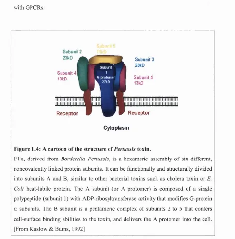
1.3.4 Other covalent modifications on G-protein subunits
GgΠis ADP-ribosylated by cholera toxin, resulting in a constitutively active a
subunit due to impaired GTP hydrolysis. The a subunits of the Gj, Go and Gt
subfamilies are ADP-ribosylated by bacterial Bordetella Pertussis toxin (PTx), at a
cysteine residue close to the C-terminus that prevents interaction of these subunits
with GPCRs.
S ub u n it 2 23kD
S u b u n it 4 13kD
S ub u n it 3 23kD
S ubunit 4 13kD
Receptor Receptor
Cytoplasm
Figure 1.4: A cartoon of the structure of Pertussis toxin.
PTx, derived from Bordetella Pertussis, is a hexameric assembly of six different, noncovalently linked protein subunits. It can be functionally and structurally divided
into subunits A and B, similar to other bacterial toxins such as cholera toxin or E. Cali heat-labile protein. The A subunit (or A protomer) is composed of a single polypeptide (subunit 1 ) with ADP-ribosyltransferase activity that modifies G-protein
a subunits. The B subunit is a pentameric complex of subunits 2 to 5 that confers
cell-surface binding abilities to the toxin, and delivers the A protomer into the cell.
Both Cholera and Pertussis toxins have been widely used experimentally to
demonstrate the involvement o f a particular subtype o f G-protein in a cell-signalling
pathway and to label G a subunits with a radioactive AD? ribose moiety (Downes &
Gautam, 1999). In the study o f Gj/o G-protein signalling pathways, each o f the six
members (G a i, G a ], Gag, GoaA, Goag, and G%a ) with the exception o f G%a can be
modified by the PTx-mediated transfer of ADP-ribose onto a cysteine residue four
amino acids from their C-terminus. This results in the loss o f coupling between the
receptor and the modified G-protein. PTx-resistant versions o f these G a subunits
have been generated by the mutation of the cysteine to a glycine or isoluceine residue
(Hunt et al., 1994; Wise et al., 1997). PTx treatment o f cells that have been
transfected with a particular PTx-resistant G a subunit abolishes coupling between
GPCRs and the cell’s endogenous G/o G-proteins, allowing one to study coupling
between receptor and the modified G/oa subunit (Wise et al., 1999; Leaney &
Tinker, 2000). This exceptionally useful strategy is used widely in these studies,
particularly with the generation o f CFP-tagged, PTx-resistant Gi/oa subunits in our
laboratory (Leaney et al., 2001).
1.3.6 Effectors activated by the Gpy subunit
Once Ga-GTP has dissociated from Gpy, free Gpy interacts with a growing list of
effector proteins (Clapham & Neer, 1997). Six different Gps and twelve different Gy
amongst their -340 amino acids; the GPs subunit is set apart with only 53% identity
to other Gps and an additional 13 amino acids (Clapham & Neer, 1997). The twelve
Gy subunit isoforms are more diverse with approximately 50% amino acid identity
between them (Downes & Gautam, 1999). Some P- and y- combinations fail to form
functional dimers, and a 14 a.a. region in the middle o f the Gy subunit confers this
specificity for Gp (Spring & Neer, 1994). The implications o f the diversity generated
by the large number o f Gpy subunit combinations that can form are not fully
understood, but antisense studies suggest that certain combinations are required to
interact with different effectors (reviewed by Hildebrandt, 1997).
The p and y polypeptides form functional monomers o f high thermal stability that
are resistant to tryptic digestion. GPy subunits were first demonstrated to regulate
effectors by Logothetis et al. (1987) who showed that Gpy could activate the G-
protein-gated muscarinic (Kir 3.1/3.4 or KACh) channel in cardiac atrial cells.
Despite initial controversy, it is now firmly established that it is the release o f Gpy
from activated Gi/o G-proteins that activates the GIRK channel, and not the Ga-GTP
subunit (Reuveny et al., 1994; Huang et al., 1995; Yamada et al., 1998).
Revealingly, the crystal structure of Gpy - solved by Sondek et al. in 1996 - showed
that Gpy does not change conformation when it dissociates from Ga-GTP. Together
with the finding that Ga-GDP inhibits the interaction o f Gpy with all o f its effectors,
this suggests a common interface on Gpy responsible for binding to Ga-GDP and for
overlapping residues on Gp that interact with effectors and with Ga-GDP subunits
(Ford et al., 1998) (Figure 1.5).
Effector enzymes activated by GPy subunits include: certain p-isozymes o f
Phospholipase-C (PLC-pl-3, not PLC-p4), Bruton tyrosine kinase, Tsk tyrosine
kinase, enzymes o f the MAP kinase cascade, certain isoforms o f adenylate cyclase
(Gpy stimulates Gas-activated AC-II, -IV and VII whereas it inhibits AC-I),
phosphoinositide-3 kinase (PI3K) and two members of the p-adrenergic kinase
family (also known as GRK2 and GRK3) (Clapham and Neer, 1997).
Ion channel modulation by G-proteins has been proposed for many different channels
(including: K v l.l, Kir 2.3, ATP-sensitive inwardly rectifying channels encoded
by Kir 6.1 and 6.2, and P/Q- and R-type voltage-dependent Ca^^ channels), but it has
only been irrefutably established for two families (reviewed by Dascal, 2001):
Gpy dimers activate the G-protein gated K^ ion channel (GIRK, encoded by Kir 3.1-
3.4 subunits), and Gpy dimers directly inhibit the N-type voltage-dependent Ca^^
channel, containing am (Cav2.3) subunits (Herlitze et al., 1996; Ikeda, 1996).
GPCR-mediated inhibition o f the high-voltage activated Ca^^ channels o f the Cav
family is complex, with voltage-independent (VI) and voltage-dependent (VD)
mechanisms. Different components o f the VI inhibition have been suggested to be
imparted by G q/n, Goa and by Gpy (Delmas et al, 1998) but the key criteria for a
a) it is membrane-delimited,
b) it can be mimicked by overexpression of GPy,
c) it can be blocked by expression o f Gpy scavengers (eg. C-terminus o f GRX2)
d) Gpy binds to fusion proteins made from regions on the proposed effector, and
e) mutations o f the regions described above can block Gpy regulation.
For the VD inhibition o f voltage-gated Ca^^ channels, all o f the above have been
satisfied except for a direct demonstration o f VD inhibition on Gpy application to an
inside-out patch (Dascal, 2001). A recent report suggests that the inhibitory
ionotropic glycine receptor, in both recombinant and native settings, may be
positively regulated by GPy subunits (Yevenes et al., 2003). While this has yet to be
independently verified, it would provide an additional mechanism for Gi/o-coupled
GPCRs to exert inhibitory effects on synaptic transmission in the peripheral and
1.3.7 The structure of the G(3y subunit
The crystal structure of the Gp subunit was first solved by Sondek and colleagues in
1996. The Op polypeptide folds into a beautiful propeller structure of seven “blades”,
based upon a repeating sequence called a WD repeat. These blades are arranged
about a central, water-filled tunnel with a protruding a-helical N-terminus.
Figure 1.5: The structure of the Gpy subunit.
Ribbon diagrams showing the Gp subunit in silver, and the Gy subunit in pink. Gp consists
of a seven-bladed propeller structure and protruding 20 amino acid N-terminus.
Left: Critical residues on the propeller of the Gp subunit for interactions with the G a
subunit are shown in red.
Right: Residues important for Gp interaction with the GIRK channel are shown in blue.
1.4 Inwardly rectifying potassium ion channels
Ion channels are macromclecular protein complexes, spanning the cell membrane
lipid bilayer, that create an aqueous pore for the passage o f specific ions to travel
along their electrical gradients, either into or out o f the cell. Ion channels that are
selectively permeable to Na^, K^, Ca^^ or Cl' ions are responsible for excitation and
electrical signaling in the nervous system. They can be viewed as excitable
molecules that aie either open or closed. The channel’s response to a specific
stimulus - a voltage change across the membrane, a neurotransmitter, a cytoplasmic
second messenger, and so on - is known as gating and simply involves opening or
closing the pore. The vast number o f potassium channel families now known can be
divided into two major classes: those gated by voltage (Kv) and those that exhibit
inward rectification (Kir). Inward rectification is a property first described by Katz in
1949 in skeletal muscle. Briefly, when the direction of K^ flow is outward, Kir
channels pass very little current, but when the K^ gradient is inward Kir channels
pass large currents into the cell.
The property o f inward rectification allows the cell to preserve and to encourage
K^ entry into the cell. The high conductance o f Kir channels at very negative
(hyperpolarised) membrane potentials tends to stablise the membrane potential near
the Nemst equilibrium potential for K^ (Ek), around -80mV. Conversely, at
depolarised membrane potentials, the low Kir conductance prevents K^ efflux fi*om
short-circuiting the action potential and serves as an economy for the cell (Nichols &
Kir channel pore from the cytoplasmic side by either a ion (for weak inward
rectifiers) or by polyamines, which are metabolites of amino acids, such as spermine,
spermidine and putrescine that mediate strong inward rectification (Nichols &
Lopatin, 1997). Activation o f Kir channels is independent o f voltage changes and
depends on the difference between the membrane potential and the Nemst
equilibrium potential for K^, (V m - E k ) , but this applies only for changes in external
concentration (Hagiwara & Yoshi, 1979; Leech & Stanfield, 1981).
1.4.1 Discovery of Kir channels in the heart: an historical perspective
Early physiologists found that stimulation of the vagal nerve slowed the heart rate
and weakened the strength of contraction. In the 1920s, an historical discovery made
by Otto Loewi established the idea o f chemical synaptic transmission. He found that
on parasympathetic stimulation, a substance that he termed Vagusstoff was released
from vagal nerve terminals onto the heart (now known to be acetylcholine (ACh)
acting at M2 muscarinic receptors that are present on all types o f cardiac muscle) and
this substance produced bradycardia and reduced contractility (Loewi, 1921). Next
Del Castillo and Katz (1955) and Rutter and Trautwein (1955) found that ACh led to
hyperpolarisation o f the membrane potential o f frog cardiac cells, accompanied by a
decrease in the rate of action potential firing, and this was mediated by an increase in
the flux o f K^ ions across the cardiac cell membrane. In 1958, Trautwein and Dudel
demonstrated an increase in membrane conductance to K^ ions in response to ACh
channel recordings of K^ch channels were made (Sakmann, Noma & Trautwein,
1983) in studies tha showed that Kach channels are very different to the background
cardiac inwardly retifying conductance, termed Iki and corresponding to the
cloned Kir2.0 chaniel. [Sakmann and colleagues (1983) found that the mean open
time o f Kach (l-2 m ) was much shorter than that of Iki (10-100ms)]. Adenosine also
slows the heart rate md Belardinelli and Isenberg (1983) demonstrated an increase in
an inwardly rectifyiig current by both adenosine and ACh and suggested that
both agonists regulafed the same channel in cardiac atrial myocytes.
The nature o f the second messenger linking the adenosine and M2 receptors to the
Kach channel stimilated intensive research in the 1980s. Receptor-activated ion
channels can be placed in one o f two major groups: channels with an intrinsic sensor
and channels with 2. remote sensor (Hille, 1984). For channels with an intrinsic
sensor, such as the nicotinic acetylcholine receptor and the glycine and GABAa
receptor channels, the receptor and ion channel pore are contained within the same
macromolecule. On the other hand, for channels with a remote sensor, the receptor
and ion channel are different membrane proteins that are linked by intracellular
second messengers such as cyclic AMP or Ca^^. Studies using rapid applications o f
ACh revealed an intrinsic delay in Kach activation, implicating a multistep process
linking receptor binding and channel activation (reviewed by Wickman & Clapham,
1995). Thus it was asked: which second messenger mediates the activation o f Kach?
They found that when ACh was perfused onto the cell in the bath solution, the
channels within tie patch were not activated. However, when ACh was included in
the pipette solution there was a dramatic opening of channels in the patch. Therefore
it appeared that ACh acted locally, in a membrane-delimited manner, and not via a
cytoplasmic secord messenger that would be expected to diffuse from bath-activated
receptors to charnels contained within the patch. This result led, briefly, to the view
that the Kach charnel had an intrinsic sensor like the nicotinic acetylcholine receptor.
< - 5 a
^ -10
-1 5
Control
«
ACh (bath)
ACh (pipette)
50 ms
Figure 1.6: Channel activation is membrane-delimited.
Single Kach channels were measured in the cell-attached configuration of the patch- clamp in symmetrical from rabbit atrial cells. The top trace shows a control
recording before ACh addition. The middle trace shows no real increase in channel
activity when ACh (lOOnM) was perfused in the bath. The lower trace shows a huge