A norm- and control-referenced comparative study of the
neuropsychological profiles of shift workers and patients with
obstructive sleep apnoea (OSA)
Jacen Man Kwan Lee BSocSci(Hons)
Submitted in partial fulfillment of the requirements of the degree of Doctor of Psychology (Clinical Neuropsychology)
School of Social Sciences & Psychology
ii
ABSTRACT
Shift work and Obstructive sleep apnoea (OSA) have been associated with excessive daytime sleepiness and increased risk of road traffic accidents. There is evidence that daytime sleepiness does not provide a satisfactory explanation for accidents, and occupational and social failures associated with sleep disorders. The possibility arises that intermittent hypoxemia and sleep deprivation due to sleep fragmentation in OSA and sleep deprivation secondary to sleep cycle disruption in shift work may underlie neuropsychological deficits, which in turn meditate these functional impairments. The current study uses a control-referenced and norm-referenced design to explore in detail the subcomponents of attention/executive functions and motor coordination of patients with OSA and shift workers with an aim to outline and compare the profiles of any cognitive impairment between these groups. Each of the attentional and executive sub-functions investigated are substantiated by theory-based models and are matched with one or more standardized subtests, which are also in accord with a theory and ecological validity. The Tests of Everyday Attention, selected subtests of the Wide Range Assessment of Memory and Learning, the Stroop Test Interference Score, and the Austin Maze were used to assess
selective attention, sustained attention, divided attention, set-shifting, working memory, and inhibition of prepotent responses, as well as complex spatial learning, planning, error utilization, behavioural inhibition and motor coordination. Fifteen patients (13 men and 2 women aged between 34 and 58), who had previously undergone a polysomnographic sleep study and a diagnosis of moderate to severe obstructive sleep apnoea (Apnoea-Hypopnoea Index (AHI) > 20/hr and Epworth Sleepiness Scale (ESS) > 8) had been established and verified by a respiratory
physician, were recruited from the Austin and Repatriation, Medical Centre. Fifteen shift workers (9 men and 6 women aged between 25 and 49) and fifteen healthy controls (6 men and 9 women aged between 25 and 69), screened for sleep disorders and excessive sleepiness by Maislin Apnoea Prediction Index and ESS, were recruited from the community. Participants were closely matched for age and educational level. More pervasive and severe attentional and executive function impairments were demonstrated in patients with OSA relative to shift workers, both in
control-referenced comparison and norm-referenced comparison. In comparison to controls, shift workers demonstrated significant reductions in the abilities of complex visual selective attention, divided attention, auditory set-shifting, verbal and symbolic working memory, and inhibition of prepotent responses, as well as a
iii
visual and auditory set-shifting, verbal and symbolic working memory, and inhibition of prepotent responses, as well as impaired spatial learning due to poor planning, error utilization, behavioural inhibition and possibly poor motor coordination, as compared to controls. A pattern of predominant attentional deficiency with a mild verbal working memory deficiency in shift workers and a dual pattern of attentional deficiency and pervasive executive dysfunctions in patients with OSA were revealed in norm-referenced analysis. By comparing the neuropsychological profiles of the two groups in standardized scaled score, it can be deduced that sleep deprivation may be the more important contributing factor to the selective inattention, the trend of reduced sustained attention, and the reduced verbal working memory in patients with OSA; whereas intermittent hypoxemia may be the more important contributing factor to the deficits in divided attention, and the trends of mildly reduced visual and auditory set-shifting abilities and inhibition of prepotent responses. Based on the incremental deficiencies in the divided attention and set-shifting sub-functions evident in the comparative control-referenced analysis between shift workers and patients with OSA, it is possible that sleep deprivation and intermittent hypoxemia may contribute additively/synergistically to these two neuropsychological
sub-functions of patients with OSA. Austin Maze results support the notion that the pathophysiology of OSA involves subcortical brain structures and the associated frontostriatal pathways. Overall, results of the current study support the Executive dysfunction model and the Microvascular theory, but not a pure Attentional deficits model. The measured attentional and executive sub-functions are separable constructs and are not in a simple hierarchical relationship. The current study exemplifies how a neuropsychological comparative study using standardized tests may serve as an experimental paradigm allowing detailed contrast of the differences in cognitive sub-functions between clinical groups that share a common
pathophysiological factor, so that enriched information about the linking of each factor with various neurocognitive deficits can be deduced. Clinical monitoring of the objective indicators of neuropsychological functions is possible by using
repeatable standardized tests with high ecological validity. To conclude, the functional impairment in shift workers in this study was significant enough to be presented as a similar profile as patients with OSA, albeit somewhat less pervasive and less severe. The results indicated the potential hazard of shift work as
iv
DECLARATION
I, Jacen Man Kwan Lee, declare that the Doctor of Psychology (Clinical
Neuropsychology) thesis entitled “A norm- and control-referenced comparative study of the neuropsychological profiles of shift workers and patients with
obstructive sleep apnoea (OSA)” is no more than 40,000 words in length including quotes and exclusive of tables, figures, appendices, references and footnotes. This thesis contains no material that has been submitted previously, in whole or in part, for the award of any other academic degree or diploma. Except where otherwise indicated, this thesis is my own work.
v
DEDICATION
vi
ACKNOWLEDGEMENTS
This thesis would not have been possible without the support and guidance of my wonderful supervisors Associate Professor Gerard Kennedy and Dr Mark Howard. It has been an incredible privilege to work with both of you, thank you for your
mentorship and endless support. I am deeply indebted to your sympathetic ear and close supervision, and awed by the depth and breadth of your knowledge in the subjects of sleep, neuropsychology and clinical psychology.
I would also like to thank my mother, Yuet-Hing Yiu Lee, and my supervisor, Dorothy Frei, who shared triumph and tribulation of the writing process and provided much needed optimism when mine was waning. Thank you to my special family in 166 Tin Sam Village, for the unflagging belief in my abilities.
Thank you to my colleagues in the 2006 neuropsychology doctoral intake for sharing the colourful experience of the post-graduate scientist-practitioner journey. A special thank you to our course coordinator, Dr Alan Tucker, whose incredible wisdom and mentorship will never be forgotten.
Thank you to the participants of this study for their contributions to further the scientific understanding of shift work and obstructive sleep apnoea.
vii
TABLE OF CONTENTS
LIST OF TABLES……… xii
LIST OF FIGURES………. xiv
LIST OF ABBREVIATIONS……….. xvi
CHAPTER ONE: INTRODUCTION………. 1
1.1 Driver sleepiness and risk of road traffic accidents (RTAs)……… 1
1.2 Cognitive impairments in sleep disorders, risk of driving and social occupational failures………. 1
1.3 Aims of current study……….………. 3
CHAPTER TWO: LITERATURE REVIEW………. 4
2.1 Shift work and Shift Work Disorder (SWD) ………. 4
2.2 Obstructive Sleep Apnoea-Hypopnoea Syndrome (OSAHS)……… 6
2.3 Sleep fragmentation = Sleep deprivation……….. 8
2.4 Sleep deprivation and neuropsychological function (The common denominator between shift workers and patients with OSAHS)……….. 10
2.5 Hypoxemia experienced by patients with OSAHS………. 13
2.6 Circadian misalignment or desynchronization in shift workers…………. 16
2.7 Neuropsychology of Obstructive Sleep Apnoea (OSA)……….. 19
2.7.1 General intellectual functioning……… 19
2.7.2 Attentional function……….. 20
2.7.3 Vigilance……… 22
2.7.4 Executive function……….. 23
2.7.5 Learning and Memory………. 25
2.7.6 Working memory……… 26
2.7.7 Procedural memory……….. 29
2.7.8 Psychomotor performance and Motor coordination…………. 30
2.7.9 Meta-analysis and implication for the present study – focusing on attentional and executive functioning, and motor coordination………. 31
2.8 Potential mechanisms for neurobehavioural dysfunction in OSA…….. 32
2.8.1 Executive dysfunction model………. 32
2.8.2 Attentional deficits model……… 34
2.8.3 Microvascular theory………. 35
2.9 Rationale behind the choice of neuropsychological sub-functions studied……….. 36
viii
2.9.2 A theoretical based test of attention with ecological
validity……….. 37
2.9.3 Latent variables of traditional executive function tasks…….. 38
2.9.4 Rationale behind the selection of attentional and executive function measures………. 39
2.9.4.1 Measuring Attentional functioning………. 39
2.9.4.2 Measuring Executive Functions………. 40
2.9.5 Maze learning test to specifically explore the effect of intermittent hypoxia hypothesis and to capture other aspects of executive functions………. 41
2.9.6 Overall goals of the current study as a function of the choice of neuropsychological sub-functions and their corresponding tests………. 42
2.10 Rationale for the current study……… 43
2.10.1 Aim 1……….. 44
2.10.2 Aim 2……….. 44
2.10.3 Aim 3……….. 45
2.10.4 Aim 4……….. 46
2.11 Research design……….. 46
2.11.1 Hypothesis 1……….. 48
2.11.1.1 Hypothesis 1a……….… 48
2.11.1.2 Hypothesis 1b……….… 49
2.11.1.3 Hypothesis 1c………. 49
2.11.2 Hypothesis 2……….. 50
2.11.2.1 Hypothesis 2a……….………… 50
2.11.2.2 Hypothesis 2b………. 51
2.11.2.3 Hypothesis 2c………. 51
2.11.3 Hypothesis 3……….. 52
2.11.4 Hypothesis 4……….. 53
CHAPTER THREE: METHOD……… 54
3.1 Participants……… 54
3.2 Research design and procedure……….. 55
3.3 Measures………. 56
3.3.1 Participant Information Statement (Plain Language Statement) ……… 56
3.3.2 Consent Form………. 56
ix
3.3.3.1 Maislin Apnoea Prediction Questionnaire………. 57
3.3.3.2 Epworth Sleepiness Scale (ESS) ……… 58
3.3.3.3 Karolinska Sleepiness Scale (KSS)………. 58
3.3.4 Stroop Colour and Word Test..……….. 59
3.3.5 Wide Range Assessment of Memory and Learning – Second Edition (WRAML-2)………. 60
3.3.5.1 Verbal Working Memory……… 60
3.3.5.2 Symbolic Working Memory………. 61
3.3.6 The Test of Everyday Attention (TEA)……… 62
3.3.6.1 Map Search………. 62
3.3.6.2 Telephone Search……… 62
3.3.6.3 Elevator Counting with Distraction………. 62
3.3.6.4 Lottery………. 62
3.3.6.5 Telephone Search while Counting (Dual Task)………. 63
3.3.6.6 Visual Elevator……….. 63
3.3.6.7 Auditory Elevator with Reversal……… 64
3.3.7 Austin Maze……… 64
CHAPTER FOUR: RESULTS……… 66
4.1 Statistical analysis………. 66
4.2 Data screening………. 67
4.3 Data analysis………. 69
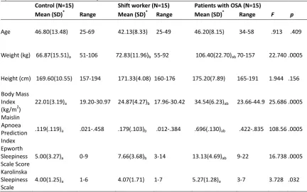
4.3.1 Demographic variables, BMI, MAPI, and subjective sleepiness scale……….. 69
4.3.2 Neuropsychological measures……… 71
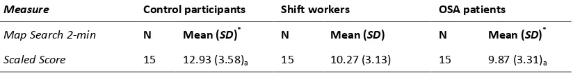
4.3.2.1 Map Search Scaled Score - Visual selective attention measure……….. 77
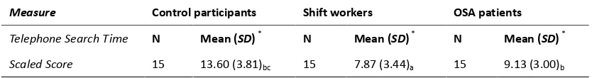
4.3.2.2 Telephone Search Scaled Score - Visual selective attention measure……… 79
4.3.2.3 Elevator Counting with Distraction Scaled Score - Auditory selective attention measure………. 81
4.3.2.4 Lottery Auditory Scaled Score - Sustained attention measure……….. 83
4.3.2.5 Telephone Search while Counting Dual Task Decrement Scaled Score - Divided attention measure……… 85
x
4.3.2.7 Visual Elevator Time Scaled Score - Visual set-shifting measure (efficiency) ………. 89
4.3.2.8 Elevator with Reversal Scaled Score - Auditory
set-shifting measure……… 91
4.3.2.9 Verbal Working Memory Scaled Score - Updating of
verbal information measure………. 93
4.3.2.10 Symbolic Working Memory Scaled Score - Updating of symbolic information measure……… 95
4.3.2.11 Stroop Interference Chafetz T Score - Inhibition of
prepotent responses measure………. 97
4.3.2.12 Austin Maze 10th-Trial Total Errors - Complex spatial learning measure – Planning, Error utilization,
Behavioural regulation (reliability)……….. 99
4.3.2.13 The differential relationships between various measured neuropsychological functions and Austin
Maze 10th-Trial Total Error across different groups…….. 101
4.3.2.14 Austin Maze 10th-Trial Total Time - Complex spatial learning – Planning, Error utilization, Behavioural
regulation (efficiency) ……….. 102
CAPTER FIVE: DISCUSSION OF RESULTS………. 104
5.1 Selective Attention - Map Search, Telephone Search, and Elevator Counting with Distraction………
104
5.2 Sustained Attention or Vigilance - Lottery……….. 106
5.3 Divided Attention – Telephone Search while Counting (Dual Task Decrement) ……….. 107
5.4 Set-Shifting or Attentional Switching - Visual Elevator and (Auditory) Elevator Counting with Reversal………. 110
5.5 Updating – Working Memory - Verbal Working Memory and Symbolic Working Memory……… 112
5.6 Inhibition of Prepotent Responses – Stroop Interference………. 114
5.7 Complex Spatial Learning – Planning, Error Utilization, and Behavioural Regulation – Austin Maze……… 116
CHAPTER SIX: GENERAL DISCUSSION……….. 123
6.1 More pervasive and severe attentional function impairments in patients with OSA relative to shift workers, both in control-referenced comparison and norm-referenced comparison…… 123
xi
and norm-referenced comparison, affecting complex spatial
learning……… 124
6.3 The measured attentional and executive sub-functions are separable constructs and are not in a simple hierarchical relationship……….. 125
6.4 Summary of control-referenced analyses..……….. 126
6.5 A pattern of predominant attentional deficiency in shift workers and a dual pattern of attentional deficiency and pervasive executive dysfunction in patients with OSA in norm-referenced analysis…………. 126
6.6 Sleep deprivation and intermittent hypoxemia………. 127
6.7 Austin Maze results support the notion that the pathophysiology of OSA involves subcortical brain structures and the associated frontostriatal pathways………. 127
6.8 The relative merits of the three OSA models………. 128
6.9 Strengths and weaknesses………. 129
6.10 Conclusions and implications on clinical practice and future research……….. 131
REFERENCES………. 133
Appendix 1: Recruitment Advertisement……… 152
Appendix 2: Participant Information Statement and Informed Consent Form. 154 Appendix 3: Demographics Questionnaire………. 164
Appendix 4: Driving Information Questionnaire……… 167
Appendix 5: Maislin Apnoea Prediction Questionnaire………. 170
Appendix 6: Epworth Sleepiness Scale ………. 172
Appendix 7: Karolinska Sleepiness Scale ………. 174
Appendix 8: Sleep Diary……… 176
Appendix 9: Stroop Colour and Word Test Instructions………..……….. 178
Appendix 10: Verbal Working Memory Test Instructions………. 181
Appendix 11: Symbolic Working Memory Test Instructions……….. 184
Appendix 12: Map Search Test Instructions……….. 187
Appendix 13: Telephone Search Test Instructions……….…… 189
Appendix 14: Elevator Counting with Distraction Test Instructions…….…………. 191
Appendix 15: Lottery Test Instructions……….. 194
Appendix 16: Telephone Search while Counting (Dual Task) Test Instructions.. 196
Appendix 17: Visual Elevator Test Instructions……… 199
Appendix 18: Elevator Counting with Reversal Test Instructions……….……… 201
xii
LIST OF TABLES
Table 1.
Summary of cognitive testing conditions.……… 56
Table 2.
Means, standard deviations, and ranges for demographic variables, Body Mass Index, Maislin Apnoea Prediction Index, and subjective sleepiness scales………… 70
Table 3.
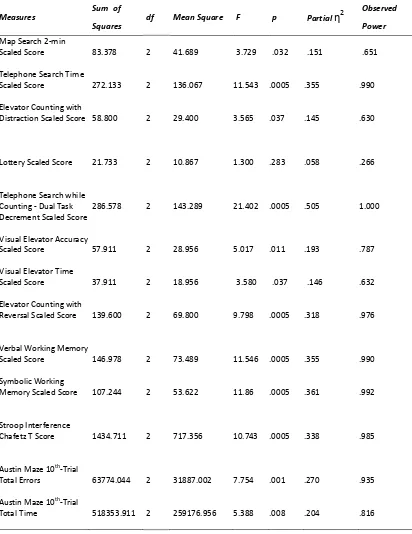
Univariate analyses of variance for neuropsychology tests performance, with
participant Group as independent variable.………. 72
Table 4.
Post hoc comparison of means of Map Search 2-min Scaled Score - Tukey HSD
test………. 77
Table 5.
Post hoc comparison of means of Telephone Search Time Scaled Score - Tukey HSD test……….. 79
Table 6.
Post hoc comparison of means of Elevator Counting with Distraction Scaled
Score - Tukey HSD test……….. 81
Table 7.
Post hoc comparison of means of Lottery Scaled Score - Tukey HSD test…………. 83
Table 8.
Post hoc comparison of means of Telephone Search while Counting Dual Task Decrement Scaled Score - Tukey HSD test……… 85
Table 9.
Post hoc comparison of means of Visual Elevator Accuracy Scaled Score -
xiii
Table 10.
Post hoc comparison of means of Visual Elevator Time Scaled Score - Tukey
HSD test……….. 89
Table 11.
Post hoc comparison of means of Elevator Counting with Reversal Scaled
Score - Tukey HSD test……….. 91
Table 12.
Post hoc comparison of means of Verbal Working Memory Scaled Score -
Tukey HSD test……… 93
Table 13.
Post hoc comparison of means of Symbolic Working Memory Scaled Score -
Tukey HSD test……… 95
Table 14.
Post hoc comparison of means of Stroop Interference Chafetz T Score - Tukey
HSD test……….. 97
Table 15.
Post hoc comparison of means of Austin Maze 10th-Trial Total Errors - Tukey
HSD test……….. 99
Table 16.
Post hoc comparison of means of Austin Maze 10th-Trial Total Time - Tukey
xiv
LIST OF FIGURES
Figure 1.
A highly simplified representation showing the relationships among the pathophysiological mechanisms, the cognitive deficits profiles and the
functional impairments in the participant groups………. 47
Figure 2.
Comparison of attentional function profiles for each participant group……… 73
Figure 3.
Comparison of executive function profiles for each participant group……… 74
Figure 4.
Comparison of performances on Austin Maze for each participant
group.……… 75
Figure 5.
Means for Map Search 2-min Scaled Score for patients with OSA, shift
workers, and controls.……… 78
Figure 6.
Means for Telephone Search Time Scaled Score for patients with OSA, shift
workers, and controls. ………. 80
Figure 7.
Means for Elevator Counting with Distraction Scaled Score for patients with
OSA, shift workers, and controls.……….. 82
Figure 8.
Means for Lottery Scaled Score for patients with OSA, shift workers, and
controls. ………. 84
Figure 9.
Means for Telephone Search while Counting Dual Task Decrement Scaled
xv
Figure 10.
Means for Visual Elevator Accuracy Scaled Score for patients with OSA, shift
workers, and controls.……… 88
Figure 11.
Means for Visual Elevator Time Scaled Score for patients with OSA, shift
workers, and controls.……… 90
Figure 12.
Means for Elevator Counting with Reversal Scaled Score for patients with OSA,
shift worker, and controls.……….. 92
Figure 13.
Means for Verbal Working Memory Scaled Score for patients with OSA, shift
workers, and controls.……… 94
Figure 14.
Means for Symbolic Working Memory Scaled Score for patients with OSA, shift
workers, and controls.……… 96
Figure 15.
Means for Stroop Interference Chafetz T Score for patients with OSA, shift
workers, and controls.……… 98
Figure 16.
Means for Austin Maze 10th-Trial Total Errors for patients with OSA, shift
workers, and controls.……… 100
Figure 17.
Means for Austin Maze 10th-Trial Total Time for patients with OSA, shift
xvi
LIST OF ABBREVIATIONS
ACTH Adrenocorticotropic hormone
AASM American Academy of Sleep Medicine
ANOVA Analysis of Variance
AHI Apnoea-Hypopnoea Index
PaCO2 Arterial partial pressure of carbon dioxide
PaO2 Arterial partial pressure of oxygen
ADHD Attention-deficit/hyperactivity disorder
BMI Body mass index
CANTAB Cambridge Neuropsychological Test Automated Battery
CFA Confirmatory factor analysis
CPT Continuous Performance Test
CRH Corticotrophin releasing hormone
DADT Divided Attention Driving Test
EEG Electroencephalograph
EOG Electrooculography
ESS Epworth Sleepiness Scale
EDS Excessive daytime sleepiness
FCRTT Four Choice Reaction Time Test
HPA Hypothalamic-pituitary-adrenocortical
iNOS Inducible NOS
IQ Intelligence Quotient
IH Intermittent hypoxia
ICSD-2 International Classification of Sleep Disorders, 2nd edition
JLD Jet Lag Disorder
KSS Karolinska Sleepiness Scale
LTP Long-term potentiation
MRI Magnetic resonance imaging
MAPI Maislin Apnoea Prediction Index
MTT Mirror Tracing Task
MID Multiple Infarct Dementia
MSLT Multiple Sleep Latency Test
MANOVA Multivariate analyses of variance
NO Nitric oxide
NOS Nitric oxide synthase
xvii
OSAHS Obstructive Sleep Apnoea-Hypopnoea Syndrome
OSA Obstructive Sleep Apnoea
PASAT Paced Auditory Serial Additional Test
PET Positron Emission Tomography
PVT Psychomotor Vigilance Task
RNG Random number generation
RT Reaction time
RDI Respiratory disturbance index
RNA Ribonucleic acid
RTA Road traffic accidents
RMSEA Root mean square error of approximation
SWD Shift Work Disorder
SWS Slow wave sleep
SPSS Statistical Package for Social Sciences
SDMT Symbol Digit Modalities Test
TEA Test of Everyday Attention
TOH Tower of Hanoi
TOL Tower of London
WAIS-R Wechsler Adult Intelligence Scale-Revised WAIS-III Wechsler Adult Intelligence Scale-Third Edition WISC-R Wechsler Intelligence Scale for Children-Revised
WMS-III Wechsler Memory Scale-Third Edition
WRAML-2 Wide Range Assessment of Memory and Learning – Second Edition
1
CHAPTER ONE: INTRODUCTION
1.1 Driver sleepiness and risk of road traffic accidents (RTAs)
Shift work has been associated with the experience of driver sleepiness
(Adam-Guppy & Guppy, 2003; Hakkanen, Summala, Partinen, Tihonen, & Silvo, 1999). The combination of homeostatic and circadian influences produces increased
behavioural, subjective and physiological sleepiness (Akerstedt, 1988; Akerstedt, 1990; Akerstedt, 2003; Akerstedt, Kecklund, & Knutsson, 1991). Shift workers commonly suffer with disturbed sleep and decreased sleep duration (Akerstedt & Torsavall, 1981). This sleep reduction also causes daytime sleepiness, inability to concentrate and misperception (Paley & Tepas, 1994). However, obstructive sleep apnoea (OSA) is another condition leading to sleep fragmentation and daytime sleepiness (Stradling & Crosby, 1991; Young, Palta, Dempsey, Skatrud, Weber, & Badr, 1993). OSA has been found to be associated with a significantly increased
frequency of falling asleep while driving and increased risk of RTAs (Aldrich, 1989; Barbe et al., 1998; Findley, Unverzagt & Suratt, 1988).
1.2 Cognitive impairments in sleep disorders, risk of driving and social occupational failures
Although sleepiness while driving is believed to be an important cause of accidents, recent evidence suggests that actually falling asleep is much less likely to be the causal event than making attentional and judgmental errors (Philip & Mitler, 2000). There is evidence suggesting that perceived sleepiness as measured by the Epworth Sleepiness Scale (ESS), and the objective sleepiness measured in the Multiple Sleep Latency Test (MSLT) are poor predictors of the accident rates in sleep apnoea patients (Young, Blustein, Finn, & Palta, 1997). Moreover, ESS was not correlated with driving simulator performance in OSA patients (Turkington, Sircar, Allgar, & Elliott, 2001).
If sleep disorders are frequently associated with accidents, but daytime sleepiness does not provide a satisfactory explanation (Philip & Mitler, 2000), it could be that factors such as sleep fragmentation and hypoxemia in OSA (Bedard, Montplaisir, Richer, Rouleau, & Malo, 1991) and sleep deprivation resulting from sleep cycle disruption in shift work (Paley & Tepas, 1994) may underlie both the daytime
2
judgment errors (Harrison & Horne, 1999), and which, in turn, may mediate the higher accident rate (Harrison & Horne, 2000a).
Basic cognitive functions traditionally found to be associated with sleep deprivation, such as alertness, reaction time, attention and vigilance (Dinges et al., 1997; Horne, Anderson, & Wilkinson, 1983) can be important mediating factors for making
performance errors and hence causing accidents. OSA patients have been shown to have more electroencephalograph (EEG) monitored attention lapses and higher lane position variability on a driving test, presumably due to their delayed responses to lane drifts during lapses (Risser, Ware, & Freeman, 2000). Recent research suggests that tests sensitive to sleep deprivation need not necessarily be monotonous and simple; they can be short, stimulating and rely on accuracy rather than speed (Wilkinson, 1992). For example, sleep loss is found to impair certain types of executive functions, such as supervisory control (Nilsson et al., 2005), problem solving, divergent thinking capacity (Horne, 1988; Linde & Bergstrom, 1992), verbal creativity, flexibility, response inhibition (Harrison & Horne, 1998,2000a), and cognitive set shifting (Wimmer, Hoffmann, Bonato, & Moffitt, 1992). Studies have shown that sleep deprivation is associated with perseverations, working memory problems, increased distractibility and concern with irrelevancies (Harrison & Horne, 2000a). Sleep deprivation also significantly reduces prefrontal metabolic activity with associated decrements in executive function task performance (Thomas et al., 2000) and biases the person toward risky decision-making, especially with increasing age, with the pattern resembling that of ventromedial prefrontal cortex lesions (Killgore, Balkin & Wesensten, 2006).
Sleep deprivation alone does affect cognitive performance; however, the fact that deficits related to executive function still persist despite treatment-related resolution of daytime sleepiness (Bedard, Montplaisir, Richer, Malo & Rouleau, 1993; Naegele et al., 1998) suggests non-sleep factors may be contributing to the development of some of the cognitive impairments. Comparison of hypoxemic and non-hypoxemic apnoea patients provides evidence to show that sleep fragmentation is a less
important cause of cognitive impairment than hypoxemia (Findley et al., 1986). Moreover, OSA in adults is associated with occupational and social failures
3
daytime sleepiness.
1.3 Aims of current study
In the present study, the aim was to investigate the neuropsychological profile of OSA patients who were affected by hypoxemia and sleep deprivation secondary to sleep fragmentation and that of shift workers who were mainly affected by sleep
deprivation due to disruption of their sleep cycle. Sustained attention, selective attention, divided attention, executive functions including inhibition of prepotent responses, set-shifting, verbal and symbolic working memory, planning, error
utilization, behavioural inhibition, as well as fine-motor coordination were measured using a battery of neuropsychology tests. Potentially, attentional and executive functions together with motor coordination can serve, besides sleepiness, as mediating factors for the real-life consequences of OSA.
4
CHAPTER TWO: LITERATURE REVIEW
2.1 Shift work and Shift Work Disorder (SWD)
Shift work is a term that applies to a broad spectrum of non-standard work schedules including occasional on-call over-night duty, rotating schedules, steady and
permanent night work, and schedules demanding an early awakening from nocturnal sleep. Shift work is very common; in fact, about one in five workers in the United States do some form of shift work (women more than men) (Presser, 1995). In 2004, for approximately 22 million US adults, shift work was an integral part of their
professional life; of these individuals, about 3.8 million regularly performed
night-shift work on a rotating basis (McMenamin, 2007; US Bureau of Labor Statistics, 2004).
SWD is experienced by individuals whose work schedule overlaps with the normal sleep period, causing misalignment between the body’s endogenous circadian clock and the time at which the worker is able to rest. The International Classification of Sleep Disorders, 2nd edition (ICSD-2) defines SWD as the presence of excessive daytime sleepiness (EDS) and/or insomnia for at least one month, in association with a shift-work schedule (American Academy of Sleep Medicine, 2005). Recent
practice parameters from the American Academy of Sleep Medicine (AASM)
recommend the use of a sleep diary for at least seven days to aid in the diagnosis of SWD and to rule out other sleep/wake disorders (Morgenthaler et al., 2007; Sack et al., 2007); but there are no standard sleep diaries as yet. ESS is helpful in measuring EDS in the primary care setting (Johns, 1991). This brief questionnaire asks the respondent to subjectively rate his or her chances of dozing in eight sedentary situations, such as reading a book or sitting in a meeting. A score of at least 10 out of a maximum 24 is indicative of clinically significant EDS (Johns, 1991). The diagnosis of SWD is based on patient history and it does not require confirmation with a sleep study (Sack et al., 2007). EDS is also a symptom of the other
sleep/wake disorders, including OSA (Aloia, Arnedt, Davis, Riggs, & Byrd, 2004). Exclusion of OSA can be done by a screening questionnaire, the Maislin Apnoea Prediction Questionnaire (Maislin et al., 1995). A high Maislin Apnoea Prediction Index (MAPI) (> 0.5) warrants a sleep study or polysomnogram to confirm the differential diagnosis (Maislin et al., 1995).
5
in research studies, and the validity and reproducibility of the AASM diagnosis criteria need testing (Sack et al., 2007). This classification results in the shift work population being separated into three distinct groups: (1) those who have no impairment; (2) those who have impairment (social, occupational or other), but do not meet the ICSD-2 criteria for the diagnosis of SWD based on history taking; and (3) those who have SWD. Drake and colleagues (2004), using questionnaire data from an epidemiological survey, found that 32.1% of night workers and 26.1% of rotating workers met the minimal criteria for SWD. The boundary between a normal and a pathological response to the circadian stress of an unnatural sleep schedule
associated with shift work remains unclear (Sack et al., 2007). Since the latter two groups are intolerant of shift work, it is likely that a much larger number of intolerant shift workers have some impairment and may or may not meet the SWD criteria, remain in the workforce.
Insomnia and EDS (drowsiness and a propensity to sleep) are the defining symptoms of SWD and can result in fatigue (weariness and depleted energy), difficulty
concentrating, reduced work performance, headache, irritability, or depressive mood, and hence constitute a significant burden of illness on society (Schwartz & Roth, 2006; Shen et al., 2006). The circadian system functions adequately under usual circumstances, but when an imposed shift in the timing of sleep exceeds the limits of circadian adaptation, misalignment occurs. Being classified under Circadian Rhythm Sleep Disorder, circadian misalignment is considered to play an important part in the primary pathophysiology of SWD, causing a constellation of symptoms that
characterize the disorder (Sack et al., 2007). This, however, does not preclude other endogenous factors, such as individual differences in the ability to sleep at an
unfavourable circadian phase, from contributing to SWD. Indeed, attempts to sleep at an unusual time are often interrupted by noise, and social factors (Sack et al., 2007). There is also an inevitable degree of sleep deprivation associated with sudden transitions in sleep schedule, for example, a night worker who stays awake for 24 hours on the first night of a rotating roster is acutely sleep deprived in the morning. In fact, the major consequences of shift-work are disturbed sleep and decreased sleep duration (Akerstedt & Torsvall, 1981), producing a cumulative sleep loss, or chronic partial sleep deprivation (Scott, 2000).
6
symptoms of Jet Lag Disorder (JLD) are considered to be generated by circadian misalignment, the inevitable consequences of crossing time zones too rapidly for the circadian system to keep pace (Sack et al., 2007). Cho, Ennaceur, Cole, and Kook Suh (2000) demonstrated that chronic jet lag experienced by cabin crew is associated with depressed nonverbal short-term memory processing, and possibly attenuated working memory, whereas short-term verbal memory is spared. Nevertheless, memory consolidation, learning, alertness, and performance are found to be severely affected by sleep deprivation, even in the absence of circadian misalignment (Dijk, Duffy, & Czeisler, 1992; Walker & Stickgold, 2005).
2.2 Obstructive Sleep Apnoea-Hypopnoea Syndrome (OSAHS)
OSAHS is a clinical condition that occurs because the upper airway collapses
intermittently and repeatedly during sleep, being characterized by recurrent episodes of partial or complete upper airway obstruction during sleep. This manifests as a reduction in (hypopnoea) or complete cessation (apnoea) of airflow despite ongoing inspiratory efforts (AASM, 1999). An apnoea is arbitrarily defined in adults as a ten second breathing pause and a hypopnoea as a ten second event where there is continued breathing, but ventilation is reduced by at least 50% from the previous baseline during sleep (Bassiri & Guilleminault, 2000). The lack of adequate alveolar ventilation usually results in arterial blood oxygen desaturation (decrease in arterial partial pressure of oxygen, PaO2) and in cases of prolonged events, a gradual increase in arterial partial pressure of carbon dioxide (PaCO2)(AASM, 1999).
As the sufferer falls asleep, the muscle tone in the upper pharyngeal airway
decreases leading to upper airway narrowing. This, in turn, produces an increase in inspiratory effort in an attempt to overcome airway narrowing, which then leads to a transient arousal from deep sleep to wakefulness or a lighter sleep phase allowing restoration of normal airway muscular tone and calibre. The patient then falls more deeply asleep again and the whole cycle repeats itself. This can occur many
hundreds of times throughout the night leading to fragmentation of normal sleep architecture and a reduction in the quality of sleep with the generation of restless, disturbed and unsatisfying sleep. Daytime symptoms such as excessive sleepiness, poor concentration, and a reduced alertness are thought to be related to sleep disruption associated with recurrent arousals (sleep fragmentation) and possibly also to recurrent hypoxemia (AASM, 1999).
7
end, moving through worsening levels of snoring, to increased upper airways resistance, and to hypopnoeas and apnoeas at the other end. The frequency of apnoeas and hypopnoeas hourly is used to assess the severity of the OSAHS and is called the apnoea/hypopnoea index (AHI) or the respiratory disturbance index (RDI) (Bennett, Langford, Stradling, & Davis, 1998). In an attempt to standardize
definitions of apnoeas/hypopnoeas and related indices, the AASM (1999) has published an arbitrary operational guideline to stratify the severity of OSAHS by varying degrees of breathing abnormality, or sleep related obstructive breathing events as defined by AHI:
Mild: AHI 5 to 14 events/hour Moderate: AHI 15 to 30 events/hour Severe: AHI greater than 30 events/hour
To fulfill the diagnostic criteria, the individual must have an overnight monitoring and demonstrate five or more obstructed breathing events per hour during sleep.
Recorded events may include any combination of obstructive apnoeas/hypopnoeas or respiratory effort related arousals. In addition, the individual must show either excessive daytime sleepiness that is not better explained by other factors, or two of the other features of OSAHS, including choking or gasping during sleep, recurrent awakenings from sleep, unrefreshed sleep, daytime fatigue, or impaired
concentration.
Stratification is used to assign patients to an approximate level of severity when considering treatment strategies. Stratification also depends on the severity of symptoms and the level of impairment of social and occupational function. In general, the more severe the breathing abnormality, the more symptomatic the patient becomes, but there may be cases where the severity of the symptoms does not correlate with the degree of breathing abnormality (Duran, Esnaola, Rubio, & Iztueta, 2001).
8
half the night. Hence, in middle-aged men, both snoring and sleep apnoea are extremely common, and it was also found that in this age range both are associated more with obesity than with age itself (Bearpark et al., 1995).
In terms of the pathophysiology of OSAHS, during the repeated complete (apnoea) or partial (hypopnoea) cessations of breathing, blood oxygen saturation can drop to dangerously low levels, resulting in increased respiratory effort and arousals from sleep to resume breathing. Recurrent hypoxemia and fragmented sleep are therefore significant consequences of the disorder (Bassiri & Guilleminault, 2000). The primary daytime sequelae of the disorder include EDS, mood changes and self-reported cognitive problems (Aloia et al., 2004).
2.3 Sleep fragmentation = Sleep deprivation
A number of studies have shown that both increased daytime sleepiness in healthy subjects and EDS in patients, whether due to total sleep deprivation, sleep restriction, sleep disruption or sleep fragmentation, impairs cognitive functions (Bonnet, 1986a, 1986b; Downey & Bonnet, 1987; Stepanski, Lamphere, Roehrs, Zorick & Roth, 1987).
Sleep fragmentation refers to the punctuation of sleep with frequent, brief arousals characterized by increases in EEG frequency or bursts of alpha activity, and
occasionally, transient increases in skeletal muscle tone (Roth, Hartse, Zorick, & Conway, 1980). These arousals last approximately 3-15 seconds, usually do not result in prolonged wakefulness, and sometimes may not even alter standard sleep stage scoring. In some sleep disordered patients, the arousing stimulus (e.g., apnoeas) can be identified (Miles & Dement, 1980; Roth et al., 1980). In other situations, the arousing stimulus cannot be identified. For example, the sleep of healthy “normal” elderly is often fragmented (Carskason, Brown & Dement, 1982), and out-of-phase sleep, such as occurs in shift work or jet lag, is also fragmented (Wegman et al., 1986). Thus, sleep fragmentation is a common cause of EDS.
9
observed, without increased wakefulness during the sleep period (i.e., subjects were not awakened behaviourally) (Stepanski et al., 1987). This was accomplished by terminating the tones upon arousal as defined by a speeding of the EEG or a burst of alpha activity of at least 3 seconds in duration, rather than causing behavioural wakefulness. In this study, sleepiness was measured repeatedly throughout the day with the MSLT, which has been shown to be a reliable measure of daytime sleepiness, and is systematically related to the amount of prior sleep (in sleep deprivation and sleep restriction studies) (Carskadon & Harvey, 1982; Carskadon, Harvey, & Dement, 1981; Roth, Roehrs, & Zorick, 1982). These studies demonstrated that sleep fragmentation, whether actually causing awakening or not, can result in increased EDS even when the “total sleep time” appears normal. Hence sleep fragmentation, which may be regarded as a kind of frequent sleep disruption, results in sleep
deprivation in effect.
Results from Bonnet’s studies (1985, 1986a) suggest that sleep continuity may be more integral to restoration of cognitive performance than “total sleep time” or specific sleep stage durations. In accordance with the sleep continuity theory (Bonnet, 1985, 1986a), the sleep process must continue undisturbed for a period of at least 10 minutes in order for sleep to be restorative. This theory is based on brain research findings that high sensory thresholds following sleep deprivation are instituted to maintain the continuity of sleep in order to allow sufficient time for effective protein synthesis (Adam, 1980; Oswald, 1980). Thus, it suggests that specific amounts of sleep stages are not important independent of sleep continuity.
Performances on psychomotor, vigilance, mental arithmetics tasks and daytime sleepiness have been shown to be a function of frequency and placement of sleep disruption (Bonnet, 1986a). It was found that arousals occurring at a rate of one per minute (sleep fragmentation) lead to daytime cognitive impairments associated with one night of sleep deprivation (Bonnet, 1986a). Bonnet, Downey, Wilms, and Dexter (1986) showed the number of arousing events and the periodic placement of these events are highly related to the severity of OSA. For example, patients with EDS rarely had a period of sleep as long as 10 minutes without an apnoea.
10
fragmenting events produced similar daytime function whether the events were evenly spaced or clustered, supporting that sleep continuity is more important than the specific amount of sleep stages (Martin, Brander, Deary, & Douglas, 1999).
2.4 Sleep deprivation and neuropsychological function (The common denominator between shift workers and patients with OSAHS)
Sleep fragmentation diminishes tremendously the recuperative value of sleep (Levine, Roehrs, Stepanski, Zorick, & Roth, 1987) and results effectively in sleep deprivation as discussed previously. This occurs in patients with OSAHS despite the fact that total daily sleep time may be greatly increased due to excessive daytime somnolence in these patients (Downey & Bonnet, 1987). For shift workers, out-of-phase sleep is often fragmented too (Wegman et al., 1986). The disturbed sleep and the almost inevitable decrease in sleep duration due to different biopsychosocial reasons also amounts to a cumulative sleep loss or chronic sleep deprivation as discussed
previously. Hence, significant sleep deprivation is a common denominator between shift workers and patients with OSAHS, albeit due to different pathophysiologies.
In general terms, excessive sleepiness is found to be associated with poor memory performance, poor concentration, and impaired learning and work performance, regardless of its etiology (Alapin et al., 2000; Rajaratnam & Arendt, 2000; Reimer & Flemons, 2003).
Basic cognitive functions traditionally found to be associated with sleep deprivation, such as alertness, reaction time, attention and vigilance (Dinges et al., 1997; Horne et al., 1983) can be important mediating factors leading to performance errors and hence accidents. For example, patients with OSA have more EEG monitored attention lapses and higher lane position variability on simulated driving tasks
presumably due to delayed responses to lane drifts during lapses (Risser et al., 2000).
The underlying mechanisms through which sleep deprivation produces deficits in neurobehavioural and cognitive functioning have yet to be fully elucidated. One early explanation was termed a lapse hypothesis. Williams, Lubin, & Goodnow (1959) suggested that transient lapses in attention and performance occur following sleep deprivation, interspersed among periods of optimal performance and alertness. Others suggested a more global decrease in performance, such as a reduction in
11
performance of fighter pilots on computerized cockpit simulation tasks assessing reaction time and vigilance and on a flight simulator was shown to deteriorate
significantly during 37 hours of sleep deprivation (Caldwell, Caldwell, Brown, & Smith, 2004). In fact, one night of total sleep deprivation has been shown to affect
reaction times and response accuracy to the same extent as having a blood alcohol concentration of .05% (Falleti, Maruff, Collie, Darby, & McStephen, 2003).
In the attention domain, significant deficits have been reported in vigilance (Blagrove, Alexander, & Horne, 1995; Caldwell et al., 2004; Orton & Gruzelier, 1989), sustained attention, attentional switching and short-term attention span (Frey, Badia, & Wright, 2004).
Sleep deprivation not only affects performances on monotonous and simple tasks, tasks which are short, stimulating and rely on accuracy rather than speed are also affected (Wilkinson, 1992). Performance on a number of tasks thought to be putatively subserved by the prefrontal cortex has been reported as significantly impaired following sleep loss, both total and chronic partial and the impairment was found to be reversible following recovery sleep (Doran et al., 2001; Mullaney, Kripke, Fleck, & Johnson, 1983; Harrison & Horne, 1998; Harrison, Horne, & Rothwell, 2000). That is, sleep loss has been found to impair certain types of executive functions such as supervisory control (Nilsson et al., 2005), problem solving, divergent thinking capacity (Horne, 1988; Linde & Bergstrom, 1992), temporal memory, verbal creativity, flexibility, response inhibition (Harrison & Horne, 1998; Harrison & Horne, 2000b) or inhibition of prepotent responses on a Go/No-Go task (Chuah, Venkatraman, Dinges, & Chee, 2006; Drummond, Paulus, & Tapert, 2006), and cognitive set shifting
(Wimmer et al., 1992). Studies have shown that sleep deprivation is related to perseverations, working memory problems, increased distractibility and concern with irrelevancies (Harrison & Horne, 2000a).
Other higher order cognitive abilities such as logical reasoning have also been shown to be affected (Blagrove et al., 1995). Temporal memory, memory of when events occur, for visual stimuli (Harrison & Horne, 2000b) and verbal memory (Deary & Tait, 1987) were found to be impaired following sleep deprivation. However,
12
found to be severely affected by sleep deprivation (Walker & Stickgold, 2005).
In addition to behavioural outputs of the prefrontal cortex demonstrating changes following sleep loss, brain imaging studies on sleep deprived subjects demonstrated decreased prefrontal activation associated with poorer performance on both
arithmetic tasks involving symbolic working memory (Drummond & Brown, 2001; Drummond et al., 1999; Thomas et al., 2000) and verbal working memory tasks (Mu et al., 2005b). Sleep deprivation was also found to significantly reduce prefrontal metabolic activity with associated decrement in performance on executive function tasks (Thomas et al., 2000) and bias the person toward risky decision-making, especially with increasing age, with patterns resembling those of ventromedial prefrontal cortex lesions (Killgore et al., 2006). On the other hand, it has been reported that learning and divided attention tasks produced increased levels of prefrontal activation following sleep deprivation (Drummond & Brown, 2001), as well as in complex cognitive tasks, such as planning, relationship reasoning, and spatial working memory (Dagher, Owen, Boecker, & Brooks, 1999; Diwadkar, Carpenter, & Just, 2000; Dorrian, Rogers, Ryan, Szuba, & Dinges, 2002; Kroger et al., 2002;
Mottaghy, Gangitano, Sparin, Krause, & Pascual-Leone, 2002). Moreover, a positive relationship between increased level of sleepiness and increased prefrontal
activation has been reported. It is possible that this differential activation of
prefrontal cortex may reflect task specific effects during sleep loss (Drummond et al., 2000) and compensatory effort to perform under sleep deprivation-induced
sleepiness and fatigue. These alterations in prefrontal cortex dynamics following sleep deprivation are consistent with neurobehavioural studies showing deficits in attention, working memory and higher-order cognitive processes known to be mediated by the frontal lobes and various frontal reciprocal connections to brain regions, which are activated during tasks requiring integrated executive functioning (Nilsson et al., 2005).
13
during sleep from other regions with which it normally interacts during daytime
hours (Braun et al., 1997, 1998; Hobson, Stickgold, & Pace-Scott 1998; Maquet, 2000). Dahl (1996) suggested that these findings may reflect a unique requirement for
‘recalibration’ of prefrontal cortex circuits without input interference from other brain regions. The prefrontal cortex is one of the most active brain regions while humans are awake, even during conscious rest, necessitating the greatest recovery during sleep; and sleep may be the only time when such restoration is possible (Binder et al., 1999; Harrison & Horne, 2000a). Finelli and colleagues (2001) using a quantitative EEG technique found that frontal regions are differentially sensitive to sleep deprivation and recovery sleep, and this effect appears to be related to time awake rather than circadian rhythmicity (Cajochen et al., 2001). In addition, by using magnetic resonance spectroscopy sensitive enough to study markers of neuronal integrity, it was revealed that neurochemical changes may be particularly prominent in the frontal lobes after sleep deprivation (Dorsey et al., 2000). Benington (2000) reviewed several hypotheses and concluded such restorative processes remain poorly understood at a cellular level. However, it is reasonable to assume that, these restorative processes require an extended period of sleep, and that disruption of sleep continuity can prevent homeostatic processes from taking place.
2.5 Hypoxemia experienced by patients with OSAHS
Benington (2000) suggested that limitation in tissue oxygen delivery (i.e., hypoxia) and decreases in intra- and extra-cellular pH (both hypoxia and hypercarbia) could also adversely affect sleep-related functions by creating a suboptimal environment for any number of cellular processes that have been implicated in restoration (e.g., mitochondrial integrity, protein synthesis, gene regulation). Bedard and colleagues (1991) reviewed research suggesting that synthesis of monoamines and acetylcholine may be disrupted by brief or intermittent hypoxemia.
David Gozal and his colleagues have been using experimentally-induced intermittent hypoxia in a rodent model of OSA to suggest potential mechanisms for
14
receptor neurons. Row, Liu, Xu, Kheirandish, and Gozal (2003) demonstrated spatial learning deficits with the Morris water maze (Morris, 1984) and hippocampal
ribonucleic acid (RNA) oxidant damage in a rodent model of sleep-disordered breathing, by exposure to intermittent hypoxia (IH), suggesting the episodic hypoxic-reoxygenation cycles of IH exposure is associated with increased oxidative stress, which is likely to play an important role in the behavioural impairments observed in patients with sleep-disordered breathing.
Li and colleagues (2004) demonstrated that IH selectively triggered one of the nitric oxide synthase (NOS) isoforms, inducible NOS (iNOS), which in turn led to excessive nitric oxide (NO) production and spatial learning deficits with the Morris water maze. Li and colleagues (2004) reported that IH exposures will also lead to substantial
up-regulation of pro-inflammatory cytokines (Interleukin-1 beta, Tumor Necrosis Factor-alpha, and Interleukin-6) in the rat cortex. The putative mechanisms of neurotoxicity caused by excessive NO formation, include activation of glutamate receptors, especially the NMDA receptors, oxygen and glucose deprivation, protein nitrosylation, mitochrondrial dysfunction, and cortical neuronal cell death or apoptosis (Li et al., 2004). Xu and colleagues (2004) hypothesized that the oscillation of oxygen concentrations during chronic IH mimics the processes of ischemia-reoxygenation and could therefore increase cellular production of reactive oxygen species (ROS). Xu and colleagues (2004) demonstrated that long-term exposure of mice to intermittent hypoxia increased ROS production and oxidative stress propagation, which at least partially contribute to chronic IH-mediated cortical neuronal apoptosis. Together, IH during sleep has been shown to induce cortical neuronal apoptosis and spatial learning deficits on a water maze task in adult rats.
15
production of iNOS was inhibited by targeted deletion of iNOS gene.
Consistent with this model, there is accumulating evidence for increased levels of inflammatory markers in adults and children with OSA (Mills & Dimsdale, 2004; Larkin et al., 2005), as well as precursors of such inflammation, including increased sympathetic nervous system activation and decreased parasympathetic activity (Mills & Dimsdale, 2004; O’Brien & Gozal, 2005). Moreover, inflammatory cytokine markers correlate with daytime sleepiness and neurobehavioural dysfunction among adults (Mills & Dimsdale, 2004; Haensel et al., 2009) and children (Gozal et al., 2009) with OSA. Although these human studies have focused on peripheral inflammatory markers, the rodent findings suggest the occurrence of parallel processes in the central nervous system. In addition, peripheral inflammation has been implicated in vascular disease, which may have cerebrovascular consequences (Aloia et al., 2004).
Another potential mechanism of neuronal damage involves the neurotransmitter glutamate. During transient hypoxia, increased glutamate release occurs into the synaptic cleft, and can lead to overstimulation of excitatory glutamate receptors. These glutamate receptors, and more specifically excitatory NMDA receptors, have been extensively implicated in neuronal excitotoxicity and neurodegeneration
(Englesen, 1986; Fung, 2000; Schousboe, Belhage, & Frandsen, 1997). Rats exposed to chemical hypoxia with carbon monoxide displayed an immediate and significant increase in glutamate release, followed days later by neuronal change that was particularly striking in the frontal cortex (Piantadosi, Zhang, Levin, Folz, & Schmeche, 1997).
16
and difficult to appreciate in the context of gross anatomical change.
Magnetic resonance spectroscopy study has found metabolic abnormalities in the left hippocampus similar to those seen in ischemic preconditioning, and this may reflect the differential susceptibility of these tissues to hypoxic damage in OSA. (Barlett et al., 2004). Other magnetic resonance spectra studies showed metabolic impairments in the frontal white matter (but not the prefrontal cortex or parietal white matter) of patients with OSA when compared to controls (Alchanatis et al., 2004; Kamba, Suto, Ohta, Inoue, & Matsuda, 1997; Kamba et al., 2001). Alchanatis and colleagues (2004) concluded that as frontal lobe white matter lesions are known to be associated with cognitive executive dysfunction, these findings may offer an explanation for the sometimes irreversible cognitive deficits, usually in the executive function domain, associated with OSA. Thus, cerebral metabolic changes occur in apparently normal brain tissue in patients with moderate to severe OSA. Some metabolic abnomalities suggest the presence of damage in frontal white matter, probably caused by repeated apnoeic episodes (Kamba et al., 1997). In contrast, functional MRI data suggest poor activation of dorsolateral prefrontal cortex in untreated adults with OSA when faced with a working memory task (Thomas, Rosen, Stern, Weiss, & Kwong, 2005).
2.6 Circadian misalignment or desynchronization in shift workers
One hypothetical mediating mechanism between circadian desynchronization or misalignment and cognitive dysfunction involves the impact of psychological stress on the brain via the hypothalamic-pituitary-adrenocortical (HPA) system with the increased secretion of cortisol (Lundberg, 2005). Briefly, stress causes the
hypothalamus to release a corticotrophin releasing hormone (CRH) which stimulates the pituitary gland to produce adrenocorticotropic hormone (ACTH). ACTH causes the adrenal cortex to release cortisol into the blood circulation, activating the sympathetic nervous system. Negative feedback to the pituitary gland via a loop incorporating the hippocampus and amygdala via glucocorticoid receptors
terminates the stress response. Chronic stress appears to cause down-regulation of glucocorticoid receptors, impairing the negative feedback mechanism, which results in over-activation of the HPA axis (Jameison & Dinan, 2001).
Disruptions of the sleep-wake cycle, such as sleep deprivation, night shift work and jet lag following rapid transmeridian flight, cause transient internal
17
Constant or prolonged sleep disruption, resulting in repeated disturbance of
synchronization of the circadian system to the environment, can be considered as a physiological stressor (Winget et al., 1984).
Cognitive and neuroendocrine effects of chronic jet lag have been reported by Cho and colleagues (Cho, 2001; Cho et al., 2000). Cho and colleagues (2000) showed that flight attendants experiencing transmeridian flights, whereby crossing of several time zones results in desynchronization internal circadian rhythm from external light-dark cycle, had significantly higher average daily cortisol secretion (as measured by salivary cortisol level) than ground crew and cortisol elevation in female flight attendants, but not ground crew, was significantly correlated (r = -.78) with poorer visual working memory performance on visual delayed-match-to-sample tasks. This evidence supports the hypothesis that chronic circadian rhythm disruption resulting from repeated exposure to jet lag leads to significantly elevated cortisol levels and related neurocognitive deficits.
Cho (2001) compared temporal lobe volume (MRI scans corrected for head size), performance responses to an experimental visual spatial cognitive task and cortisol levels between two groups of female flight attendants, one had less than five days between transmeridian flights, whereas the other had more than 14 days in between, controlling for five working years and total flight exposure during this period. The results showed that the short recovery group, as compared to the long recovery group, had significantly reduced right temporal lobe volume, made more errors and were significantly slower on the visual-spatial task. There was also a strong and significant negative correlation between chronic elevation of cortisol levels and right temporal lobe atrophy (r = -.78) for the short recovery group only, suggesting a possible association between chronic jet lag induced stress and right temporal lobe atrophy, although longer periods between transmeridian flights may circumvent this effect.
Studies on the nature of circadian dysregulation of rotating night shift workers showed mixed results. For example, Lac and Chamoux (2003) demonstrated a significant increase in overall cortisol production while Zuzewicz, Kwarecki, and Waterhouse (2000) found lower cortisol level in night shift workers. Similarly, while Touitou and colleagues (1990) found dysregulation of the circadian markers of
18
circadian markers of the cortisol rhythm (Lac & Chamoux, 2004). Moreover, Roden, Koller, Pirich, Vierhapper, and Waldhauser (1993) reported no differences in plasma cortisol rhythm characteristics (acrophase, amplitude, average secretion, and phase relationship with melatonin) between seven male controls and nine long-term, full-time, male night shift workers with high levels of work satisfaction. Overall, there is a general trend for cortisol rhythm dysregulation associated with shift work but the relationships between different circadian markers and different shift systems are complex. In addition, there seem to be large inter-individual differences in the tolerance of different shift schedules.
Notwithstanding this, it has become increasingly clear from research on HPA axis reactivity that chronically high or low levels of cortisol and problems with the up- or down-regulation of cortisol in response to stress are associated with difficulties in cognitive and behavioural self-regulation. The relation between cortisol and these brain functions generally follows an inverted U-shaped (Blair, Granger, & Razza, 2005). In children, moderate increase in cortisol followed by down-regulation of this
increase, in mildly challenging situations, was positively associated with measures of executive function and self-regulation (Blair et al., 2005).
Wright, Hull, Hughes, Ronda, and Czeisler (2006) assessed learning in healthy patients who lived under shift-work conditions in a laboratory devoid of time cues. They compared improvements on the Mathematical Addition Test and the Digit Symbol Substitution Task between a synchronized group, where the normal relationship between sleep-wakefulness and internal circadian time was maintained, and a non-synchronized group mimicking the shift work condition, with both groups allowed to have 8 hours of scheduled sleep. Cognitive performance improved (i.e., learning) in the synchronized group, whereas learning was significantly impaired in the non-synchronized group. Hence, short-term circadian misalignment was found to be detrimental to learning in subjects who failed to adapt to their imposed
schedule of sleep and wake, even though the total sleep time appears to be sufficient; in other words, proper alignment between sleep-wakefulness and internal circadian time is crucial for enhancement of cognitive performance (Wright et al., 2006).
19
affect the response times on tests of visual selective attention in a shift-work simulation study (Santhi et al., 2007).
Nevertheless, as mentioned previously, memory consolidation, learning, alertness and performance have been shown to be negatively affected by sleep deprivation, even in the absence of circadian misalignment (Dijk et al., 1992; Walker & Stickgold, 2005).
2.7 Neuropsychology of Obstructive Sleep Apnoea (OSA)
OSA can cause significant daytime behavioural and adaptive deficits. Functional impairments like sleepiness, impaired driving, increased risk of accidents, and decreased quality of life are common consequences of sleep apnoea (Engleman & Douglas, 2004; George & Smiley, 1999). Behavioural effects of OSA are often referred to as ‘neurobehavioural’ consequences because they are presumed to be directly related to brain function (Beebe, 2005). Neurobehavioural functioning is a broad term that includes several specific cognitive functions. Numerous studies have examined these specific cognitive functions and some have attempted to identify a “pattern” of cognitive dysfunction in OSA. Such patterns, as have been identified, are summarized below. Following that summary, theoretical models describing potential mechanisms involved in these relationships are discussed.
Cognition in OSA has been examined as both a unitary function (general intellectual functioning) and one divided into several specific domains (e.g., memory, attention, executive functioning, etc.).
2.7.1 General intellectual functioning
20
In Aloia and colleagues’ (2004) review, four out of seven group comparison studies using standardized neuropsychological measures found that global cognitive
functioning was spared in OSA. In other words, OSA patients exhibit relatively few deficits in the global cognitive domain when compared to normal controls suggesting that cognitive impairment among OSA patients, if it exists, is not detectable on global measures. From another perspective, studies that limit themselves to global
functioning would appear to lack a true appreciation of the various components of cognition that contribute to a global score, such that specific cognitive deficits can be masked. This masking effect may be present in Bedard, Montplaisir, Malo, Richer, & Rouleau’ (1993) study in which the authors found no differences between untreated apnoea patients and controls on the WAIS-R Full Scale IQ and Verbal IQ, but reported a significantly lower WAIS-R Performance IQ in untreated apnoea patients. It is apparent that simply reporting a global score or Full Scale IQ score, which summarizes Verbal IQ score and Performance IQ score, would have masked significant changes in specific cognitive domains. Generally speaking, subtests relying on previously learned material or on verbal associations are more resistant to pathological processes or advancing age. Subtests requiring immediate memory, concentration, psychomotor speed, abstract concept formation or problem solving are vulnerable to such processes (Heaton, Baade, & Johnson, 1978).
Domain-specific hypotheses may remedy this problem. Domains can be delineated in several ways, but common domain names include executive functioning, attention, vigilance, visuospatial ability, constructional ability, psychomotor functioning,
memory, and language. Each of these domains may also have subdomains that further break apart their complex nature and furthermore domains are not mutually exclusive in their functions (e.g., executive functioning and attention can overlap). For patients with OSA, the domains of cognitive functioning may be differentially affected.
2.7.2 Attentional function
21
Findley, 1991), whereas, others relate them to daytime somnolence (Bedard, Montplaisir et al., 1991; Naegele et al., 1995).
The concept of attention is complex and multifaceted (Johnson & Dark, 1986). Several aspects of attention can be distinguished, including selective attention (or concentration), sustained attention (or vigilance) and divided attention as measured by dual tasks (Sohlberg & Mateer, 1989; van Zomeren & Brouwer, 1990).
Performances on the Digit Symbol Modality Test (Bedard et al., 1991), the Letter Cancellation Test (Bedard et al., 1991; Greenberg et al., 1987), auditory reaction time (Scheltens et al., 1991), the Paced Auditory Serial Additional Test (PASAT) (Engleman, Cheshire, Deary, & Douglas, 1993; Findley et al., 1986; Presty et al., 1991) have been found to be impaired and the impairment was interpreted in terms of attention and concentration deficits in OSA patients. However, the interpretation of what is being measured varies from one study to another. Limitations have been identified with established measures of attention, which may be contributing to these problems, namely, their multifactorial nature, poor ecological validity, and lack of a theoretical basis.
Most of these established measures, commonly employed by researchers to study a particular attentional function, were not originally designed with reference to any particular theory of attention (Sohlberg & Mateer, 1989). Many of these tests require upon the mental manipulation of complicated verbal or mathematical concepts, as well as making significant demands upon short-term memory (Sohlberg & Mateer, 1989). For example, although the Symbol Digit Modalities Test (SDMT; Smith, 1982) has been used as a test of divided attention (Ponsford & Kinsella, 1992), it also requires complex visual scanning and tracking abilities (Shum, McFarland, & Bain, 1990), in addition to motor speed and memory (Lezak, Howieson, & Loring, 2004). Similarly, the PASAT (Gronwall, 1977), often cited as a measure of divided attention (Kinsella, 1998; van Zomeren & Brouwer, 1994), relies heavily upon speed of information processing (Ponsford & Kinsella, 1992). Therefore, the multifactorial nature of many established tests of attention is a significant confounding problem in the interpretation of the results. The resulting variation in interpretation could lead to divergent conclusions.
Ecological validity refers to the ability of the assessment task to mimic the types of tasks that individuals are faced with in their everyday life and is particularly